Báo cáo khoa học: Production of a recombinant mouse monoclonal antibody in transgenic silkworm cocoons pptx
Bạn đang xem bản rút gọn của tài liệu. Xem và tải ngay bản đầy đủ của tài liệu tại đây (575.83 KB, 15 trang )
Production of a recombinant mouse monoclonal antibody
in transgenic silkworm cocoons
Masashi Iizuka
1
, Shingo Ogawa
2
, Atsushi Takeuchi
1
, Shinichi Nakakita
3
, Yuhki Kubo
4
,
Yoshitaka Miyawaki
4
, Jun Hirabayashi
3
and Masahiro Tomita
1
1 Neosilk Co., Ltd, Higashihiroshima, Hiroshima, Japan
2 Research Institute, Koken Co., Ltd, Kita-ku, Tokyo, Japan
3 Life Science Research Center, Kagawa University, Kita-gun, Kagawa, Japan
4 Masuda Chemical Industries Co., Ltd, Takamatsu, Japan
Introduction
mAbs comprise the fastest growing class of therapeutic
proteins; thus, there is an increasing need for their cost-
effective production. Current standard procedures for
the production of recombinant mAbs rely on mamma-
lian cell lines as hosts [1] because their use meets current
regulatory requirements. However, enormous invest-
ment is required for the construction of the bioreactors
used to culture the cells and to run the reactors. On the
other hand, numerous production systems for mAbs
have been developed using non-mammalian hosts,
Keywords
IgG; monoclonal antibody; N-glycosylation;
recombinant protein; silk gland
Correspondence
M. Tomita, Neosilk Co., Ltd, 3-13-26
Kagamiyama, Higashihiroshima, Hiroshima
739-0046, Japan
Fax: +81 82 431 0654
Tel: +81 82 431 0652
E-mail:
(Received 15 June 2009, revised 3 August
2009, accepted 5 August 2009)
doi:10.1111/j.1742-4658.2009.07262.x
In the present study, we describe the production of transgenic silkworms
expressing a recombinant mouse mAb in their cocoons. Two transgenic
lines, L- and H-, were generated that carried cDNAs encoding the L- and
H-chains of a mouse IgG mAb, respectively, under the control of the
enhancer-linked sericin-1 promoter. Cocoon protein analysis indicated that
the IgG L- or H-chain was secreted into the cocoons of each line. We also
produced a transgenic line designated L ⁄ H, which carried both cDNAs, by
crossing the L- and H-lines. This line efficiently produced the recombinant
mAb as a fully assembled H
2
L
2
tetramer in its cocoons, with negligible
L- or H-chain monomer and H-chain dimer production. Thus, the H
2
L
2
tetramer was synthesized in, and secreted from, the middle silk gland cells.
Crossing of the L⁄ H-line with a transgenic line expressing a baculovirus-
derived trans-activator produced a 2.4-fold increase in mAb expression.
The recombinant mAb was extracted from the cocoons with a buffer
containing 3 m urea and purified by protein G affinity column chromato-
graphy. The antigen-binding affinity of the purified recombinant mAb was
identical to that of the native mAb produced by a hybridoma. Analysis of
the structure of the N-glycans attached to the recombinant mAb revealed
that the mAb contained high mannose-, hybrid- and complex-type N-gly-
cans. By contrast, insect-specific paucimannose-type glycans were not
detected. Fucose residues a-1,3- and a-1,6-linked to the core N-acetylglu-
cosamine residue, both of which are found in insect N-glycans, were not
observed in the N-glycans of the mAb.
Abbreviations
AAL, Aleuria aurantia lectin; CBB, Coomassie brilliant blue; DsRed, red fluorescent protein; GnT, N-acetylglucosaminyltransferase; HRP,
horseradish peroxidase; MGFP, monster green fluorescent protein; MSG, middle silk gland; PA-N-glycans, pyridylaminated-N-glycans;
PNGaseF, peptide-N-glycosidase F; PSG, posterior silk gland
5806 FEBS Journal 276 (2009) 5806–5820 ª 2009 The Authors Journal compilation ª 2009 FEBS
including plants [2–4], filamentous fungi [5], chickens [6]
and insect cells [7–9]. A single IgG molecule is a tetra-
mer consisting of two H- and two L-chains. The recom-
binant mAbs produced by the above non-mammalian
production systems are intact H
2
L
2
tetramers with nor-
mal antigen-binding ability. N-glycans are attached to
Asn297 of the H-chain constant region in IgG mAbs.
Because differences in the structures of these N-glycans
can cause allergic reactions [10] or lead to rapid clear-
ance of the mAbs from the human body [11–13], it is
important to humanize them when the mAbs produced
by non-mammalian hosts are to be used for therapeutic
applications. Several attempts have been made to pro-
duce recombinant mAbs with humanized N-glycans
using plants as hosts. For example, immunogenic b-1,2-
xylose and a-1,3-fucose residues have been removed
from the glycans by inhibiting b-1,2-xylosyltransferase
and a-1,3-fucosyltransferase, respectively, using RNA
interference or knockout technology [4,14,15].
The silkworm Bombyx mori synthesizes large
amounts of silk proteins in its silk glands and spins
them into silk fibers to build a cocoon. This ability to
synthesize silk proteins in large quantities may be use-
ful for the production of recombinant proteins. By
increasing the number of reared silkworms, the proce-
dure for protein production can be scaled up with
ease. Therefore, the silkworm might be suited as a host
for the mass production of recombinant mAbs com-
pared to mammalian cultured cells and non-mamma-
lian organisms. The silk fibers are composed of the
proteins fibroin and sericin, which constitute approxi-
mately 75% and 25%, respectively, of the fiber weight.
Fibroin, which constitutes the silk fiber core, is synthe-
sized in the posterior silk gland (PSG) [16]. Sericin,
which comprises a group of hydrophilic glue proteins
that surround the fibroin core, is synthesized in the
middle silk gland (MSG). Two sericin genes are known
(ser1 and ser2); however, most sericin proteins are
encoded by ser1 [17–21]. One method for generating
germline transgenic silkworms involves the use of pig-
gyBac transposon-derived vectors [22,23]. By taking
advantage of PSG- and MSG-specific promoters, we
developed two recombinant expression systems using
transgenic silkworms. On the one hand, the recombi-
nant proteins were expressed as fusion proteins with
fibroin in the PSG under control of the fibroin
promoter [23–25]. The silk fibers produced by these
silkworms exhibited the properties of both the silk and
the recombinant proteins because the recombinant pro-
teins were embedded in the fibroin fibers. On the other
hand, the ser1 promoter was used to express recombi-
nant proteins in the MSG. In this case, the recombi-
nant proteins were secreted into the hydrophilic sericin
layers without being fused to the silk proteins; thus,
they were extractable from the cocoons with mild neu-
tral aqueous solutions such as NaCl ⁄ P
i
or NaCl ⁄ Tris
[26,27]. We previously reported an increase in the
expression of recombinant proteins in the MSG using
the baculovirus-derived enhancer hr3, the trans-activa-
tor IE1 [27] and the 5¢-UTR of baculovirus polyhedrin
mRNA [28]. Recombinant mRNAs were efficiently
transcribed from their transgenes in MSG cells by
using both the above enhancer and trans-activator; the
amounts of the mRNAs observed reached 30–40% of
the endogenous ser1 mRNA level. On the other hand,
the 5¢-UTR enhanced recombinant protein expression
in the MSG cells at the level of translation, leading to
a 1.5-fold increase in recombinant protein synthesis.
In the present study, we generated germline trans-
genic silkworms that synthesize both the L- and
H-chains of a mouse IgG mAb in their MSG cells and
secrete the mAb as an H
2
L
2
tetramer into the sericin
layer of their silk fibers. Expression of the mAb was
increased by introducing the gene encoding the baculo-
virus-derived trans-activator. The recombinant mAb
was extracted and purified from the silk fibers, and the
antigen-binding properties of the purified mAb were
compared with those of a natural mAb from a hybrid-
oma that had been used as the source of the intro-
duced IgG genes, demonstrating that the binding
properties of the recombinant mAb were identical to
those of the hybridoma-derived natural mAb. The
structures of the N-glycans attached to the recombi-
nant mAb were also determined. Paucimannose-type
N-glycans were not detected, whereas high mannose-,
hybrid- and complex-type N-glycans were detected. No
core fucosylations were found in the N-glycans of the
recombinant mAb. Major N-glycans in insect cells
have paucimannose structures with core fucosylations
and high mannose structures, although some variations
in the glycan structure are observed depending on the
synthesized glycoproteins. Further analysis of the
N-glycans from silkworm tissues revealed that the
above-described N-glycan structures in the recombinant
mAb are a result of the tissue specificity of silk glands.
Results
Generation of transgenic silkworms carrying
cDNAs encoding a mouse IgG mAb
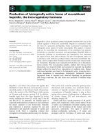
We constructed two vectors, pIgGL ⁄ M1.1MG and pIg-
GH ⁄ M1.1R, for the generation of transgenic silkworms
expressing a mouse IgG mAb (Fig. 1). The former vector
contained the cDNA for monster green fluorescent
protein (MGFP) as a marker under the control of an
M. Iizuka et al. Production of mouse mAb by transgenic silkworms
FEBS Journal 276 (2009) 5806–5820 ª 2009 The Authors Journal compilation ª 2009 FEBS 5807
eye- and nervous tissue-specific promoter, 3xP3, plus the
cDNA for the IgG L-chain under the control of the ser1
promoter. The latter vector contained the cDNAs for red
fluorescent protein (DsRed) and the IgG H-chain under
the control of the 3xP3 and ser1 promoters, respectively
(Fig. 1). pIgGL ⁄ M1.1MG and pIgGH ⁄ M1.1R were
injected into 3154 and 2854 eggs, respectively, and the
hatched G0 larvae were allowed to develop to moths. G1
embryos from the G0 moths were screened for MGFP or
DsRed fluorescence to obtain transgenic silkworms.
Genomic Southern blot analysis of the transgenic
silkworms demonstrated the existence of 13 and 17
independent transgenic lines, respectively, for pIg-
GL ⁄ M1.1MG- and pIgGH ⁄ M1.1R in relation to the
chromosomal insertion positions and copy numbers of the
transgenes. Transgenic lines with a single-copy transgene
were selected, and the cocoon proteins of the lines were
analyzed by SDS ⁄ PAGE. The lines with the highest levels
of IgG L- and H-chain expression were used in the subse-
quent experiments as the L- and H-lines, respectively.
To generate transgenic silkworms bearing both the
L- and H-chain cDNAs, an L-line worm was crossed
with an H-line worm, and the silkworms in the subse-
quent generation that expressed both MGFP and
DsRed in their eyes were selected. The silkworms car-
rying both the L- and H-chain cDNAs were referred
to as L ⁄ H-line silkworms.
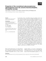
Analysis of recombinant mouse IgG in cocoons
To analyze secreted proteins in the sericin layer of the
silk fibers, all proteins in the layer were dissolved in a
buffer containing 8 m urea, electrophoresed under
reducing conditions, and analyzed by western blotting
using polyclonal anti-mouse IgG serum. Recombinant
mouse IgG L- and H-chain was detected in the L- and
H-lines, respectively (Fig. 2A, lanes 8 and 9). The
H-chain was also identified in the H-line proteins by
Coomassie brilliant blue (CBB) staining (Fig. 2A,
lane 4), whereas the L-chain was not, as a result of the
presence of endogenous silk proteins with a similar
molecular weight (Fig. 2A, lane 3). Both L- and
H-chains were detected in the cocoon proteins from
the L ⁄ H-line by CBB staining and western blotting
(Fig. 2A, lanes 5 and 10). The amount of L- or
H-chain in the L ⁄ H-line appeared to be higher than
that in the L- or H-line. The intensity of the H-chain
on the CBB-stained gels was quantified by densitome-
try. The mean ± SEM amount of H-chain present in
0.1 mg of cocoons from the L ⁄ H- and H-lines was
319 ± 1 ng (n = 3) and 139 ± 9 ng (n = 3), respec-
tively.
To investigate the assembly of the recombinant
L- and H-chains, cocoon proteins were analyzed by
electrophoresis under nonreducing conditions. No
L- or H-chain was detected among the cocoon proteins
from the L- and H-lines by CBB staining, respectively
(Fig. 2B, lanes 3 and 4). Western blotting revealed that
the L-chain in the L-line cocoons existed as a mono-
mer (Fig. 2B, lane 8). By contrast, the H-chain was
detected as a dimer in the H-line cocoon proteins
(Fig. 2B, lane 9). Intense bands with an apparent
molecular weight that exceeded that of the H-chain
dimer were also visible on the blot. Although the
ie1
P
3 xP3
P
ser1
DsRed
SV40
polyA
ie1
polyA
piggyBac
right arm
piggyBac
left arm
IgG H-chain
hr3
P
3 xP3
P
ser1
DsRed
SV40
polyA
fibL
polyA
piggyBac
right arm
piggyBa
c
left arm
BmNPVpol 5′-UTR
pIgGH/M1.1R
IgG L-chain
hr3
P
3xP3
P
ser1
MGFP
SV40
polyA
fibL
polyA
piggyBac
right arm
piggyBac
left arm
BmNPVpol 5′-UTR
pIgGL/M1.1MG
pIE1
Fig. 1. Structures of the transformation vectors. Three transformation vectors (pIgGL ⁄ M1.1MG, pIgGH ⁄ M1.1R and pIE1) were constructed,
each of which contained expression units for selection markers and the recombinant proteins between the right and left arms of piggyBac.
In the selection marker units, the gene encoding DsRed (DsRed) or MGFP (MGFP) was placed between the 3xP3 promoter (P
3xP3
) and
SV40 polyA signal sequence (SV40 polyA). The recombinant protein units were designed to express IgG L- and H-chains in pIgGL ⁄ M1.1MG
and pIgGH ⁄ M1.1R, respectively; thus, the L-chain (IgG L-chain) or H-chain (IgG H-chain) cDNA was placed between the BmNPV hr3 enhan-
cer, (hr3)-ser1 promoter (P
ser1
) and fibroin L-chain polyA signal sequence (fibL polyA). The recombinant protein unit in pIE1 was composed
of the ser1 promoter (P
ser1
), IE1 gene (ie1) and ie1 polyA signal sequence (ie1 polyA).
Production of mouse mAb by transgenic silkworms M. Iizuka et al.
5808 FEBS Journal 276 (2009) 5806–5820 ª 2009 The Authors Journal compilation ª 2009 FEBS
detailed structures of these high molecular weight
products were unclear, the products most likely were
random aggregates of H-chain connected by inter-
chain disulfide bonds. CBB staining of the cocoon pro-
teins from the L ⁄ H-line revealed a band co-migrating
with the standard IgG H
2
L
2
tetramer (Fig. 2B, lanes 5
and 6). In addition to the H
2
L
2
tetramer, small
amounts of H
2
L and H
2
were detected among the pro-
teins from the L ⁄ H-line by western blotting, which
were also detectable in the standard mouse IgG
(Fig. 2B, lanes 10 and 11). Bands with the higher
molecular weight than H
2
L
2
, which were assumed to
be derived from random aggregates of the chains, were
also detected on the blot as the single expression of the
H-chain. These aggregates appear in much smaller
amounts than the H
2
L
2
because the H
2
L
2
was detected
as a major product on the CBB-stained gel. No
L-chain monomer was present among the cocoon
proteins (Fig. 2B, lane 10). These results suggest that a
fully assembled mouse IgG mAb with an H
2
L
2
-subunit
structure was synthesized in the MSG cells and
secreted into the sericin layer of the silk fibers in silk-
worms carrying both the IgG L- and H-chain cDNAs.
Quantification of L- and H-chain mRNAs in MSGs
As described above, the amounts of L- and H-chain in
the cocoons were increased by the co-expression of both
chains compared to the expression of either chain. We
therefore investigated whether these increases arose
from an increase in the corresponding mRNAs in the
cells. Total RNA was extracted from the MSGs of
fifth-instar larvae of the L-, H- and L ⁄ H-lines, and the
L- and H-chain mRNA levels were measured by quanti-
tative RT-PCR. The amount of sericin-1 mRNA was
also determined to allow for normalization of the
expression of the L- and H-chains. As shown in
Table 1, the amount of L- or H-chain mRNA in the
L ⁄ H-line was lower than that of the corresponding
chain in the L- or H-line, most likely as a result of the
co-expression of the two genes from the same promoter.
These results suggest that the increases in IgG L- and
H-chain in the L ⁄ H-line cocoons were not caused by
the transcriptional regulation of mRNA expression, but
by the regulation of protein synthesis and secretion.
Enhanced transgene expression using
trans-activator IE1
We previously demonstrated that the baculovirus-
derived trans-activator IE1 stimulates the transcrip-
CBB Western
Reducing
1 2 3 4 5 6 7 8 9 1 0 11
H
L
20
30
40
80
50
60
kD a
H
L
M W L H L/H St W L H L / H S t
Nonreducing
H
2
L
2
H
2
L
2
H
2
L
H
2
L
W L H S t M W L H L / H St L / H
20
30
40
80
50
60
120
100
kD a
220
1 2 3 4 5 6 7 8 9 1 0 1 1
CBB Western
A
B
Fig. 2. Analysis of the cocoon proteins in the L-, H- and L ⁄ H-lines.
The proteins in the cocoons of wild-type (W), L- (L), H- (H) or L ⁄ H-line
(L ⁄ H) silkworms were extracted with (A) 8
M urea containing 2%
(v ⁄ v) b-mercaptoethanol and 50 m
M Tris–HCl, pH 8.0 (i.e. reducing
conditions) or (B) 8
M urea containing 50 mM Tris–HCl, pH 8.0 (i.e.
nonreducing conditions). Aliquots of the extracts were subjected to
SDS ⁄ PAGE. Some of the gels were stained with CBB, whereas
others were subjected to western blotting using the rabbit anti-
(mouse IgG) as a primary antibody (western). ‘H
2
L
2
’, ‘H
2
L’, ‘H
2
’, ‘H’
and ‘L’ to the right of the gel indicate the H
2
L
2
tetramer, H
2
L trimer,
H
2
dimer, H monomer and L monomer, respectively. The numbers to
the left of the gel are the molecular masses (kDa) as determined by
the migration of the markers (M). St, commercially available standard
mouse IgG.
Table 1. Copy numbers of mRNAs of the L-chain, H-chain and
sericin-1 in MSG cells.
Line L-chain
a
H-chain
a
Sericin-1
a
Percentage
of
L-chain to
sercin-1
Percentage
of
H-chain to
sercin-1
L 2.4 ± 0.4
b
0.0 58.3 ± 1.8 4.1 ± 0.6 0.0
H 0.0 3.0 ± 0.5 67.7 ± 4.6 0.0 4.4 ± 0.7
L ⁄ H 1.4 ± 0.2 1.6 ± 0.1 47.0 ± 2.5 3.4 ± 0.3 2.9 ± 0.1
a
Copy numbers of mRNAs of L-chain and H-chains of the recombi-
nant IgG, and sericin-1 per 10 ng of total RNA. The indicated values
are 10
)5
of the actual copy numbers.
b
Data are the mean ± SEM
of the results obtained from three MSGs.
M. Iizuka et al. Production of mouse mAb by transgenic silkworms
FEBS Journal 276 (2009) 5806–5820 ª 2009 The Authors Journal compilation ª 2009 FEBS 5809
tional activity of the ser1 promoter in the presence of
the baculovirus-derived enhancer hr3 in MSG cells
[27]. This mechanism was used to express recombinant
proteins in transgenic silkworms [26,27]. However, the
simultaneous introduction of hr3 and ie1 using a single
transformation vector induced the leaky expression of
ie1 in tissues other than the MSG because of the self-
activation of ie1 expression through an interaction
between IE1 and hr3, resulting in high silkworm mor-
tality. In the present study, ie1-bearing silkworms were
generated using a transformation vector lacking hr3
but containing ser1 promoter-linked ie1 and crossed
with the L ⁄ H-line to obtain silkworms carrying the
genes encoding L-chain, H-chain and IE1. The resul-
tant silkworms, which were designated the L ⁄ H⁄ IE1-
line, showed no lethality or abnormalities (data not
shown), and were therefore used in the subsequent
experiments aiming to investigate the increases in
L- and H-chain in the cocoons.
The proteins contained in the cocoons of the L ⁄ H-
and L ⁄ H ⁄ IE1-lines were separated by SDS ⁄ PAGE
under reducing conditions and stained with CBB (data
not shown). L- and H-chains in the L ⁄ H ⁄ IE1-line
cocoons were more highly expressed than those in the
L ⁄ H-line cocoons. The intensities of the H-chain bands
on the gels were quantified by densitometry. The
mean ± SEM amount of H-chain per 0.1 mg of
cocoon in the L ⁄ H- and L ⁄ H ⁄ IE1-lines was
319 ± 1 ng (n = 3) and 754 ± 36 ng (n = 3), respec-
tively. Thus, the expression of IE1 induced an approxi-
mate 2.4-fold increase in the expression of the IgG
mAb in the silkworms. The mAb content in the
cocoons of the L ⁄ H ⁄ IE1-line was estimated to be 1.1%.
Extraction and purification of recombinant mouse
mAb from cocoons
Recombinant mAb was extracted from L ⁄ H ⁄ IE1-line
cocoons at 4 °C with NaCl ⁄ Pi or a buffered solution
containing urea at a variety of concentrations (in the
range 2–8 m), and the resultant extracts were analyzed
by SDS ⁄ PAGE (Fig. 3A, lanes 4–10). All the proteins
in the sericin layers were solubilized using 8 m urea and
2% b-mercaptoethanol with heating and then subjected
to SDS ⁄ PAGE (Fig. 3A, lane 3). The ratios of the
amount of mAb extracted with NaCl ⁄ P
i
or the urea-
containing solutions to the total amount of mAb in the
sericin layers were calculated by quantifying the band
intensities of the CBB-stained H-chains. When the
extracted proteins with NaCl ⁄ P
i
were analyzed, faint
bands of the H- and L-chains were detected. The ratio
of the extracted H-chains to all H-chains in the sericin
layers was estimated to be 8% (Fig. 3A, lane 10). In
the case of the extraction with a buffered solution con-
taining urea at a concentration of 4 m or less, the
amount of the mAb increased as the urea concentration
increased; the extraction efficiencies were 18%, 25%
and 40% at 2, 3 and 4 m urea, respectively (Fig. 3A,
lanes 4–6). In the previous studies, recombinant
enhanced green fluorescent protein and human serum
albumin were efficiently extracted with NaCl ⁄ P
i
or
NaCl ⁄ Tris saline from cocoons of transgenic silkworms
[26,27]. In the present study, however, the addition of
urea in the saline was required for the efficient extrac-
tion of the mAb. This difference in the protein extrac-
tion may be a result of the difference in the structure of
recombinant proteins or their affinities for sericin.
Endogenous sericin variants were hardly solubilized by
urea at concentrations of less than 3 m (Fig. 3A, lanes
4, 5 and 10). More than 80% of the mAb was recover-
able using a solution containing more than 5 m urea
(Fig. 3A, lanes 7–9). Under these conditions, however,
a large proportion of the sericin variants were solubi-
lized. Thus, in our subsequent purification experiment,
L ⁄ H ⁄ IE1-line cocoons were treated with a buffered
solution containing 3 m urea to reduce the level of
contamination in the extract by sericin variants.
An L⁄ H ⁄ IE1-line cocoon extract prepared with 3 m
urea and 50 mm Tris-HCl (pH 7.4) was dialyzed
against 20 mm phosphate buffer (pH 7.0) and sub-
jected to protein G affinity column chromatography.
As shown in Fig. 3B, this process was sufficient to pur-
ify the mAb to apparent homogeneity (Fig. 3B,
lane 5). SDS ⁄ PAGE under nonreducing conditions
revealed that the purified mAb was fully assembled
H
2
L
2
(Fig. 3B, lane 6). As shown in Table 2, we were
able to obtain 1.2 mg of purified mAb from 500 mg of
L ⁄ H ⁄ IE1-line cocoons.
Antigen-binding properties of recombinant mAb
The IgG L- and H-chain cDNAs used in the present
study were cloned from a mouse hybridoma that pro-
duces an IgG mAb against human IgG. Therefore,
binding of the recombinant mAb to human IgG was
analyzed by ELISA to compare the antigen-binding
properties of the recombinant mAb with those of the
hybridoma-derived one. As depicted in Fig. 4, both
mAbs produced almost identical binding curves to the
antigen with similar EC
50
values. The mouse mAb to
human surfactant protein D that was used as a
negative control did not bind human IgG at all. We
also surveyed the binding of the recombinant and
hybridoma-derived mAbs to human IgM as a negative
control; no binding was detected in either case (data
not shown).
Production of mouse mAb by transgenic silkworms M. Iizuka et al.
5810 FEBS Journal 276 (2009) 5806–5820 ª 2009 The Authors Journal compilation ª 2009 FEBS
Structures of N-glycans attached to recombinant
mAb
The N-glycan profiles of the recombinant mAb pro-
duced by the silkworms were determined. The purified
mAb with a protein G column was used for this deter-
mination. Protein G, as well as protein A, binds the
CH
2
and CH
3
domain interface region distal to the
glycosylation site in the CH
2
domain of IgG [29], and
the affinity of the IgG-binding proteins for IgG is
unchanged by deglycosylation of IgG [30]. Thus, it is
unlikely that any specific mAb glycoform is preferen-
tially selected by purification using protein G.
When the pyridylaminated-N-glycans (PA-N-gly-
cans) prepared from the mAb were separated by
anion exchange chromatography, the glycans were
detected only in a flow-through fraction (data not
shown), suggesting the absence of negatively-charged
saccharides such as sialic acids. Subsequently, the
PA-N-glycans in the fraction were separated by size
fractionation and RP-HPLC. Six major PA-N-glycan
fractions were obtained, and their structures were
analyzed by MALDI-TOF-MS. The results obtained
are summarized in Table 3. The six PA-N-glycan
fractions were identified as GlcNAcMan
3
GlcNAc
2
-PA
(GNb), Man
2
Man
3
GlcNAc
2
-PA (M5), GlcNAc
2
Man
3
GlcNAc
2
-PA (GN2), Man
3
Man
3
GlcNAc
2
-PA (M6),
Man
4
Man
3
GlcNAc
2
-PA (M7) and Man
5
Man
3
Glc-
NAc
2
-PA (M8). Most of the major N-glycans
were high mannose-types such as M5 (51.1%) and
M6 (11.9%). On the other hand, significant amounts
of hybrid-type (GNb) and complex-type N-glycans
(GN2) having one and two GlcNAc residues at
their nonreducing termini, respectively, were detected
at ratios of 18.1% and 11.7%, respectively. Pauci-
20
30
40
80
50
60
120
100
kDa
220
Extr
ac
ti
o
n
sM
sA
sP
H
L
P
u
rifi
ca
ti
o
n
20
30
40
80
50
60
120
100
kDa
220
H
L
H
2
L
2
Urea conc.(M)
NaCl/P
i
2 3 4 5 6 8
Heating
W
L/H/IE1
M
W
L/H/IE1
Extraction
Purification
Heating
M
1 2 3 4 5 6 7 8 9 10 6 1 2 3 4 5
A
B
Fig. 3. Extraction and purification of recombinant mAb from cocoons. (A) Extraction of recombinant mAb from L ⁄ H ⁄ IE1-line cocoons. The
proteins in the sericin layer of the silk fibers from wild-type (lane 2) or L ⁄ H ⁄ IE1-line (lane 3) silkworms were extracted by maintaining the
cocoons at 80 °C for 5 min in 8
M urea, 2% (v ⁄ v) b-mercaptoethanol and 50 mM Tris–HCl (pH 8.0), at 10 mg dry weightÆmL
)1
. The proteins
from the L ⁄ H ⁄ IE1-line cocoons were also extracted with 50 m
M Tris–HCl (pH 7.4) containing 2, 3, 4, 5, 6 or 8 M urea (lanes 4–9) or NaCl ⁄ Pi
(lane 10) at 4 °C for 24 h. The extracted proteins were separated by SDS ⁄ PAGE and stained with CBB. (B) Purification of the recombinant
mAb from the L ⁄ H ⁄ IE1-line cocoons. Recombinant mAb extracted with 50 m
M Tris–HCl (pH 7.4) containing 3 M urea was purified using a
protein G column. The extract (lane 4) and purified mAb (lane 5) were electrophoresed under reducing conditions. The purified mAb was also
subjected to SDS ⁄ PAGE under nonreducing conditions (lane 6). The electrophoresed proteins were stained with CBB. The numbers to the
left of the gel indicate the molecular masses (kDa) as determined by the migration of the markers (M). ‘sM’, ‘sA’, and ‘sP’ to the right of
the gel represent sericin M, A and P, respectively. ‘H
2
L
2
’, ‘H’ and ‘L’ represent the H
2
L
2
tetramer, H-chain monomer and L-chain monomer
of the IgG, respectively.
Table 2. Purification of the recombinant mAb from 500 mg of
cocoons.
Purification step
Amount
of mAb (mg)
Recovery
(%)
Cocoons 5.5 100
Extract 1.4 25
Eluate from protein G column 1.2 22
M. Iizuka et al. Production of mouse mAb by transgenic silkworms
FEBS Journal 276 (2009) 5806–5820 ª 2009 The Authors Journal compilation ª 2009 FEBS 5811
mannose-type N-glycans such as Man
3
GlcNAc
2
-PA
(M3), which are typically found in insects, were not
observed [31]. It is also noteworthy that fucose resi-
dues linked to the core GlcNAc residue were not
detected.
To confirm the absence of fucose residues in the
N-glycans attached to the mAb, SDS ⁄ PAGE with
lectin blotting using Aleuria aurantia lectin (AAL) or
concanavalin A was performed on the purified recom-
binant mAb and standard human IgG from human
serum treated with or without peptide-N-glycosidase F
(PNGaseF). The AAL lectin used in this analysis rec-
ognizes fucose residues a-1,3- and a-1,6-linked to the
GlcNAc residue [32]. Concanavalin A lectin recogniz-
ing mannose was also used as a control. CBB staining
showed that PNGaseF-treated H-chains in both the
recombinant mAb and standard human IgG were
slightly lower in molecular mass than the correspond-
ing untreated chains (Fig. 5, lanes 1–4). This indicates
that PNGaseF actually removed N-glycans from the
H-chains. Concanavalin A reacted with both the
recombinant and standard H-chains (Fig. 5, lanes 5
and 7); however, this reaction was not observed after
PNGaseF treatment (Fig. 5, lanes 6 and 8), confirming
the presence of mannose residues in the N-glycans of
both H-chains. On the other hand, AAL did not react
Table 3. N-glycan structures of the recombinant mouse mAb produced by silkworms. The proposed structure is illustrated using symbols:
closed square, open circle and closed circle indicate N-acetylglucosamine, mannose and aminopyridine, respectively. ODS, octadecyl silane.
Abbreviation
of N-glycan
structure
Proposed
structure
b
Theoretical m ⁄ z
(mass + H
+
)
a
Observed m ⁄ z
(mass + H
+
)
b
% Peak area
obtained from
HPLC (ODS)
c
GNb 1192.469 1192.576 18.1
M5
1313.495 1313.591 51.1
GN2
1395.548 1395.444 11.7
M6
1475.548 1475.654 11.9
M7
1637.601 1637.597 2.7
M8
1799.654 1799.650 4.5
a
Theoretical m ⁄ z of PA-N-linked glycans was calculated as the monoisotopic mass of (mass + H
+
).
b
Observed m ⁄ z (mass + H
)
) were
obtained from reflector mode MALDI-TOF mass spectra of the labeled N-glycans.
c
% Peak area calculated from the result of RP-HPLC.
Concentration (ng·mL
–1
)
Absorbance at 490 nm
Recombinant EC
50
: 73.9 ng·mL
–1
Hybridoma EC
50
: 99.4 ng·mL
–1
Negative control
1.25
1.00
0.75
0.50
0.25
0.00
10
–1
10
0
10
1
10
2
10
3
10
4
Fig. 4. Antigen binding of recombinant mouse mAb. The binding of
recombinant mouse mAb to human IgG was analyzed by ELISA.
Recombinant mAb (closed triangles), hybridoma-derived natural
mAb (closed squares) and negative control (anti-human surfactant
protein D mouse IgG
1
; closed circles) at various concentrations
(3333.33, 1111.11, 370.37, 123.46, 41.15, 13.72, 4.57, 1.52, 0.51
and 0.17 ngÆmL
)1
) were reacted against human IgG. The EC
50
val-
ues for the binding of the mAbs to the antigen were determined
from binding curves.
Production of mouse mAb by transgenic silkworms M. Iizuka et al.
5812 FEBS Journal 276 (2009) 5806–5820 ª 2009 The Authors Journal compilation ª 2009 FEBS
with the recombinant H-chain, whereas the standard
H-chain was clearly stained with this lectin (Fig. 5, lanes
9 and 11). On the basis of these results, together with
our structural data, we conclude that the N-glycans
attached to the recombinant mouse mAb contained no
detectable a-1,3-linked or a-1,6-linked fucose residues.
N-Glycan structures of endogenous proteins in
cocoons and larval tissues
As described above, the recombinant mAb contained
high mannose, hybrid and complex N-glycans. On the
other hand, major N -glycans synthesized in insect cell
lines have paucimannose structures with a-1,3- and ⁄ or
a-1,6-fucose residues and high mannose structures,
although some variations in the N-glycan structure are
observed depending on the synthesized glycoproteins
[33]. To investigate the reason for this difference in
N-glycan structure, we analyzed the N-glycans con-
tained in the cocoons and two larval tissues (MSGs
and fat bodies) of wild-type pnd-w1 silkworms. The
results obtained are shown in Table 4.
The major N-glycans in the cocoons were M5
(48.5%) and GN2 (36.2%), with small amounts of M3
(1.2%). Fucosylated glycans were not detected in the
cocoons, as in the case of the recombinant mAb. Thus,
the N-glycans attached to the endogenous cocoon
proteins were similar to those attached to the mAb.
Similar N-glycan structures were noted in the MSG
N-glycans, suggesting that the structural features of
the N-glycans in the cellular glycoproteins of the MSG
cells are comparable to those in the secreted cocoon
glycoproteins. The N-glycan structures from the fat
bodies were different from the MSG. The major fat
body glycans had a fucosylated paucimannose struc-
ture (Man
2
[Fuc
1
]GlcNAc
2
-PA [FM2; 37.4%]) with
high mannose structures having more than six man-
noses. Because FM2 was identified only by MS, it was
not possible to determine whether the fucose residues
were a-1,3- or a-1,6-linked to the GlcNAc residues.
The M5 observed in the cocoons and MSGs as a
major high mannose-type glycan was not present in
the fat bodies. We also analyzed the N-glycan struc-
tures in the tissues of another silkworm strain, Kinshu,
and found that they were essentially the same as those
in the pnd-w1 silkworms (data not shown). From these
results, we conclude that the structural features of the
N-glycans in the recombinant mAb are attributed to
the tissue specificity of the silk glands.
Discussion
In the present study, we generated three transgenic
lines, L-, H- and L ⁄ H-, that synthesized mouse IgG
L-chain and H-chain, or both L- and H-chains, respec-
tively. The L-line silkworms secreted L-chain as a
monomer into their cocoons, whereas the H-line silk-
worms secreted H-chain as a dimer and higher mole-
cular aggregates. In the case of the L ⁄ H-line, the
co-expressed L- and H-chains formed H
2
L
2
tetramers
that were secreted as a major product into the cocoons.
L-chain monomers and H-chain dimers were hardly
detected in the L ⁄ H-line cocoons. The amount of
H-chain in the L ⁄ H-line cocoons was approximately
2.3-fold higher than that in the H-cocoons. Quantita-
tive analysis of the H-chain mRNA in the MSG cells
revealed that the increase in H-chain in the L⁄ H-line
cocoons was not the result of a rise in the mRNA level.
Thus, H
2
L
2
tetramers were preferentially synthesized
and secreted through post-transcriptional regulation.
In vertebrate antibody-producing cells, H-chain
dimers synthesized in the absence of L-chain expres-
sion are not secreted, but are retained within the cells.
This inhibition of secretion is caused by the stable
association of an endoplasmic reticulum-resident stress
protein, BiP, with the H-chain dimer [34–36]. This
mechanism could be present in the MSG cells of silk-
worms. However, the regulation of IgG secretion may
be insufficient in MSG cells because the L-chain
monomers or H-chain dimers were secreted from the
cells in the case of the single expression of each chain.
Similar observations were reported in Drosophila cells
transfected with the genes encoding humanized IgG
[37]. When the H-chain gene was expressed in these
cells, H-chain was efficiently secreted as a dimer into
the culture medium. Furthermore, a Drosophila BiP
homolog, hsc72, transiently interacts with the H-chain
PNGase
+–– –– ––+++
KDa
50
25
35
CBB
H
L
AAL
++
Con A
St
1234
R
91011125678
StRStR
Fig. 5. Analysis of N-glycans in the recombinant mAb by lectin blot-
ting. Recombinant mAb was subjected to lectin blotting with AAL
and concanavalin A. The purified recombinant mAb (R) and standard
human IgG (St) treated with (+) or without ()) PNGaseF were elec-
trophoresed on polyacrylamide gradient gels. One gel was stained
with CBB (lanes 1–4), whereas the others were subjected to conca-
navalin A (lanes 5–8) or AAL blotting (lanes 9–12). ‘H’ and ‘L’ to the
right of the gel represent the H- and L-chains of IgG, respectively.
The numbers to the left of the gel correspond to the molecular
masses (kDa) as determined by the migration of the markers.
M. Iizuka et al. Production of mouse mAb by transgenic silkworms
FEBS Journal 276 (2009) 5806–5820 ª 2009 The Authors Journal compilation ª 2009 FEBS 5813
during its secretion [37]. Unlike vertebrate BIP,
Drosophila hsc72 dissociates from the H-chain inde-
pendently of the L-chain association, allowing the
secretion of the H-chain as the dimer. The silkworm
genome contains a homolog of BiP (NCBI accession
number AB016836), and this gene is expressed in
MSG and PSG cells [38]. It is also suggested that
endoplasmic reticulum-resident chaperone proteins
such as BiP are involved in the synthesis and secre-
tion of fibroin in PSG cells [16]. Therefore, it is rea-
sonable to assume that the silkworm BiP homolog
and ⁄ or other chaperone proteins involved in the
secretion of silk proteins might also function in that
of recombinant IgG. Although the function of these
factors in IgG-secretion is not sufficient in MSG cells,
as observed in the Drosophila cells, the factors might
have selectively enhanced the secretion of the IgG
H
2
L
2
tetramers from the cells into the cocoons.
Accordingly, we were able to collect the mAb as fully
assembled H
2
L
2
tetramers from the cocoons.
Previous studies have shown that the major N-gly-
cans in insects have paucimannose- and high mannose-
structures [9,33,39]. Paucimannose-type N-glycans such
as M3 are characteristic of insects and are not found in
mammals. On the other hand, in the present study,
paucimannose-type N-glycans were detected at very low
levels in the cocoons and MSGs, whereas N-glycans of
this type were present at high levels in the fat bodies.
Paucimannose-type N-glycans arise from GNb by the
removal of a GlcNAc residue by the Golgi membrane-
associated enzyme b-N-acetylglucosaminidase [40]. In
the MSG cells of silkworms, b-N-acetylglucosaminidase
activity might be absent or very low, resulting in the
nondetection of paucimannose structures in the N-gly-
cans of the cocoons. On the other hand, it is likely that
N-acetylglucosaminyltransferase (GnT)-I and II activity
Table 4. Structures of N-glycans from cocoons, MSGs and fat bodies. The proposed structure is illustrated using symbols: closed square,
open circle, open diamond, closed triangle and closed circle indicate N-acetylglucosamine, mannose, galactose, fucose and aminopyridine,
respectively. ODS, octadecyl silane.
Abbreviation of N-glycan structure Proposed structure
% Peak area obtained from HPLC (ODS)
a
Cocoons MSGs Fat bodies
FM2
0.0 0.0 37.4
M3
1.2 5.7 7.9
GNb
4.5 10.9 0.0
GNa
1.7 7.2 0.0
M5
48.5 42.3 0.0
GN2
36.2 24.0 0.0
M6
3.8 4.9 7.2
M7
2.5 3.1 13.7
M8
1.6 0.0 16.5
M9
0.0 1.9 17.3
GAa ⁄ b
0.0 0.0 0.0
GA2
0.0 0.0 0.0
a
% Peak area calculated from the result of RP-HPLC.
Production of mouse mAb by transgenic silkworms M. Iizuka et al.
5814 FEBS Journal 276 (2009) 5806–5820 ª 2009 The Authors Journal compilation ª 2009 FEBS
is present in the MSG cells of silkworms as in other
insect cells or tissues [41–43]. Therefore, it is reasonable
to find that significant amounts of N-glycans having
GlcNAc residues at their nonreducing termini were
detected among the MSG-synthesized glycoproteins.
We also detected large amounts of M5, which is a pos-
sible substrate for GnT-I [44]. The accumulation of M5
suggests relatively low GnT-I activity in the MSG cells.
No b-1,4-galactose-containing N-glycans were detected
among the MSG-synthesized glycoproteins, implying
little or no b-1,4-galactosyltransferase activity in the
cells. This is consistent with previous observations in
other insect cells and tissues [45–47].
One surprising finding obtained from our N-glycan
analysis was the absence of fucose residues among the
MSG-synthesized glycoproteins. Previously, the N-gly-
cans of insects such as silkworms were reported to
contain considerable amounts of fucose residues a-1,3-
and ⁄ or a-1,6-linked to the core GlcNAc residue
[33,48]. For example, the ratios of N-glycans with
a-1,3-fucose and a-1,6-fucose, and both a-1,3- and
a-1,6-fucoses, to the total amount of N-glycans among
the membrane glycoproteins in Sf-21 cells were found
to be 1.8%, 15.1% and 8.8%, respectively [33]. The
absence of fucose residues is not attributable to the
silkworm strain used in the present study, but to the
tissue specificity of the silk glands. The Drosophila gen-
ome contains genes for a-1,3-fucosyltransferase
(FucTA) and a-1,6- fucosyltransferase (FucT6) [49,50].
These Drosophila enzymes preferred N-glycans with
nonreducing terminal GlcNAc residues as substrates
[49,50]. Gene homologs of the Drosophila
fucosyltransferases were also identified from the silk-
worm genome (FucTA homolog, NCBI accession
number CK537398; FucT6 homolog, NCBI accession
number BB987128). Our preliminary analysis demon-
strated that the fucosyltransferase mRNAs were
expressed in the MSG cells (data not shown), suggest-
ing that the absence of the core fucosylation is not a
result of the absence of the fucosyltransferase expres-
sion. The shortage of GDP-fucose in the cells might
lead to the prevention of fucosylation.
The present study highlights several advantages of
using transgenic silkworms as hosts for the production
of recombinant mAbs. Cocoons of transgenic silk-
worms contained fully formed H
2
L
2
tetramers with
appropriate antigen-binding ability. The expression of
the mAb was increased up to 1.1% by the introduction
of ie1 into the mAb-expressing silkworms. The mAb
was easily extracted and purified from the cocoons.
Thus, the present study demonstrated the feasibility of
using transgenic silkworms for the mass production of
recombinant mAbs. Silkworms have been used for the
manufacture of silk in the sericultural industry. There-
fore, the industrial production of recombinant mAbs
could be achieved by taking advantage of technologies
employed in the sericultural industry, although quality
control of the product must be taken into consider-
ation. The observed structures of the N-glycans further
highlight the potential use of transgenic silkworms for
mAb production. Although the presence of oligoman-
nose N-glycans such as M5 is not always favorable for
the therapeutic use of the mAb, the absence of the core
fucosylation is beneficial. Fucose residues a-1,3-linked
to GlcNAc show high antigenicity when administrated
to humans [10]. Therefore, the presence of a-1,3-fucose
in the recombinant glycoproteins produced by insect
cells has been recognized as an important issue for
their use in therapeutic applications. In our system,
this issue may be solved because no a-1,3-fucose resi-
dues were detected among the N-glycans in the recom-
binant proteins. The absence of a-1,6-fucose comprises
yet another reason supporting the use of this system in
the production of mAbs. The absence of a-1,6-fucose
enhances the activity of antibody-dependent cellular
cytotoxicity of IgG [51,52]. Thus, the present system
might be particularly beneficial for the production of
therapeutic mAbs, whose main mechanism of action is
antibody-dependent cellular cytotoxicity activity.
Experimental procedures
Experimental animals
B. mori strain pnd-w1 was obtained from the National
Institute of Agrobiological Science (Tsukuba, Japan). The
larvae were reared at 25 °C on an artificial diet (Silk Mate
PM, Nosan Corp, Kanagawa, Japan).
Construction of the vectors used to generate the
transgenic silkworms
A mouse hybridoma that produces a mouse IgG
1
mAb to
human IgG was kindly provided by Dr S. Usuda (Institute
of Immunology, Tokyo, Japan). Total RNA prepared from
the hybridoma using an RNeasy kit (Qiagen, Valencia, CA,
USA) was reverse-transcribed to produce cDNA fragments.
Two cDNA fragments encoding the IgG L- and H-chain
variable regions were obtained from the hybridoma cDNAs
using a SMART RACE cDNA Amplification Kit (Clon-
tech, Palo Alto, CA, USA) in accordance with the manu-
facturer’s instructions, and the obtained fragments were
sequenced. The sequences were used to design PCR
primers, and the cDNAs encoding the full-length L- and
H-chain ORFs were cloned by PCR from the hybridoma
cDNAs. The 5¢-UTR sequence of BmNPV polyhedrin [53]
M. Iizuka et al. Production of mouse mAb by transgenic silkworms
FEBS Journal 276 (2009) 5806–5820 ª 2009 The Authors Journal compilation ª 2009 FEBS 5815
was inserted just upstream of the cDNAs for the L- and
H-chain ORFs, as described previously [28]. The H-chain
ORF cDNA with the 5¢-UTR was then inserted down-
stream of the hr3-linked ser1 promoter in pMSG1.1R [28]
to generate pIgGH ⁄ M1.1R (Fig. 1). Similarly, the L-chain
ORF cDNA with the 5¢-UTR was introduced into
pMSG1.1MG, which was created from pMSG1.1R by
replacing the DsRed cDNA with the cDNA for MGFP
(Promega, San Luis Obispo, CA, USA). The resulting
vector was dubbed pIgGL ⁄ M1.1MG (Fig. 1).
Generation of transgenic silkworms
pIgGL ⁄ M1.1MG and pIgGH ⁄ M1.1R were each injected
with the helper vector pHA3PIG [23] into eggs, as described
previously [23]. The hatched G0 larvae were reared at 25 °C
to moths. G1 embryos obtained by mating among siblings
or with pnd-w1 were screened for MGFP and DsRed
expression in the eyes to obtain transgenic silkworms bear-
ing the IgG L- and H-chain genes, respectively. To generate
silkworms bearing both the L- and H-chain genes, an
L-chain silkworm was crossed with an H-chain silkworm.
pBac[Ser1 IE1 ⁄ 3xP3-DsRed] [27], which was renamed
pIE1 (Fig. 1), was used to produce IE1 gene (ie1)-carrying
transgenic silkworms. pIE1 was injected into eggs, and
transgenic silkworms were created as described above. The
resulting ie1 silkworms were crossed with silkworms carry-
ing both the IgG L- and H-chain genes to obtain silkworms
bearing ie1 and the L- and H-chain genes.
Analysis of recombinant proteins in cocoons
Cocoon fragments were suspended in an extraction buffer
comprised of 8 m urea, 2% (v ⁄ v) b-mercaptoethanol and
50 mm Tris-HCl (pH 8.0) at 10 mg dry weightÆmL
)1
, and
maintained at 80 °C for 5 min. The extracted proteins were
then electrophoresed under reducing conditions on 0.1%
(w ⁄ v) SDS ⁄ 5–20% (w ⁄ v) polyacrylamide gradient gels
(Atto, Tokyo, Japan). For the analysis of subunit assembly,
cocoon proteins were extracted with b-mercaptoethanol-free
extraction buffer and electrophoresed under nonreducing
conditions. The gels for protein staining were treated with
CBB R250. In some cases, gel images were captured after
CBB staining and analyzed using imagej (o.
nih.gov/ij/). For western blotting, the proteins on the gels
were transferred to nitrocellulose membranes (BA85; Schlei-
cher and Schuell, Dassell, Germany) and reacted with
AffiniPure Rabbit Anti-Mouse IgG (H + L; Jackson
ImmunoResearch Laboratories, Inc., West Grove, PA,
USA) and then with horseradish peroxidase (HRP)-linked
anti-(rabbit IgG) sera (Cell Signaling Technology, Danvers,
MA, USA). The antigen–antibody complexes were visual-
ized using the ECL Western Blotting Detection System (GE
Healthcare, Little Chalfont, UK).
Quantification of mRNA in MSG cells
Total RNA was extracted from the MSGs of the transgenic
silkworms using Isogen (Nippon Gene, Tokyo, Japan).
cDNAs were synthesized from the RNAs using PowerScript
Reverse Transcriptase (BD Bioscience, Rockville, MD,
USA). The mRNAs of the IgG L- and H-chains and seri-
cin-1 were quantified using an ABI PRISM 7700 Sequence
Detector (Applied Biosystems, Foster City, CA, USA), as
described previously [25].
Extraction and purification of recombinant mAb
Pieces of cocoons were suspended in 50 mm Tris–HCl (pH
7.4) containing urea at various concentrations and stirred
at 4 °C for 24 h. The extracted proteins were then ana-
lyzed by SDS ⁄ PAGE to determine the optimal conditions
for extraction of the recombinant mAb. For purification
of the mAb, 50 mm Tris–HCl (pH 7.4) containing 3 m
urea was used for extraction of the cocoon proteins. The
extract was centrifuged at 20 000 g for 15 min, and the
obtained supernatant was dialyzed against 20 mm phos-
phate buffer (pH 7.0). Next, the recombinant mAb was
purified by protein G affinity column chromatography
according to the manufacturer’s instructions (GE Health-
care). The purified mAb was quantified using a mouse
IgG
1
ELISA quantitation kit (Bethyl Laboratories, Mont-
gomery, TX, USA).
Binding affinity assay
Commercially available purified human IgG from human
serum (Cappel, Irvine, CA, USA) was diluted to a final
concentration of 5 lgÆmL
)1
with 150 mm NaCl containing
0.1% (w ⁄ v) NaN
3
, and aliquots were dispensed into the
wells of 96-well polystyrene microplates (Greiner Bio-One,
Frickenhausen, Germany). The plates were then incubated
for 16 h at room temperature, washed with 150 mm NaCl
containing 0.05% (w ⁄ v) Tween 20, and blocked with 2%
(v ⁄ v) fetal calf serum in NaCl ⁄ Tris for 30 min. After
washing, mAb at various concentrations was added to
the wells and incubated for 80 min at room temperature.
The wells were then washed with 150 mm NaCl
containing 0.05% (w ⁄ v) Tween 20 and incubated with
peroxidase-labeled rabbit anti-(mouse IgG) serum (Jack-
son ImmunoResearch Laboratories, Inc.). O-phenylenedi-
amine dihydrochloride was used as a substrate for
peroxidase. The reaction was stopped by adding 2 m
H
2
SO
4
, and A
490
was measured using a microplate
reader. The effector concentration for half-maximum
response (EC
50
) values for the mAb bound to human
IgG was determined from binding curves using graphpad
prism 4.0 (GraphPad Software, Avenida de La Jolla, CA,
USA).
Production of mouse mAb by transgenic silkworms M. Iizuka et al.
5816 FEBS Journal 276 (2009) 5806–5820 ª 2009 The Authors Journal compilation ª 2009 FEBS
Preparation of PA-N-glycans
The purified mAb and cocoons or larval tissues were
hydrazinolyzed at 100 °C for 10 h. The liberated N-glycans
were then N-acetylated and pyridylaminated, as described
previously [54]. All excess reagents were removed by phe-
nol ⁄ chloroform extraction [55] and subsequent solid-phase
extraction using a Sep-PAK Plus C18 cartridge (Waters,
Milford, MA, USA) [56].
HPLC
A Waters 2694 separation module liquid chromatograph
was used to conduct HPLC analyses. PA-N-glycans were
separated by anion exchange chromatography using a Mono
Q5⁄ 5 HR column (0.5 · 5 cm; GE Healthcare). Subse-
quently, size fractionation and RP-HPLC chromatography
were performed on a Shodex Asahipak NH2P-50 column
(0.2 · 15 cm; Showa Denko, Tokyo, Japan) and Cosmosil
5C18-P column (0.2 · 25 cm; Nacalai Tesque, Kyoto,
Japan), respectively, as described previously [57]. The frac-
tionated PA-N-glycans were quantified from the peak areas
in comparison with those of standard PA-N-glycans.
MS
For MALDI-TOF-MS, the PA-N-glycans were co-crystal-
lized in a matrix of 2,5-dihydroxybenzoic acid and analyzed
with an Autoflex II mass spectrometer (Bruker Daltonics,
Billerica, MA, USA) operated in the reflector mode, as
described previously [58]. Peptide standards were used to
achieve a six-point external calibration for mass assignment
of the ions.
Digestion with PNGaseF
The purified recombinant mouse mAb and standard human
IgG (Cappel) were digested with PNGaseF (Takara Bio
Inc., Shiga, Japan) at 37 °C for 17 h under denaturing
conditions. The reaction was terminated by boiling in
SDS ⁄ PAGE sample buffer.
Lectin blotting
mAb treated with or without PNGaseF was electrophore-
sed under reducing conditions as described above and
transferred to ImmobilonÒ poly(vinylidene difluoride)
membranes (Millipore Corp., Billerica, MA, USA). The
membranes were then treated with TBST buffer [NaCl ⁄ Tris
containing 0.05% (w ⁄ v) Tween 20] and reacted with bioti-
nylated AAL (Seikagakukogyo, Tokyo, Japan) at a concen-
tration of 1 lgÆmL
)1
or concanavalin A (Seikagakukogyo)
at a concentration of 0.3 lgÆmL
)1
at room temperature for
1 h. After three washes with TBST, the membranes were
incubated with 1 lgÆmL
)1
HRP-linked streptavidin (Jack-
son ImmunoResearch Laboratories, Inc.) in TBST at room
temperature for 1 h. After washing with TBST, the HRP
was detected with 3,3¢-diaminobenzidine (Wako Chemicals,
Osaka, Japan) at a concentration of 0.2 mgÆmL
)1
in
NaCl ⁄ P
i
.
References
1 Chadd HE & Chamow SM (2001) Therapeutic antibody
expression technology. Curr Opin Biotechnol 12, 188–
194.
2 Bakker H, Rouwendal GJ, Karnoup AS, Florack DE,
Stoopen GM, Helsper JP, van ReeR, van Die I &
Bosch D (2006) An antibody produced in tobacco
expressing a hybrid b-1,4-galactosyltransferase is
essentially devoid of plant carbohydrate epitopes. Proc
Natl Acad Sci USA 103, 7577–7582.
3 Bakker H, Bardor M, Molthoff JW, Gomord V, Elbers
I, Stevens LH, Jordi W, Lommen A, Faye L, Lerouge
P et al. (2001) Galactose-extended glycans of antibodies
produced by transgenic plants. Proc Natl Acad Sci USA
98, 2899–2904.
4 Cox KM, Sterling JD, Regan JT, Gasdaska JR, Frantz
KK, Peele CG, Black A, Passmore D, Moldovan-
Loomis C, Srinivasan M et al. (2006) Glycan optimiza-
tion of a human monoclonal antibody in the aquatic
plant Lemna minor. Nat Biotechnol 24, 1591–1597.
5 Ward M, Lin C, Victoria DC, Fox BP, Fox JA, Wong
DL, Meerman HJ, Pucci JP, Fong RB, Heng MH et al.
(2004) Characterization of humanized antibodies
secreted by Aspergillus niger. Appl Environ Microbiol 70,
2567–2576.
6 Zhu L, van de Lavoir MC, Albanese J, Beenhouwer
DO, Cardarelli PM, Cuison S, Deng DF, Deshpande S,
Diamond JH, Green L et al. (2005) Production of
human monoclonal antibody in eggs of chimeric chick-
ens. Nat Biotechnol 23, 1159–1169.
7 Johansson DX, Drakenberg K, Hopmann KH, Schmidt
A, Yari F, Hinkula J & Persson MA (2007) Efficient
expression of recombinant human monoclonal antibod-
ies in Drosophila S2 cells. J Immunol Methods 318, 37–46.
8 Hsu TA, Eiden JJ, Bourgarel P, Meo T & Betenbaugh
MJ (1994) Effects of co-expressing chaperone BiP on
functional antibody production in the baculovirus
system. Protein Expr Purif 5, 595–603.
9 Hsu TA, Takahashi N, Tsukamoto Y, Kato K,
Shimada I, Masuda K, Whiteley EM, Fan JQ, Lee YC
& Betenbaugh MJ (1997) Differential N-glycan patterns
of secreted and intracellular IgG produced in
Trichoplusia ni cells. J Biol Chem 272, 9062–9070.
10 Bencu´ rova
´
M, Hemmer W, Focke-Tejkl M, Wilson IB
& Altmann F (2004) Specificity of IgG and IgE anti-
bodies against plant and insect glycoprotein glycans
M. Iizuka et al. Production of mouse mAb by transgenic silkworms
FEBS Journal 276 (2009) 5806–5820 ª 2009 The Authors Journal compilation ª 2009 FEBS 5817
determined with artificial glycoforms of human transfer-
rin. Glycobiology 14, 457–466.
11 Wright A & Morrison SL (1994) Effect of altered
CH2-associated carbohydrate structure on the func-
tional properties and in vivo fate of chimeric mouse-
human immunoglobulin G1. J Exp Med 180,
1087–1096.
12 Wright A & Morrison SL (1998) Effect of C2-associated
carbohydrate structure on Ig effector function: studies
with chimeric mouse-human IgG1 antibodies in glyco-
sylation mutants of Chinese hamster ovary cells.
J Immunol 160, 3393–3402.
13 Wright A, Sato Y, Okada T, Chang K, Endo T &
Morrison S (2000) In vivo trafficking and catabolism
of IgG1 antibodies with Fc associated carbohydrates of
differing structure. Glycobiology 10, 1347–1355.
14 Schuster M, Jost W, Mudde GC, Wiederkum S, Schwa-
ger C, Janzek E, Altmann F, Stadlmann J, Stemmer C
& Gorr G (2007) In vivo glyco-engineered antibody with
improved lytic potential produced by an innovative
non-mammalian expression system. Biotechnol J 2,
700–708.
15 Strasser R, Stadlmann J, Scha
¨
hs M, Stiegler G, Quen-
dler H, Mach L, Glo
¨
ssl J, Weterings K, Pabst M &
Steinkellner H (2008) Generation of glyco-engineered
Nicotiana benthamiana for the production of monoclo-
nal antibodies with a homogeneous human-like N-gly-
can structure. Plant Biotechnol J 6, 392–402.
16 Inoue S, Tanaka K, Arisaka F, Kimura S, Ohtomo K
& Mizuno S (2000) Silk fibroin of Bombyx mori is
secreted, assembling a high molecular mass elementary
unit consisting of H-chain, L-chain, and P25, with a
6:6:1 molar ratio. J Biol Chem 275, 40517–40528.
17 Okamoto H, Ishikawa E & Suzuki Y (1982) Structural
analysis of sericin genes. Homologies with fibroin gene
in the 5¢ flanking nucleotide sequences. J Biol Chem
257, 15192–15199.
18 Couble P, Michaille JJ, Garel A, Couble ML &
Prudhomme JC (1987) Developmental switches of seri-
cin mRNA splicing in individual cells of Bombyx mori
silkgland. Dev Biol 124 , 431–440.
19 Michaille JJ, Garel A & Prudhomme JC (1990) Cloning
and characterization of the highly polymorphic Ser2
gene of Bombyx mori. Gene 86, 177–184.
20 Garel A, Deleage G & Prudhomme JC (1997) Structure
and organization of the Bombyx mori sericin 1 gene and
of the sericins 1 deduced from the sequence of the Ser
1B cDNA. Insect Biochem Mol Biol 27, 469–477.
21 Takasu Y, Yamada H & Tsubouchi K (2002) Isolation
of three main sericin components from the cocoon of
the silkworm, Bombyx mori. Biosci Biotechnol Biochem
66, 2715–2718.
22 Tamura T, Thibert C, Royer C, Kanda T, Abraham E,
Kamba M, Komoto N, Thomas JL, Mauchamp B,
Chavancy G et al. (2000) Germline transformation of
the silkworm Bombyx mori L. using a piggyBac transpo-
son-derived vector. Nat Biotechnol 18, 81–84.
23 Tomita M, Munetsuna H, Sato T, Adachi T, Hino R,
Hayashi M, Shimizu K, Nakamura N, Tamura T &
Yoshizato K (2003) Transgenic silkworms produce
recombinant human type III procollagen in cocoons.
Nat Biotechnol 21, 52–56.
24 Hino R, Tomita M & Yoshizato K (2006) The genera-
tion of germline transgenic silkworms for the produc-
tion of biologically active recombinant fusion proteins
of fibroin and human basic fibroblast growth factor.
Biomaterials 27, 5715–5724.
25 Adachi T, Tomita M, Shimizu K, Ogawa S & Yoshizato
K (2006) Generation of hybrid transgenic silkworms that
express Bombyx mori prolyl-hydroxylase alpha-subunits
and human collagens in posterior silk glands: production
of cocoons that contained collagens with hydroxylated
proline residues. J Biotechnol 126, 205–219.
26 Ogawa S, Tomita M, Shimizu K & Yoshizato K (2007)
Generation of a transgenic silkworm that secretes
recombinant proteins in the sericin layer of cocoon:
production of recombinant human serum albumin.
J Biotechnol 128, 531–544.
27 Tomita M, Hino R, Ogawa S, Iizuka M, Adachi T, Shi-
mizu K, Sotoshiro H & Yoshizato K (2007) A germline
transgenic silkworm that secretes recombinant proteins
in the sericin layer of cocoon. Transgenic Res 16, 449–
465.
28 Iizuka M, Tomita M, Shimizu K, Kikuchi Y & Yoshiz-
ato K (2008) Translational enhancement of recombinant
protein synthesis in transgenic silkworms by a 5¢-
untranslated region of polyhedrin gene of Bombyx mori
Nucleopolyhedrovirus. J Biosci Bioeng 105, 595–603.
29 Stone GC, Sjo
¨
bring U, Bjo
¨
rck L, Sjo
¨
quist J, Barber CV
& Nardella FA (1989) The Fc binding site for strepto-
coccal protein G is in the C gamma 2-C gamma 3 inter-
face region of IgG and is related to the sites that bind
staphylococcal protein A and human rheumatoid fac-
tors. J Immunol 143, 565–570.
30 Nose M & Wigzell H (1983) Biological significance of
carbohydrate chains on monoclonal antibodies. Proc
Natl Acad Sci USA 80, 6632–6636.
31 Tomiya N, Narang S, Lee YC & Betenbaugh MJ
(2004) Comparing N-glycan processing in mammalian
cell lines to native and engineered lepidopteran insect
cell lines. Glycoconj J 21, 343–360.
32 Wimmerova M, Mitchell E, Sanchez JF, Gautier C &
Imberty A (2003) Crystal structure of fungal lectin:
six-bladed b-propeller fold and novel fucose recognition
mode for Aleuria aurantia lectin. J Biol Chem 278,
27059–27067.
33 Kubelka V, Altmann F, Kornfeld G & Ma
¨
rz L (1994)
Structures of the N-linked oligosaccharides of the mem-
brane glycoproteins from three lepidopteran cell lines
Production of mouse mAb by transgenic silkworms M. Iizuka et al.
5818 FEBS Journal 276 (2009) 5806–5820 ª 2009 The Authors Journal compilation ª 2009 FEBS
(Sf-21, IZD-Mb-0503, Bm-N). Arch Biochem Biophys
308, 148–157.
34 Bole DG, Hendershot LM & Kearney JF (1986) Post-
translational association of immunoglobulin heavy
chain binding protein with nascent heavy chains in non-
secreting and secreting hybridomas. J Cell Biol 102,
1558–1566.
35 Hendershot L, Bole D, Ko
¨
hler G & Kearney JF (1987)
Assembly and secretion of heavy chains that do not
associate posttranslationally with immunoglobulin
heavy chain-binding protein. J Cell Biol 104 , 761–767.
36 Haas IG & Meo T (1988) cDNA cloning of the immu-
noglobulin heavy chain binding protein. Proc Natl Acad
Sci USA 85, 2250–2254.
37 Kirkpatrick RB, Ganguly S, Angelichio M, Griego S,
Shatzman A, Silverman C & Rosenberg M (1995)
Heavy chain dimers as well as complete antibodies
are efficiently formed and secreted from Drosophila via
a BiP-mediated pathway. J Biol Chem 270,
19800–19805.
38 Hou Y, Xia Q, Zhao P, Zou Y, Liu H, Guan J, Gong J
& Xiang Z (2007) Studies on middle and posterior silk
glands of silkworm (Bombyx mori) using two-dimen-
sional electrophoresis and mass spectrometry. Insect
Biochem Mol Biol 37, 486–496.
39 Kulakosky PC, Hughes PR & Wood HA (1998)
N-linked glycosylation of a baculovirus-expressed
recombinant glycoprotein in insect larvae and tissue
culture cells. Glycobiology 8, 741–745.
40 Altmann F, Schwihla H, Staudacher E, Glo
¨
ssl J &
Ma
¨
rz L (1995) Insect cells contain an unusual, mem-
brane-bound b-N-acetylglucosaminidase probably
involved in the processing of protein N-glycans. J Biol
Chem 270, 17344–17349.
41 Velardo MA, Bretthauer RK, Boutaud A, Reinhold B,
Reinhold VN & Castellino FJ (1993) The presence of
UDP-N-acetylglucosamine: a-3-d-mannoside b 1,2-
N-acetylglucosaminyltransferase I activity in Spodopter-
a frugiperda cells (IPLB-SF-21AE) and its enhancement
as a result of baculovirus infection. J Biol Chem 8,
17902–17907.
42 Altmann F, Kornfeld G, Dalik T, Staudacher E &
Glo
¨
ssl J (1993) Processing of asparagine-linked oligo-
saccharides in insect cells. N-acetylglucosaminyltransfer-
ase I and II activities in cultured lepidopteran cells.
Glycobiology 3, 619–625.
43 Tsitilou SG & Grammenoudi S (2003) Evidence for
alternative splicing and developmental regulation of the
Drosophila melanogaster Mgat2 (N-acetylglucosaminyl-
transferase II) gene.
Biochem Biophys Res Commun 312,
1372–1376.
44 Harpaz N & Schachter H (1980) Control of glycopro-
tein synthesis. Processing of asparagine-linked oligosac-
charides by one or more rat liver Golgi
a-d-mannosidases dependent on the prior action of
UDP-N-acetylglucosamine: a-d-mannoside b 2-N-acetyl-
glucosaminyltransferase I. J Biol Chem 255, 4894–4902.
45 van Die I, van Tetering A, Bakker H, van den Eijnden
DH & Joziasse DH (1996) Glycosylation in lepidopteran
insect cells: identification of a b1 fi 4-N-acetylgalactos-
aminyltransferase involved in the synthesis of complex-
type oligosaccharide chains. Glycobiology 6, 157–164.
46 Hollister JR, Shaper JH & Jarvis DL (1998) Stable
expression of mammalian b 1,4-galactosyltransferase
extends the N-glycosylation pathway in insect cells.
Glycobiology 8, 473–480.
47 Hollister JR & Jarvis DL (2001) Engineering lepidop-
teran insect cells for sialoglycoprotein production by
genetic transformation with mammalian b 1,4-galacto-
syltransferase and a 2,6-sialyltransferase genes. Glycobi-
ology 11, 1–9.
48 Staudacher E, Kubelka V & Ma
¨
rz L (1992) Distinct N-
glycan fucosylation potentials of three lepidopteran cell
lines. Eur J Biochem 207, 987–993.
49 Fabini G, Freilinger A, Altmann F & Wilson IB (2001)
Identification of core alpha 1,3-fucosylated glycans and
cloning of the requisite fucosyltransferase cDNA from
Drosophila melanogaster. Potential basis of the neural
anti-horseadish peroxidase epitope. J Biol Chem 276,
28058–28067.
50 Paschinger K, Staudacher E, Stemmer U, Fabini G &
Wilson IB (2005) Fucosyltransferase substrate specificity
and the order of fucosylation in invertebrates. Glycobi-
ology 15, 463–474.
51 Shields RL, Lai J, Keck R, O’Connell LY, Hong K,
Meng YG, Weikert SH & Presta LG (2002) Lack of
fucose on human IgG1 N-linked oligosaccharide
improves binding to human FccRIII and antibody-
dependent cellular toxicity. J Biol Chem 277, 26733–
26740.
52 Shinkawa T, Nakamura K, Yamane N, Shoji-Hosaka
E, Kanda Y, Sakurada M, Uchida K, Anazawa H,
Satoh M, Yamasaki M et al. (2003) The absence of
fucose but not the presence of galactose or bisecting
N-acetylglucosamine of human IgG1 complex-type
oligosaccharides shows the critical role of enhancing
antibody-dependent cellular cytotoxicity. J Biol Chem
278, 3466–3473.
53 Iatrou K, Ito K & Witkiewicz H (1985) Polyhedrin gene
of Bombyx mori nuclear polyhedrosis virus. J Virol 54,
436–445.
54 Nakakita S, Natsuka S, Okamoto J, Ikenaka K & Hase
S (2005) Alteration of brain type N-glycans in neurolog-
ical mutant mouse brain. J Biochem 138, 277–283.
55 Tokugawa K, Oguri S & Takeuchi M (1996) Large
scale preparation of PA-oligosaccharides from glycopro-
teins using an improved extraction method. Glycoconj J
13, 53–56.
56 Natsuka S, Adachi J, Kawaguchi M, Ichikawa A &
Ikura K (2002) Method for purification of fluorescence-
M. Iizuka et al. Production of mouse mAb by transgenic silkworms
FEBS Journal 276 (2009) 5806–5820 ª 2009 The Authors Journal compilation ª 2009 FEBS 5819
labeled oligosaccharides by pyridylamination. Biosci
Biotechnol Biochem 66, 1174–1175.
57 Nakakita S, Menon KK, Natsuka S, Ikenaka K &
Hase S (1999) b1-4Galactosyltransferase activity of
mouse brain as revealed by analysis of brain-specific
complex-type N-linked sugar chains. J Biochem 126,
1161–1169.
58 Kamoda S, Ishikawa R & Kakehi K (2006) Capillary
electrophoresis with laser-induced fluorescence detection
for detailed studies on N-linked oligosaccharide profile
of therapeutic recombinant monoclonal antibodies.
J Chromatogr A 1133, 332–339.
Production of mouse mAb by transgenic silkworms M. Iizuka et al.
5820 FEBS Journal 276 (2009) 5806–5820 ª 2009 The Authors Journal compilation ª 2009 FEBS