Báo cáo khoa học: Biochemical analysis of the human EVL domains in homologous recombination doc
Bạn đang xem bản rút gọn của tài liệu. Xem và tải ngay bản đầy đủ của tài liệu tại đây (408.01 KB, 8 trang )
Biochemical analysis of the human EVL domains in
homologous recombination
Motoki Takaku, Shinichi Machida, Shugo Nakayama, Daisuke Takahashi and Hitoshi Kurumizaka
Laboratory of Structural Biology, Graduate School of Advanced Science and Engineering, Waseda University, Tokyo, Japan
Introduction
Chromosomal DNA is constantly exposed to various
DNA-damaging agents, including ionizing radiation,
crosslinking reagents, and oxidative stress. A double-
strand break (DSB), which is induced by such DNA-
damaging agents and ⁄ or failure of DNA replication,
results in chromosome aberrations and tumorigenesis,
if it is not properly repaired [1–3]. Homologous recom-
binational repair (HRR) is a major pathway for the
repair of DSBs in higher eukaryotes [4].
RAD51, an essential enzyme for the HRR pathway,
promotes the homologous pairing and strand exchange
reactions, which are key steps in homologous recombi-
nation. To efficiently promote the homologous pairing
and strand exchange reactions, RAD51 requires several
auxiliary factors, which directly or indirectly interact
with RAD51 [5,6]. In humans, RAD52, RAD54,
RAD54B, RAD51B–RAD51C, RAD51AP1, BRCA2
and PSF have been reported to be RAD51-binding
proteins that modulate the homologous pairing and ⁄ or
strand exchange reactions promoted by RAD51 [7–10].
In addition to these RAD51-binding proteins, we
previously reported that human EVL binds directly to
RAD51 and RAD51B, and stimulates RAD51-medi-
ated homologous pairing and strand exchange in vitro
[11]. Human EVL is a member of the ENA ⁄ VASP
family, which is involved in actin-remodeling processes,
and is composed of 418 amino acids forming three dis-
tinct domains, EVH1, Pro-rich, and EVH2 [12]. Two
other ENA ⁄ VASP family proteins, MENA and VASP,
also contain these three domains, but the significance
of these domains in homologous recombination has
not been elucidated.
Keywords
DSB; ENA/VASP; EVL; homologous
recombination; RAD51
Correspondence
H. Kurumizaka, Laboratory of Structural
Biology, Graduate School of Advanced
Science and Engineering, Waseda
University, 2-2 Wakamatsu-cho, Shinjuku-ku,
Tokyo 162-8480, Japan
Fax: +81 3 5367 2820
Tel: +81 3 5369 7315
E-mail:
(Received 3 June 2009, revised 3 August
2009, accepted 6 August 2009)
doi:10.1111/j.1742-4658.2009.07265.x
EVL is a member of the ENA ⁄ VASP family, which is involved in actin-
remodeling processes. Previously, we reported that human EVL directly
interacts with RAD51, which is an essential protein in the homologous
recombinational repair of DNA double-strand breaks, and stimulates
RAD51-mediated recombination reactions in vitro. To identify the EVL
domain required for the recombination function, we purified the EVL
fragments EVL(1–221) and EVL(222–418), which contain the EVH1 and
Pro-rich domains and the EVH2 domain, respectively. We found that
EVL(222–418) possesses DNA-binding and RAD51-binding activities, and
also stimulates RAD51-mediated homologous pairing. In contrast,
EVL(1–221) did not exhibit any of these activities. Therefore, the EVH2
domain, which is highly conserved among the ENA ⁄ VASP family proteins,
may be responsible for the recombination function of EVL.
Structured digital abstract
l
MINT-7239394: EVL (uniprotkb:Q9UI08) binds (MI:0407)toRAD51 (uniprotkb:Q06609)by
pull down (
MI:0096)
Abbreviations
DSB, double-strand break; HRR, homologous recombinational repair; MMC, mitomycin C.
FEBS Journal 276 (2009) 5841–5848 ª 2009 The Authors Journal compilation ª 2009 FEBS 5841
To identify the functional domain for the recombi-
nation-related activity of EVL, we purified two EVL
fragments, corresponding to the EVH1 and Pro-rich
domains and the EVH2 domain, as well as the full-
length EVL protein. We found that the EVH2
domain, but not the EVH1 and Pro-rich domains, is
responsible for the recombination-related activity of
EVL.
Results
Preparation of EVL(1–221) and EVL(222–418)
To identify the functional domain responsible for the
recombination activity of human EVL, we purified two
EVL fragments, EVL(1–221) and EVL(222–418), con-
taining amino acids 1–221 and 222–418, respectively
(Fig. 1A). EVL(1–221) contained both the EVH1 and
Pro-rich domains, and EVL(222–418) contained the
EVH2 domain (Fig. 1A). EVL(1–221) and EVL(222–
418) were purified as recombinant proteins by a five-
step procedure (Fig. 1B,C). In this procedure, the His6
tag was removed by thrombin protease treatment, and
the proteins without the His6 tag consequently
migrated slightly faster than the His6-tagged proteins
upon SDS ⁄ PAGE (Fig. 1B,C, lane 6). EVL(1–221) and
EVL(222–418) were further purified by gel filtration
chromatography and MonoS chromatography
(Fig. 1B,C, lanes 7 and 8).
The EVH2 domain is responsible for the
DNA-binding activity of EVL
We first tested the ssDNA-binding and dsDNA-bind-
ing activities of EVL(1–221) and EVL(222–418),
because the DNA-binding activity of EVL was
reported previously [11]. As shown in Fig. 2A,B,
EVL(222–418) bound to both ssDNA and dsDNA,
although its activity was somewhat reduced as com-
pared with the DNA-binding activity of full-length
EVL. The apparent EVH2 ⁄ nucleotide ratio in EVH2–
ssDNA binding was about 1 : 20. In contrast,
EVL(1–221) bound to neither ssDNA nor dsDNA
(Fig. 2A,B). A competitive binding assay with ssDNA
and dsDNA revealed that EVL(222–418), like full-
length EVL [11], bound preferentially to ssDNA
rather than dsDNA (Fig. 2C). Therefore, the EVH2
domain is responsible for the DNA-binding activity
of EVL.
The EVH2 domain binds RAD51
Previously, we found that EVL directly binds to
RAD51 [11], which is an essential protein for the
HRR pathway in eukaryotes. We then tested whether
EVL(1–221) and EVL(222–418) could bind to RAD51.
To do this, we performed Ni
2+
–nitrilotriacetic acid
bead pull-down assays with the His6-tagged EVL,
EVL(1–221) and EVL(222–418) proteins (Fig. 3A). As
shown in Fig. 3A (lane 6) and Fig. 3B, His6-tagged
EVL copelleted with RAD51, indicating that this assay
could be useful for the detection of EVL–RAD51
binding. Interestingly, a significant amount of RAD51
was captured by the Ni
2+
–nitrilotriacetic acid agarose
beads in the presence of His6-tagged EVL(222–418)
(Fig. 3A, lane 8, and Fig. 3B). Careful titration experi-
ments revealed that the RAD51 ⁄ EVL(222–418) bind-
ing stoichiometry was 1 : 2 (Fig. 3C,D). In contrast,
only background levels of His6-tagged EVL(1–221)
copelleted with RAD51 (Fig. 3A, lane 7, and Fig. 3B).
These results indicate that the EVH2 domain, but not
the EVH1 and Pro-rich domains, directly binds to
RAD51.
B
A
C
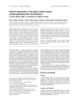
Fig. 1. Purification of EVL(1–221) and EVL(222–418). (A) Schematic
representation of the EVL fragments. Boxes denoted as EVH1,
P-rich and EVH2 represent regions corresponding to the EVH1,
Pro-rich and EVH2 domains, respectively. (B) Purification of
EVL(1–221). (C) Purification of EVL(222–418). Proteins from each
purification step were analyzed by 15% SDS ⁄ PAGE with Coomas-
sie Brilliant Blue staining. Lane 1: molecular mass markers. Lanes 2
and 3: whole cell lysates before and after induction with isopropyl
thio-b-
D-galactoside (IPTG), respectively. Lanes 4–8: samples from
the peak Ni
2+
–nitrilotriacetic acid agarose (Invitrogen) fraction, the
hydroxyapatite (Bio-Rad) flow-through or peak fraction, the fraction
after removal of the His6 tag, the peak Superdex 75 or Superdex
200 fraction (GE Healthcare), and the MonoS (GE Healthcare) flow-
through or peak fraction, respectively.
Domain analysis of human EVL M. Takaku et al.
5842 FEBS Journal 276 (2009) 5841–5848 ª 2009 The Authors Journal compilation ª 2009 FEBS
The EVH2 domain stimulates RAD51-mediated
homologous pairing
As we reported previously [11], EVL stimulates homolo-
gous pairing by RAD51. We then tested whether the
EVH2 domain also possesses this activity. To do this, we
employed the D-loop formation assay, in which an
ssDNA 50-mer and superhelical dsDNA were used as
substrates. This combination of DNA substrates gener-
ates D-loops as a product of homologous pairing by
RAD51 (Fig. 4A). As shown in Fig. 4B (lanes 4–7), EVL
significantly stimulated RAD51-mediated homologous
pairing in the presence of a suboptimal concentration of
RAD51 (50 nm). In contrast, this RAD51 stimulation by
EVL was less obvious in the presence of an optimal con-
centration of RAD51 (340 nm) (Fig. 4B, lanes 8–11).
Therefore, we employed the suboptimal RAD51 condi-
tions to evaluate the activities of the EVL domains. As
shown in Fig. 4C (compare lane 4 with lanes 8–10) and
Fig. 4D, EVL(222–418) stimulated homologous pairing
by RAD51, although its efficiency was not significant as
compared with that of full-length EVL (Fig. 4D). In con-
trast, EVL(1–221) did not stimulate homologous pairing
by RAD51 (Fig. 4E,F). These results indicate that the
EVH2 domain is responsible for the stimulation of
RAD51-mediated homologous pairing.
Discussion
EVL was originally identified as a member of the
ENA ⁄ VASP family, and it reportedly functions in
cytoplasmic actin remodeling [12]. In addition to its
cytoplasmic function, we previously found that EVL
binds to the human recombination protein, RAD51,
and stimulates RAD51-mediated homologous pairing
and strand exchange reactions in vitro [11]. EVL also
possesses DNA-binding activity [11], and was found as
a nuclear phosphoprotein by large-scale characteriza-
tion of the HeLa cell nuclear fraction [13]. These facts
suggested that EVL may be a novel factor that func-
tions in the HRR pathway. Human EVL is composed
of 418 amino acids, and contains three distinct
domains, EVH1, Pro-rich, and EVH2 [12]. To identify
the functional domain that is responsible for the
recombination-related activity of EVL, we performed a
deletion analysis with EVL, which revealed that the
EVH2 domain is responsible for the recombination
activity of EVL. We found that the EVH2 domain: (a)
contains the ssDNA-binding and dsDNA-binding
activities; (b) binds to RAD51; and (c) stimulates
homologous pairing by RAD51.
These new findings related to the recombination
activities of the EVH2 domain suggest that the ENA ⁄
VASP family proteins, which contain the EVH2
domain at their C-terminus, may be involved in DSB
repair by homologous recombination in cells. In the
EVL-knockdown cells, we previously observed a
20–30% reduction in RAD51 foci formation after
DSB induction. However, the EVL-knockdown cells
exhibited slightly increased sensitivity to a DSB-induc-
ing agent, mitomycin C (MMC) [11]. The weak MMC
sensitivity in the EVL-knockdown cells may be due to
ABC
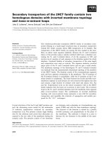
Fig. 2. DNA-binding activities of EVL(1–221) and EVL(222–418). uX174 ssDNA (20 lM) and uX174 linear dsDNA (20 lM) were each incu-
bated with the EVL protein at 37 °C for 15 min. The samples were then separated by 0.8% agarose gel electrophoresis in TAE buffer, and
were visualized by ethidium bromide staining. (A) The ssDNA-binding assay. (B) The dsDNA-binding assay. Lane 1: negative control experi-
ments without the protein. Lanes 2–4, 5–7, and 8–10: experiments with full-length EVL, EVL(1–221), and EVL(222–418), respectively. The
concentrations of the protein used in the DNA-binding experiments were 0.25 l
M (lanes 2, 5, and 8), 0.5 lM (lanes 3, 6, and 9), and 1 lM
(lanes 4, 7, and 10). (C) Competitive DNA-binding assay. uX174 ssDNA (20 lM) and uX174 linear dsDNA (20 lM) were used as substrates in
this assay. The concentrations of EVL(222–418) were 0.1 l
M (lane 2), 0.2 lM (lane 3), 0.4 lM (lane 4), 0.8 lM (lane 5), and 1.2 lM (lane 6).
Lane 1 indicates the negative control experiment without the protein.
M. Takaku et al. Domain analysis of human EVL
FEBS Journal 276 (2009) 5841–5848 ª 2009 The Authors Journal compilation ª 2009 FEBS 5843
the presence of EVL paralogs, such as the MENA and
VASP proteins. Another explanation for the weak
MMC sensitivity in the EVL-knockdown cells is that
the reduced HRR pathway may be complemented by
the nonhomologous DNA end-joining pathway, which
functions as a complementary DSB repair pathway
[14]. MENA and VASP are members of the ENA⁄
VASP family [12], and the EVH2 domain, which is
responsible for the RAD51-binding, DNA-binding and
homologous pairing stimulation, is highly conserved
between them (Fig. 5). Therefore, the EVH2 domains
of MENA and VASP may also possess these recombi-
AB
CD
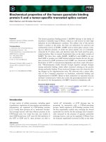
Fig. 3. EVL(222–418) binds to RAD51. (A) His6-tagged EVL, His6-tagged EVL(1–221) and His6-tagged EVL(222–418) were each incubated
with RAD51. The RAD51 protein bound to the His6-tagged EVL, EVL(1–221) or EVL(222–418) protein was captured by the Ni
2+
–nitrilotriace-
tic acid agarose beads. The proteins bound to the Ni
2+
–nitrilotriacetic acid agarose beads were then analyzed by 12% SDS ⁄ PAGE with Coo-
massie Brilliant Blue staining. Lane 1: molecular mass markers. Lanes 2–5: proteins (0.5 lg) used in this assay. Lanes 6–8: experiments with
His6-tagged EVL, His6-tagged EVL(1–221), and His6-tagged EVL(222–418), respectively. Lane 9: control experiment with RAD51 in the
absence of His6-tagged EVL, His6-tagged EVL(1–221), and His6-tagged EVL(222–418). (B) Graphic representation of the experiments shown
in (A). The averages of three independent experiments are shown with the standard deviations. In this Ni
2+
–nitrilotriacetic acid bead pull-
down assay, RAD51 nonspecifically bound to the Ni
2+
–nitrilotriacetic acid beads, and gave a background signal [(A), lane 9]. This background
signal was reduced in the presence of EVL(1–221), resulting in a negative value in (B) (1–221). (C) RAD51 titration. Lane 1: molecular mass
markers. Lanes 2 and 3: proteins (0.5 lg) used in this assay. Lanes 4–7: experiments with RAD51 in the presence of His6-tagged EVL(222–
418). Lanes 8–11: negative control experiments with RAD51 in the absence of His6-tagged EVL(222–418). The RAD51 concentrations were
2 lg (lanes 4 and 8), 4 lg (lanes 5 and 9), 6 lg (lanes 6 and 10), and 8 lg (lanes 7 and 11). (D) Graphic representation of the experiments
shown in (C). RAD51 and His6-tagged EVL(222–418), shown in (C), were quantitated. The amounts of RAD51 nonspecifically bound to the
Ni
2+
–nitrilotriacetic acid beads were subtracted to determine the amounts of RAD51 bound to EVL(222–418). The vertical axis indicates the
EVL(222–418) ⁄ RAD51 molar ratios.
Domain analysis of human EVL M. Takaku et al.
5844 FEBS Journal 276 (2009) 5841–5848 ª 2009 The Authors Journal compilation ª 2009 FEBS
nation-related activities, and may complement the
functions of EVL in vivo. A comparative study of these
ENA ⁄ VASP family proteins is required to clarify the
contributions of these proteins in the HRR pathway.
During the cytoplasmic actin-remodeling process,
the ENA ⁄ VASP family proteins bind to both the
barbed end and sides of actin filaments [15–18]. These
actin-binding activities are conserved in the EVH2
domain [19,20]. On the other hand, the EVH1 domain
binds to proteins containing the consensus sequence
D ⁄ E FPPPPXD ⁄ E [21–23]. The Pro-rich domain also
binds to profilin and proteins containing the SH3 and
WW domains [17,24–26]. These facts suggest that the
EVH1, Pro-rich and EVH2 domains all have the
potential to function in protein binding. The present
study revealed that the EVH2 domain is the RAD51-
binding domain. Minimal RAD51 binding was
observed with EVL(1–221), which contains both the
EVH1 and Pro-rich domains, also supporting the con-
clusion that the EVH2 domain is the RAD51-binding
domain in EVL. RAD51 and actin, which both bind
ATP, may share a common structural property that is
recognized by the EVH2 domain. Further studies are
required to identify the common sequence or structure
that may be recognized by EVH2.
Experimental procedures
Protein preparation
The DNA fragments encoding EVL(1–221) and EVL(222–
418) were amplified by PCR, and were cloned in the NdeI
site of the pET15b vector (Novagen, Darmstadt, Germany).
In this construct, the His6-tag sequence was fused to the
N-terminus of the protein. The EVL fragments were expressed
in the Escherichia coli BL21(DE3) strain, which also carried
an expression vector for the minor tRNAs [Codon(+)RP;
Stratagene]. The cells producing the EVL fragments were
resuspended in 20 mm potassium phosphate buffer (pH 8.5),
containing 700 mm NaCl, 5 mm 2-mercaptoethanol, 10 mm
imidazole, and 10% glycerol, and were disrupted by sonica-
tion. The cell debris was removed by centrifugation for
A
C
EF
D
B
Fig. 4. EVL(222–418) stimulates RAD51-mediated homologous pair-
ing. (A) Schematic representation of the D-loop formation assay.
Superhelical dsDNA and a 50-mer ssDNA were used as substrates
for this assay. Asterisks indicate the
32
P-labeled end of the 50-mer
ssDNA. (B) The D-loop formation assay with EVL. Lane 1: control
experiment without proteins. Lanes 2 and 3: control experiments
with EVL and EVL(222–418), respectively, in the absence of
RAD51. Lanes 4–7 and 8–11: experiments with a suboptimal
RAD51 concentration (50 n
M) and an optimal RAD51 concentration
(340 n
M), respectively. Lanes 4 and 8: experiments with 50 and
340 n
M RAD51, respectively, in the absence of EVL. Lanes 5–7 and
9–11: experiments with EVL. The EVL concentrations were 0.1 l
M
(lanes 5 and 9), 0.5 lM (lanes 6 and 10), and 1 lM (lanes 7 and 11).
(C) The D-loop formation assay with EVL(222–418). Lane 1: control
experiment without proteins. Lanes 2 and 3: control experiments
with EVL and EVL(222–418), respectively, in the absence of
RAD51. Lane 4: experiment with RAD51 in the absence of EVL or
EVL(222–418). Lanes 5–7 and 8–10: experiments with EVL and
EVL(222–418), respectively, in the presence of RAD51 (50 n
M). The
EVL and EVL(222–418) concentrations were 0.1 l
M (lanes 5 and 8),
0.5 l
M (lanes 6 and 9), and 1 lM (lanes 2, 3, 7, and 10). (D) Graphic
representation of the experiments shown in (C). Open and closed
circles represent the experiments with EVL and EVL(222–418),
respectively. The average values of three independent experiments
are shown with the standard deviations. (E) The D-loop formation
assay with EVL(1–221). The same procedure as shown in (C) was
used, except that EVL(1–221) was used instead of EVL(222–418).
(F) Graphic representation of the experiments shown in (E).
Open and closed circles represent the experiments with EVL and
EVL(1–221), respectively.
M. Takaku et al. Domain analysis of human EVL
FEBS Journal 276 (2009) 5841–5848 ª 2009 The Authors Journal compilation ª 2009 FEBS 5845
20 min at 30 000 g, and the lysates were then mixed gently
by the batch method with Ni
2+
–nitrilotriacetic acid agarose
beads (4 mL; Invitrogen, Carlsbad, CA, USA) at 4 °C for
1 h. The beads were washed with 20 mm potassium phos-
phate buffer (pH 8.5), containing 700 mm NaCl, 5 mm
2-mercaptoethanol, 30 mm imidazole, and 10% glycerol, and
were then washed with 20 mm potassium phosphate buffer
(pH 8.5), containing 700 mm NaCl, 5 mm 2-mercaptoetha-
nol, 60 mm imidazole, and 10% glycerol. The beads were
washed again with 20 mm potassium phosphate buffer (pH
8.5), containing 700 mm NaCl, 5 mm 2-mercaptoethanol,
30 mm imidazole, and 10% glycerol, and were then packed
into an Econo-column (Bio-Rad Laboratories, Hercules,
CA, USA). The Ni
2+
–nitrilotriacetic acid column was
washed with 38 column volumes of 20 mm potassium phos-
phate buffer (pH 8.5), containing 100 mm NaCl, 5 mm
2-mercaptoethanol, 30 mm imidazole, and 10% glycerol.
The His6-tagged EVL fragments were eluted with a 7.5
column volume linear gradient of 30–300 mm imidazole, in
20 mm potassium phosphate buffer (pH 8.5), containing
100 mm NaCl, 5 mm 2-mercaptoethanol, and 10% glycerol.
The fractions containing the His6-tagged EVL(1–221) frag-
ment or the His6-tagged EVL(222–418) fragment were
diluted with the same volume of 10 mm potassium phos-
phate buffer (pH 8.5), containing 100 mm NaCl, 5 mm
2-mercaptoethanol, and 10% glycerol, and were mixed
gently by the batch method with hydroxyapatite resin (5 mL;
Bio-Rad) at 4 °C for 1 h. The His6-tagged EVL(1–221) frag-
ment did not bind to the hydroxyapatite resin. Therefore,
the supernatants, which contained the His6-tagged EVL(1–
221) fragment, were collected, and the His6 tag was uncou-
pled from the EVL(1–221) portion by digestion with 4 units
of thrombin protease (GE Healthcare Biosciences, Uppsala,
Sweden) per milligram of the protein. On the other hand,
the His6-tagged EVL(222–418) fragment bound to hydroxy-
apatite resin. Therefore, the resin containing the His6-tagged
EVL(222–418) fragment was packed into an Econo-column.
The resin was further washed with 6 column volumes of
10 mm potassium phosphate buffer (pH 8.5), containing
225 mm NaCl, 5 mm 2-mercaptoethanol, and 10% glycerol,
and the His6-tagged EVL(222–418) fragment was then
eluted with a 6 column volume linear gradient of 225–
1000 mm NaCl and 10–300 mm potassium phosphate (pH
8.5). The His6 tag was uncoupled from the EVL(222–418)
portion by digestion with 4 units of thrombin protease per
milligram of the protein. The EVL(1–221) and EVL(222–
418) fragments were then immediately dialyzed against
20 mm potassium phosphate buffer (pH 7.5), containing
200 mm NaCl, 5 mm 2-mercaptoethanol, and 10% glycerol,
at 4 °C. After uncoupling of the His6 tag, the EVL(1–221)
and EVL(222–418) fragments were further purified by
Superdex 75 and Superdex 200 gel filtration column
(HiLoad 16 ⁄ 60 or HiLoad 26 ⁄ 60 prep grade; GE Health-
care) chromatography, respectively, followed by MonoS
column chromatography (MonoS HR5 ⁄ 5; GE Healthcare).
The peak fractions were diluted with two volumes of 20 mm
potassium phosphate buffer (pH 7.5), containing 5 mm
2-mercaptoethanol and 10% glycerol, and were subjected
to MonoS (GE Healthcare) column chromatography. The
column was washed with 20 column volumes of 20 mm
potassium phosphate buffer (pH 7.5), containing 67 mm
NaCl, 5 mm 2-mercaptoethanol, and 10% glycerol, and the
EVL(222–418) fragment was eluted with a four column vol-
ume linear gradient of 67–600 mm NaCl. The EVL(1–221)
fragment was obtained in the flow-through fraction of the
MonoS column, and was concentrated. The purified EVL
fragments were dialyzed against 20 mm Hepes ⁄ NaOH buf-
fer (pH 7.3), containing 100 mm NaCl, 5 mm 2-mercapto-
ethanol, and 30% glycerol, and were stored at ) 80 °C.
Human RAD51 was expressed in
E. coli cells [27], and was
purified by methods described previously [28,29]. The con-
centrations of the RAD51 and EVL proteins were determined
with the Bradford method, using BSA as the standard. The
concentrations of the EVL(1–221) and EVL(222–418) pro-
teins were determined by quantitative SDS ⁄ PAGE analysis,
using full-length EVL as the standard. We then confirmed
that the Bradford and quantitative SDS ⁄ PAGE methods
generated the same results.
Assays for DNA binding
The u X174 circular ssDNA (20 lm) or the uX174 linear
dsDNA (20 l m) was mixed with EVL, EVL(1–221) or
B
A
Fig. 5. Comparison of amino acid sequences of human ENA ⁄ VASP
family proteins. (A) Schematic representation of human EVL,
MENA, and VASP. Boxes denoted as EVH1, P-rich and EVH2 repre-
sent regions corresponding to the EVH1, Pro-rich and EVH2
domains, respectively. (B) The EVH2 sequences were aligned with
CLUSTALW.
Domain analysis of human EVL M. Takaku et al.
5846 FEBS Journal 276 (2009) 5841–5848 ª 2009 The Authors Journal compilation ª 2009 FEBS
EVL(222–418) in 10 lL of a standard reaction solution,
containing 36 mm Hepes ⁄ NaOH buffer (pH 7.5) with 1 mm
dithiothreitol, 4 mm 2-mercaptoethanol, 80 mm NaCl,
1mm MgCl
2
, 24% glycerol, and 0.1 mgÆmL
)1
BSA. The
reaction mixtures were incubated at 37 °C for 15 min, and
were then analyzed by 0.8% agarose gel electrophoresis in
1 · TAE buffer (40 mm Tris ⁄ acetate and 1 mm EDTA) at
3VÆcm
)1
for 2 h. The bands were visualized by ethidium
bromide staining.
The D-loop formation assay
In the D-loop formation assay, superhelical dsDNA
(pB5Sarray DNA) was prepared by a method that prevents
the irreversible denaturation of the dsDNA substrate by
alkaline treatment of the cells harboring the plasmid DNA.
The cells were gently lysed using sarkosyl, as described pre-
viously [30]. The pB5Sarray DNA contained 11 repeats of a
sea urchin 5S rRNA gene (207 bp fragment) within the
pBlueScript II SK(+) vector. The indicated amount of
EVL, EVL(1–221) or EVL(222–418) was incubated in the
presence of RAD51 (50 nm or 340 nm)at37°C for 5 min,
in a reaction buffer containing 26 mm Hepes ⁄ NaOH (pH
7.5), 40 mm NaCl, 0.02 mm EDTA, 0.9 mm 2-mercaptoeth-
anol, 5% glycerol, 1 m m MgCl
2
,1mm dithiothreitol, 2 mm
AMPPNP, and 0.1 mgÆmL
)1
BSA. After this incubation,
the
32
P-labeled 50-mer oligonucleotide (1 lm) was added,
and the samples were further incubated at 37 °C for 5 min.
For the ssDNA substrate used in the D-loop assay, the
following HPLC-purified oligonucleotide was purchased:
50-mer, 5¢-GGA ATT CGG TAT TCC CAG GCG GTC
TCC CAT CCA AGT ACT AAC CGA GCC CT-3¢ (Nihon
Gene Research Laboratory, Sendai, Japan). The reactions
were then initiated by the addition of the pB5Sarray super-
helical dsDNA (30 lm) along with 9 mm MgCl
2
, and were
continued at 37 °C for 30 min. The reactions were stopped
by the addition of 0.2% SDS and 1.5 mgÆmL
)1
proteinase
K, and were further incubated at 37 °C for 15 min. After
addition of six-fold loading dye, the deproteinized reaction
products were separated by 1% agarose gel electrophoresis
in 1 · TAE buffer at 3.6 VÆ cm
)1
for 2 h. The gels were
dried, exposed to an imaging plate, and visualized using an
FLA-7000 imaging analyzer (Fujifilm, Tokyo, Japan).
The pull-down assay with Ni
2+
–nitrilotriacetic
acid beads
Purified His6-tagged EVL, His6-tagged EVL(1–221) or
His6-tagged EVL(221–418) (2 lm) was mixed with RAD51
(2 lm)in50lL of binding buffer, composed of 20 mm
Hepes (pH 7.3), containing 95 mm NaCl, 0.1 mm EDTA,
0.042% Triton X-100, 1.7 mm ammonium sulfate, 2 mm
2-mercaptoethanol, 4.2 mm imidazole, and 28% glycerol.
After a 30 min incubation at room temperature, a 1.5 lL
aliquot of the Ni
2+
–nitrilotriacetic acid agarose beads was
added to the reaction mixture, and the RAD51 bound to
the His6-tagged EVL fragments was captured by the beads.
The beads were washed two times with 100 lL of washing
buffer, containing 20 mm Hepes ⁄ NaOH (pH 7.3), 90 mm
NaCl, 0.1 mm EDTA, 0.05% Triton X-100, 2 mm ammo-
nium sulfate, 2 mm 2-mercaptoethanol, 5 mm imidazole,
and 30% glycerol. The proteins that copelleted with the
Ni
2+
–nitrilotriacetic acid beads were eluted with a buffer,
containing 14 mm Hepes ⁄ NaOH (pH 7.3), 100 mm NaCl,
3.5 mm 2-mercaptoethanol, 300 mm imidazole, and 21%
glycerol. The eluted fractions were analyzed by 12%
SDS ⁄ PAGE with Coomassie Brilliant Blue staining.
Acknowledgements
This work was supported in part by a Grant-in-Aid
from the Ministry of Education, Sports, Culture, Sci-
ence, and Technology, Japan. H. Kurumizaka is a
research fellow in the Waseda Research Institute for
Science and Engineering.
References
1 Agarwal S, Tafel AA & Kanaar R (2006) DNA double-
strand break repair and chromosome translocations.
DNA Repair (Amst) 5, 1075–1081.
2 Weinstock DM, Richardson CA, Elliott B & Jasin M
(2006) Modeling oncogenic translocations: distinct roles
for double-strand break repair pathways in transloca-
tion formation in mammalian cells. DNA Repair
(Amst) 5, 1065–1074.
3 Wyman C & Kanaar R (2006) DNA double-strand
break repair: all’s well that ends well. Annu Rev Genet
40, 363–383.
4 West SC (2003) Molecular views of recombination
proteins and their control. Nat Rev Mol Cell Biol 4,
435–445.
5 Symington LS (2002) Role of RAD52 epistasis group
genes in homologous recombination and double-strand
break repair. Microbiol Mol Biol Rev 66, 630–670.
6 Sung P, Krejci L, Van Komen S & Sehorn MG (2003)
Rad51 recombinase and recombination mediators.
J Biol Chem 278, 42729–42732.
7 San Filippo J, Sung P & Klein H (2008) Mechanism of
eukaryotic homologous recombination. Annu Rev Bio-
chem 77, 229–257.
8 Modesti M, Budzowska M, Baldeyron C, Demmers JA,
Ghirlando R & Kanaar R (2007) RAD51AP1 is a struc-
ture-specific DNA binding protein that stimulates joint
molecule formation during RAD51-mediated homolo-
gous recombination. Mol Cell 28, 468–481.
9 Wiese C, Dray E, Groesser T, San Filippo J, Shi I,
Collins DW, Tsai MS, Williams GJ, Rydberg B, Sung P
et al. (2007) Promotion of homologous recombination
M. Takaku et al. Domain analysis of human EVL
FEBS Journal 276 (2009) 5841–5848 ª 2009 The Authors Journal compilation ª 2009 FEBS 5847
and genomic stability by RAD51AP1 via RAD51
recombinase enhancement. Mol Cell 28, 482–490.
10 Morozumi Y, Takizawa Y, Takaku M & Kurumizaka
H (2009) Human PSF binds to RAD51 and modulates
its homologous-pairing and strand-exchange activities.
Nucleic Acids Res 37, 4296–4307.
11 Takaku M, Machida S, Hosoya N, Nakayama S,
Takizawa Y, Sakane I, Shibata T, Miyagawa K &
Kurumizaka H (2009) Recombination activator
function of the novel RAD51- and RAD51B-binding
protein, human EVL. J Biol Chem 284, 14326–14336.
12 Kwiatkowski AV, Gertler FB & Loureiro JJ (2003)
Function and regulation of Ena ⁄ VASP proteins. Trends
Cell Biol 13, 386–392.
13 Beausoleil SA, Jedrychowski M, Schwartz D, Elias JE,
Villen J, Li J, Cohn MA, Cantley LC & Gygi SP (2004)
Large-scale characterization of HeLa cell nuclear phos-
phoproteins. Proc Natl Acad Sci USA 101, 12130–
12135.
14 Takata M, Sasaki MS, Sonoda E, Morrison C,
Hishimoto M, Utsumi H, Yamaguchi-Iwai Y,
Shinohara A & Takeda S (1998) Homologous
recombination and non-homologous end-joining
pathways of DNA double-strand break repair have
overlapping roles in the maintenance of chromosomal
integrity in vertebrate cells. EMBO J 17, 5497–5508.
15 Hu
¨
ttelmaier S, Harbeck B, Steffens O, Messerschmidt
T, Illenberger S & Jockusch BM (1999) Characteriza-
tion of the actin binding properties of the vasodilator-
stimulated phosphoprotein VASP. FEBS Lett 451,
68–74.
16 Walders-Harbeck B, Khaitlina SY, Hinssen H, Jock-
usch BM & Illenberger S (2002) The vasodilator-stimu-
lated phosphoprotein promotes actin polymerisation
through direct binding to monomeric actin. FEBS Lett
529, 275–280.
17 Krause M, Dent EW, Bear JE, Loureiro JJ & Gertler
FB (2003) Ena ⁄ VASP proteins: regulators of the actin
cytoskeleton and cell migration. Annu Rev Cell Dev Biol
19, 541–564.
18 Barzik M, Kotova TI, Higgs HN, Hazelwood L,
Hanein D, Gertler FB & Schafer DA (2005) Ena ⁄ VASP
proteins enhance actin polymerization in the presence of
barbed end capping proteins. J Biol Chem 280,
28653–28662.
19 Loureiro JJ, Rubinson DA, Bear JE, Baltus GA,
Kwiatkowski AV & Gertler FB (2002) Critical roles of
phosphorylation and actin binding motifs, but not
the central proline-rich region, for Ena ⁄ vasodilator-
stimulated phosphoprotein (VASP) function during cell
migration. Mol Biol Cell 13, 2533–2546.
20 Applewhite DA, Barzik M, Kojima S, Svitkina TM,
Gertler FB & Borisy GG (2007) Ena ⁄ VASP proteins
have an anti-capping independent function in filopodia
formation. Mol Biol Cell 18, 2579–2591.
21 Niebuhr K, Ebel F, Frank R, Reinhard M, Domann E,
Carl UD, Walter U, Gertler FB, Wehland J & Chakr-
aborty T (1997) A novel proline-rich motif present in
ActA of Listeria monocytogenes and cytoskeletal pro-
teins is the ligand for the EVH1 domain, a protein
module present in the Ena ⁄ VASP family. EMBO J 16,
5433–5444.
22 Carl UD, Pollmann M, Orr E, Gertlere FB, Chakraborty
T & Wehland J (1999) Aromatic and basic residues
within the EVH1 domain of VASP specify its interaction
with proline-rich ligands. Curr Biol 9, 715–718.
23 Fedorov AA, Fedorov E, Gertler F & Almo SC (1999)
Structure of EVH1, a novel proline-rich ligand-binding
module involved in cytoskeletal dynamics and neural
function. Nat Struct Biol 6, 661–665.
24 Gertler FB, Comer AR, Juang JL, Ahern SM, Clark
MJ, Liebl EC & Hoffmann FM (1995) Enabled, a dos-
age-sensitive suppressor of mutations in the Drosophila
Abl tyrosine kinase, encodes an Abl substrate with SH3
domain-binding properties. Genes Dev 9, 521–533.
25 Ermekova KS, Zambrano N, Linn H, Minopoli G,
Gertler F, Russo T & Sudol M (1997) The WW domain
of neural protein FE65 interacts with proline-rich
motifs in Mena, the mammalian homolog of Drosophila
enabled. J Biol Chem 272, 32869–32877.
26 Ahern-Djamali SM, Bachmann C, Hua P, Reddy SK,
Kastenmeier AS, Walter U & Hoffmann FM (1999)
Identification of profilin and src homology 3 domains
as binding partners for Drosophila enabled. Proc Natl
Acad Sci USA 96, 4977–4982.
27 Kurumizaka H, Aihara H, Kagawa W, Shibata T &
Yokoyama S (1999) Human Rad51 amino acid residues
required for Rad52 binding. J Mol Biol 291, 537–548.
28 Matsuo Y, Sakane I, Takizawa Y, Takahashi M &
Kurumizaka H (2006) Roles of the human Rad51 L1
and L2 loops in DNA binding. FEBS J 273, 3148–3159.
29 Ishida T, Takizawa Y, Sakane I & Kurumizaka H
(2008) The Lys313 residue of the human Rad51 protein
negatively regulates the strand-exchange activity. Genes
Cells 13, 91–103.
30 Kagawa W, Kurumizaka H, Ikawa S, Yokoyama S &
Shibata T (2001) Homologous pairing promoted by the
human Rad52 protein. J Biol Chem 276, 35201–35208.
Domain analysis of human EVL M. Takaku et al.
5848 FEBS Journal 276 (2009) 5841–5848 ª 2009 The Authors Journal compilation ª 2009 FEBS