Báo cáo khoa học: Viral entry mechanisms: the increasing diversity of paramyxovirus entry doc
Bạn đang xem bản rút gọn của tài liệu. Xem và tải ngay bản đầy đủ của tài liệu tại đây (589.13 KB, 11 trang )
MINIREVIEW
Viral entry mechanisms: the increasing diversity of
paramyxovirus entry
Everett C. Smith, Andreea Popa, Andres Chang, Cyril Masante and Rebecca Ellis Dutch
Department of Molecular and Cellular Biochemistry, University of Kentucky, Lexington, KY, USA
Introduction
The paramyxovirus family is composed of enveloped,
negative-stranded RNA viruses, many of which are
major human pathogens [1]. Members of this family
include human respiratory syncytial virus (hRSV), the
leading cause of viral lower respiratory tract infections
in infants and children worldwide, and the measles
virus, which remains a significant source of morbidity
and mortality in developing countries. In recent years,
a number of new paramyxoviruses have been recog-
nized, including the Hendra and Nipah viruses, which
are highly pathogenic in humans and are the only
identified zoonotic members of the paramyxovirus
family [2].
Paramyxoviruses contain between six and ten genes,
encoding proteins involved in critical processes such as
transcription ⁄ replication (large polymerase, nucleocap-
sid, phosphoprotein), assembly (matrix protein) and
viral entry. Paramyxovirus entry into target cells is
mediated by two glycoproteins present on the viral
membrane: the attachment protein (termed HN for
hemagglutinin-neuraminidase, H for hemagglutinin, or
G for glycoprotein, depending on the virus) and the
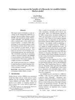
fusion (F) protein (Fig. 1A). Recent examination by
cryo-electron microscopy indicated that these glycopro-
teins are packed in a dense layer on the viral surface
[3]. Primary adsorption of the virus to the target cell is
Keywords
fusion proteins; paramyxovirus; receptor
binding; viral entry
Correspondence
R. E. Dutch, Department of Molecular and
Cellular Biochemistry, University of
Kentucky, College of Medicine, Biomedical
Biological Sciences Research Building,
741 South Limestone, Lexington,
KY 40536-0509, USA
Fax: +1 859 323 1037
Tel: +1 859 323 1795
E-mail:
(Received 17 June 2009, revised 11
September 2009, accepted 22 September
2009)
doi:10.1111/j.1742-4658.2009.07401.x
The paramyxovirus family contains established human pathogens such as
the measles virus and human respiratory syncytial virus, as well as emerg-
ing pathogens including the Hendra and Nipah viruses and the recently
identified human metapneumovirus. Two major envelope glycoproteins, the
attachment protein and the fusion protein, promote the processes of viral
attachment and virus-cell membrane fusion required for entry. Although
common mechanisms of fusion protein proteolytic activation and the mech-
anism of membrane fusion promotion have been shown in recent years,
considerable diversity exists in the family relating to receptor binding and
the potential mechanisms of fusion triggering.
Abbreviations
F, fusion; G, glycoprotein; H, hemagglutinin; HMPV, human metapneumovirus; HN, hemagglutinin-neuraminidase; hPIV3, human
parainfluenza virus 3; HRA, heptad repeat A; HRB, heptad repeat B; hRSV, human respiratory syncytial virus; N, neuraminidase; NDV,
Newcastle Disease virus; PIV5, parainfluenza virus 5; SLAM, signal lymphocyte-activating molecule.
FEBS Journal 276 (2009) 7217–7227 ª 2009 The Authors Journal compilation ª 2009 FEBS 7217
generally promoted by the attachment protein, with
sialic acid residues or cell surface proteins serving as
receptors. The F protein is then responsible for fusion
of the viral membrane with a host cell membrane.
Paramyxovirus F proteins are trimeric type I integral
membrane proteins initially synthesized as nonfuso-
genic F
0
precursors, which require subsequent cleavage
into the fusogenic disulfide-linked F
1
+F
2
heterodimer
(Fig. 1B). This cleavage event places the conserved
fusion peptide at the N-terminus of the newly-created
F
1
subunit, priming the protein for fusion activity.
Most paramyxoviruses require their homotypic attach-
ment protein for membrane fusion activity, suggesting
a role for F-attachment protein interactions in control
of fusion [4–9]. The Hendra and Nipah F proteins
interchangeably utilize the Hendra and Nipah G pro-
teins in the fusion process, and this fully functional
bidirectional heterotypic fusion activity is unique
among paramyxoviruses [10]. Interestingly, some para-
myxovirus fusion proteins can promote membrane
fusion in the absence of their homotypic attachment
protein [8,11,12], making the role of paramyxovirus
attachment proteins in membrane fusion unclear and
potentially virus specific. Despite varying sequence
homology among paramyxoviruses and the diverse
requirement for the attachment protein, the positional
conservation of a number of structural elements sug-
gests a similar mechanism of fusion. Membrane fusion
is considered to be driven by very large conformational
changes [13] following the triggering of the F protein,
leading to exposure and insertion of the fusion peptide
into the target membrane and subsequent fusion of the
viral and cellular membranes.
Attachment proteins and receptors
For the majority of paramyxoviruses, interaction of the
attachment protein with a cellular receptor is necessary
for virus binding to target cells, and for the triggering
of F protein-promoted fusion. All paramyxovirus
attachment proteins characterized to date are type II
integral membrane proteins that form homotetramers
[1,14] (Fig. 1B). Attachment protein nomenclature is
defined by two characteristics: (a) the ability or inabil-
ity to bind sialic acid and (b) the presence or absence
of neuramidase activity (or the ability to cleave sialic
acid). The Respirovirus, Rubulavirus and Avulavirus
attachment proteins are denoted HN, because they
bind sialic acid-containing glycoproteins or glycolipids
on the cell surface (H activity) and also remove sialic
acid from carbohydrates on viral glycoproteins and
other cell surface molecules (N activity), thus prevent-
ing viral self-agglutination during budding [15]. The
HN proteins differ in their binding affinity for varying
sialic acid-containing molecules [15], likely contributing
to their differing pathogenesis. The Morbillivirus
attachment proteins (H) lack N activity and utilize pro-
tein cellular receptors instead of sialic acid. Measles
virus H binds to CD46 or signal lymphocyte-activating
molecule (SLAM) receptors [16,17], potentially
accounting for the restriction of measles infection to
higher primates. Down-regulation of CD46 and SLAM
A
B
Fig. 1. Schematic of paramyxovirus virion
and surface glycoproteins. (A) Schematic of
a paramyxovirus; viral membrane shown in
blue. (B) Conserved domains of paramyxo-
virus fusion and attachment proteins.
Domain abbreviations: fusion peptide
(FP, orange); HRA (blue); HRB (red);
transmembrane domain (TMD, black);
cytoplasmic tail (C-Tail, dotted box);
disulfide bond (S-S).
The increasing diversity of paramyxovirus entry Everett C. Smith et al.
7218 FEBS Journal 276 (2009) 7217–7227 ª 2009 The Authors Journal compilation ª 2009 FEBS
in infected cells presumably prevents viral aggregation
during budding [18]. The Pneumovirus and Henipavi-
rus attachment proteins lack both H and N activity,
and are therefore termed G (for glycoprotein) proteins.
The Hendra and Nipah G proteins have been shown to
bind EphrinB2 and EphrinB3 cellular receptors [19,20].
The hRSV G protein has been shown to bind heparin
[21] and cell surface proteoglycans [22].
The crystal structures of a number of paramyxovirus
attachment proteins have been determined, including
the HN proteins from Newcastle Disease virus (NDV),
parainfluenza virus 5 (PIV5) and human parainfluenza
virus 3 (hPIV3), the H protein from measles virus and
the G protein from Nipah virus [23–29]. In all cases, a
C-terminal globular head that contains the receptor
binding and the enzymatic activity site is observed to
sit on top of a membrane-proximal stalk domain. The
globular head is composed of four identical monomers
arranged with four-fold symmetry, with each of the
monomers consisting of a six-blade b-propeller fold
[23–28]. For the majority of HN proteins, a single bind-
ing site on top of the globular head domain has both H
and N activity [24]. However, NDV HN has been
demonstrated to contain two sialic acid binding sites:
one in the globular head and one at an interface
between two dimers [28]. Interestingly, for measles virus
H protein, the CD46 ⁄ SLAM binding sites are located
toward the sides of the H protein b-barrel [26,29]. This
altered placement of the receptor binding domain led
to the suggestion that differences in sialic acid versus
protein receptor binding may lead to different mecha-
nisms of fusion initiation [30]. However, the binding
site for ephrinB2 ⁄ B3 on Nipah G was recently shown
to reside at the top of the globular head domain, in a
similar position to HN protein sialic acid binding sites,
and a co-complex with ephrin-B3 revealed extensive
protein–protein interactions, including the insertion of
a portion of ephrin-B3 into the central cavity of Nipah
G [27]. Thus, conserved positioning of the binding site
is seen for at least some protein-binding and sialic-acid
binding attachment proteins.
Interestingly, recent data suggest that the Pneumo-
virus attachment protein may not be obligatory for
attachment and entry in all cases. An attenuated hRSV
missing the G protein or hRSV and bovine respiratory
syncytial virus recombinants lacking the G protein
were found to replicate in cell culture [31–33], indicat-
ing that the RSV F protein can provide sufficient bind-
ing to allow viral entry. Similarly, the G protein from
the recently identified human metapneumovirus
(HMPV) has been shown to be dispensible for growth
in both cell culture and animal models [34]. The hRSV
F protein has been shown to bind to heparin [35],
although a recombinant hRSV virus lacking the G
protein has been found to be less dependent on glyco-
saminoglycans for attachment than the wild-type virus
[36], suggesting interactions with a receptor in addition
to glycosaminoglycans. No specific receptor for the
RSV F protein has been identified, although a recent
study indicates a role for aVb1 integrin-HMPV F pro-
tein interactions in HMPV entry [37]. Finally, studies
have shown that the human asialoglycoprotein recep-
tor (a mammalian lectin) may be an attachment factor
for the Sendai F protein [38]. Thus, it is possible that
the process of paramyxovirus attachment may be more
complex than had previously been considered, poten-
tially involving interactions beyond those of the well-
characterized attachment protein-receptor. Interaction
between the F protein and the target cell might allow
for a final selection step prior to triggering fusion.
Proteolytic processing of
paramyxovirus F proteins
Proteolytic processing of the nonfusogenic precursor
forms (F
0
) of paramyxovirus fusion proteins into the
disulfide-linked heterodimer F
1
+F
2
is essential for for-
mation of fusogenically active proteins because it
primes the protein for fusion by positioning the fusion
peptide at the newly-formed N-terminus of F
1
[39].
Although the requirement for proteolytic processing is
conserved among paramyxoviruses, the protease
responsible for cleavage of the F
0
precursor varies.
Many paramyxovirus F proteins are cleaved during
transport through the trans-Golgi network by the
ubiquitous subtilisin-like cellular protease, furin [40].
Furin-mediated proteolytic cleavage occurs following
R-X-K ⁄ R-R sequences and has been demonstrated to
occur in the F proteins of several paramyxoviruses,
including hRSV [41], PIV5 [40] and mumps virus [42].
Interestingly, hRSV F has recently been shown to
undergo two N-terminal furin-mediated cleavage
events, both of which are required for fusion promo-
tion [43,44]. The Hendra and Nipah F proteins, how-
ever, lack the R-X-K ⁄ R-R consensus sequence for
furin-mediated cleavage. Instead, both the Hendra and
Nipah F proteins are cleaved by the endosomal ⁄ lyso-
somal protease cathepsin L following a single basic
residue in the N-terminal sequences VGDVK
109
and
VGDVR
109
, respectively [45–47]. Finally, some viral F
proteins, including F proteins from HMPV [48,49]
and Sendai virus [50], are cleaved by tissue-specific
extracellular proteases such as tryptase Clara and mini-
plasmin. Despite containing a minimal furin cleavage
sequence (R-X-X-R), HMPV is not cleaved intracellu-
larly but requires exogenous protease addition for
Everett C. Smith et al. The increasing diversity of paramyxovirus entry
FEBS Journal 276 (2009) 7217–7227 ª 2009 The Authors Journal compilation ª 2009 FEBS 7219
activation [51,52], although intracellular cleavage has
been observed in laboratory-expanded strains [52].
Regardless of the protease responsible for F cleav-
age, this step is essential for both virulence and patho-
genicity. The presence of single or multiple basic
residues has been demonstrated to modulate proteo-
lytic processing and thus acts to determine pathogen
virulence. NDV F proteins containing multiple basic
residues in proximity to the cleavage site are more
virulent and exhibit higher levels of dissemination
throughout the host compared to their F counterparts
containing only one basic residue [53,54]. Proteolytic
cleavage of F proteins can also result in structural
rearrangement because peptide antibodies directed to
the PIV5 heptad repeats recognize primarily the
uncleaved form [55]. Interestingly, insertion of both
multi-basic cleavage sites present in RSV F into Sendai
F leads to a decreased dependency on the Sendai
attachment protein and increased cell–cell fusion [56].
Thus, cleavage of viral F proteins constitutes a pivotal
point in the viral life cycle affecting both pathogenesis
and virulence, most likely by reducing the energy
required to promote the structural rearrangements of
the protein required for membrane fusion activity.
Triggering of membrane fusion
Many viral fusion proteins contain both receptor-bind-
ing and fusion activities, suggesting a straightforward
model indicating how fusion is triggered by receptor
binding. However, the separation of these two func-
tions in paramyxoviruses makes the control of fusion
triggering more complex. Fusion-associated conforma-
tional changes in the F protein are considered to be
irreversible, leading to a nonfusogenically active post-
fusion form of the protein. Thus, it is extremely impor-
tant that triggering is properly regulated both spatially
and temporally [57]. The majority of paramyxovirus F
proteins promote membrane fusion at neutral pH, with
the exception of F proteins from certain HMPV strains
that were shown to be triggered by exposure to low
pH [11,58]. Thus, alterations in pH are not the univer-
sal trigger for paramyxovirus F protein fusion. Sub-
stantial evidence suggests that, for most members of
the family, fusion triggering involves specific inter-
actions of the cleaved, metastable F protein with its
homotypic attachment protein [59–64]. Upon receptor
binding, the attachment protein ‘transmits’ a signal
to the F protein, potentially through conformational
changes in the attachment protein and ⁄ or changes
in the F protein–attachment protein interaction.
Structural analysis of the NDV HN protein suggested
significant conformational changes upon ligand bind-
ing [23,28], although similar changes were not observed
in the PIV5 or hPIV3 HN following sialic acid binding
[24,25], or in Nipah G following ephrin B3 binding
[27]. Thus, a model where receptor engagement results
in subtle rearrangements and reposition of the fusion
and attachment proteins has been proposed [27].
The requirement for a homotypic attachment protein
for fusion triggering suggests a specific interaction
between the fusion and attachment proteins, and con-
siderable research has focused on characterizing the
physical interaction between these key proteins. Both
co-immunoprecipitation studies and antibody-induced
co-capping analyses have demonstrated interactions
for the fusion and attachment proteins from a number
of paramyxoviruses [59,60,62,64,65]. Numerous studies
indicate that the membrane proximal stalk domain of
the attachment protein is important for interaction
with the fusion protein [6,9,65–68], but residues present
in the globular head domain [60,69,70] or the trans-
membrane domain [14,71] have also been implicated.
Studies have also indicated a role for the F protein
TM-proximal heptad repeat B (HRB) region [72] or a
region within the F protein globular head [73] in these
critical glycoprotein interactions.
Triggering of F protein-promoted membrane fusion
is clearly also modulated by factors beyond the attach-
ment protein. A number of F protein mutations have
been shown to affect fusion triggering and ⁄ or the
requirement for a homotypic attachment protein. The
NDV F protein requires its homotypic HN protein,
although a single amino acid change (L289A) [12] can
remove this requirement in some cell types [74]. Substi-
tution of the extended hRSV cleavage-site into the
Sendai F protein can modulate attachment protein
dependence [56]. Mutations in the cytoplasmic tail of
the SER virus have also been found to confer HN
independence to this F protein [75]. Several specific
regions in paramyxovirus F proteins have also been
implicated in triggering, including the linker region
immediately preceding HRB [76,77], portions of hep-
tad repeat A (HRA) [78] and a conserved region of
F
2
that interacts with HRA in the prefusion form [79].
The F protein from the PIV5 strain WR, which
normally requires the presence of an HN protein for
function, can promote HN-independent membrane
fusion when present at elevated temperature [80], sug-
gesting that the requirement for HN triggering of F
can also be replaced by conditions which destabilize
the F protein. For the HMPV F protein, low pH can
efficiently trigger fusion for some strains, and no
requirement for an attachment protein is observed
[11,58]. Additionally, hRSV, PIV5 strain W3A and
Sendai virus F proteins can also mediate membrane
The increasing diversity of paramyxovirus entry Everett C. Smith et al.
7220 FEBS Journal 276 (2009) 7217–7227 ª 2009 The Authors Journal compilation ª 2009 FEBS
fusion even in the absence of their attachment protein
[36,38,81], suggesting that their F proteins have a lower
energy requirement to transition from their metastable
state [39], and do not require the presence of an attach-
ment protein to stabilize the prefusion form.
The time and place where the fusion and attachment
proteins interact is critical to understanding the mecha-
nism of fusion control, but the details of these inter-
actions are still under investigation, and may vary
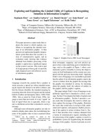
between viruses. One proposed model (Fig. 2, Model 1)
suggests that the initial interaction between the two
glycoproteins occurs within the endoplasmic reticu-
lum at the time of synthesis, potentially allowing the
attachment protein to hold the F protein in its prefu-
sion conformation until after receptor binding. Studies
of measles virus [82,83] and NDV [62] support this
model, although recent studies of the Henipavirus gly-
coproteins suggest differential trafficking through the
secretory pathway [84,85]. In addition, fusion proteins
that do not require their attachment protein for func-
tion do not fit this model because they clearly maintain
their prefusion state independently. The fusion and
attachment proteins may instead traffic separately
through the secretory pathway, arriving at the cell
Fig. 2. Potential mechanisms of paramyxovirus fusion protein triggering. Attachment protein shown with orange head domain and blue stalk;
fusion protein shown in blue ⁄ green head domain and red stalk region; receptor shown in grey.
Everett C. Smith et al. The increasing diversity of paramyxovirus entry
FEBS Journal 276 (2009) 7217–7227 ª 2009 The Authors Journal compilation ª 2009 FEBS 7221
surface independently. Interaction could then occur,
with subsequent disruption of the F protein–attachment
protein interaction by receptor binding leading to
fusion triggering (Fig. 2, Model 2). Recent studies of
Hendra and Nipah fusion support this model because
it was shown that G mutations that inhibit F–G inter-
action also inhibit the fusion process [66], and that
fusion promotion also correlates inversely with F–G
avidity [59,60]. Alternatively, an interaction between
the two proteins may not occur until after the attach-
ment protein binds its receptor (Fig. 2, Model 3).
Interactions between the NDV F and HN protein have
been demonstrated only in the presence of receptor,
and mutations that alter receptor binding decrease
both fusion and F–HN interactions [86,87], supporting
this model. Finally, the attachment protein is not
required to interact with F for fusion promotion in
some cases, although receptor binding likely facilitates
the process by bringing the two membranes into close
proximity (Fig. 2, Model 4). The HMPV F protein has
replaced the requirement for an attachment protein
with a low pH-induced triggering [11], with electro-
static repulsion in the HRB linker domain shown to be
critical for the triggering process [77]. It is unclear
which factors drive triggering of other attachment
protein-independent paramyxovirus fusion proteins.
Paramyxovirus F protein-mediated
membrane fusion
Fusion between the viral envelope and cell membrane
presents a daunting challenge for enveloped viruses.
To drive membrane merger, the virus must provide
sufficient energy to deform opposing bilayers, ulti-
mately resulting in the formation of a fusion pore and
the release of the viral genome inside the cell (Fig. 3A).
Promotion of this energetically demanding process is
driven by viral fusion proteins, including HIV envelope
protein, influenza virus HA and the paramyxovirus F
proteins, which act as molecular machines driving
fusion through a series of dramatic conformational
changes [88]. Despite little sequence homology between
these disparate class I fusion proteins, all share com-
mon features, including glycosylation, trimerization,
the need for proteolytic cleavage and conserved
sequence motifs [39]. Thus, it is likely that they medi-
ate membrane fusion through very similar mecha-
nisms.
Paramyxovirus F proteins, similar to other class I
fusion proteins, are present in their metastable, prefu-
sion conformation prior to fusion activation [88]. Sub-
sequent to proteolytic processing and triggering, a
series of conformational changes lead to the formation
of a more stable, post-fusion form of the protein, with
the energy released utilized to drive the fusion process.
An understanding of paramyxovirus F protein-medi-
ated membrane fusion has increased greatly with the
elucidation of the crystal structures of the prefusion
form of the PIV5 F protein [89] and of the postulated
postfusion forms of the NDV and hPIV3 F proteins
[90–92]. Despite these advances, many important ques-
tions related to key intermediates remain. Research to
date on a number of paramyxovirus F proteins sug-
gests a model for membrane fusion that demonstrates
the importance of key conserved regions within the F
protein (Fig. 3B). In the prefusion form, the HRA
A
B
Fig. 3. Models of lipid and protein fusion intermediates. (A) Lipid intermediates culminating in the formation of a full fusion pore. (B) Pro-
posed fusion protein intermediates with subsequent formation of the post-fusion six-helix bundle. FP, orange; HRA, blue; HRB, red; TMD,
black.
The increasing diversity of paramyxovirus entry Everett C. Smith et al.
7222 FEBS Journal 276 (2009) 7217–7227 ª 2009 The Authors Journal compilation ª 2009 FEBS
domains (Fig. 3B, blue) are separated, the hydrophobic
fusion peptide is buried, and the HRB regions
(Fig. 3B, red) interact in a coiled-coil conformation.
Subsequent to triggering, conformational changes
result in the release of the fusion peptide, formation of
a long HRA coiled-coil, and subsequent insertion of
the fusion peptide into the target membrane [93]. The
HRB regions separate, and subsequent refolding leads
to formation of a hairpin structure that positions HRB
in an anti-parallel fashion within the grooves of the
HRA trimeric coiled-coil. It is hypothesized that the
formation of this six-helix bundle complex provides at
least a portion of the energy required for the merging
of the lipid bilayers [13]. Subsequently, the fusion pore
expands, and this expansion step is hypothesized to be
the most energetically costly stage of the membrane
fusion process [94].
Route of paramyxovirus entry
Enveloped viruses can enter cells either via receptor-
mediated endocytosis or by direct fusion between the
viral envelope and the plasma membrane. Viruses that
require low pH for fusion, such as the influenza virus
and vesicular stomatitis virus, utilize the cellular endo-
cytic machinery to enter cells as vesicles from the
major endocytic pathways converge into acidified
endosomes [95]. Other viruses such as Ebola require
endocytosis to expose their fusion proteins to pH-
dependent proteases before membrane fusion can
occur [96,97]. In these cases, virus–cell fusion occurs
somewhere within the endocytic pathway. Viruses
with pH-independent fusion proteins, such as para-
myxoviruses and retroviruses, are generally considered
to enter cells at the plasma membrane because the
majority of viruses from these families can efficiently
infect cells in the presence of agents such as ammo-
nium chloride that raise the endosomal pH. However,
recent studies suggest that some viruses with pH-inde-
pendent fusion proteins may still utilize endosomal
entry routes [98]. Most paramyxovirus F proteins can
induce cell–cell fusion when expressed on the cell sur-
face at neutral pH, leading to the formation of giant
multinucleated cells termed syncytia. These experi-
ments clearly indicate that the triggering for most
paramyxovirus F proteins is pH-independent, with
the exception of the HMPV F protein [11]. However,
these experiments do not directly address the site of
virus–cell fusion.
Although paramyxoviruses have generally been con-
sidered to enter at the plasma membrane, recent evi-
dence points towards a more complex mechanism of
cell entry for at least some members of the family.
Internalization of viral particles prior to fusion has
been noted for Sendai virus [99] and Nipah virus [100].
Chemical agents that sequester cholesterol have
recently been shown to disrupt NDV infection, indicat-
ing that this paramyxovirus could be utilizing caveolin-
mediated endocytosis as an entry pathway [101]. Endo-
cytosis has also been implicated in hRSV entry because
hRSV infection is decreased in cells expressing siRNAs
against key components of the clatrhin-mediated endo-
cytosis pathway, namely the clathrin light chain, the
clathrin-adapter complex, dynamin 3, and the small
GTPase Rab5A. Further experiments utilizing chemi-
cal inhibitors as well as dominant negative proteins
further support the hypothesis that hRSV may at least
partially utilize clathrin-dependent endocytosis to
establish an active infection [102]. Recent work indi-
cates that HMPV may utilize the cellular endocytic
machinery for entry because treatment with chlor-
promazine, an inhibitor of clathrin-mediated endo-
cytosis, conferred protection against this virus.
Furthermore, dynasore, a small molecule inhibitor of
dynamin, comprising a protein required in the final
step of vesicle formation in both clathrin- and caveo-
lin-mediated endocytosis, was highly effective in block-
ing HMPV infection, reducing infection levels by up to
90% [77]. For some strains, HMPV F protein trigger-
ing is strongly stimulated by low pH [11], suggesting a
role for the lower endosomal pH in entry, and inhibi-
tors of endosomal acidification such as bafilomycin
A1, concanamycin, ammonium chloride and monensin
have all shown some efficacy in preventing HMPV
infection [77]. Thus, to date, at least some members of
the paramyxovirus family appear to utilize endocytic
entry routes. Endosomal entry could potentially pro-
tect viruses from the host immune system and provide
unique environments, in addition to lowered pH, that
assist in productive infection. Further work is needed
to more fully characterize the entry pathways utilized
by paramyxoviruses.
Acknowledgements
We thank the members of the Dutch laboratory for
their careful reading of the manuscript. This work was
supported by NIAID ⁄ NIH grants R01AI051517 and
R21AI074783 to R.E.D.
References
1 Lamb RA & Parks GD (2007) Paramyxoviridae: the
viruses and their replication. In Fields Virology (Knipe
DM & Howley PM eds), pp. 1449–1496. Lippincott,
Williams and Wilkins, Philadelphia, PA.
Everett C. Smith et al. The increasing diversity of paramyxovirus entry
FEBS Journal 276 (2009) 7217–7227 ª 2009 The Authors Journal compilation ª 2009 FEBS 7223
2 Eaton BT, Broder CC, Middleton D & Wang LF
(2006) Hendra and Nipah viruses: different and dan-
gerous. Nat Rev Microbiol 4, 23–35.
3 Ludwig K, Schade B, Bottcher C, Korte T, Ohlwein N,
Baljinnyam B, Veit M & Herrmann A (2008) Electron
cryomicroscopy reveals different F1+F2 protein states
in intact parainfluenza virions. J Virol 82, 3775–3781.
4 Tong S & Compans RW (1999) Alternative mecha-
nisms of interaction between homotypic and hetero-
typic parainfluenza virus HN and F proteins. J Gen
Virol 80, 107–115.
5 Cattaneo R & Rose JK (1993) Cell fusion by the enve-
lope glycoproteins of persistent measles viruses which
cause lethal human brain disease. J Virol 67, 1493–
1502.
6 Deng R, Wang Z, Mirza AM & Iorio RM (1995)
Localization of a domain on the paramyxovirus
attachment protein required for the promotion of
cellular fusion by its homologous fusion protein spike.
Virol 209, 457–469.
7 Ebata SN, Cote MJ, Kang CY & Dimock K (1991)
The fusion and hemagglutinin-neuraminidase glycopro-
teins of human parainfluenza virus 3 are both required
for fusion. Virol 183, 437–441.
8 Horvath CM & Lamb RA (1992) Studies on the
fusion peptide of a paramyxovirus fusion glycopro-
tein: roles of conserved residues in cell fusion. J Virol
66, 2443–2455.
9 Tanabayashi K & Compans RW (1996) Functional
interaction of paramyxovirus glycoproteins: identifica-
tion of a domain in Sendai virus HN which promotes
cell fusion. J Virol 70, 6112–6118.
10 Bossart KN, Wang LF, Flora MN, Chua KB, Lam
SK, Eaton BT & Broder CC (2002) Membrane fusion
tropism and heterotypic functional activities of the
Nipah virus and Hendra virus envelope glycoproteins.
J Virol 76, 11186–11198.
11 Schowalter RM, Smith SE & Dutch RE (2006) Char-
acterization of human metapneumovirus F protein-
promoted membrane fusion: critical roles for proteo-
lytic processing and low pH. J Virol 80, 10931–10941.
12 Sergel TA, McGinnes LW & Morrison TG (2000) A
single amino acid change in the Newcastle disease
virus fusion protein alters the requirement for HN
protein in fusion. J Virol 74, 5101–5107.
13 Baker KA, Dutch RE, Lamb RA & Jardetzky TS
(1999) Structural basis for paramyxovirus-mediated
membrane fusion. Mol Cell 3, 309–319.
14 McGinnes L, Sergel T & Morrison T (1993) Mutations
in the transmembrane domain of the HN protein of
Newcastle disease virus affect the structure and activity
of the protein. Virol 196, 101–110.
15 Villar E & Barroso IM (2006) Role of sialic acid-
containing molecules in paramyxovirus entry into the
host cell: a minireview. Glycoconj J 23, 5–17.
16 Dorig RE, Marcil A, Chopra A & Richardson CD
(1993) The human CD46 molecule is a receptor for
measles virus (Edmonston strain). Cell 75, 295–305.
17 Tatsuo H, Ono N, Tanaka K & Yanagi Y (2000)
SLAM (CDw150) is a cellular receptor for measles
virus. Nature 406, 893–897.
18 Welstead GG, Hsu EC, Iorio C, Bolotin S &
Richardson CD (2004) Mechanism of CD150
(SLAM) down regulation from the host cell surface
by measles virus hemagglutinin protein. J Virol 78,
9666–9674.
19 Bonaparte MI, Dimitrov AS, Bossart KN, Crameri G,
Mungall BA, Bishop KA, Choudhry V, Dimitrov DS,
Wang LF, Eaton BT et al. (2005) Ephrin-B2 ligand is
a functional receptor for Hendra virus and Nipah
virus. Proc Natl Acad Sci USA 102
, 10652–10657.
20 Negrete OA, Levroney EL, Aguilar HC, Bertolotti-
Ciarlet A, Nazarian R, Tajyar S & Lee B (2005)
EphrinB2 is the entry receptor for Nipah virus, an
emergent deadly paramyxovirus. Nature 436, 401–405.
21 Krusat T & Streckert HJ (1997) Heparin-dependent
attachment of respiratory syncytial virus (RSV) to host
cells. Arch Virol 142, 1247–1254.
22 Escribano-Romero E, Rawling J, Garcia-Barreno B &
Melero JA (2004) The soluble form of human respira-
tory syncytial virus attachment protein differs from the
membrane-bound form in its oligomeric state but is
still capable of binding to cell surface proteoglycans.
J Virol 78, 3524–3532.
23 Crennell S, Takimoto T, Portner A & Taylor G (2000)
Crystal structure of the multifunctional paramyxovirus
hemagglutinin-neuraminidase. Nat Struct Biol 7, 1068–
1074.
24 Yuan P, Thompson TB, Wurzburg BA, Paterson RG,
Lamb RA & Jardetzky TS (2005) Structural studies of
the parainfluenza virus 5 hemagglutinin-neuraminidase
tetramer in complex with its receptor, sialyllactose.
Structure 13, 803–815.
25 Lawrence MC, Borg NA, Streltsov VA, Pilling PA,
Epa VC, Varghese JN, McKimm-Breschkin JL &
Colman PM (2004) Structure of the haemagglutinin-
neuraminidase from human parainfluenza virus type
III. J Mol Biol 335, 1343–1357.
26 Hashiguchi T et al. (2007) Crystal structure of
measles virus hemagglutinin provides insight into
effective vaccines. Proc Natl Acad Sci USA 104,
19535–19540.
27 Xu K, Rajashankar KR, Chan YP, Himanen JP,
Broder CC & Nikolov DB (2008) Host cell recognition
by the henipaviruses: crystal structures of the Nipah G
attachment glycoprotein and its complex with ephrin-
B3. Proc Natl Acad Sci USA 105, 9953–9958.
28 Zaitsev V, von Itzstein M, Groves D, Kiefel M,
Takimoto T, Portner A & Taylor G (2004) Second
sialic acid binding site in Newcastle disease virus
The increasing diversity of paramyxovirus entry Everett C. Smith et al.
7224 FEBS Journal 276 (2009) 7217–7227 ª 2009 The Authors Journal compilation ª 2009 FEBS
hemagglutinin-neuraminidase: implications for fusion.
J Virol 78, 3733–3741.
29 Colf LA, Juo ZS & Garcia KC (2007) Structure of the
measles virus hemagglutinin. Nat Struct Mol Biol 14,
1227–1228.
30 Iorio RM & Mahon PJ (2008) Paramyxoviruses:
different receptors - different mechanisms of fusion.
Trends Microbiol 16, 135–137.
31 Karron RA et al. (1997) Respiratory syncytial virus
(RSV) SH and G proteins are not essential for viral
replication in vitro: clinical evaluation and molecular
characterization of a cold-passaged, attenuated RSV
subgroup B mutant. Proc Natl Acad Sci USA 94,
13961–13966.
32 Karger A, Schmidt U & Buchholz UJ (2001) Recombi-
nant bovine respiratory syncytial virus with deletions
of the G or SH genes: G and F proteins bind heparin.
J Gen Virol 82, 631–640.
33 Techaarpornkul S, Barretto N & Peeples ME (2001)
Functional analysis of recombinant respiratory syncy-
tial virus deletion mutants lacking the small hydro-
phobic and ⁄ or attachment glycoprotein gene. J Virol
75, 6825–6834.
34 Biacchesi S, Pham QN, Skiadopoulos MH, Murphy
BR, Collins PL & Buchholz UJ (2005) Infection of
nonhuman primates with recombinant human meta-
pneumovirus lacking the SH, G, or M2-2 protein cate-
gorizes each as a nonessential accessory protein and
identifies vaccine candidates. J Virol 79, 12608–12613.
35 Feldman SA, Audet S & Beeler JA (2000) The fusion
glycoprotein of human respiratory syncytial virus
facilitates virus attachment and infectivity via an
interaction with cellular heparan sulfate. J Virol 74,
6442–6447.
36 Techaarpornkul S, Collins PL & Peeples ME (2002)
Respiratory syncytial virus with the fusion protein as
its only viral glycoprotein is less dependent on cellular
glycosaminoglycans for attachment than complete
virus. Virol 294, 296–304.
37 Cseke G, Maginnis MS, Cox RG, Tollefson SJ,
Podsiad AB, Wright DW, Dermody TS & Williams JV
(2009) Integrin alphavbeta1 promotes infection by
human metapneumovirus. Proc Natl Acad Sci USA
106, 1566–1571.
38 Leyrer S, Bitzer M, Lauer U, Kramer J, Neubert WJ
& Sedlmeier R (1998) Sendai virus-like particles devoid
of haemagglutinin-neuraminidase protein infect cells
via the human asialoglycoprotein receptor. J Gen Virol
79, 683–687.
39 Dutch RE, Jardetsky TS & Lamb RA (2000) Virus
membrane fusion proteins: biological machines that
undergo a metamorphosis. Biosci Rep 20, 597–612.
40 Garten W, Hallenberger S, Ortmann D, Schafer W,
Vey M, Angliker H, Shaw E & Klenk HD (1994)
Processing of viral glycoproteins by the subtilisin-like
endoprotease furin and its inhibition by specific
peptidylchloroalkylketones. Biochimie 76, 217–225.
41 Ortmann D, Ohuchi M, Angliker H, Shaw E, Garten
W & Klenk H-D (1994) Proteolytic cleavage of wild
type and mutants of the F protein of human parainflu-
enza virus type 3 by two subtilisin-like endoproteases,
furin and KEX2. J Virol 68, 2772–2776.
42 Watanabe M, Hirano A, Stenglein S, Nelson J,
Thomas G & Wong TC (1995) Engineered serine
protease inhibitor prevents furin-catalyzed activation
of the fusion glycoprotein and production of infectious
measles virus. J Virol 69, 3206–3210.
43 Begona Ruiz-Arguello M et al. (2002) Effect of proteo-
lytic processing at two distinct sites on shape and
aggregation of an anchorless fusion protein of human
respiratory syncytial virus and fate of the intervening
segment. Virol 298, 317–326.
44 Gonzalez-Reyes L et al. (2001) Cleavage of the human
respiratory syncytial virus fusion protein at two
distinct sites is required for activation of membrane
fusion. Proc Natl Acad Sci USA 98
, 9859–9864.
45 Diederich S, Moll M, Klenk HD & Maisner A (2005)
The nipah virus fusion protein is cleaved within the
endosomal compartment. J Biol Chem 280, 29899–
29903.
46 Pager CT, Craft WW Jr, Patch J & Dutch RE (2006)
A mature and fusogenic form of the Nipah virus
fusion protein requires proteolytic processing by
cathepsin L. Virol 346, 251–257.
47 Pager CT & Dutch RE (2005) Cathepsin L is involved
in proteolytic processing of the Hendra virus fusion
protein. J Virol 79, 12714–12720.
48 van den Hoogen BG, de Jong JC, Groen J, Kuiken T,
de Groot R, Fouchier RA & Osterhaus AD (2001) A
newly discovered human pneumovirus isolated from
young children with respiratory tract disease. Nat Med
7, 719–724.
49 Biacchesi S, Skiadopoulos MH, Yang L, Lamirande
EW, Tran KC, Murphy BR, Collins PL & Buchholz
UJ (2004) Recombinant human Metapneumovirus
lacking the small hydrophobic SH and ⁄ or attachment
G glycoprotein: deletion of G yields a promising
vaccine candidate. J Virol 78, 12877–12887.
50 Murakami M, Towatari T, Ohuchi M, Shiota M, Akao
M, Okumura Y, Parry MA & Kido H (2001) Mini-
plasmin found in the epithelial cells of bronchioles
triggers infection by broad-spectrum influenza A viruses
and Sendai virus. Eur J Biochem 268, 2847–2855.
51 Biacchesi S, Pham QN, Skiadopoulos MH, Murphy
BR, Collins PL & Buchholz UJ (2006) Modification of
the trypsin-dependent cleavage activation site of the
human metapneumovirus fusion protein to be trypsin
independent does not increase replication or spread in
rodents or nonhuman primates. J Virol 80, 5798–5806.
Everett C. Smith et al. The increasing diversity of paramyxovirus entry
FEBS Journal 276 (2009) 7217–7227 ª 2009 The Authors Journal compilation ª 2009 FEBS 7225
52 Schickli JH, Kaur J, Ulbrandt N, Spaete RR & Tang
RS (2005) An S101P substitution in the putative
cleavage motif of the human metapneumovirus fusion
protein is a major determinant for trypsin-independent
growth in vero cells and does not alter tissue tropism
in hamsters. J Virol 79 , 10678–10689.
53 Nagai Y & Klenk H-D (1977) Activation of precursors
to both glycoproteins of Newcastle disease virus by
proteolytic cleavage. Virol 77, 125–134.
54 Nagai Y, Klenk H-D & Rott R (1976) Proteolytic cleav-
age of the viral glycoproteins and its significance for the
virulence of Newcastle disease virus. J Virol 20, 501–508.
55 Dutch RE, Hagglund RN, Nagel MA, Paterson RG &
Lamb RA (2001) Paramyxovirus fusion (F) protein: a
conformational change on cleavage activation. Virol
281, 138–150.
56 Rawling J, Garcia-Barreno B & Melero JA (2008)
Insertion of the two cleavage sites of the respiratory
syncytial virus fusion protein in Sendai virus fusion
protein leads to enhanced cell-cell fusion and a
decreased dependency on the HN attachment protein
for activity. J Virol 82 , 5986–5998.
57 Lamb RA (1993) Paramyxovirus fusion: a hypothesis
for changes. Virol 197, 1–11.
58 Herfst S, Mas V, Ver LS, Wierda RJ, Osterhaus AD,
Fouchier RA & Melero JA (2008) Low pH induced
membrane fusion mediated by human metapneumo-
viruses F protein is a rare, strain dependent phenome-
non. J Virol 82, 8891–8895.
59 Aguilar HC, Matreyek KA, Choi DY, Filone CM,
Young S & Lee B (2007) Polybasic KKR motif in the
cytoplasmic tail of Nipah virus fusion protein
modulates membrane fusion by inside-out signaling.
J Virol 81, 4520–4532.
60 Bishop KA et al. (2007) Identification of hendra virus
g glycoprotein residues that are critical for receptor
binding. J Virol 81, 5893–5901.
61 Plemper RK, Hammond AL, Gerlier D, Fielding AK
& Cattaneo R (2002) Strength of envelope protein
interaction modulates cytopathicity of measles virus.
J Virol 76, 5051–5061.
62 Stone-Hulslander J & Morrison TG (1997) Detection
of an interaction between the HN and F proteins in
Newcastle disease virus-infected cells. J Virol 71, 6287–
6295.
63 Takimoto T, Taylor GL, Connaris HC, Crennell SJ &
Portner A (2002) Role of the hemagglutinin-neuramin-
idase protein in the mechanism of paramyxovirus-cell
membrane fusion. J Virol 76 , 13028–13033.
64 Yao Q, Hu X & Compans RW (1997) Association of
the parainfluenza virus fusion and hemagglutinin-
neuraminidase glycoproteins on cell surfaces. J Virol
71, 650–656.
65 Melanson VR & Iorio RM (2006) Addition of N-gly-
cans in the stalk of the Newcastle disease virus HN
protein blocks its interaction with the F protein and
prevents fusion. J Virol 80, 623–633.
66 Bishop KA et al. (2008) Residues in the stalk domain
of the hendra virus g glycoprotein modulate conforma-
tional changes associated with receptor binding.
J Virol 82, 11398–11409.
67 Porotto M, Murrell M, Greengard O & Moscona A
(2003) Triggering of human parainfluenza virus 3
fusion protein (F) by the hemagglutinin-neuraminidase
(HN) protein: an HN mutation diminishes the rate of
F activation and fusion. J Virol 77, 3647–3654.
68 Sergel T, McGinnes LW, Peeples ME & Morrison TG
(1993) The attachment function of the Newcastle
disease virus hemagglutinin-neuraminidase protein can
be separated from fusion promotion by mutation.
Virol 193, 717–726.
69 Aguilar HC, Ataman ZA, Aspericueta V, Fang AQ,
Stroud M, Negrete OA, Kammerer RA & Lee B (2009)
A novel receptor-induced activation site in the Nipah
virus attachment glycoprotein (G) involved in triggering
the fusion glycoprotein (F). J Biol Chem 284, 1628–1635.
70 Mirza AM, Deng R & Iorio RM (1994) Site-directed
mutagenesis of a conserved hexapeptide in the para-
myxovirus hemagglutinin-neuraminidae glycoprotein:
effects on antigenic structure and function. J Virol 68,
5093–5099.
71 Bousse T, Takimoto T, Gorman WL, Takahashi T &
Portner A (1994) Regions on the hemagglutinin-
neuraminidase proteins of human parainfluenza virus
type-1 and Sendai virus important for membrane
fusion. Virol 204, 506–514.
72 Gravel KA & Morrison TG (2003) Interacting
domains of the HN and F proteins of newcastle
disease virus. J Virol 77, 11040–11049.
73 Lee JK, Prussia A, Paal T, White LK, Snyder JP &
Plemper RK (2008) Functional interaction between
paramyxovirus fusion and attachment proteins. J Biol
Chem 283, 16561–16572.
74 Li J, Melanson VR, Mirza AM & Iorio RM (2005)
Decreased dependence on receptor recognition for the
fusion promotion activity of L289A-mutated newcastle
disease virus fusion protein correlates with a mono-
clonal antibody-detected conformational change.
J Virol 79, 1180–1190.
75 Seth S, Vincent A & Compans RW (2003) Mutations
in the cytoplasmic domain of a paramyxovirus fusion
glycoprotein rescue syncytium formation and eliminate
the hemagglutinin-neuraminidase protein requirement
for membrane fusion. J Virol 77, 167–178.
76 Russell CJ, Kantor KL, Jardetzky TS & Lamb RA
(2003) A dual-functional paramyxovirus F protein
regulatory switch segment: activation and membrane
fusion. J Cell Biol 163, 363–374.
77 Schowalter RM, Chang A, Robach JG, Buchholz UJ
& Dutch RE (2009) Low-pH triggering of human
The increasing diversity of paramyxovirus entry Everett C. Smith et al.
7226 FEBS Journal 276 (2009) 7217–7227 ª 2009 The Authors Journal compilation ª 2009 FEBS
metapneumovirus fusion: essential residues and
importance in entry. J Virol 83, 1511–1522.
78 Luque LE & Russell CJ (2007) Spring-loaded heptad
repeat residues regulate the expression and activation
of paramyxovirus fusion protein. J Virol 81, 3130–
3141.
79 Gardner AE & Dutch RE (2007) A conserved region
in the F(2) subunit of paramyxovirus fusion proteins is
involved in fusion regulation. J Virol 81, 8303–8314.
80 Paterson RG, Russell CJ & Lamb RA (2000) Fusion
protein of the paramyxovirus SV5: destabilizing and
stabilizing mutants of fusion activation. Virol 270,
17–30.
81 Feldmann H, Kretzschmar E, Klingeborn B, Rott R,
Klenk H-D & Garten W (1988) The structure of sero-
type H10 hemagglutinin of influenza A virus: Compar-
ison of an apathogenic avian and a mammalian strain
pathogenic for mink. Virol 165, 577–583.
82 Corey EA & Iorio RM (2009) Measles virus attach-
ment proteins with impaired ability to bind CD46
interact more efficiently with the homologous fusion
protein. Virol 383, 1–5.
83 Plemper RK, Hammond AL & Cattaneo R (2001)
Measles virus envelope glycoproteins hetero-oligomer-
ize in the endoplasmic reticulum. J Biol Chem 276,
44239–44246.
84 Whitman SD & Dutch RE (2007) Surface density of
the Hendra G protein modulates Hendra F protein-
promoted membrane fusion: Role for Hendra G pro-
tein trafficking and degradation. Virol 363, 419–429.
85 Whitman SD, Smith EC & Dutch RE (2009) Differen-
tial rates of protein folding and cellular trafficking for
the Hendra virus F and G proteins: implications for
F-G complex formation. J Virol 83, 8998–9001.
86 Corey EA & Iorio RM (2007) Mutations in the stalk
of the measles virus hemagglutinin protein decrease
fusion but do not interfere with virus-specific inter-
action with the homologous fusion protein. J Virol 81,
9900–9910.
87 Melanson VR & Iorio RM (2004) Amino acid substi-
tutions in the F-specific domain in the stalk of the
newcastle disease virus HN protein modulate fusion
and interfere with its interaction with the F protein.
J Virol 78, 13053–13061.
88 White JM, Delos SE, Brecher M & Schornberg K
(2008) Structures and mechanisms of viral membrane
fusion proteins: multiple variations on a common
theme. Crit Rev Biochem Mol Biol 43, 189–219.
89 Yin HS, Wen X, Paterson RG, Lamb RA & Jardetzky
TS (2006) Structure of the parainfluenza virus 5 F
protein in its metastable, prefusion conformation.
Nature 439, 38–44.
90 Chen L, Gorman JJ, McKimm-Breschkin J, Lawrence
LJ, Tulloch PA, Smith BJ, Colman PM & Lawrence
MC (2001) The structure of the fusion glycoprotein of
Newcastle disease virus suggests a novel paradigm for
the molecular mechanism of membrane fusion. Struc-
ture (Camb) 9, 255–266.
91 Colman PM & Lawrence MC (2003) The structural
biology of type I viral membrane fusion. Nat Rev Mol
Cell Biol 4 , 309–319.
92 Yin HS, Paterson RG, Wen X, Lamb RA & Jardetzky
TS (2005) Structure of the uncleaved ectodomain of
the paramyxovirus (hPIV3) fusion protein. Proc Natl
Acad Sci USA 102, 9288–9293.
93 Asano K & Asano A (1985) Why is a specific amino
acid sequence of F glycoprotein required for the mem-
brane fusion reaction between envelope of HVJ (Sen-
dai virus) and target cell membranes? Biochem Int 10,
115–122.
94 Chernomordik LV, Zimmerberg J and Kozlov MM
(2006) Membranes of the world unite!
J Cell Biol 175,
201–207.
95 Pelkmans L, Bu
¨
rli T, Zerial M & Helenius A (2004)
Caveolin-stabilized membrane domains as multifunc-
tional transport and sorting devices in endocytic
membrane traffic. Cell 118, 767–780.
96 Chandran K, Sullivan NJ, Felbor U, Whelan SP &
Cunningham JM (2005) Endosomal Proteolysis of the
Ebola Virus Glycoprotein Is Necessary for Infection.
Science.
97 Schornberg K, Matsuyama S, Kabsch K, Delos S,
Bouton A & White J (2006) Role of endosomal
cathepsins in entry mediated by the Ebola virus
glycoprotein. J Virol 80, 4174–4178.
98 Miyauchi K, Kim Y, Latinovic O, Morozov V &
Melikyan GB (2009) HIV enters cells via endocytosis
and dynamin-dependent fusion with endosomes. Cell
137, 433–444.
99 Rasmusson BJ, Flanagan TD, Turco SJ, Epand RM &
Petersen NO (1998) Fusion of Sendai virus and indi-
vidual host cells and inhibition of fusion by lipo-
phosphoglycan measured with image correlation
spectroscopy. Biochimica et Biophysica Acta (BBA) –
Molecular Cell Research 1404, 338–352.
100 Diederich S, Thiel L & Maisner A (2008) Role of
endocytosis and cathepsin-mediated activation in
Nipah virus entry. Virol 375, 391–400.
101 Cantin C, Holguera J, Ferreira L, Villar E & Munoz-
Barroso I (2007) Newcastle disease virus may enter
cells by caveolae-mediated endocytosis. J Gen Virol 88,
559–569.
102 Kolokoltsov AA, Deniger D, Fleming EH, Roberts NJ
Jr, Karpilow JM & Davey RA (2007) Small interfering
RNA profiling reveals key role of clathrin-mediated
endocytosis and early endosome formation for
infection by respiratory syncytial virus. J Virol 81,
7786–7800.
Everett C. Smith et al. The increasing diversity of paramyxovirus entry
FEBS Journal 276 (2009) 7217–7227 ª 2009 The Authors Journal compilation ª 2009 FEBS 7227