Báo cáo khoa học: Lack of stabilized microtubules as a result of the absence of major maps in CAD cells does not preclude neurite formation pot
Bạn đang xem bản rút gọn của tài liệu. Xem và tải ngay bản đầy đủ của tài liệu tại đây (556.72 KB, 14 trang )
Lack of stabilized microtubules as a result of the absence
of major maps in CAD cells does not preclude neurite
formation
C. Gasto
´
n Bisig
1
, Marı
´
a E. Chesta
1
, Guillermo G. Zampar
1
, Silvia A. Purro
1
, Vero
´
nica S. Santander
2
and Carlos A. Arce
1
1 Centro de Investigaciones en Quı
´
mica Biolo
´
gica de Co
´
rdoba (CIQUIBIC), UNC-CONICET, Departamento de Quı
´
mica Biolo
´
gica, Facultad de
Ciencias Quı
´
micas, Universidad Nacional de Co
´
rdoba, Argentina
2 Departamento de Biologı
´
a Molecular, Facultad de Ciencias Exactas, Fı
´
sico-Quı
´
micas y Naturales, Universidad Nacional de Rı
´
o Cuarto,
Argentina
Introduction
Correct functioning of the nervous system requires the
proper development of neuronal circuits and the estab-
lishment of synapses. Although neurons from different
regions of the nervous system acquire diverse morpho-
Keywords
CAD cells; microtubule-associated proteins;
microtubule dynamics; microtubules;
neurites
Correspondence
C. A. Arce, Centro de Investigaciones en
Quı
´
mica Biolo
´
gica de Co
´
rdoba (CIQUIBIC),
UNC-CONICET, Departamento de Quı
´
mica
Biolo
´
gica, Facultad de Ciencias Quı
´
micas,
Universidad Nacional de Co
´
rdoba,
5000-Co
´
rdoba, Argentina
Fax: +54 0351 4334074
Tel: +54 0351 000000
E-mail:
(Received 19 May 2009, revised
28 September 2009, accepted 2 October
2009)
doi:10.1111/j.1742-4658.2009.07422.x
In many laboratories, the requirement of microtubule-associated proteins
(MAPs) and the stabilization of microtubules for the elongation of neurites
has been intensively investigated, with controversial results being obtained.
We have observed that the neurite microtubules of Cath.a-differentiated
(CAD) cells, a mouse brain derived cell, are highly dynamic structures, and
so we analyzed several aspects of the cytoskeleton to investigate the molecu-
lar causes of this phenomenon. Microtubules and microfilaments were pres-
ent in proportions similar to those found in brain tissue and were
distributed similarly to those in normal neurons in culture. Neurofilaments
were also present. Analysis of tubulin isospecies originating from post-trans-
lational modifications revealed an increased amount of tyrosinated tubulin,
a diminished amount of the detyrosinated form and a lack of the Delta2
form. This tyrosination pattern is in agreement with highly dynamic micro-
tubules. Using western blot analyses with specific antibodies, we found that
CAD cells do not express several MAPs such as MAP1b, MAP2, Tau, dou-
blecortin, and stable-tubule-only-peptide. The presence of the genes corre-
sponding to these MAPs was verified. The absence of the corresponding
mRNAs confirmed the lack of expression of these proteins. The exception
was Tau, whose mRNA was present. Among the several MAPs investigated,
LIS1 was the only one to be expressed in CAD cells. In addition, we
determined that neurites of CAD cells form and elongate at the same rate
as processes in a primary culture of hippocampal neurons. Treatment with
nocodazol precluded the formation of neurites, and induced the retraction
of previously formed neurites. We conclude that the formation and elon-
gation of neurites, at least in CAD cells, are dependent on microtubule
integrity but not on their stabilization or the presence of MAPs.
Abbreviations
CAD, Cath.a-differentiated; dCAD, diffentiated CAD; MAP, microtubule-associated protein; STOP, stable-tubule-only-peptide; TSA,
trichostatin A.
7110 FEBS Journal 276 (2009) 7110–7123 ª 2009 The Authors Journal compilation ª 2009 FEBS
logies and abilities, there are certain basic features
common to all neurons (e.g. the initiation and elonga-
tion of membrane protrusions for neurite formation,
and their stabilization and differentiation into
dendrites and axons). Other processes, such as the
organization of the internal cytoskeleton, migration,
guidance, and selective synaptogenesis, differ depend-
ing on the type of neuron [1,2].
The cytoskeleton is a critical structure for the elon-
gation of neurites and the maintenance of neuronal
architecture [3], and the stabilization of microtubules is
considered to be essential for neurons to maintain their
asymmetry and to transport materials required for
neurite elongation [4,5]. The mechanism by which neu-
rons regulate microtubule assembly, stability, and
interactions with other cell structures is considered to
depend on the presence of microtubule-associated pro-
teins (MAPs), among which the most prominent are
MAP1b, MAP2, Tau, and stable-tubule-only-peptide
(STOP) [6–9]. There are also other MAPs that have
been studied to a lesser extent (spectraplakins, adeno-
matous polyposis coli, doublecortin, dishevelled), as
well as other proteins binding to the plus end of micro-
tubules that could be involved in this process [10–12].
Transfection analyses have shown that Tau and
MAP2 induce the elongation of processes of non-neu-
ronal cells [13,14]. Suppressed expression of MAP1b,
MAP2, and Tau using antisense and siRNA technol-
ogy in several studies [15–17] caused a reduction of
neurite outgrowth. The microinjection of anti-Tau
antibodies into cultured neurons did not inhibit axonal
extension [18]. Tau knockout mice showed a decreased
number of microtubules in small-diameter axons, but
extended axons were indistinguishable from those of
wild-type controls [19]. MAP1b deficient mice show an
abnormal brain architecture, whereas, in MAP2 defi-
cient mice, the cytoarchitecture was normal, suggesting
an overlapping function of MAP2 with MAP1b [20].
Lack of functional alteration in cases when only one
gene for MAP was silenced was generally attributed to
other proteins that provide additional redundancy with
MAP functions [21–25]. The conflicting conclusions
made in different studies may be related to the use of
different technologies or different cell or tissue systems,
or the presence of MAPs with redundant functions. In
any case, a requirement of MAPs and stabilized micro-
tubules for neurite formation has not yet been clearly
demonstrated. In the present study, we characterized
cytoskeleton and neurite formation in Cath.a-differen-
tiated (CAD) cells, adding new information regarding
this particular subject.
CAD originated as a subclone of the cathecolamin-
ergic cell line CATH.a, which was derived from a
neuronal brain tumor in a transgenic mouse expressing
SV40 large T antigen under the control of the tyrosine
hydroxylase promoter [26]. CAD cells proliferate with
a rounded or polygonal shape in the presence of
serum. When serum is removed, they stop proliferating
and differentiate, acquiring a neuron-like morphology,
and, when serum is re-added, a rapid shortening of
neurites is observed, such that most cells present a
rounded morphology within approximately 40 min
[27]. Studies from several laboratories have shown that
these cells contain synaptic vesicle proteins and express
neuron-specific proteins such as b-tubulin III, GAP-43,
SNAP-25, synaptostagmin, and other neuropeptides
[27,28]. The intracellular traffic powered by kinesins
and dynein in these cells functions similarly to other
neuronal systems [29–31]. After differentiation, cell
processes contain numerous varicosities similar to
those of neurons [27,32]. Single-cell electrophysiologi-
cal studies have demonstrated that CAD cells can be
induced to fire action potentials, and that voltage-
dependent sodium and potassium currents can be
elicited [33].
The rapid retraction of neurites after the addition of
serum led us to consider the possibility that the cyto-
skeleton of CAD cells should have peculiar properties.
Thus, in the present study, we investigated the main
constituents of this structure and found that neurites
have highly dynamic microtubules and lack stabilized
microtubules because major MAPs are not expressed
in these cells. However, neurites elongate at the same
rate as those of normal neurons in culture.
Results
Cytoskeletal proteins
As noted in the Introduction, the stabilization of
microtubules is recognized as an essential process dur-
ing the elongation of neurites, presumably to assure
cell asymetry and the transport of materials to the
growth cone. Consistently, this process of stabilization
has been described in several types of neurons in cul-
ture [10]. The rapid shortening of CAD cell neurites
after the addition of serum led us to presume that
there are alterations in the cytoskeleton. Accordingly,
we investigated the presence, amount, and distribution
of the main components of this structure. Immunofluo-
rescence using specific antibodies revealed that actin
microfilaments (Fig. 1A) are present in CAD cells dis-
playing the typical localization, positioned along the
shaft and in the apical region of the growth cone pre-
ceding the microtubules (Fig. 1A). The three major
cytoplasmic growth cone domains [i.e. central (C),
C. G. Bisig et al. Neurite formation in CAD cells
FEBS Journal 276 (2009) 7110–7123 ª 2009 The Authors Journal compilation ª 2009 FEBS 7111
transition (T) and peripheral (P) zones] can be clearly
distinguished (Fig. 1A). The localization of these pro-
teins is the same as that previously reported in
cultured hyppocampal cells [10]. Western blot and
subsequent determination of optical density confirmed
the presence of actin in CAD cells (Fig. 1B, lanes 1
and 2) in an actin ⁄ tubulin proportion slightly higher
than that determined in mouse brain [0.16 ± 0.05 and
0.12 ± 0.03 for diffentiated CAD (dCAD) cells and
brain, respectively, n = 3]. We found no significant
difference in the amount of the neurofilament 100 kDa
constituent in relation to tubulin in CAD cells com-
pared to mouse brain (Fig. 1B, lanes 3 and 4).
Acetylation and tyrosination states of tubulin
The tubulin molecule is subject to a variety of post-
translational modifications [14,34]. One of them com-
prises the reversible acetylation of its a-chain at the
e-amino group of Lys40 [35]. Although its physiological
role is unclear, we have previously presented evidence
demonstrating that acetylation is necessary for tubulin
to interact with Na,K-ATPase [36]. In living cells, acet-
ylated and deacetylated tubulin coexist in variable pro-
portions depending on the cell type [37]. Microtubules
containing a high degree of acetylated tubulin were
found to be more stable [35]. In addition, microtubules
DE
C
AB
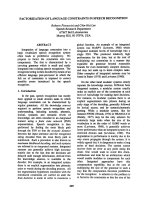
Fig. 1. Tubulin, actin, and neurofilament
protein expression in CAD cells. (A) CAD
cells differentiated for 5 days were stained
for double immunofluorescence using rhoda-
mine-conjugated phalloidin to detect actin
microfilaments (Actin) and anti-total tubulin
(Tubulin) to detect microtubules. The
merged image shows that actin microfila-
ments invades the growth cone, whereas
microtubules remain behind. The central (C),
transition (T) and peripheral (P) zones are
also indicated. Scale bar = 5 lm. (B) CAD
cells (80% confluence) were differentiated
for 5 days, collected, and dissolved in
Laemmli’s sample buffer for immunoblot in
parallel with samples of mouse brain tissue.
Blots were stained simultaneously with anti-
tubulin (DM1A) and anti-actin (lanes 1 and
2). Other samples were stained with anti-
neurofilament protein (lanes 3 and 4). The
volume of each sample was adjusted to
load a similar amount of tubulin. (C) CAD
cells differentiated for 5 days were treated
with 5 l
M TSA for 0, 3, and 6 h, and imme-
diately processed for immunofluorescence
with anti-acetylated tubulin (clone 6-11B-1).
(D) CAD cells differentiated for 5 days were
treated with TSA for the indicated times and
immunoblotted with anti-acetylated- and
anti-total-tubulin. (E) CAD cells differentiated
for 5 days were treated for 12 h with 10 l
M
Taxol. A control without Taxol was also run.
Cells were collected and processed for wes-
tern blotting using antibodies against acety-
lated and total tubulin. The lane labeled
+Taxol was overloaded to highlight the
absence of acetylated tubulin.
Neurite formation in CAD cells C. G. Bisig et al.
7112 FEBS Journal 276 (2009) 7110–7123 ª 2009 The Authors Journal compilation ª 2009 FEBS
were shown to be the preferred substrate for the acety-
lating enzyme [35]. We found that the acetylated form
of tubulin was essentially absent in CAD cells (Fig. 1C,
t = 0; Fig. 1D, lane 0). This could be a result of the
predominance of highly dynamic microtubules versus
stable microtubules, to the predominance of tubulin-
deacetylase activity (histone deacetylase 6) versus tubu-
lin-acetyltransferase activity, or to absence or inhibition
of the latter enzyme. Treatment of cells with the non-
specific deacetylase inhibitor trichostatin A (TSA)
resulted in the appearance of a significant amount of
acetylated tubulin (Fig. 1C, 3 and 6 h; Fig. 1D), indi-
cating that both acetylase and deacetylase were present
in CAD cells. Treatment of cells with Taxol induces an
increment in acetylated microtubules because the acetyl
transferase acts preferentially on these structures [35].
Stabilization of microtubules by treating CAD cells
with 10 lm Taxol did not cause increase of acetylated
tubulin (Fig. 1E), indicating that the acetylation state
of tubulin depends mainly on the relative activities of
the acetylating and deacetylating enzymes rather than
on microtubule dynamics.
Tyrosination ⁄ detyrosination at the COOH-terminus
of a-tubulin is another post-translational modification
that has been extensively studied, although its physio-
logical role also remains unclear [38–40]. As a result of
this cyclic modification, different isotypes of tubulin
exist: tyrosinated (Tyr-tubulin), detyrosinated (Glu-
tubulin), and Delta2 (a-tubulin lacking the two
COOH-terminal amino acids). Glu-tubulin and Delta2-
tubulin have been used as markers of stable micro-
tubules [41]. Immunofluorescence images of CAD cells
using an antibody against total tubulin (which does
not discriminate different states of tubulin tyrosina-
tion) showed a bright, typical microtubule network in
the cell body and neurites (Fig. 2A). A similar pattern
was observed using an antibody specific to tyrosinated
tubulin. Antibody against Glu-tubulin revealed scarce,
curly microtubules, whereas antibody against Delta2-
tubulin revealed no microtubules. These results were
confirmed by immunoblots using the same antibodies
(Fig. 2B). Mouse brain tissue was used as a positive
control. The reduced amount of Glu-tubulin in CAD
cells was not a result of a lack of (or inhibition of) the
putative detyrosinating enzyme (tubulin carboxypepti-
dase) because a significant increase of Glu-tubulin was
observed in differentiated and nondifferentiated cells
treated with Taxol (Fig. 2C).
Microtubule dynamics
The rapid shortening of neurites found in CAD cells,
along with the absence of markers of stable micro-
tubules (Glu-tubulin, and Delta2-tubulin), led us to
consider the possibility that microtubules are highly
dynamic structures in these cells. By measuring the
rate of microtubule depolymerization after nocodazole
treatment [4,5], microtubule dynamics in CAD cells
was compared with that of other cell types. Micro-
tubules of CAD cells were as dynamic as those of
Chinese hamster ovary and PC12 cells in active prolif-
eration. The time required for 50% depolymerization
was 1–2 min (Fig. 3A, B, empty circles). On the other
hand, the depolymerization curve for 7-day-old
chicken embryo brain cells showed a two-phase behav-
ior, suggesting the presence of two microtubule popu-
lations: one with a half-life of 1–2 min and the other
being more stable (Fig. 3A, B, solid triangle). As a
negative control of microtubule disassembly by noco-
dazole treatment, CAD cells were pre-treated with
sodium azide, which stabilizes microtubules by deplet-
ing cells of ATP [42]. Under these conditions, microtu-
bules were not disassembled by nocodazole treatment
(Fig. 3A, bottom; Fig. 3B, solid circles).
Several MAPs are not expressed in CAD cells
From a mechanistic point of view, there is a general
consensus that MAPs are the proteins responsible of
microtubule stabilization [10,22,43,44]. Thus, we
investigated whether the occurrence of highly dynamic
microtubules in CAD cells is the result of some alter-
ation in one or more MAPs. The presence of neuro-
nal structural MAPs (i.e. MAP1b, MAP2, Tau, and
STOP) was investigated in 10-day-differentiated CAD
cells by immunoblotting using appropriate antibodies.
In the case of Tau, immunoblots were revealed with
antibodies that recognize dephosphorylated and phos-
phorylated epitopes and a nonphosphorylable region
of the protein (Tau-1, Tau-2 and 134d). For compari-
son, soluble fractions from 30-day-old mouse brain
were simultaneously run. All the MAPs investigated
were present in brain samples, but not in samples
from CAD cells (Fig. 4). Brain and CAD samples
run in each lane contained similar amounts of
a-tubulin (Fig. 4, lower panels). The experiment was
repeated, running overloaded samples of CAD cells
and using a more sensitive chemiluminescent method
(Femtomolar detection system), with similar results
being obtained (i.e. no band was observed in lanes
corresponding to CAD cells). This is exemplified by
an overloaded dCAD cell sample being revealed with
134d antibody (Fig. 4, lane dCAD ⁄ Overload). More-
over, treatment of nitrocellulose membrane with alka-
line phosphatase prior to incubation with anti-
Tau-1, aiming to increase the epitopes that can be
C. G. Bisig et al. Neurite formation in CAD cells
FEBS Journal 276 (2009) 7110–7123 ª 2009 The Authors Journal compilation ª 2009 FEBS 7113
recognized, produced a significant increase in Tau
bands in the Br lane, but no band appeared in the
dCAD lane (Fig. S1).
To concentrate MAPs eventually diluted in the cell
extract, we performed immunoprecipitation with
Sepharose beads linked to antibodies specific to each
MAP. As a control, mouse brain samples were also
analyzed in parallel. The amount of the brain soluble
fraction and dCAD cell extract used in these experi-
ments as input material was 30-fold higher than those
loaded on each lane shown in Fig. 4. For each MAP,
most of the protein in brain samples was found in the
pellet, whereas, in dCAD cells samples, no MAP band
was observed (not shown).
Gene and mRNA analyses of MAP1b, MAP2, Tau,
and STOP
The finding that apparently normal neurites are formed
even when CAD cells lack MAP1b, MAP2, Tau, and
STOP proteins was surprising. This led us to investigate
the presence of their respective genes and messenger
RNAs, using a PCR technique with specifically designed
primers (Table 1). In every case, the PCR products
A
C
B
Fig. 2. Tyrosination state of tubulin. (A)
Cells differentiated for 5 days (dCAD) and
nondifferentiated cells (CAD) were visual-
ized by immunofluorescence using antibod-
ies specific to a-tubulin (total tubulin), Tyr-,
Glu-, and Delta2-tubulin. The inset shows
embryonic chicken brain cells differentiated
for 6 days in culture and revealed with anti-
body to Delta2-tubulin. Scale bar = 10 lm.
(B) Cells obtained as in (A) were subjected
to western blotting and stained with the
same antibodies as in (C). For staining with
each antibody, identical volumes of CAD
cell samples were run. As positive controls,
samples of mouse brain (Br) were included.
(C) Nondifferentiated CAD cells and cells
differentiated for 7 days were treated (+) or
not ()) with 10 l
M Taxol for 12 h and then
subjected to western blotting using
antibody to detyrosinated tubulin (Glu-
tubulin). All lanes were loaded with samples
containing the same amount of total tubulin.
Neurite formation in CAD cells C. G. Bisig et al.
7114 FEBS Journal 276 (2009) 7110–7123 ª 2009 The Authors Journal compilation ª 2009 FEBS
AB
Fig. 3. Nocodazole sensitivity of microtubules of CAD and different cell types. CAD cells differentiated for 7 days, PC12 cells (80% conflu-
ence), Chinese hamster ovary cells (80% confluence), a primary culture of 7-day-old chicken brain cells, and differentiated CAD cells treated
with 20 m
M sodium azide (in culture medium without glucose) for 1 h, were incubated in the presence of 10 lM nocodazole for the indicated
times and immediately processed to isolate the cytoskeletal fraction, which remained bound to the plastic dish (see Experimental proce-
dures). The cytoskeletal fraction remaining after nocodazole treatment was processed for western blotting and stained with antibodies to
total tubulin (DM1A) and actin (as a loading control). (A) Immunoblots from a typical experiment. (B) Optical density values for total tubulin
corresponding to bands from three independent experiments (mean ± SD). For each type of cell, the attenuance of the tubulin band at time
zero of nocodazole treatment is considered to be 100%.
Fig. 4. Analysis of microtubule-associated proteins in differentiated CAD cells. CAD cells were grown on 10 cm dishes (to 80% confluence)
and differentiated over 10 days in fetal bovine serum-free culture medium. Cells were collected, dissolved in a small volume of Laemli’s
sample buffer, and subjected to SDS ⁄ PAGE (6% acrylamide for MAP1B, MAP2 and STOP; and 10% for Tau) and immunoblotting using anti-
bodies to various MAPs as indicated. As positive controls, samples of supernatant fractions from mouse brain homogenates centrifuged at
100 000 g were processed in parallel (Br) and revealed with antibodies to each of the MAPs. For brain and CAD cells, the volume loaded in
each lane was adjusted to contain equivalent amounts of total tubulin, as revealed with DM1A antibody (bottom panel), except for the lane
on the right, which was revealed with 134d (dCAD ⁄ Overload) in which a triple amount of total tubulin was loaded. The positions of mole-
cular mass markers are indicated.
C. G. Bisig et al. Neurite formation in CAD cells
FEBS Journal 276 (2009) 7110–7123 ª 2009 The Authors Journal compilation ª 2009 FEBS 7115
obtained represent approximately 30% of the complete
genes. At least these portions of the genes corresponding
to each of the MAPs were present in CAD cells
(Fig. 5A). We consider it most likely that the complete
sequences of the respective genes are present in the cell
genome because it would be an extreme coincidence that
the rest of each gene had been missed. Analysis of the
respective mRNAs by RT-PCR, using the same primers,
indicated that the mRNAs of MAP1b, MAP2, and
STOP were absent in nondifferentiated cells, whereas, in
differentiated cells, a weak band was observed for
MAP1b and STOP. On the other hand, the quantity of
RT-PCR product corresponding to Tau in both differ-
entiated and nondifferentiated CAD cells was similar to
that in brain tissue (Fig. 5B).
LIS1 but not doublecortin is expressed in CAD
cells
Other proteins, such as a-Lis 1 and doublecortin, have
been shown to interact, directly or indirectly, with
microtubules and to stabilize them in vitro [11,12,45].
Investigations on the biochemical basis of lissencephaly,
a human neurological disease characterized by an
abnormal layering of brain cortex, led to the discovery
of these two proteins, which are lacking or mutated in
patients [46]. Although they are not major MAPs of
neurons (based on their quantity in total brain), we
investigated their presence in CAD cells. Immunoblots
using the corresponding antibodies revealed the
presence of a-Lis 1 and the absence of doublecortin in
these cells (Fig. 6A). Similarly, RT-PCR using specifi-
cally designed primers (Table 1) revealed the absence
of mRNA corresponding to doublecortin and the
presence of a-Lis 1 mRNA (Fig. 6B).
Neurite formation in CAD cells
CAD cells were grown under differentiating conditions
as described previously [27], microphotographs were
taken on various days, and neurite length was mea-
sured. On day 0, cells were rounded, with only minor
membrane protrusions. Numerous processes subse-
quently appeared, and grew rapidly to form a dense
meshwork (Fig. 7A, 15 days). Varicosities, similar to
those of neurons in primary culture, were observed in
all of the processes (not shown). On day 15, cells were
changed to culture medium containing 10% fetal
bovine serum, and photographed 24 h later (Fig. 7A,
+FBS 24 h). As reported previously [27], fetal bovine
serum treatment induces the retraction of processes,
and cells assume a rounded or polygonal form with
scarce, short processes and resume proliferation (not
shown). Neurite length was quantified as a function of
days in culture under differentiating conditions
(Fig. 7B). During days 1–8, neurites elongated at an
average rate of approximately 40 lm per day. This
rate is very similar to that of axons in central nervous
system cells in culture [47,48]. For statistical measure-
ment of neurite retraction, at day 7 under differentiat-
ing conditions, cells were changed to culture medium
containing 10% fetal bovine serum, and cultured for
an additional 24 h. Neurite length determination
demonstrated that the processes retracted almost
completely (Fig. 7B, open square).
The peculiar properties of the CAD cell cytoskeleton
compelled us to investigate to what extent neurite for-
mation is a microtubule-dependent process. We found
that treatment of nondifferentiated cells with nocodaz-
ole precluded neurite outgrowth, and a similar treat-
ment after differentiation led to the retraction of
Table 1. PCR primer sequences used for screening expression of different MAPs genes by CAD cells.
Primers Sequence (5¢-to3¢) Location GenBank accession number
MAP1b-for
MAP1b-rev
GAGCTGGAGCCAGTTGAGAAGCAGGG
GTTGGTCTCGTCGCTCATCACATCACGAGG
82898–82923
83581–83552
NC_000076 Idem
MAP2-for
MAP2-rev
GCTTGAAGGCGCTGGATCTGCGACAATAG
GACTGGGCTTTCATCAGCGACAGGTGGC
91489–91517
92431–92404
NC_000067 Idem
Tau-for
Tau-rev
GTGAACCACCAAAATCGGAGAACGAAGC
CAGGTTCTCAGTAGAGCCAATCTTCGACCTGAC
78772–78800
79013–78981
NC_000077 Idem
STOP-for
STOP-rev
AGAGTCGGATGCAGTTGCCCGGGCAACA
GGCTCCTCCAGCACCCTCCGGGTCCCG
210–237
657–631
NC_000073 Idem
Doublecortin-for
Doublecortin-rev
CCCCAAACTTGTGACCATCATTC
GGAGAAATCATCTTGAGCATAGCG
705–728
967–943
NM_010025 Idem
LIS1-for
LIS1-rev
CGAACTCTCAAGGGC
ATGCATCAGAACCATGCACG
1288–1303
1427–1407
NM_95116 Idem
Tubulin a6-for
Tubulin a6-rev
AGCCCTACAATTCCATCCTCACC
GCTGAAGGAGACGATGAGGGTGA
6854–6876
7646–7624
NC_000081 Idem
Neurite formation in CAD cells C. G. Bisig et al.
7116 FEBS Journal 276 (2009) 7110–7123 ª 2009 The Authors Journal compilation ª 2009 FEBS
neurites (results not shown), indicating that micro-
tubule integrity is necessary for both elongation and
sustaining neurites.
Discussion
Our understanding of neurogenesis, neuronal plasticity,
and the establishment of correct synapses and circuits
in the central and peripheral nervous systems has
advanced greatly over the past decade. The most stud-
ied MAPs (i.e. MAP1b, MAP2, Tau, and STOP) have
been shown to promote the polymerization and stabil-
ization of microtubules, and therefore these proteins
and microtubules are involved in the elongation of
neural processes (i.e. the establishment of neuronal
polarity) [10,22,43,44].
We found that MAP1b, MAP2, Tau, STOP, and
doublecortin are not expressed in CAD cells (Fig. 4).
This was observed by an immunoblot using specific
antibodies against each MAP. Complementary experi-
ments [immunoprecipitation, overloaded gels, highly
sensitive chemiluminescent method (Femtomolar
detection system) and the use of different antibodies
against Tau] confirmed the absence of these proteins.
Molecular biology techniques showed the presence of
the genes corresponding to each MAP and the absence
of their mRNAs (with the exception of that of Tau)
(Fig. 5). mRNA corresponding to Tau was detected in
CAD cells in amounts similar to that in brain tissue
(Fig. 5), suggesting that Tau expression is inhibited at
the translational level, whereas other MAPs are down-
regulated at the transcriptional level.
A study showing the expression of MAP1b in
CAD cells using a polyclonal antibody was recently
published [32]. However, when we tested the same
antibody (a gift from I. Fisher, Drexel University,
Philadelphia, PA, USA) on either mouse brain or
CAD cells samples, we obtained a complex and con-
fusing pattern of bands (not shown). Thus, we were
unable to draw any conclusions regarding this anti-
body. This observation, in addition to the absence of
any band on the immunoblot stained with a com-
mercial anti-MAP1b (Fig. 4) and the strong evidence
about the absence of MAP1b mRNA (Fig. 5), leads
us to conclude that MAP1b is not expressed in
CAD cells. Even if this protein were expressed at a
very low level, as suggested by the trace amount of
MAP1b mRNA shown in Fig. 5B, it is evident (from
the results provided in Fig. 3) that the amount of
this MAP is insufficient to stabilize microtubules.
Tubulin, actin, neurofilament protein (Fig. 1), LIS1
(Fig. 6), and the other proteins tested (not shown) are
present in CAD cells in normal amounts and with nor-
mal cellular distribution, suggesting that these proteins
are not involved in the mechanism that leads to the
peculiar behaviour of CAD cells. It is a remarkable
coincidence that only those proteins having the ability
to associate directly with microtubules (structural
AB
Fig. 5. Analysis of genes and mRNAs corresponding to MAP1b, MAP2, Tau, and STOP in CAD cells. (A) Genomic DNA from CAD cells dif-
ferentiated for 10 days, and from mouse brain, was purified and subjected to PCR using primers specifically designed to detect each of the
MAPs (see Experimental procedures and Table 1). Products were electrophoretically separated on agarose gels and stained with ethidium
bromide. For each MAP, single bands were obtained in each lane. Standard molecular masses are shown on the right. (B) Total RNA from
mouse brain and 10 day-differentiated (dCAD) and nondifferentiated (CAD) cells were purified and subjected to RT-PCR with the same prim-
ers used in (A). As a positive control of expression, primers designed to detect the presence of a-tubulin 6 mRNA (a protein of constitutive
expression) were also used (Table 1).
C. G. Bisig et al. Neurite formation in CAD cells
FEBS Journal 276 (2009) 7110–7123 ª 2009 The Authors Journal compilation ª 2009 FEBS 7117
MAPs) and stabilize them are absent in CAD cells. A
possible explanation is that the expression of all these
MAPs is under a common regulatory mechanism.
Alternatively, the expression of each MAP could be
sequential, so that the expression of each MAP would
depend on the regulation of the previous one in the
sequence.
Dynamic and stable microtubules coexist in neu-
rons. For example, Baas et al. [49] reported a half-life
of 3.5 and 130 min for dynamic and stable subpopu-
lations, respectively. Proximal microtubules in axons
are more stable than distal ones [50], suggesting that
microtubules become stabilized as the process elon-
gates. On the basis of sensitivity to nocodazol treat-
ment, microtubules in CAD cells were shown to be
highly dynamic (half-life = 2 min) (Fig. 3). Similarly,
these microtubules contain a very low level of detyro-
sinated tubulin and no Delta2 tubulin, which are
markers of stable microtubules (Fig. 1C, D). Further-
more, the level of tyrosinated tubulin (a marker of
dynamic microtubules) was high (Fig. 1C, D). Taken
together, these results clearly indicate that microtu-
bules in CAD cells are highly dynamic structures.
This is consistent with the lack of microtubule-stabi-
lizing MAPs in these cells.
The hypothesis underlying most of the numerous
experiments that have been performed to elucidate the
physiological role of MAPs assumes that these proteins
stabilize microtubules, and thus are therefore required
for the extension of membrane protrusions such as
axons and dendrites. We found that apparently normal
neurites in CAD cells elongate similarly to neurites in
primary culture (Fig. 7), even though the microtubules
lack most MAPs (Figs 4 and 5), and are highly
dynamic structures (Fig. 3). With regard to neurite
elongation, MAPs could theoretically be ‘substituted’
by other yet-undescribed proteins having redundant
functions. However, the finding in the present study
that microtubules in CAD cells are highly dynamic
indicates that no mechanism is operating to compen-
sate for the absence of the microtubule-stabilizing
function of MAPs.
The results obtained in the present study are consis-
tent with the idea that even though intact microtubules
are necessary for neurite elongation, neither stabiliza-
tion of these structures nor the presence of MAPs is
required. The only MAP that we found to be
expressed in CAD cells is LIS1 (Fig. 6). This protein
belongs to a unique class of microtubule-binding pro-
teins termed +TIPS (for plus-end tracking proteins)
[51] and is a regulated adapter between CLIP-170 and
cytoplasmic dynein. In addition, LIS1 forming a com-
plex with other proteins (e.g. dynein ⁄ dynactin and
Clip170) was suggested to be necessary for the elonga-
tion of the growth cone, cell migration, prevention of
catastrophe events, docking of the growing microtu-
bule to specific cortical sites, tethering microtubules to
the cell cortex, etc. [45,52,53]. In this scenario, we can
imagine that the +TIPs complex is responsible for the
elongation of the neural processes without the need for
microtubule stabilization or the expression of struc-
tural MAPs. In normal neurons, MAPs may regulate
B
A
Fig. 6. LIS1 but not doublecortin is expressed in CAD cells. (A) Dif-
ferentiated (dCAD) and nondifferentiated (CAD) cells were sub-
jected to SDS ⁄ PAGE and immunoblot with antibodies to
doublecortin (A, left) and to LIS1 (A, right). As positive controls,
samples of cytosolic fractions from adult or newborn mouse brain
(for LIS1 or doublecortin, respectively) were included (Br). For com-
parison, total tubulin (as revealed with the monoclonal DM1A anti-
body) contained in each sample was also determined (A, bottom
panel). (B) Total RNA from mouse brain (Br) and 10 day-dCAD cells
were purified and subjected to RT-PCR with primers specifically
designed to detect doublecortin or a-Lis 1 (Table 1). After 46 cycles
of PCR, samples were loaded in an agarose gel, and stained with
ethidium bromide.
Neurite formation in CAD cells C. G. Bisig et al.
7118 FEBS Journal 276 (2009) 7110–7123 ª 2009 The Authors Journal compilation ª 2009 FEBS
microtubule dynamics not for the purpose of initiating
or sustaining neurite elongation, but to modulate other
more subtle functions (e.g. spatial organization of
microtubules, interaction with other structures, growth
cone guidance, synaptogenesis, etc.). Because five
major MAPs are absent in CAD cells, these cells
provide a useful model for studying the roles of
other cytoskeletal proteins in neurite formation at the
molecular level.
Experimental procedures
Chemicals
Nocodazole, paclitaxel (Taxol), TSA, rhodamine-conju-
gated phalloidin, sodium butyrate, and culture media were
obtained from Sigma-Aldrich (St Louis, MO, USA). Fetal
bovine serum was obtained from Natocor (Co
´
rdoba,
Argentina).
Soluble mouse brain extract preparation
Brains from 15- to 30-day-old mice were homogenized in
1 vol (w ⁄ v) of cold MEM buffer (100 mm Mes adjusted
with NaOH to pH 6.7, containing 1 mm EGTA and
1mm MgCl
2
). The homogenate was centrifuged at
100 000 g for 1 h, and the supernatant fraction was col-
lected.
Cell culture
Brain cells from 7-day-old chicken embryos were isolated
and cultured as described previously [54]. Chinese hamster
ovary and PC12 cells were grown in DMEM containing
10% fetal bovine serum (fetal bovine serum) at 37 °Cinan
air ⁄ CO
2
(19 : 1) incubator. CAD cells were grown on
35 mm dishes in DMEM ⁄ F12 (50 : 50, v ⁄ v) with 10% fetal
bovine serum and 2 mm glutamine. The differentiation of
these cells was accomplished by replacing the medium with
the same medium lacking fetal bovine serum. Under these
conditions, neurites longer than five soma diameters are
visualized after 24–48 h. In all experiments, the differen-
tiation status of cells was confirmed by microscopic exami-
nation.
Antibodies
Rabbit polyclonal antibodies specific to Glu-tubulin (anti-
Glu) and to Delta2-tubulin were prepared in our laboratory
as described previously [55]. Mouse monoclonal antibodies
against Tyr-tubulin (Tub 1A2, 1 : 1000), total a-tubulin
(DM1A, 1 : 1000), b-actin (Clone AC-15; 1 : 500), acety-
lated tubulin (6-11B-1, 1 : 1000), peroxidase-conjugated
rabbit anti-(mouse IgG) (1 : 800), rhodamine-conjugated
goat anti-(rabbit IgG) (1 : 600) and fluorescein-conjugated
goat anti-(mouse IgG) (1 : 600) were obtained from
Sigma-Aldrich. Mouse monoclonal antibody mainly specific
B
A
0 day
1 day
8 days3 days
15 days +FBS 24hs
Fig. 7. Elongation and retraction of neurites in CAD cells. CAD cells
were grown under proliferating conditions on coverslips, up to
approximately 40% confluence, and transferred to culture medium
without fetal bovine serum (FBS). (A) Images were taken from
0–15 days of differentiation. At day 15, fetal bovine serum was
added (10% final concentration), and cells were photographed 24 h
later. Scale bar = 100 lm. (B) At the indicated days of culture, five
different areas from three different plates were analyzed to mea-
sure the length of the processes. The sum of the lengths of all the
measured processes was divided by the number of cells. Cells with
no process were excluded from the analysis. At day 7 under differ-
entiating conditions, cells were changed to culture medium contain-
ing 10% fetal bovine serum and, after 24 h, neurite length was
measured as described above (open square). Values are the
mean ± SD of three independent experiments.
C. G. Bisig et al. Neurite formation in CAD cells
FEBS Journal 276 (2009) 7110–7123 ª 2009 The Authors Journal compilation ª 2009 FEBS 7119
to dephosphorylated Tau protein (Tau-1, 1 : 1000) was
obtained from Chemicon (Temecula, CA, USA). Mouse
monoclonal antibody to phosphorylated Tau protein (Tau-
2, 1 : 1000) was obtained from Sigma-Aldrich. A polyclonal
antibody (134d, 1 : 800) (a gift from Dr A. Alonso, New
York State Institute for Basic Research in Developmental
Disabilities, New York, NY, USA) that recognizes Tau
independently of its phosphorylation state was also used
[56]. Mouse monoclonal antibodies against MAP2
(2a + 2b, clone AP20) (anti-MAP2, 1 : 1000), and against
MAP1b, clone AA6 (anti-MAP1b, 1 : 500), were obtained
from Sigma-Aldrich. For some experiments, we also used a
rabbit polyclonal antibody to MAP1b (1 : 5000) produced
in the laboratory of I. Fischer (Drexel University, Philadel-
phia, PA, USA). Rabbit polyclonal antibodies against
STOP (23C and 23N; 1 : 5000) that specifically recognize
central repeats coded by exon 1 of STOP cDNA were a gift
from Dr D. Job (INSERM, Grenoble, France). Rabbit
polyclonal antibody against doublecortin (1 : 5000) was a
gift from Dr F. Francis (Institut Cochin, Paris, France).
Mouse monoclonal antibody against LIS1(1 : 1000) was a
gift from Dr O. Reiner (Weizmann Institute of Science,
Rehovot, Israel).
Immunofluorescence
Cells were cultured on coverslips and fixed with anhy-
drous methanol at )20 ° C for 10 min. The samples were
washed, incubated with 5% (w ⁄ v) BSA in NaCl ⁄ Pi for
1 h, and incubated with the primary antibody for 4 h at
37 °C. After three washes with NaCl ⁄ Pi, cells were incu-
bated for 1 h at 37 °C with fluorescein- or rhodamine-
conjugated anti-mouse IgG at 1 : 400 dilution. Coverslips
were mounted in FluorSave (Calbiochem, San Diego,
CA, USA) and epifluorescence was observed on an Axio-
plan microscope (Carl Zeiss, Oberkochen, Germany).
When a comparison of different preparations was nec-
essary, photographs were taken using the same gain
value.
Isolation of cytoskeletal fraction
Cells were washed with microtubule-stabilizing buffer
(90 mm Mes, pH 6.7, 1 mm EGTA, 1 mm MgCl
2
, 10%
glycerol), then extracted with 2.5 or 6 mL (for 6 or 10 cm
dishes, respectively) of microtubule-stabilizing buffer con-
taining 10 lm Taxol, 0.5% Triton X-100, and protease
inhibitors (10 lgÆmL
)1
aprotinin, 0.5 mm benzamidine,
5 lgÆmL
)1
O-phenanthroline, 0.2 mm phenylmethanesulfo-
nyl fluoride) at 37 °C for 4 min with frequent gentle agita-
tion. The detergent extract was discarded. Cytoskeletons,
which remained attached to the dishes, were rapidly washed
twice with 5 or 12 mL (for 6 or 10 cm dishes respectively)
of pre-warmed microtubule-stabilizing buffer, and subjected
to SDS ⁄ PAGE.
SDS/PAGE and immunoblotting
After elimination of culture medium, cells were rapidly
washed with MEM buffer, immediately solubilized in a
small volume of sample buffer ·1 [57], and heated at 90 °C
for 5 min. Soluble fractions of mouse brain were mixed
with 1 vol (v ⁄ v) of sample buffer ·2, and heated as above.
Samples were subjected to SDS ⁄ PAGE, immunoblotting,
and quantification of bands as described previously [58].
Briefly, cytoskeleton fractions were dissolved in 100 lLof
sample buffer and subjected to SDS ⁄ PAGE, and the
proteins were transferred to nitrocellulose sheets. The sheets
were reacted overnight at 4 °C with the corresponding pri-
mary antibody. After washing, sheets were incubated with
peroxidase-conjugated secondary antibody, and then incu-
bated for 1 h at room temperature. Color was developed
using 4-chloronaphth-1-ol or ECL reactive (Pierce Biotech-
nology, Rockford, IL, USA). After washing, immunoblots
were partially dried by pressing the sheet between tissue
paper sheets and immediately scanned with a Duoscan
T1200 (Agfa, Mortsel, Belgium) connected to a personal
computer. Where indicated, optical density values were
determined using the scion image program (Scion Corp.,
Frederick, MD, USA).
Phosphatase alkaline treatment
When indicated, prior to incubation with anti-TAU 1 anti-
body, the nitrocellulose membrane was treated with phos-
phatase alkaline as described previously [59].
Immunoprecipitation
Samples (300 lL) of soluble cell extracts (cells dissolved
with buffer containing 1% Triton X-100 and centrifuged at
100 000 g) and mouse brain extracts (containing 1% Triton
X-100) were mixed with 25 lL of Sepharose beads previ-
ously linked to MAP antibody and incubated overnight at
4 °C with gentle agitation. After centrifugation, the pellet
was washed twice with an excess of 0.5 m NaCl. Finally,
the pellet was resuspended in 25 lL of sample buffer (·2),
incubated for 5 min at 90 °C and centrifuged to sediment
the beads. A 20 lL aliquot of the supernatant fraction was
subjected to SDS ⁄ PAGE and immunoblotted with antibody
to the same MAP.
Analysis of genes for MAP
Genes for MAP were detected by PCR. Briefly, genomic
DNA from CAD cells was purified using the cetyltrimethy-
lammonium bromide method [60] and subjected to PCR
(35 cycles) using primers (Table 1) specifically designed to
obtain fragments corresponding to approximately 30% of
the coding region of each of the genes for MAP. Resulting
Neurite formation in CAD cells C. G. Bisig et al.
7120 FEBS Journal 276 (2009) 7110–7123 ª 2009 The Authors Journal compilation ª 2009 FEBS
fragments were separated on 1.3% agarose gels and stained
with ethidium bromide.
RNA purification and cDNA synthesis
mRNA corresponding to each of the microtubule-associ-
ated proteins was detected by RT-PCR. Total mRNA from
CAD cells (nondifferentiated or differentiated for 10 days)
was purified using Trizol (Invitrogen, Carlsbad, CA, USA).
cDNA was synthesized from 2 lg of total RNA using the
Superscript III first-strand synthesis system, followed by
RNase H step (Invitrogen) according to the manufacturer’s
instructions, and subjected to PCR using the primers listed
in Table 1.
Acknowledgements
We thank Dr J. L. Barra for critically reading the
manuscript, Mrs S. N. Deza and Mrs M. G. Schachner
for technical assistance, and Dr S. Anderson for editing
the English. This work was supported by grants from
Agencia Nacional de Promocio
´
n Cientı
´
fica y Tecnolo
´
g-
ica de la Secretarı
´
a de Ciencia y Tecnologı
´
a del Minis-
terio de Cultura y Educacio
´
n en el marco del Programa
de Modernizacio
´
n Tecnolo
´
gica (BID 802-OC ⁄ AR),
CONICET, Secretarı
´
a de Ciencia y Te
´
cnica de la Uni-
versidad Nacional de Co
´
rdoba and Ministerio de Cien-
cia y Tecnologı
´
a (Provincia de Co
´
rdoba), Argentina.
References
1 da Silva JS & Dotti CG (2002) Breaking the neuronal
sphere: regulation of the actin cytoskeleton in neurito-
genesis. Nat Rev Neurosci 3, 694–704.
2 Goldberg JL (2003) How does an axon grow? Genes
Dev 17, 941–958.
3 Mitchison T & Kirschner M (1988) Cytoskeletal
dynamics and nerve growth. Neuron 1, 761–772.
4 Baas PW & Ahmad FJ (1992) The plus ends of
stable microtubules are the exclusive nucleating
structures for microtubules in the axon. J Cell Biol
116, 1231–1241.
5 Baas PW & Heidemann SR (1986) Microtubule
reassembly from nucleating fragments during the
regrowth of amputated neurites. J Cell Biol 103,
917–927.
6 Bosc C, Cronk JD, Pirollet F, Watterson DM, Haiech
J, Job D & Margolis RL (1996) Cloning, expression,
and properties of the microtubule-stabilizing protein
STOP. Proc Natl Acad Sci USA 93, 2125–2130.
7 Chilton JK & Gordon-Weeks PR (2007) Role of
microtubules and MAPs during neuritogenesis. In
Intracellular mechanisms for neuritogenesis (de Curtis I,
ed.), pp 57–88. Springer US, New York, NY.
8 Hirokawa N (1994) Microtubule organization and
dynamics dependent on microtubule-associated proteins.
Curr Opin Cell Biol 6, 74–81.
9 Matus A (1988) Microtubule-associated proteins: their
potential role in determining neuronal morphology.
Annu Rev Neurosci 11, 29–44.
10 Conde C & Caceres A (2009) Microtubule assembly,
organization and dynamics in axons and dendrites. Nat
Rev Neurosci 10, 319–332.
11 Horesh D, Sapir T, Francis F, Wolf SG, Caspi M,
Elbaum M, Chelly J & Reiner O (1999) Doublecortin,
a stabilizer of microtubules. Hum Mol Genet 8, 1599–
1610.
12 Sapir T, Cahana A, Seger R, Nekhai S & Reiner O
(1999) LIS1 is a microtubule-associated phosphoprotein.
Eur J Biochem 265, 181–188.
13 Kanai Y, Takemura R, Oshima T, Mori H, Ihara Y,
Yanagisawa M, Masaki T & Hirokawa N (1989)
Expression of multiple tau isoforms and microtubule
bundle formation in fibroblasts transfected with a single
tau cDNA. J Cell Biol 109, 1173–1184.
14 Westermann S & Weber K (2003) Post-translational
modifications regulate microtubule function. Nat Rev
Mol Cell Biol 4, 938–947.
15 Caceres A & Kosik KS (1990) Inhibition of neurite
polarity by tau antisense oligonucleotides in primary
cerebellar neurons. Nature 343, 461–463.
16 Gonzalez-Billault C, Engelke M, Jimenez-Mateos EM,
Wandosell F, Caceres A & Avila J (2002) Participation
of structural microtubule-associated proteins (MAPs) in
the development of neuronal polarity. J Neurosci Res
67, 713–719.
17 Tint I, Fischer I & Black M (2005) Acute inactivation
of MAP1b in growing sympathetic neurons destabilizes
axonal microtubules. Cell Motil Cytoskeleton 60, 48–65.
18 Tint I, Slaughter T, Fischer I & Black MM (1998)
Acute inactivation of tau has no effect on dynamics of
microtubules in growing axons of cultured sympathetic
neurons. J Neurosci 18, 8660–8673.
19 Harada A, Oguchi K, Okabe S, Kuno J, Terada S,
Ohshima T, Sato-Yoshitake R, Takei Y, Noda T &
Hirokawa N (1994) Altered microtubule organization
in small-calibre axons of mice lacking tau protein.
Nature 369, 488–491.
20 Teng J, Takei Y, Harada A, Nakata T, Chen J & Hirok-
awa N (2001) Synergistic effects of MAP2 and MAP1B
knockout in neuronal migration, dendritic outgrowth,
and microtubule organization. J Cell Biol 155, 65–76.
21 Arimura N & Kaibuchi K (2007) Neuronal polarity:
from extracellular signals to intracellular mechanisms.
Nat Rev Neurosci 8, 194–205.
22 Dehmelt L & Halpain S (2005) The MAP2 ⁄ Tau family
of microtubule-associated proteins. Genome Biol 6, 204.
23 Guillaud L, Bosc C, Fourest-Lieuvin A, Denarier E,
Pirollet F, Lafanechere L & Job D (1998) STOP
C. G. Bisig et al. Neurite formation in CAD cells
FEBS Journal 276 (2009) 7110–7123 ª 2009 The Authors Journal compilation ª 2009 FEBS 7121
proteins are responsible for the high degree of micro-
tubule stabilization observed in neuronal cells. J Cell
Biol 142, 167–179.
24 Rochlin MW, Wickline KM & Bridgman PC (1996)
Microtubule stability decreases axon elongation but
not axoplasm production. J Neurosci 16, 3236–3246.
25 Witte H & Bradke F (2008) The role of the cytoskele-
ton during neuronal polarization. Curr Opin Neurobiol
18, 479–487.
26 Suri C, Fung BP, Tischler AS & Chikaraishi DM
(1993) Catecholaminergic cell lines from the brain and
adrenal glands of tyrosine hydroxylase-SV40 T antigen
transgenic mice. J Neurosci 13, 1280–1291.
27 Qi Y, Wang JK, McMillian M & Chikaraishi DM
(1997) Characterization of a CNS cell line, CAD, in
which morphological differentiation is initiated by
serum deprivation. J Neurosci 17, 1217–1225.
28 Li Y, Hou LX, Aktiv A & Dahlstrom A (2007) Studies
of the central nervous system-derived CAD cell line, a
suitable model for intraneuronal transport studies?
J Neurosci Res 85 , 2601–2609.
29 Muresan Z & Muresan V (2005) Coordinated transport
of phosphorylated amyloid-beta precursor protein and
c-Jun NH2-terminal kinase-interacting protein-1. J Cell
Biol 171, 615–625.
30 Verhey KJ, Meyer D, Deehan R, Blenis J, Schnapp BJ,
Rapoport TA & Margolis B (2001) Cargo of kinesin
identified as JIP scaffolding proteins and associated
signaling molecules. J Cell Biol 152, 959–970.
31 Szebenyi G, Hall B, Yu R, Hashim AI & Kramer H
(2007) Hook2 localizes to the centrosome, binds directly
to centriolin ⁄ CEP110 and contributes to centrosomal
function. Traffic 8, 32–46.
32 Li W, Xia JT & Feng Y (2006) Microtubule stability
and MAP1B upregulation control neuritogenesis in
CAD cells. Acta Pharmacol Sin 27, 1119–1126.
33 Wang H & Oxford GS (2000) Voltage-dependent ion
channels in CAD cells: a catecholaminergic neuronal
line that exhibits inducible differentiation. J Neuro-
physiol 84, 2888–2895.
34 Hammond JW, Cai D & Verhey KJ (2008) Tubulin
modifications and their cellular functions. Curr Opin
Cell Biol 20, 71–76.
35 Piperno G, LeDizet M & Chang XJ (1987) Microtu-
bules containing acetylated alpha-tubulin in mammalian
cells in culture. J Cell Biol 104, 289–302.
36 Santander VS, Bisig CG, Purro SA, Casale CH, Arce
CA & Barra HS (2006) Tubulin must be acetylated in
order to form a complex with membrane Na(+),K
(+)-ATPase and to inhibit its enzyme activity. Mol Cell
Biochem 291, 167–174.
37 Arce CA, Casale CH & Barra HS (2008) Submembra-
neous microtubule cytoskeleton: regulation of ATPases
by interaction with acetylated tubulin. Febs J 275,
4664–4674.
38 Barra HS, Arce CA & Argarana CE (1988) Posttransla-
tional tyrosination ⁄ detyrosination of tubulin. Mol
Neurobiol 2, 133–153.
39 Peris L, Thery M, Faure J, Saoudi Y, Lafanechere L,
Chilton JK, Gordon-Weeks P, Galjart N, Bornens M,
Wordeman L et al. (2006) Tubulin tyrosination is a
major factor affecting the recruitment of CAP-Gly
proteins at microtubule plus ends. J Cell Biol
174,
839–849.
40 Utreras E, Jimenez-Mateos EM, Contreras-Vallejos E,
Tortosa E, Perez M, Rojas S, Saragoni L, Maccioni
RB, Avila J & Gonzalez-Billault C (2008) Microtubule-
associated protein 1B interaction with tubulin tyrosine
ligase contributes to the control of microtubule tyrosi-
nation. Dev Neurosci 30, 200–210.
41 Kreis TE (1987) Microtubules containing detyrosinated
tubulin are less dynamic. EMBO J 6, 2597–2606.
42 Bershadsky AD & Gelfand VI (1981) ATP-dependent
regulation of cytoplasmic microtubule disassembly. Proc
Natl Acad Sci USA 78, 3610–3613.
43 Halpain S & Dehmelt L (2006) The MAP1 family of
microtubule-associated proteins. Genome Biol 7, 224.
44 Takei Y, Teng J, Harada A & Hirokawa N (2000)
Defects in axonal elongation and neuronal migration in
mice with disrupted tau and map1b genes. J Cell Biol
150, 989–1000.
45 Sapir T, Elbaum M & Reiner O (1997) Reduction of
microtubule catastrophe events by LIS1, platelet-acti-
vating factor acetylhydrolase subunit. EMBO J 16,
6977–6984.
46 Francis F, Koulakoff A, Boucher D, Chafey P, Schaar
B, Vinet MC, Friocourt G, McDonnell N, Reiner O,
Kahn A et al. (1999) Doublecortin is a developmentally
regulated, microtubule-associated protein expressed in
migrating and differentiating neurons. Neuron 23, 247–
256.
47 Baas PW, Black MM & Banker GA (1989) Changes in
microtubule polarity orientation during the develop-
ment of hippocampal neurons in culture. J Cell Biol
109, 3085–3094.
48 Dotti CG, Sullivan CA & Banker GA (1988) The estab-
lishment of polarity by hippocampal neurons in culture.
J Neurosci 8, 1454–1468.
49 Baas PW, Slaughter T, Brown A & Black MM (1991)
Microtubule dynamics in axons and dendrites. J Neuro-
sci Res 30, 134–153.
50 Contin MA & Arce CA (2000) Tubulin carboxypepti-
dase ⁄ microtubules association can be detected in the
distal region of neural processes. Neurochem Res 25,
27–36.
51 Schuyler SC & Pellman D (2001) Microtubule ‘plus-
end-tracking proteins’: the end is just the beginning.
Cell 105, 421–424.
52 Grabham PW, Seale GE, Bennecib M, Goldberg DJ &
Vallee RB (2007) Cytoplasmic dynein and LIS1 are
Neurite formation in CAD cells C. G. Bisig et al.
7122 FEBS Journal 276 (2009) 7110–7123 ª 2009 The Authors Journal compilation ª 2009 FEBS
required for microtubule advance during growth cone
remodeling and fast axonal outgrowth. J Neurosci 27,
5823–5834.
53 Ligon LA & Holzbaur EL (2007) Microtubules tethered
at epithelial cell junctions by dynein facilitate efficient
junction assembly. Traffic 8, 808–819.
54 Pettmann B, Louis JC & Sensenbrenner M (1979) Mor-
phological and biochemical maturation of neurones cul-
tured in the absence of glial cells. Nature 281, 378–380.
55 Gundersen GG, Kalnoski MH & Bulinski JC (1984)
Distinct populations of microtubules: tyrosinated and
nontyrosinated alpha tubulin are distributed differently
in vivo. Cell 38, 779–789.
56 Grundke-Iqbal I, Vorbrodt AW, Iqbal K, Tung YC,
Wang GP & Wisniewski HM (1988) Microtubule-asso-
ciated polypeptides tau are altered in Alzheimer paired
helical filaments. Brain Res 464, 43–52.
57 Laemmli UK (1970) Cleavage of structural proteins
during the assembly of the head of bacteriophage T4.
Nature 227, 680–685.
58 Bisig CG, Purro SA, Contin MA, Barra HS & Arce CA
(2002) Incorporation of 3-nitrotyrosine into the C-ter-
minus of alpha-tubulin is reversible and not detrimental
to dividing cells. Eur J Biochem 269, 5037–5045.
59 Maya R & Oren M (2000) Unmasking of phosphoryla-
tion-sensitive epitopes on p53 and Mdm2 by a simple wes-
tern-phosphatase procedure. Oncogene 19, 3213–3215.
60 Allen GC, Flores-Vergara MA, Krasynanski S,
Kumar S & Thompson WF (2006) A modified
protocol for rapid DNA isolation from plant tissues
using cetyltrimethylammonium bromide. Nat Protoc 1,
2320–2325.
Supporting information
The following supplementary material is available:
Fig. S1. Alkaline phosphatase prior to staining with
anti-Tau-1.
This supplementary material can be found in the
online version of this article.
Please note: As a service to our authors and readers,
this journal provides supporting information supplied
by the authors. Such materials are peer-reviewed and
may be re-organized for online delivery, but are not
copy-edited or typeset. Technical support issues arising
from supporting information (other than missing files)
should be addressed to the authors.
C. G. Bisig et al. Neurite formation in CAD cells
FEBS Journal 276 (2009) 7110–7123 ª 2009 The Authors Journal compilation ª 2009 FEBS 7123