Báo cáo khoa học: The multi-replication protein A (RPA) system – a new perspective ppt
Bạn đang xem bản rút gọn của tài liệu. Xem và tải ngay bản đầy đủ của tài liệu tại đây (604.18 KB, 21 trang )
REVIEW ARTICLE
The multi-replication protein A (RPA) system – a new
perspective
Kengo Sakaguchi, Toyotaka Ishibashi*, Yukinobu Uchiyama and Kazuki Iwabata
Department of Applied Biological Science, Tokyo University of Science, Chiba, Japan
Replication protein A (RPA) is a single-stranded DNA
(ssDNA)-binding protein complex comprising a hetero-
trimeric combination of a large (70 kDa), middle
(32 kDa) and small (14 kDa) subunit [1,2]. Function-
ally, RPA corresponds to an alternative form of a
bacterial ssDNA-binding protein (SSB). Until 2005,
only one copy of the RPA complex was thought to be
present in eukaryotes [1–9]. Indeed, preliminary
analysis of the genomes of mammals and yeast
indicated that they encoded a single copy of each
subunit of the RPA complex [1,2]. However, we
recently found that higher plants have at least three
different species of complex (types A, B and C), each
displaying a different biological function [10–12]. Orig-
inally, we intended to investigate the plant repair
system [13–43], but during the course of this study we
Keywords
convergent evolution; DNA polymerases;
eukaryotic DNA metabolism; meiotic pairing
and recombination; multi-RPA system;
O. sativa and A. thaliana; paralog ⁄ ortholog/
analog/heterolog; Rad51 ⁄ DMC1 ⁄ Lim15;
replication protein A; RPA subunits (70, 32
and 14 kDa)
Correspondence
K. Sakaguchi, Department of Applied
Biological Science, Faculty of Science and
Technology, Tokyo University of Science,
2641 Yamazaki, Noda, Chiba 278 8510,
Japan
Fax: +81 471 23 9767
Tel: +81 471 24 1501 (ext. 3409)
E-mail:
*Present address
Department of Biochemistry and
Microbiology, University of Victoria, Victoria,
Canada
(Received 11 September 2008, revised 26
November 2008, accepted 5 December
2008)
doi:10.1111/j.1742-4658.2008.06841.x
Replication protein A (RPA) complex has been shown, using both in vivo
and in vitro approaches, to be required for most aspects of eukaryotic
DNA metabolism: replication, repair, telomere maintenance and homolo-
gous recombination. Here, we review recent data concerning the function
and biological importance of the multi-RPA complex. There are distinct
complexes of RPA found in the biological kingdoms, although for a long
time only one type of RPA complex was believed to be present in eukary-
otes. Each complex probably serves a different role. In higher plants, three
distinct large and medium subunits are present, but only one species of the
smallest subunit. Each of these protein subunits forms stable complexes
with their respective partners. They are paralogs as complex. Humans pos-
sess two paralogs and one analog of RPA. The multi-RPA system can be
regarded as universal in eukaryotes. Among eukaryotic kingdoms, para-
logs, orthologs, analogs and heterologs of many DNA synthesis-related
factors, including RPA, are ubiquitous. Convergent evolution seems to be
ubiquitous in these processes. Using recent findings, we review the compo-
sition and biological functions of RPA complexes.
Abbreviations
ATR, ataxia telangiectasia mutated and Rad3-related; dsDNA, double-stranded DNA; MMS, methyl methanesulfonate; NER, nucleotide
excision repair; PCNA, proliferating cell nuclear antigen; pol a, DNA polymerase a; RPA, replication protein A; SC, synaptinemal complex;
SSB, single-stranded DNA-binding protein; ssDNA, single-stranded DNA.
FEBS Journal 276 (2009) 943–963 ª 2009 The Authors Journal compilation ª 2009 FEBS 943
serendipitously discovered the involvement of RPA
[10–12]. Interestingly, RPAs are not necessarily com-
pletely independent complexes. Only one copy of the
small subunit was found, whereas there were three sets
of the large and middle subunits [10–12]. The mode of
action of these RPA complexes seems to be universal,
at least in Plantae. Each RPA complex must be inde-
pendently related to various DNA synthetic events
within the plant. Because DNA replication and repair
are generally very similar between animals and plants
[13,44–66], the role of the RPA complex should be
reconsidered in the light of this new finding. Therefore,
we retrospectively searched reports about screening for
RPA homologs in animals and fungi. Humans carry
two homologs of the middle subunit (HsRPA2 and
HsRPA4) [67–69]. Moreover, Richard et al. recently
reported that the two human SSB homologs (hSSB1
and hSSB2) possess a domain organization that is
closer to archaeal SSB than to RPA [70]. Although the
genetic and biochemical characteristics of hSSB1 are
totally different from those of human RPA, both are
critical for genomic stability [70]. Thus, like Plantae,
the human DNA repair enzymes also function as a
multiple system. Furthermore, the multi-RPA or SSB–
RPA mixed system is presumably universal in eukary-
otes. Here, in the light of these recent discoveries, we
review the function and structure of the RPA com-
plexes.
There are many reports in the literature concerning
the role of RPAs. RPA is ubiquitous and essential for
a wide variety of DNA metabolic processes, including
DNA replication, repair and recombination [1]. In par-
ticular, RPA is required for cross-over during meiosis
[71–74]. According to a recent report [75], the large
and middle subunits of human RPA may act as an
independent prognostic indicator of colon cancer, as
well as therapeutic targets for regulation by tumor sup-
pressors involved in the control of cell proliferation.
Thus, despite the previous studies on RPA, there are
many new areas of research involving this complex
that still need to be addressed.
History of RPA studies
We begin this review by summarizing studies that first
identified RPA as a factor necessary for SV40 replica-
tion in vitro [76–79]. RPA is required for activation of
the pre-replication complex to form the initiation com-
plex, and for the ordered loading of essential initiator
functions, such as DNA polymerase a–primase (pol a)
complex, to the origins of replication [76–79]. The gen-
eral role of RPA has been studied in great detail in
mammals and yeasts [1,2]. It was originally thought
that the RPA complex was evolutionarily conserved
throughout eukaryotes and that the function is funda-
mental irrespective of DNA synthesis. Many data were
obtained on the assumption that there is just one RPA
copy. RPA accumulates along stretches of ssDNA gen-
erated during DNA replication and repair (Fig. 1A)
[1,5–8,79–87]. RPA also plays an essential role in
DNA repair and is required for nucleotide excision
A
B
Fig. 1. (A) RPA in the DNA replication. (B) The role of RPA in NER.
The multi-replication protein A system K. Sakaguchi et al.
944 FEBS Journal 276 (2009) 943–963 ª 2009 The Authors Journal compilation ª 2009 FEBS
repair (NER) [88–90]. During strand elongation in
DNA replication ⁄ repair, RPA stimulates the action of
DNA polymerases such as pol a, pol d, pol e, pol k
and pol j [5–8,80,81,85–87]. Conversely, pol f is not
under the influence of RPA, suggesting that RPA-
dependent ssDNA stretching is not always essential for
DNA polymerization [88]. RPA interacts with XPA at
sites of DNA damage, stimulating XPA–DNA contact
and recruiting the incision proteins ERCC1 ⁄ XPF and
XPG to the damaged site (Fig. 1B) [89–91]. These pro-
cesses include damage detection and signaling, tran-
scriptional responses, DNA damage checkpoints and
apoptosis [4,7]. RPA is known to interact specifically
with numerous transcription, replication and repair
proteins including T antigen, the tumor suppressor
p53, the transcription factors Gal4 and VP16, DDB,
uracil DNA glycosylase, recombinases and the DNA
helicases, Bloom’s and Werner’s proteins.
RPA is also a checkpoint protein that has been iden-
tified by the generation of a mutant in the large sub-
unit in yeast [92]. In addition, RPA was found to be
necessary for the removal of oxidized base lesions from
genomic DNA in long-patch base excision repair
[93,94]. RPA also interacts with Rad51 and Rad52,
thereby playing a role in initiating homologous recom-
bination events [95–111]. In the repair of double-strand
breaks by homologous recombination in Saccharomy-
ces cerevisiae, RPA stimulates DNA strand exchange
by Rad51 protein, provided that RPA is added to a
pre-existing complex of Rad51 protein and ssDNA.
RPA is also implicated in forming the meiotic recom-
bination nodules [112–118]. Furthermore, RPA has a
specific interaction with the tumor suppressor p53
[119–121] and promotes DNA binding and chromatin
association of ataxia telangiectasia mutated and Rad3-
related (ATR) in vitro via ATR interacting protein
[122]. RPA is also required to recruit and activate
Rad17 complexes for checkpoint signaling in vivo
[123]. Thus, the functions of RPA are surprisingly
ambiguous. Namely, RPA functions in a wide range of
systems from DNA replication to DNA damage and
stress responses (biochemical and cell biological) as
well as cross-over in meiosis [1,2].
It is thought that the major interaction between
RPA and DNA occurs through the RPA70kDa sub-
unit, and the role of the RPA32kDa and RPA14kDa
subunits is supplementary [124]. Indeed, RPA70kDa is
the major subunit of the complex having four ssDNA-
binding domains in the middle of the subunit. By
contrast, RPA32kDa and RPA14kDa each possess a
single DNA-binding domain, displaying only weak
binding affinity [2,125]. The contact surfaces in RPA
have been elucidated for several of its binding part-
ners. The results of these studies suggest that proteins
from distinct processing pathways may use a small
number of common sites to bind RPA and remodel
the mode of DNA binding [124].
The RPA32kDa subunit is phosphorylated during
progression of the cell cycle and in response to a wide
variety of DNA-damaging agents, such as ionizing
radiation, UV and camptothecin [120,126–128]. RPA
phosphorylation stimulated by DNA damage promotes
DNA binding and chromatin association of ATR
in vitro via ATR interacting protein [83,122,129]. RPA
is also required for recruitment and activation of the
Rad17 complexes during checkpoint signaling in vivo.
RPA may function in the sensing of DNA damage
[111]. In budding yeast, the middle subunit (32 kDa)
becomes phosphorylated in reactions that require the
Mec1 protein kinase, a central checkpoint regulator
and homolog of human ATR [71–74]. However, the
meiosis-specific protein kinase Ime2 is required for
normal meiotic progression [130]. A natural target of
Ime2 activity is also the middle subunit of RPA [130].
Ime2-dependent RPA phosphorylation first occurs
early in meiosis. The middle subunit is not supplemen-
tary, but is a signal acceptor for sensing various struc-
turally specific DNA sites. Furthermore, RPA32kDa is
reportedly related to viral DNA replication [124,131].
There is almost no information concerning the
molecular role of the RPA14kDa subunit. It is known
that RPA14kDa contains one weak DNA-binding
domain, which may slightly modify the mode of DNA
binding of RPA.
Consequently, it was generally believed that the
major roles of RPA had been elucidated. However, at
this stage, it was not known that RPA represented
more than one molecular species. Thus, most research-
ers did not consider the possibility of orthologs, para-
logs, analogs and heterologs of the RPA complex.
Multi-RPA systems
In contrast to the intensive studies of RPA in mam-
mals and yeasts, until 2001 little was known about this
protein in plants. Plants are affected by various envi-
ronmental stress factors. For example, DNA in plants
is continuously damaged by UV irradiation from sun-
light. UV is known to induce DNA damage [13],
although plants generally have a higher tolerance for
UV than animals. Field-grown crops such as wheat are
also known to suffer continuous UV-induced DNA
damage. Furthermore, the formation of reactive
oxygen species in cells due to UV irradiation, biotic
stresses and secondary metabolism, causes cellular
components, including DNA, to be oxidized and there-
K. Sakaguchi et al. The multi-replication protein A system
FEBS Journal 276 (2009) 943–963 ª 2009 The Authors Journal compilation ª 2009 FEBS 945
fore susceptible to oxidative modification. In addition,
the fidelity and integrity of DNA are constantly chal-
lenged by chemical substances in the environment, ion-
izing radiation and errors that occur during DNA
replication or proofreading. This accumulated damage
blocks a number of critical processes, such as tran-
scription and replication, and can eventually cause cell
death. Thus, UV damage can reduce the growth and
yield of plant crops. Indeed, there is no difference
between the abilities of animals and plants to remove
damaged DNA [13]. Plants have evolved several DNA-
repair pathways [13]. Whereas previous studies on
DNA repair have focused mostly on animals and yeast
cells, recent analyses of UV tolerance and DNA repair
have addressed the responses of plants to environmen-
tal factors and the mechanisms of stress resistance in
plants [13]. An additional basis for molecular analyses
has been provided by the completion of genome-
sequencing projects in model plants such as rice and
Arabidopsis. Completed genome sequences allow the
identification of entire gene groups related to DNA
repair in higher plants. In order to better understand
the mechanisms of DNA protection and plant DNA
repair systems, we attempted to isolate the gene encod-
ing plant RPA. Surprisingly, analysis of rice revealed a
new type of RPA complex gene [10–12].
In 1997, an ortholog of the RPA70kDa subunit (Os-
RPA1) was isolated from deepwater rice (Oryza sativa
L. cv. Pin Gaew 56), and its expression was induced
by gibberellin [132]. To use the OsRPA1 protein for
plant DNA replication studies, we aimed to clone the
cDNA and obtain the recombinant protein from rice
(O. sativa L. cv. Nipponbare). Although we failed to
clone the OsRPA1 cDNA, we unexpectedly obtained
cDNA of the RPA70kDa subunit alternative. The new
alternative gene differed greatly from OsRPA1, having
closer homology with its counterpart in Arabdop-
sis thaliana reported in the database [10]. We found
that A. thaliana also has a homolog of OsRPA1, sug-
gesting that two different RPA types are universally
present in seed plants [10]. Rice has two different types
of RPA70kDa subunit, renamed OsRPA70a (newly
found) and OsRPA70b (OsRPA1), respectively [10].
We discovered their homologs in A. thaliana, and
described the substantial properties of the T-DNA
insertion lines [11]. Transcripts of OsRPA70a are
expressed in proliferating tissues, such as root tips and
young leaves that contain meristem, but also more
weakly in the mature leaves, whereas OsRPA70b is
expressed mostly in proliferating tissues [10].
The existence of these genes gives rise to an intrigu-
ing evolutionary question. Why do mammals and yeast
have only one copy of the gene for the RPA70kDa
subunit in their genome? Furthermore, is only the larg-
est subunit of the RPA complex duplicated in plant,
and what are the roles of the two RPA types? Interest-
ingly, when the RPA70a subunit lacked the T-DNA
insertion or RNA interference (RNAi), the line could
be viable [10–12]. The surviving mutant was morpho-
logically normal except for hypersensitivity towards
some mutagens, such as UV and methyl methanesulfo-
nate (MMS) [10–12]. Plants are naturally exposed to
UV for much longer than animals or yeast [133–135]
and depend on sunlight for their development. Because
seed plants synthesize DNA under relatively high levels
of UV irradiation, the RPA system might be more
complicated in plants than in animals.
Therefore, we attempted to screen for rice RPA genes
in the genome (O. sativa L. cv. Nipponbare). We found
three different genes encoding the largest (RPA70kDa)
and middle subunits (RPA32kDa), but only one
gene encoding the smallest (RPA14kDa) [12]. Each
OsRPA70s and OsRPA32s gene was not a pseudogene
or redundant gene. We designated the subunits from rice
as OsRPA70a, OsRPA70b, OsRPA70c, OsRPA32-1,
OsRPA32-2, OsRPA32-3 and OsRPA14 [12]. The
RPA70bsubunit is the ubiquitous RPA70 subunit found
in all eukaryotes [10]. The various subunits do not ran-
domly associate with other subunits, but form a distinct
complex. Three different RPA complexes (A, B or C
type) were composed of these subunits in vivo. Types A,
B and C were OsRPA70a–OsRPA32-2–OsRPA14,
OsRPA70b–OsRPA32-1–OsRPA14 and OsRPA70c–
OsRPA32-3–OsRPA14, respectively [11,12]. Only the
smallest subunit is common to all the complexes.
Because the system was also present in A. thaliana
[11,12], these properties may be universal in higher
plants. In conclusion, higher plants have a multi-RPA
system [11,12].
The RPA complexes are spatially segregated in
plants. Type A is localized to the chloroplast, whereas
types B and C are found in the nuclear region [11]. In
human and yeast cells, the middle subunit exists in the
nucleus and cytoplasm, whereas the large subunit is
present only in the nucleus [11]. The RPA32kDa sub-
units probably exist as each protein alone (OsRPA32-
1, OsRPA32-2, OsRPA32-3 or OsRPA14) or as free
heterodimer complexes such as OsRPA32-1–OsRPA14,
OsRPA32-2–OsRPA14 and OsRPA32-3–OsRPA14
[11,12].
In rice, co-regulation of OsRPA70b and OsRPA32-1
during the cell cycle, and regulation of OsRPA32-1 in
response to UV has been reported [43]. RPA70kDa
has been reported to be unstable when not in a com-
plex. Because expression of OsRPA70a was observed
at both the mRNA and protein levels, we suggest that
The multi-replication protein A system K. Sakaguchi et al.
946 FEBS Journal 276 (2009) 943–963 ª 2009 The Authors Journal compilation ª 2009 FEBS
the rice genome contains another protein, distinct from
OsRPA32-2 that might form a stable complex with
OsRPA70a. As described earlier, the RPA32kDa sub-
unit is phosphorylated in response to cell-cycle phase
transitions and a wide variety of DNA-damaging
agents, suggesting that RPA activities are regulated by
the extent of phosphorylation [120,126–128]. Rice had
three different RPA32kDa subunits. This infers the
existence of independent phosphorylation systems that
control each type of RPA complex. Does the phos-
phorylation occur on the same RPA complex?
Are such phenomena limited in the RPA system?
Drosophila has two paralogs of proliferating cell
nuclear antigen (PCNA) and a ‘heterolog’ (Rad9–
Rad1–Hus1) [65,136,137]. Moreover, the fungus
Coprinus cinereus generates two different PCNAs by
alternative splicing, although there is only a single
copy of the gene in the genome [138]. Even the plural-
izing recipe of PCNA is also phylogenetically diversi-
fied. The roles of PCNA are probably diversified, and
a division of labor occurs [65]. Like RadA and hSSB,
we also found another FEN-1-like analog, SEND-1
and GEN [25,63,66]. All are transcribed and translated
and therefore do not represent pseudogenes. Knock-
down of one of their genes in the same category seems
to lead to lethality, although there is little published
data on this subject. The diversification must be closely
related to the point at which biochemical control sys-
tems divide [65]. Similar considerations probably apply
to the multi-RPA system.
Phylogenetic aspects of multi-RPA
systems
Sophisticated studies are required to verify whether
a specific subunit (OsRPA32-1, OsRPA32-2 or
OsRPA32-3) is responsible for phosphorylational
control. Furthermore, which RPA complex corresponds
to the RPA found in mammals and yeast? Are no other
RPA types present in animals and yeasts? Whether
mammals and yeasts evolved a multi-RPA system,
which was subsequently lost over evolutionary time is so
far unclear. We have investigated the plant multi-RPA
system in terms of phylogenetics.
Two large RPA subunits, RPA70 and RPA32, and a
small subunit, RPA14, are relatively well conserved
among eukaryotes (Fig. 2A). The deduced amino
acid sequence among OsRPA70a, OsRPA70b and
OsRPA70c showed low identity levels ( 50%) between
them [12]. Similarly, the deduced amino acid sequence
among OsRPA32-1, OsRPA32-2 and OsRPA32-3 was
compared; each type also displayed low identity levels
[12]. In the system, the sequence homologies among the
OsRPA70kDa subunits and among the OsRPA32kDa
subunits were low [12]. The B type complex was
thought to be ubiquitous in eukaryotes [12].
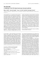
RPA70kDa has two RPA ssDNA-binding domains,
DBD-A and DBD-B for binding ssDNA, and a third,
DBD-C, which displays only weak ssDNA-binding
activity (Fig. 2B). RPA70kDa also contains the DBD-
F domain, which has been shown to interact with
multiple proteins and to interact weakly with DNA
(Fig. 2B). The primary amino acid sequences of
DBD-A, DBD-B, DBD-C and DBD-F domains are
very similar [12]. RPA32kDa has only a single ssDNA-
binding domain (DBD-D) [12]. Furthermore, all the
domains have high levels of sequence homology with
their counterparts in human and yeast RPAs [12]. The
DBD-E domain is in the RPA14kDa subunit, and is
also highly conserved [12].
In yeast, RPA1 (largest subunit) can only bind to
the RPA2 ⁄ 3 dimer (middle and smallest subunit
dimer). The DBD-C and DBD-D regions of rice are
quite similar to the DBD-C and DBD-D regions of
S. cerevisiae [139], but OsRPA14 has only low simi-
larity to RPA3. This sequence divergence may
account for the differences in binding observed
between the yeast and rice proteins. Rice DBD-A and
DBD-B domains are more conserved than DBD-C
and DBD-F, implying that the primary function of
OsRPA70a and OsRPA70b is to bind DNA, and that
this function has been conserved during evolution,
even though the secondary functions of these proteins
may have diverged. Based on this analysis the B type
complex corresponds to the mammalian and yeast
RPA.
In plant, human and yeast, the domains of DBD-A
and DBD-B are more homologous than those of
DBD-C and DBD-F, and the biochemical characteris-
tics are common among OsRPA70a, OsRPA70b and
OsRPA70c. It is well established that the RPA70kDa
subunit accumulates along stretches of ssDNA gener-
ated by stalled replication forks and ⁄ or DNA damage
[1,82–84]. In the RPA70kDa subunit, DBD-A and
DBD-B possess the strongest ssDNA-binding activity.
Indeed, DBD-A and DBD-B were the first to be iden-
tified as DNA-binding domains [12]. DBD-C and
DBD-D have a weak ssDNA–binding activity [12],
whereas DBD-F interacts physically with the tumor
suppressor p53 and nucleosome remodeling complex
FACT. The interaction with DBD-F can also contrib-
ute to an additional binding of structurally distorted
DNA (i.e. damaged DNA). By analogy, the primary
function of all the OsRPA70kDa subunits must be to
find special regions of DNA with which to bind. Is
there a divergence in biochemical function among the
K. Sakaguchi et al. The multi-replication protein A system
FEBS Journal 276 (2009) 943–963 ª 2009 The Authors Journal compilation ª 2009 FEBS 947
various domains? What is the specialization of hSSBs
(analogs of RPA), which appeared by convergent
evolution [70]?
Furthermore, why are the middle subunits diversified
phylogenetically? As discussed earlier, the major role
of the middle subunits is not to bind to DNA,
although they may be involved in the controlling signal
via phosphorylation. Indeed, in humans, HsRPA2
interacts with uracil–DNA glycosylase and XPA, but
HsRPA4 does not [67–69]. Moreover, the small sub-
unit is presumably responsible for linking the other
subunits (large and middle). The driving force behind
the diversification of the small subunit is an interesting
question that needs to be addressed.
The phylogenetic data suggest that the multi-RPA
(or the SSB–RPA mixed) systems are universal in
eukaryotes. However, it is important to establish
whether plants have paralogs or orthologs of hSSB. In
particular, we need to investigate the in vivo functions
of each of the A, B and C types of plant multi-RPA
systems.
In vivo roles of the multi-RPA system
If the multi-RPA system is unique in plants, some of
the in vivo roles may also be specific for plants.
OsRPA70a (type A complex) is localized in the chloro-
plast, but OsRPA70b (type B) and OsRPA70c (type C)
are found in the nuclear compartment [12]. The type A
system is thought to be plant specific, whereas types B
and C could be universal. Fortunately, the homologs
of OsRPA70a, OsRPA70b and OsRPA70c were found
A
B
Fig. 2. (A) Pairwise comparison of each
OsRPA subunit with human (HsRPA),
Schizosaccharomyces pombe (SpRF-A)
and Drosophila melanogaster (DmRPA). (B)
Domain structures of OsRPAs. Each color
box indicates each DBD domain shown as
the lower half of the figure. DBD domain
are classified into A, B, C, D, E and F.
The multi-replication protein A system K. Sakaguchi et al.
948 FEBS Journal 276 (2009) 943–963 ª 2009 The Authors Journal compilation ª 2009 FEBS
to be present in A. thaliana (AtRPA70a, AtRPA70b
and AtRPA70c) [11,12].
Interestingly, the AtRPA70a deletion mutant
(SALK017580) was lethal, but the AtRPA70b deletion
mutant (SALK088429) was viable and hypersensitive
to UV and MMS [12]. Therefore, type A may be
essential for DNA replication and transcription (and
also DNA repair) in the chloroplast. Type B may have
at least some role in nuclear DNA repair [12]. Intrigu-
ingly, the AtRPA70c deletion mutant does not appear
to be viable. Type C shows nuclear localization, and
the AtRPA70c deletion mutant may be lethal, suggest-
ing that type C is essential for DNA replication and
transcription (and possibly DNA repair) in the nucleus
[12].
To investigate the function of the various proteins,
RNAi of AtRPA70a and AtRPA70b were performed
[140–143]. The RNAi-mediated knockdown of
AtRPA70a also displayed lethality. However, RNAi of
AtRPA70b was viable and did not differ in phenotype
from wild-type. RT-PCR analysis was also carried out
using total RNA extract from seedlings of atrpa70b
mutant and the AtRPA70b RNAi line. No atRPA70b
transcript could be detected. Furthermore, western blot
analysis of total proteins from seedlings of wild-type
and atrpa70b mutant indicated very little AtRPA70b
[12].
These results indicated that AtRPA70a (probably,
the AtRPA70a–AtRPA32-2–AtRPA14 complex) has
an essential role, probably in DNA replication in the
chloroplast, whereas AtRPA70b (the AtRPA70b–At-
RPA32-1–AtRPA14 complex) is not essential under
normal growth conditions. However, it is known that
yeast rpa70 mutants are very sensitive to mutagens
such as UV and MMS [11,12]. To determine whether
AtRPA70b is similarly involved in mutagen tolerance,
the mutagen sensitivity of atrpa70b mutant and the
AtRPA70b RNAi line was tested. When 1-week-old
seedlings were exposed to various UV-B doses and
then grown for an additional week in the absence of
UV-B, there were no remarkable morphological differ-
ences between wild-type, atrpa70b mutant and
AtRPA70b RNAi line seedlings, although leaf yellow-
ing was somewhat increased in the mutant and RNAi
seedlings [11,12]. Compared with wild-type, the
amounts of chlorophyll (a + b) were decreased in
atrpa70b and the AtRPA70b RNAi lines [11,12]. One-
week-old seedlings were also grown on MS medium
containing various concentrations of MMS or H
2
O
2
.
After 1 week, growth of the wild-type plants was
inhibited by UV-B, MMS or H
2
O
2
. Compared with
wild-type plants, the growth of atrpa70b mutant and
AtRPA70b RNAi line seedlings was more inhibited by
UV-B, and was completely stopped by MMS [11,12].
Mutants showed little increase in sensitivity to H
2
O
2
.
Like the yeast rpa70 mutants, the atrpa70b mutant
and AtRPA70b RNAi line are more sensitive than
wild-type to UV and MMS, suggesting that At-
RPA70b is involved in the repair system for DNA
damaged by these mutagens [11,12].
The lethality of both the T-DNA insertion mutant
and the RNAi line of AtRPA70a indicate that the
AtRPA70a–AtRPA32-2–AtRPA14 complex plays an
essential role, such as DNA replication, in the chlorop-
lasts of living cells (Fig. 3). By contrast, the mutant
and RNAi line of AtRPA70b were viable but showed
high sensitivity to UV and MMS, suggesting involve-
ment of the AtRPA70b–AtRPA32-1–AtRPA14 com-
plex in the repair of damaged DNA (Fig. 3). However,
AtRPA70c deletion was thought to be lethal, suggest-
ing that the AtRPA70c–AtRPA32-3–AtRPA14 com-
plex may function mainly in nuclear DNA replication
and transcription (Fig. 3). Subcellular localization
analysis suggested that the type A RPA complex is
required for chloroplast DNA metabolism, whereas
types B and C function in nuclear DNA metabolism
[12].
Recently, RPA70 and RPA32 subunits from plants
have been reported to play a role in viral and transpo-
son DNA syntheses [131,144]. It will be intriguing to
investigate how the RPA complex functions in these
mechanisms. Higher plants may have evolved the
type A for the chloroplast to offer protection against
high levels of UV irradiation. Indeed, as mentioned
earlier, plants are exposed to UV radiation for much
longer than animals or yeast. Higher plants depend on
exposure to sunlight, including UV, for their develop-
ment because their energy is derived from photosyn-
thesis. Thus, the repair system in subcellular organelles
is presumably much more efficient in plants than in
animals and yeast.
The human homologs of RPA32, HsRPA2 and
HsRPA4 [67] may correspond to OsRPA32-1 (type B)
and OsRPA32-3 (type C) of plants, respectively,
although only the middle subunit is diversified. Inter-
estingly, hSSB1 did not localize to replication foci in
S-phase cells and hSSB1 deficiency did not influence
S-phase progression [70]. Depletion of hSSB1 abro-
gated the cellular response to DSBs, including activa-
tion of ATM and phosphorylation of ATM targets,
after ionizing radiation [70]. Ionizing radiation and
anti-cancer drugs can induce DNA DSBs, which are
highly cytotoxic lesions. Cells deficient in hSSB1 exhib-
ited increased radiosensitivity, defective checkpoint
activation and enhanced genomic instability coupled
with a diminished capacity for DNA repair. Thus,
K. Sakaguchi et al. The multi-replication protein A system
FEBS Journal 276 (2009) 943–963 ª 2009 The Authors Journal compilation ª 2009 FEBS 949
hSSB1 must influence diverse endpoints in the cellular
DNA damage response. In this way, hSSB1 resembles
the type B system.
Why are they not always found? The multi-RPA
types may resemble each other biochemically because
most of the subunits (large and ⁄ or middle) display a
significant degree of similarity. In many eukaryotes, the
multi-RPA system may diversify by exchanging some
subunits. For example, some of the non-homolog(s) of
hSSB1 are derived from convergent evolution. Further-
more, ubiquitous RPA (type B) is dispensable and can
easily be analyzed using the knockdown mutant,
whereas the type C or HsRPA complex (or hSSB2) is
lethal. However, very few researchers have studied these
mutants. Interestingly, the same phenomena was found
in Drosophila PCNAs, where the major PCNA is a
homolog of the ubiquitous PCNA in eukaryotes but is
dispensable [65]. Subsequently we analyzed the proper-
ties of these proteins in more detail. The role of the
miner subunit is not well understood because the
knockdown mutant is, as yet, unavailable [65].
A new perspective for RPA complexes
If multi-system RPAs are found to be universal each
of the corresponding functions should be reconsidered.
Nuclear RPAs may be divided into two categories: (a)
replication ⁄ transcription (plant C type), and (b)
repair ⁄ recombination (plant B type). The large subunit
may function as an agent for ssDNA stretching [1,2],
whereas the middle subunit may act as a signal trans-
duction acceptor. The small subunit may be a connect-
ing factor for forming the heterotrimeric complex.
Indeed, the small subunit mostly exists as a hetero-
dimer with the middle subunit, whereas the largest sub-
unit can be stabilized by binding to the dimer [10–12].
Genetic knockdown of the type 1 RPA increases the
lethality (i.e. the type C), but type 2 RPA can survive
unless the DNA is damaged (i.e. type B). Therefore,
subunit variety and function of the various subunits of
RPA must be reconsidered in view of these new find-
ings. For example, human RPA interacted with XPA
at sites of DNA damage, stimulated XPA–DNA inter-
action, and recruited the incision proteins
ERCC1 ⁄ XPF and XPG to the damaged site [89]. The
RPA must be a complex with HsRPA2, which corre-
sponds to type B. In NER and long-patch base exci-
sion repair, type B may be responsible for these
functions in eukaryote kingdoms.
The reported biological functions of mammalian and
yeast RPA are mostly involved in meiosis. The middle
subunit has an important role in regulating synaptine-
mal complex (SC) formation and meiotic recombina-
tion at meiotic prophase, mainly at zygotene and
pachytene [71–74,114,115,130]. The protein factors,
such as DNA polymerases and recombinases, are
major proteins involved in meiotic prophase events.
Nevertheless, RPA is known biochemically to interact
in vitro with DNA polymerases and recombinases
[6–8,13,31,40–42,44,72,85–88,138,145–169].
In fulfilling its biosynthetic roles in nuclear replica-
tion and in several types of repair, DNA polymerase is
assisted by RPA. In eukaryotes, recent investigations
have revealed at least 14 types of DNA polymerase
Fig. 3. Hypothetic model of the cellular
function of A-, B- and C-type RPA com-
plexes.
The multi-replication protein A system K. Sakaguchi et al.
950 FEBS Journal 276 (2009) 943–963 ª 2009 The Authors Journal compilation ª 2009 FEBS
(pol a, b, c, d, e, f, g, h, i, j, k, l, m and p) [45,170].
In a sense, all are analogs of each other. RPA is
reported to interact with at least pol a, d, e, k and j
[3,5–8,76,80,81,85–88]. RPA contributes to the high
fidelity of the polymerases during DNA synthesis. Of
the polymerase species, pol a, d and e replicate DNA
during S phase, but pol a is replication specific [80].
All the other polymerases are involved in DNA repair
and recombination [81]. We reported that in meiosis
two categories of DNA polymerases (a) pol a complex
and (b) pol k and l were expressed [165,168]. The
former is for replication at zygotene (or SC formation)
and the latter is for repair and recombination at late
zygotene to pachytene (Fig. 4) [155,165,168,171–173].
Using a D-loop recombination intermediate substrate,
we observed that either pol k or pol l can promote
the primer extension of an invading strand present in a
D-loop structure [168]. Both could fully extend the
primer in the D-loop substrate, suggesting that the
D-loop extension is an activity that is intrinsic to
the polymerases [168].
Two orthologs of the recombinases, Rad51 and
Lim15 ⁄ Dmc1, are present in meiosis [44,114,115,152–
154,161,162,167]. These recombinases occur at late
leptotene to early zygotene (Fig. 4). The interaction of
RPA and Rad51 is well established. Another meiotic
role of RPA was also found. At meiotic prophase (late
leptotene to early zygotene), with RPA, the homology-
search recombinase complex is involved in homologous
chromosome synapsis, preventing the formation of
superfluous reciprocal recombinant events (Fig. 4)
[114,115]. Both Rad51 and Lim15 ⁄ Dmc1 were identi-
fied as being involved in this process, although the
specific function of each protein is not yet known [44].
Are the DNA polymerase and recombinase
functions mediated by one species of RPA complex?
Interestingly, dephosphorylation of transformed nod-
ule-associated histone H2AX chromatin occurs at this
time. This suggests annealing of single strands or
repair of DSBs. By a similar mechanism, if the middle
subunit of RPA is also dephosphorylated, RPA would
lose the function of maintaining the noncross-over
condition. We must also consider the role of the multi-
RPA system during the meiotic prophase events.
It is known that a small amount of DNA replicates
at zygotene (pairing DNA synthesis) and that the
repair synthesis of DNA occurs at pachytene (cross-
over DNA synthesis) [172,173]. The two sequential
DNA synthesis reactions play a role in the progression
of meiosis. It is possible that a complex of RPA and
pol a differs from the recombination-dependent RPA.
Because DNA polymerase searches for the RPA–
Fig. 4. Hypothetic model of meiotic cell cycle and its relation to RPA.
K. Sakaguchi et al. The multi-replication protein A system
FEBS Journal 276 (2009) 943–963 ª 2009 The Authors Journal compilation ª 2009 FEBS 951
ssDNA complex structure on the DNA, RPA
complexed with pol a are probably functionally inde-
pendent from RPA complexed with other repair
polymerases. Pol k and l were thought to be involved
in the ‘crossover DNA synthesis’ for DNA recombina-
tion. Because the pol k(or the pol l)-deficient mutant
is viable, RPA may be like the type B or HsRPA2
type. However, ‘pairing DNA replication’ appears to
be specific for SC formation. At that stage, the DNA
polymerase a-catalytic subunit and primase are pre-
sumably also present [165]. This replication could be
the basis for SC extension and formation of the transi-
tion nodules [44]. Indeed, this process probably
requires RPA, such as the type C form (Fig. 4).
During prophase, DNA polymerases as well as
paralogs and orthologs of PCNA, recombinases
and perhaps RPA are required (Fig. 4) [42,44,45,
151,152,155,157,159,160,165,168,171]. Electron micros-
copy data [115,117] suggest that meiotic functions
in vivo are shared by each of the paralogs and ortho-
logs, and maybe also the analogs and heterologs.
Indeed, control of the biological process could be more
finely tuned by sharing function amongst paralogs,
orthologs, analogs and heterologs.
Background for the screening of
multiple protein systems involved in
DNA metabolism
We have studied many protein factors in DNA replica-
tion ⁄ repair and their relation to the meiotic system in
higher plants (O. sativa and A. thaliana) [13–
43,45,156,171], a fungus (C. cinereus) [44,138,145–155,
157–169] and an arthropod (Drosophila melanogaster)
[44–66]. Each of the materials represents the biological
kingdom of plant, fungus and animal, respectively.
Our research aimed to comprehensively understand
these DNA synthesis-related events in phylogenetically
diverse species. In addition to RPA, we elucidated
many of the related factors, such as Rad51,
Lim15 ⁄ Dmc1, RadA, PCNA, DDB, XRCC1, Rad2
family nucleases and special nucleases, DNA polyme-
rases, ORC1, RFC, RecQ, DNA ligases, CAF-1,
mtTFA, Rrp1, Mer3, Snm1, Rad6, SUMOylation fac-
tors (Aos1, Uba2, Ubc9, SUMO), leucine aminopepti-
dase and 26S proteasome-related factors (Jab1, Sgt1,
DnaJ) (Table 1). During the course of our experi-
ments, we frequently observed that protein factors
involved in the same DNA metabolic processes are not
always homologs in eukaryotic cells. Although the
paralogs and orthologs are ubiquitous, evolutionally
different factors were often found to be involved in
the same biosystems, which are referred to as ‘analogs’
and ‘heterologs’. Indeed, convergent evolution might
be ubiquitous in eukaryotic DNA metabolic processes.
According to definition, ‘homolog’ is a gene related to
a second gene by descent from a common ancestral DNA
sequence. ‘Ortholog’ is a gene in different species that
evolved from a common ancestral gene. ‘Paralog’ is a
gene related by duplication within a genome. Orthologs
retain the same function in the course of evolution,
whereas paralogs evolve new functions. ‘Analog’ is a gene
that has common activity but not a common origin.
‘Heterolog’ is a gene that differs in both origin and activ-
ity. Heterolog does not classify homolog, ortholog, par-
alog or analog. It may be also said that heterolog is used
as a synonym of ‘just different protein (gene)’, basically.
For example, PCNA is not one copy [65,138,159];
two PCNA paralogs and one PCNA-like heterotrimer
(Rad9–Rad1–Hus1) (‘analog’ or ‘heterolog’) were
found in Drosophila [65,136,137]. Rad9–Rad1–Hus1 is
found universally in eukaryotes. Plant SYCP1 and
yeast Zip1 mediate the same role in meiosis, despite
displaying no significant homology (‘analog’ or ‘het-
erologs’) [174,175]. Similarly, human mus81–Eme1 is
functionally the same as Escherichia coli RuvC (‘ana-
log’ or ‘heterologs’) [176–178]. In plants, two recA-like
protein paralogs (Rad51 and Lim15 ⁄ Dmc1) as well as
a prokaryotic recA homolog (RadA) were found (‘ana-
logs’) [42]. Furthermore, this is not the plastid compo-
nent [42]. As described earlier, in addition to the two
subtypes of RPA (HsRPA2 and HsRPA4) two human
SSB homologs are also present (‘analogs’) [70]. More-
over, in human, five Rad51 paralogs (Rad51B,
Rad51C, Rad51D, Xrcc2 and Xrcc3) have been found
[179–181]. Two FEN-1 paralogs (FEN-1a and FEN-
1b) and one analog (SEND-1) were found in plants
[25,26], and another FEN-1 analog occurs in Drosoph-
ila (GEN) [63,66]. DNA polymerases, especially for
DNA repair, are greatly diversified in eukaryotes
[76,182,183]. DNA polymerase b (pol b) for short
patch base excision repair are found only in verte-
brates [45]; plant short patch base excision repair uses
pol f instead [33,39,45]. However, as yet, a recBCD
homolog has not been found in the eukaryotic recom-
bination process. Prokaryotic homologs such as RadA
and hSSB are often found in eukaryotes (‘analog’ or
‘heterolog’), although there are the eukaryotic func-
tional alternatives [42,70]. All the protostomic animals
lack any X family DNA polymerases essential for
development of the nervous and immune system [45].
In Drosophila, AP endonuclease 1 homolog (Rrp1)
binds to pol f [64]. Plant XRCC1 lacks the polymer-
ase-binding domain [33,39]. Therefore, factor variation
(orthologs, paralogs, ‘analogs’ and ‘heterologs’) seems
to be ubiquitous in eukaryotic DNA metabolism.
The multi-replication protein A system K. Sakaguchi et al.
952 FEBS Journal 276 (2009) 943–963 ª 2009 The Authors Journal compilation ª 2009 FEBS
Table 1. The main role of DNA synthesis-related factor.
Protein Function Reference
RPA Required for DNA recombination, repair and replication.
The activity of RPA is mediated by ssDNA binding and protein
interactions
[1,10–12,30,43,124]
Rad51 May participate in a common DNA damage-response pathway
associated with the activation of homologous recombination and
double-strand break repair. Binds to ssDNA and dsDNA and exhibits
DNA-dependent ATPase activity. Unwinds duplex DNA and forms
helical nucleoprotein filaments
[152,154,184–186]
Lim15 ⁄ Dmc1 May participate in meiotic recombination, specifically in homologous
strand assimilation, which is required for the resolution of meiotic
double-strand breaks.
[99,152–154,159–163,
167,187]
RadA This family consists exclusively of archaeal RadA protein, a homolog of
bacterial RecA, eukaryotic Rad51 and archaeal RadB. This protein is
involved in DNA repair and recombination
[42,188]
PCNA PCNA, or cyclin, is a non-histone acidic nuclear protein that plays a key
role in the control of eukaryotic DNA replication. It acts as a co-factor
for DNA polymerase delta, which is responsible for leading strand
DNA replication
[21,30,31,34,65,138,
159,189,190]
DDB Required for DNA repair. Binds to DDB2 to form the UV-damaged
DNA-binding protein complex (the UV–DDB complex). The UV–DDB
complex may recognize UV-induced DNA damage and recruit proteins
of the NER pathway to initiate DNA repair
[27,30,36,52,54,58,
61,62,191]
XRCC1 DNA-repair protein Xrcc1 functions in the repair of ssDNA breaks in
mammalian cells and forms a repair complex with beta-Pol, ligase III
and PARP
[39,192,193]
Rad2 Single-stranded DNA endonuclease involved in excision repair of DNA
damaged with UV light, bulky adducts, or cross-linking agents.
Essential for the incision step of excision-repair
[25,63,66,194,195]
DNA polymerase DNA polymerase is an enzyme that catalyzes the polymerization of
deoxyribonucleotides into a DNA strand. Based on sequence
homology, DNA polymerases can be further subdivided into seven
different families: A, B, C, D, X, Y and RT
[16,22,24,33,38,40,45,46,
51,53,55,56,59,60,64,
76,145,151,155,157,158,
165,168,170,171,182,183]
ORC1 Component of the origin recognition complex that binds
origins of replication. It has a role in both chromosomal
replication and mating type transcriptional silencing.
Binds to the ARS consensus sequence
of origins of replication in an ATP-dependent manner
[19,41,196]
RFC A complex of five polypeptides in eukaryotes, and two in prokaryotes,
that loads the DNA polymerase processivity factor PCNA onto DNA,
thereby permitting processive DNA synthesis catalyzed by DNA
polymerase
[20,28,197]
RecQ The ATP-dependent DNA helicase RecQ is involved in genome
maintenance. All homologues tested to date unwind paired DNA,
translocating in a 3¢ to 5¢ direction and several have a preference for
forked or 4-way DNA structures (e.g. Holliday junctions) or for
G-quartet DNA
[37,198]
DNA ligase DNA ligase (polydeoxyribonucleotide synthase) is the enzyme that
joins two DNA fragments by catalyzing the formation of an
internucleotide ester bond between phosphate and deoxyribose.
It is active during DNA replication, DNA repair and DNA
recombination
[150,164,166,199]
CAF-1 Complex that is thought to mediate chromatin assembly in DNA
replication and DNA repair. Assembles histone octamers onto
replicating DNA in vitro. CAF-1 performs the first step of the
nucleosome assembly process, bringing newly synthesized histones
H3 and H4 to replicating DNA
[160,200,201]
K. Sakaguchi et al. The multi-replication protein A system
FEBS Journal 276 (2009) 943–963 ª 2009 The Authors Journal compilation ª 2009 FEBS 953
Table 1. Continued.
Protein Function Reference
mtTFA Involved in mitochondrial transcription regulation. Required for accurate
and efficient promoter recognition by the mitochondrial RNA
polymerase. Activates transcription by binding immediately upstream
of transcriptional start sites. Is able to unwind and bend DNA
[57,202,203]
Rrp1 Could promote homologous recombination at sites of DNA damage.
Has apurinic endonuclease and double-stranded DNA 3¢-exonuclease
activities and carries out single-stranded DNA renaturation in a
Mg
2+
-dependent manner. Activity is more efficient in purine-rich
regions of dsDNA than in pyrimidine-rich regions
[64,204–206]
Mer3 DNA-dependent ATPase. Required in the control of double strand
breaks transition and crossover during meiosis. Unwinds DNA in the
3¢ to 5¢ direction. Prefers ssDNA
[207–211]
Snm1 May be required for DNA interstrand cross-link repair. Also required for
checkpoint mediated cell cycle arrest in early prophase in response to
mitotic spindle poisons
[35,212,213]
Rad6 Ubiquitin-conjugating enzyme (E2), involved in postreplication repair
(with Rad18p), sporulation, telomere silencing, and ubiquitin-mediated
N-end rule protein degradation (with Ubr1p)
[32,214,215]
SUMOylation
(Aos1, Uba2, Ubc9, SUMO)
SUMO (small ubiquitin-like modifier) or SUMO proteins are a family of
small proteins that are covalently attached to and detached from
other proteins in cells to modify their function. SUMOylation is a
post-translational modification involved in various cellular processes,
such as nuclear–cytosolic transport, transcriptional regulation,
apoptosis, protein stability, response to stress and progression
through the cell cycle. In an ATP-dependent reaction, SUMO is
activated by a single specific E1-activating enzyme, the heterodimer
between Aos1 and Uba2 (or SAE1 ⁄ SAE2) resulting in an E1–SUMO
thioester linkage. SUMO is then transferred to the E2-conjugating
enzyme Ubc9, again forming a thioester. The catalytic cleft of Ubc9
directly interacts with many substrates via their SUMO consensus
motif. Target modification therefore often depends on a third class of
enzymes, the E3 ligases which enhance SUMO transfer from the E2
to the substrate
[44,163,216–219]
Leucine amino
peptidase
Presumably involved in the processing and regular turnover of
intracellular proteins. Catalyzes the removal of unsubstituted
N-terminal amino acids from various peptides
[146,220,221]
26S proteasome
related factor (Jab1)
Probable protease subunit of the COP9 signalosome complex (CSN), a
complex involved in various cellular and developmental processes.
The CSN complex is an essential regulator of the ubiquitin (Ubl)
conjugation pathway by mediating the deneddylation of the cullin
subunits of the SCF-type E3 ligase complexes, leading to decrease
the Ubl ligase activity of SCF-type complexes such as SCF, CSA or DDB2
[29,222]
26S proteasome
related factor (Sgt1)
Sgt1 is a novel subunit of the SCF ubiquitin ligase complex. Involved
in ubiquitination and subsequent proteosomal degradation of target
proteins. Required for both entry into S phase and kinetochore
function
[32,223]
26S proteasome
related factor (DnaJ)
Participates actively in the response to hyperosmotic and heat shock
by preventing the aggregation of stress-denatured proteins and by
disaggregating proteins, also in an autonomous, dnaK-independent
fashion. Unfolded proteins bind initially to dnaJ; upon interaction with
the dnaJ-bound protein, dnaK hydrolyzes its bound ATP, resulting in
the formation of a stable complex. GrpE releases ADP from dnaK;
ATP binding to dnaK triggers the release of the substrate protein,
thus completing the reaction cycle. Several rounds of ATP-dependent
interactions between dnaJ, dnaK and grpE are required for fully
efficient folding
[34,224,225]
The multi-replication protein A system K. Sakaguchi et al.
954 FEBS Journal 276 (2009) 943–963 ª 2009 The Authors Journal compilation ª 2009 FEBS
In conclusion, DNA synthetic systems are basically the
same from lower to higher eukaryotes, although related
factors are not necessarily homologs. In relation to
biological events, most, or all, of the repair factors are
involved in SC formation and meiotic recombination.
The presence of orthologs, paralogs, analogs and hetero-
logs should always be considered in the process. In meio-
sis, many of the factors require a protein that firstly
recognizes DNA structure to generate a highly organized
DNA–protein complex (i.e., replisome, repairsome or
recombinosome). Proteins such as RPA, PCNA, DDB or
FEN-1 may be responsible for defining the factor that
first recognizes the DNA structure. In our experiments,
many of the structure-recognition proteins tended to
diversify, suggesting that each of the factors can partici-
pate in generating the DNA–protein complexes.
In summary, we have used recent findings to specu-
late on the biological function of RPA. Figure 4 shows
the hypothetical model of the meiotic cell cycle at
prophase.
Acknowledgements
We thank Drs Yoichi Takakusagi, Seisuke Kimura,
Yoshihiro Kanai, Tatsushi Ruike and Taichi Yamam-
oto of our Department, for helpful discussions.
References
1 Wold MS (1997) Replication protein A: a heterotrimer-
ic, single-stranded DNA-binding protein required for
eukaryotic DNA metabolism. Annu Rev Biochem 66,
61–92.
2 Zou Y, Liu Y, Wu X & Shell SM (2006) Functions of
human replication protein A (RPA): from DNA repli-
cation to DNA damage and stress responses. J Cell
Physiol 208, 267–273.
3 Wang Y, Putnam CD, Kane MF, Zhang W, Edelmann
L, Russell R, Carrio
´
n DV, Chin L, Kucherlapati R,
Kolodner RD et al. (2005) Mutation in Rpa1 results in
defective DNA double-strand break repair, chromo-
somal instability and cancer in mice. Nat Genet 37,
750–755.
4 Li L & Zou L (2005) Sensing, signaling, and respond-
ing to DNA damage: organization of the checkpoint
pathways in mammalian cells. J Cell Biochem 94,
298–306.
5 Iftode C, Daniely Y & Borowiec JA (1999) Replication
protein A (RPA): the eukaryotic SSB. Crit Rev
Biochem Mol Biol 34, 141–180.
6 Kastan MB & Bartek J (2004) Cell-cycle checkpoints
and cancer. Nature 432, 316–323.
7 Sancar A, Lindsey-Boltz LA, Unsal-Kac¸ maz K & Linn
S (2004) Molecular mechanisms of mammalian DNA
repair and the DNA damage checkpoints. Annu Rev
Biochem 73, 39–85.
8 Maga G, Ramadan K, Locatelli GA, Shevelev I,
Spadari S & Hu
¨
bscher U (2005) DNA elongation by
the human DNA polymerase lambda polymerase and
terminal transferase activities are differentially coordi-
nated by proliferating cell nuclear antigen and replica-
tion protein A. J Biol Chem 280, 1971–1981.
9 Longhese MP, Plevani P & Lucchini G (1994) Repli-
cation factor A is required in vivo for DNA replication,
repair, and recombination. Mol Cell Biol 14, 7884–
7890.
10 Ishibashi T, Kimura S, Furukawa T, Hatanaka M,
Hashimoto J & Sakaguchi K (2001) Two types of repli-
cation protein A 70 kDa subunit in rice, Oryza sativa:
molecular cloning, characterization, and cellular and
tissue distribution. Gene 272, 335–343.
11 Ishibashi T, Koga A, Yamamoto T, Uchiyama Y,
Mori Y, Hashimoto J, Kimura S & Sakaguchi K
(2005) Two types of replication protein A in seed
plants. FEBS J 272, 3270–3281.
12 Ishibashi T, Kimura S & Sakaguchi K (2006) A higher
plant has three different types of RPA heterotrimeric
complex. J Biochem 139, 99–104.
13 Kimura S & Sakaguchi K (2006) DNA repair in plants.
Chem Rev 106, 753–766.
14 Ishibashi T, Kimura S, Furukawa T & Sakaguchi K
(2006) DNA repair mechanisms in UV-B tolerant
plants. Jpn Agric Res Q 40, 107–113.
15 Kimura S, Kai M, Kobayashi H, Suzuki A, Morioka
H, Otsuka E & Sakaguchi K (1997) A structure-specific
endonuclease from cauliflower (Brassica oleracea
var.
botrytis) inflorescence. Nucleic Acids Res 25, 4970–
4976.
16 Seto H, Hatanaka M, Kimura S, Oshige M, Tsuya Y,
Mizushina Y, Sawado T, Aoyagi N, Matsumoto T,
Hashimoto J et al. (1998) Purification and characteriza-
tion of a 100 kDa DNA polymerase from cauliflower
inflorescence. Biochem J 332(Pt 2), 557–563.
17 Kimura S, Takenouchi M, Hatanaka M, Seto H,
Kouroku Y & Sakaguchi K (1998) An ATP-inhibited
endonuclease from cauliflower (Brassica oleracea var.
botorytis) inflorescence. Planta 206 , 641–648.
18 Kimura S, Ueda T, Hatanaka M, Takenouchi M,
Hashimoto J & Sakaguchi K (2000) Plant homologue
of flap endonuclease-1: molecular cloning, characteriza-
tion, and evidence of expression in meristematic
tissues. Plant Mol Biol 42, 415–427.
19 Kimura S, Ishibashi T, Hatanaka M, Sakakibara Y,
Hashimoto J & Sakaguchi K (2000) Molecular cloning
and characterization of a plant homologue of the
origin recognition complex 1 (ORC1). Plant Sci 158,
33–39.
20 Furukawa T, Ishibashi T, Kimura S, Ueda T, Hashim-
oto J & Sakaguchi K (2001) Plant homologue of
K. Sakaguchi et al. The multi-replication protein A system
FEBS Journal 276 (2009) 943–963 ª 2009 The Authors Journal compilation ª 2009 FEBS 955
36 kDa subunit of replication factor C: molecular clon-
ing and characterization. Plant Sci 161, 99–106.
21 Kimura S, Suzuki T, Yanagawa Y, Yamamoto T,
Nakagawa H, Tanaka I, Hashimoto J & Sakaguchi K
(2001) Characterization of plant proliferating cell
nuclear antigen (PCNA) and flap endonuclease-1
(FEN-1), and their distribution in mitotic and meiotic
cell cycles. Plant J 28, 643–653.
22 Kimura S, Uchiyama Y, Kasai N, Namekawa S, Sao-
tome A, Ueda T, Ando T, Ishibashi T, Oshige M,
Furukawa T et al. (2002) A novel DNA polymerase
homologous to Escherichia coli DNA polymerase I
from a higher plant, rice (Oryza sativa L.). Nucleic
Acids Res 30, 1585–1592.
23 Yanagawa Y, Kimura S, Takase T, Sakaguchi K,
Umeda M, Komamine A, Tanaka K, Hashimoto J,
Sato T & Nakagawa H (2002) Spatial distribution of
the 26S proteasome in meristematic tissues and
primordia of rice (Oryza sativa L.). Planta 214, 703–
707.
24 Uchiyama Y, Hatanaka M, Kimura S, Ishibashi T,
Ueda T, Sakakibara Y, Matsumoto T, Furukawa T,
Hashimoto J & Sakaguchi K (2002) Characterization
of DNA polymerase delta from a higher plant, rice
(Oryza sativa L.). Gene 295, 19–26.
25 Furukawa T, Kimura S, Ishibashi T, Mori Y, Hashim-
oto J & Sakaguchi K (2003) OsSEND-1: a new RAD2
nuclease family member in higher plants. Plant Mol
Biol 51, 59–70.
26 Kimura S, Furukawa T, Kasai N, Mori Y, Kitamoto
HK, Sugawara F, Hashimoto J & Sakaguchi K (2003)
Functional characterization of two flap endonuclease-1
homologues in rice. Gene 314, 63–71.
27 Ishibashi T, Kimura S, Yamamoto T, Furukawa T,
Takata K, Uchiyama Y, Hashimoto J & Sakaguchi K
(2003) Rice UV-damaged DNA binding protein homo-
logues are most abundant in proliferating tissues. Gene
308, 79–87.
28 Furukawa T, Ishibashi T, Kimura S, Tanaka H,
Hashimoto J & Sakaguchi K (2003) Characterization
of all the subunits of replication factor C from a
higher plant, rice (Oryza sativa L.), and their relation
to development. Plant Mol Biol 53, 15–25.
29 Yamamoto T, Kimura S, Mori Y, Uchiyama Y, Ishib-
ashi T, Hashimoto J & Sakaguchi K (2003) Interaction
between proliferating cell nuclear antigen and JUN-
activation-domain-binding protein 1 in the meristem of
rice, Oryza sativa L. Planta 217, 175–183.
30 Kimura S, Tahira Y, Ishibashi T, Mori Y, Mori T,
Hashimoto J & Sakaguchi K (2004) DNA repair in
higher plants; photoreactivation is the major DNA
repair pathway in non-proliferating cells while excision
repair (nucleotide excision repair and base excision
repair) is active in proliferating cells. Nucleic Acids Res
32, 2760–2767.
31 Yamamoto T, Kimura S, Mori Y, Oka M, Ishibashi T,
Yanagawa Y, Nara T, Nakagawa H, Hashimoto J &
Sakaguchi K (2004) Degradation of proliferating cell
nuclear antigen by 26S proteasome in rice (Oryza sati-
va L.). Planta 218, 640–646.
32 Yamamoto T, Mori Y, Ishibashi T, Uchiyama Y, Sak-
aguchi N, Furukawa T, Hashimoto J, Kimura S &
Sakaguchi K (2004) Characterization of Rad6 from a
higher plant, rice (Oryza sativa L.) and its interaction
with Sgt1, a subunit of the SCF ubiquitin ligase com-
plex. Biochem Biophys Res Commun
314, 434–439.
33 Uchiyama Y, Kimura S, Yamamoto T, Ishibashi T &
Sakaguchi K (2004) Plant DNA polymerase lambda, a
DNA repair enzyme that functions in plant meriste-
matic and meiotic tissues. Eur J Biochem 271, 2799–
2807.
34 Yamamoto T, Mori Y, Ishibashi T, Uchiyama Y,
Ueda T, Ando T, Hashimoto J, Kimura S & Sakaguchi
K (2005) Interaction between proliferating cell nuclear
antigen (PCNA) and a DnaJ induced by DNA damage.
J Plant Res 118, 91–97.
35 Kimura S, Saotome A, Uchiyama Y, Mori Y, Tahira
Y & Sakaguchi K (2005) The expression of the rice
(Oryza sativa L.) homologue of Snm1 is induced by
DNA damage. Biochem Biophys Res Commun 329,
668–672.
36 Koga A, Ishibashi T, Kimura S, Uchiyama Y & Sak-
aguchi K (2006) Characterization of T-DNA insertion
mutants and RNAi silenced plants of Arabidopsis thali-
ana UV-damaged DNA binding protein 2 (AtUV-
DDB2). Plant Mol Biol 61, 227–240.
37 Saotome A, Kimura S, Mori Y, Uchiyama Y, Moroh-
ashi K & Sakaguchi K (2006) Characterization of four
RecQ homologues from rice (Oryza sativa L. cv. Nip-
ponbare). Biochem Biophys Res Commun 345, 1283–
1291.
38 Takeuchi R, Kimura S, Saotome A & Sakaguchi K
(2007) Biochemical properties of a plastidial DNA
polymerase of rice. Plant Mol Biol 64, 601–611.
39 Uchiyama Y, Suzuki Y & Sakaguchi K (2008) Charac-
terization of plant XRCC1 and its interaction with
proliferating cell nuclear antigen. Planta 227, 1233–
1241.
40 Mori Y, Kimura S, Saotome A, Kasai N, Sakaguchi
N, Uchiyama Y, Ishibashi T, Yamamoto T, Chiku H
& Sakaguchi K (2005) Plastid DNA polymerases from
higher plants, Arabidopsis thaliana. Biochem Biophys
Res Commun 334, 43–50.
41 Mori Y, Yamamoto T, Sakaguchi N, Ishibashi T,
Furukawa T, Kadota Y, Kuchitsu K, Hashimoto J,
Kimura S & Sakaguchi K (2005) Characterization of
the origin recognition complex (ORC) from a higher
plant, rice (Oryza sativa L.). Gene 353, 23–30.
42 Ishibashi T, Isogai M, Kiyohara H, Hosaka M, Chiku
H, Koga A, Yamamoto T, Uchiyama Y, Mori Y,
The multi-replication protein A system K. Sakaguchi et al.
956 FEBS Journal 276 (2009) 943–963 ª 2009 The Authors Journal compilation ª 2009 FEBS
Hashimoto J et al. (2006) Higher plant RecA-like pro-
tein is homologous to RadA. DNA Repair (Amst) 5,
80–88.
43 Marwedel T, Ishibashi T, Lorbiecke R, Jacob S, Sak-
aguchi K & Sauter M (2003) Plant-specific regulation
of replication protein A2 (OsRPA2) from rice during
the cell cycle and in response to ultraviolet light expo-
sure. Planta 217, 457–465.
44 Sakaguchi K, Koshiyama A & Iwabata K (2007)
Meiosis and small ubiquitin-related modifier
(SUMO)-conjugating enzyme, Ubc9. FEBS J 274,
3519–3531.
45 Uchiyama Y, Takeuchi R, Kodera H & Sakaguchi K
(2008) Evolution and roles of X-family DNA
polymerases. Biochemie, doi:10.1016/j.biochi.2008.
07.005 (in press).
46 Sakaguchi K & Boyd JB (1985) Purification and char-
acterization of a DNA polymerase beta from Drosoph-
ila*. J Biol Chem 260, 10406–10411.
47 Boyd JB, Sakaguchi K & Harris PV (1990) mus308
mutants of Drosophila exhibit hypersensitivity to DNA
cross-linking agents and are defective in a deoxyribo-
nuclease. Genetics 125, 813–819.
48 Sakaguchi K, Harris PV, van Kuyk R, Syngston A &
Boyd JB (1990) A mitochondrial nuclease is modified
in Drosophila mutants (mus308) that are hypersensitive
to DNA cross-linking agents. Mol Gen Genet 224,
31–38.
49 Sakaguchi K, Harris PV, Ryan C, Buchwald M &
Boyd JB (1991) Alteration of a nuclease in Fanconi
anemia. Mutat Res 255, 31–38.
50 Sakaguchi K, Zdzienicka MZ, Harris PV & Boyd JB
(1992) Nuclease modification in Chinese hamster cells
hypersensitive to DNA cross-linking agents – a model
for Fanconi anemia. Mutat Res 274, 11–18.
51 Aoyagi N, Matsuoka S, Furunobu A, Matsukage A &
Sakaguchi K (1994) Drosophila DNA polymerase delta,
purification and characterization. J Biol Chem 269,
6045–6050.
52 Kai M, Takahashi T, Todo T & Sakaguchi K (1995)
Novel DNA binding proteins highly specific to
UV-damaged DNA sequences from embryos of Dro-
sophila melanogaster. Nucleic Acids Res 23, 2600–2607.
53 Aoyagi N, Oshige M, Hirose F, Kuroda K, Matsukage
A & Sakaguchi K (1997) DNA polymerase epsilon
from Drosophila melanogaster. Biochem Biophys Res
Commun 230, 297–301.
54 Kai M, Todo T, Wada M, Ryo H, Masutani C,
Kobayashi H, Morioka H, Ohtsuka E, Hanaoka F &
Sakaguchi K (1998) A new Drosophila ultraviolet light-
damaged DNA recognition endonuclease that selec-
tively nicks a (6-4) photoproduct site. Biochim Biophys
Acta 1397, 180–188.
55 Oshige M, Aoyagi N, Harris PV, Burtis KC & Sakagu-
chi K (1999) A new DNA polymerase species from
Drosophila melanogaster: a probable mus308 gene prod-
uct. Mutat Res 433, 183–192.
56 Oshige M, Yoshida H, Hirose F, Takata KI, Inoue Y,
Aoyagi N, Yamaguchi M, Koiwai O, Matsukage A &
Sakaguchi K (2000) Molecular cloning and expression
during development of the Drosophila gene for the cat-
alytic subunit of DNA polymerase epsilon. Gene 256,
93–100.
57 Takata K, Yoshida H, Hirose F, Yamaguchi M, Kai
M, Oshige M, Sakimoto I, Koiwai O & Sakaguchi K
(2001) Drosophila mitochondrial transcription factor A:
characterization of its cDNA and expression pattern
during development. Biochem Biophys Res Commun
287, 474–483.
58 Takata K, Ishikawa G, Hirose F & Sakaguchi K
(2002) Drosophila damage-specific DNA-binding pro-
tein 1 (D-DDB1) is controlled by the DRE ⁄ DREF sys-
tem. Nucleic Acids Res 30, 3795–3808.
59 Oshige M, Takeuchi R, Ruike T, Ruike R, Kuroda K
& Sakaguchi K (2004) Subunit protein-affinity isolation
of Drosophila DNA polymerase catalytic subunit. Pro-
tein Expr Purif 35, 248–256.
60 Takeuchi R, Oshige M, Uchida M, Ishikawa G, Takat-
a K, Shimanouchi K, Kanai Y, Ruike T, Morioka H
& Sakaguchi K (2004) Purification of Drosophila DNA
polymerase zeta by REV1 protein-affinity chromatog-
raphy. Biochem J 382, 535–543.
61 Takata K, Yoshida H, Yamaguchi M & Sakaguchi K
(2004) Drosophila damaged DNA-binding protein 1 is
an essential factor for development. Genetics 168, 855–
865.
62 Takata K, Shimanouchi K, Yamaguchi M, Murakami
S, Ishikawa G, Takeuchi R, Kanai Y, Ruike T,
Nakamura R, Abe Y et al. (2004) Damaged DNA
binding protein 1 in Drosophila defense reactions.
Biochem Biophys Res Commun 323, 1024–1031.
63 Ishikawa G, Kanai Y, Takata K, Takeuchi R, Shima-
nouchi K, Ruike T, Furukawa T, Kimura S & Sakagu-
chi K (2004) DmGEN, a novel RAD2 family endo-
exonuclease from Drosophila melanogaster. Nucleic
Acids Res 32, 6251–6259.
64 Takeuchi R, Ruike T, Nakamura R, Shimanouchi K,
Kanai Y, Abe Y, Ihara A & Sakaguchi K (2006) Dro-
sophila DNA polymerase zeta interacts with recombi-
nation repair protein 1, the Drosophila homologue of
human abasic endonuclease 1. J Biol Chem 281, 11577–
11585.
65 Ruike T, Takeuchi R, Takata K, Oshige M, Kasai N,
Shimanouchi K, Kanai Y, Nakamura R, Sugawara F
& Sakaguchi K (2006) Characterization of a second
proliferating cell nuclear antigen (PCNA2) from
Drosophila melanogaster.
FEBS J 273, 5062–
5073.
66 Kanai Y, Ishikawa G, Takeuchi R, Ruike T, Nakam-
ura R, Ihara A, Ohashi T, Takata K, Kimura S &
K. Sakaguchi et al. The multi-replication protein A system
FEBS Journal 276 (2009) 943–963 ª 2009 The Authors Journal compilation ª 2009 FEBS 957
Sakaguchi K (2007) DmGEN shows a flap endonucle-
ase activity, cleaving the blocked-flap structure and
model replication fork. FEBS J 274, 3914–3927.
67 Keshav KF, Chen C & Dutta A (1995) Rpa4, a
homolog of the 34-kilodalton subunit of the
replication protein A complex. Mol Cell Biol 15,
3119–3128.
68 Nagelhus TA, Haug T, Singh KK, Keshav KF, Skor-
pen F, Otterlei M, Bharati S, Lindmo T, Benichou S,
Benarous R et al. (1997) A sequence in the N-terminal
region of human uracil-DNA glycosylase with homol-
ogy to XPA interacts with the C-terminal part of the
34-kDa subunit of replication protein A. J Biol Chem
272, 6561–6566.
69 Bouziane M, Miao F, Bates SE, Somsouk L, Sang BC,
Denissenko M & O’Connor TR (2000) Promoter struc-
ture and cell cycle dependent expression of the human
methylpurine-DNA glycosylase gene. Mutat Res 461,
15–29.
70 Richard DJ, Bolderson E, Cubeddu L, Wadsworth RI,
Savage K, Sharma GG, Nicolette ML, Tsvetanov S,
McIlwraith MJ, Pandita RK et al. (2008) Single-
stranded DNA-binding protein hSSB1 is critical for
genomic stability. Nature 453, 677–681.
71 Brush GS, Clifford DM, Marinco SM & Bartrand AJ
(2001) Replication protein A is sequentially phosphory-
lated during meiosis. Nucleic Acids Res 29, 4808–4817.
72 Soustelle C, Vedel M, Kolodner R & Nicolas A (2002)
Replication protein A is required for meiotic recombi-
nation in Saccharomyces cerevisiae. Genetics 161,
535–547.
73 Ashley T, Gaeth AP, Creemers LB, Hack AM & de
Rooij DG (2004) Correlation of meiotic events in testis
sections and microspreads of mouse spermatocytes rel-
ative to the mid-pachytene checkpoint. Chromosoma
113, 126–136.
74 Bartrand AJ, Iyasu D, Marinco SM & Brush GS
(2006) Evidence of meiotic crossover control in Saccha-
romyces cerevisiae through Mec1-mediated phosphory-
lation of replication protein A. Genetics 172, 27–39.
75 Givalos N, Gakiopoulou H, Skliri M, Bousboukea K,
Konstantinidou AE, Korkolopoulou P, Lelouda M,
Kouraklis G, Patsouris E & Karatzas G (2007) Repli-
cation protein A is an independent prognostic indicator
with potential therapeutic implications in colon cancer.
Mod Pathol 20, 159–166.
76 Hu
¨
bscher U, Maga G & Spadari S (2002) Eukaryotic
DNA polymerases. Annu Rev Biochem 71, 133–163.
77 Wold MS & Kelly T (1988) Purification and character-
ization of replication protein A, a cellular protein
required for in vitro replication of simian virus 40
DNA. Proc Natl Acad Sci USA 85, 2523–2527.
78 Wobbe CR, Weissbach L, Borowiec JA, Dean FB,
Murakami Y, Bullock P & Hurwitz J (1987) Replica-
tion of simian virus 40 origin-containing DNA in vitro
with purified proteins. Proc Natl Acad Sci USA 84,
1834–1838.
79 Fairman MP & Stillman B (1988) Cellular factors
required for multiple stages of SV40 DNA replication
in vitro. EMBO J 7, 1211–1218.
80 Kornberg A & Baker T (1992) DNA Replication. Free-
man, New York.
81 Hu
¨
bscher U, Mega G & Podust VN (1996) In DNA
replication in eukaryotic cells (DePamphilis ML, ed.),
pp. 525–543. Cold Spring Harbor Laboratory Press,
Cold Spring Harbor, NY.
82 Raderschall E, Golub EI & Haaf T (1999) Nuclear foci
of mammalian recombination proteins are located at
single-stranded DNA regions formed after DNA
damage. Proc Natl Acad Sci USA 96, 1921–1926.
83 Wang H, Guan J, Wang H, Perrault AR, Wang Y &
Iliakis G (2001) Replication protein A
2
phosphoryla-
tion after DNA damage by the coordinated action of
ataxia telangiectasia-mutated and DNA-dependent
protein kinase. Cancer Res 61, 8554–8563.
84 Oakley GG, Loberg LI, Yao J, Risinger MA, Yunker
RL, Zernik-Kobak M, Khanna KK, Lavin MF, Carty
MP & Dixon K (2001) UV-induced hyperphosphoryla-
tion of replication protein a depends on DNA replica-
tion and expression of ATM protein. Mol Biol Cell 12,
1199–1213.
85 Haracska L, Unk I, Johnson RE, Phillips BB, Hurwitz
J, Prakash L & Prakash S (2002) Stimulation of DNA
synthesis activity of human DNA polymerase kappa by
PCNA. Mol Cell Biol 22, 784–791.
86 Maga G, Shevelev I, Villani G, Spadari S & Hu
¨
bscher
U (2006) Human replication protein A can suppress
the intrinsic in vitro mutator phenotype of human
DNA polymerase lambda. Nucleic Acids Res 34, 1405–
1415.
87 Fortune JM, Stith CM, Kissling GE, Burgers PM &
Kunkel TA (2006) RPA and PCNA suppress forma-
tion of large deletion errors by yeast DNA polymerase
delta. Nucleic Acids Res 34, 4335–4341.
88 Zhong X, Garg P, Stith CM, Nick McElhinny SA,
Kissling GE, Burgers PM & Kunkel TA (2006) The
fidelity of DNA synthesis by yeast DNA polymerase
zeta alone and with accessory proteins. Nucleic Acids
Res 34, 4731–4742.
89 Wakasugi M, Reardon JT & Sancar A (1997) The
non-catalytic function of XPG protein during dual inci-
sion in human nucleotide excision repair. J Biol Chem
272, 16030–16034.
90 Buschta-Hedayat N, Buterin T, Hess MT, Missura M
& Naegeli H (1999) Recognition of nonhybridizing
base pairs during nucleotide excision repair of DNA.
Proc Natl Acad Sci USA 96, 6090–6095.
91 Aboussekhra A, Biggerstaff M, Shivji MK, Vilpo JA,
Moncollin V, Podust VN, Protic
´
M, Hu
¨
bscher U, Egly
JM & Wood RD (1995) Mammalian DNA nucleotide
The multi-replication protein A system K. Sakaguchi et al.
958 FEBS Journal 276 (2009) 943–963 ª 2009 The Authors Journal compilation ª 2009 FEBS
excision repair reconstituted with purified protein com-
ponents. Cell 80, 859–868.
92 Longhese MP, Neecke H, Paciotti V, Lucchini G &
Plevani P (1996) The 70 kDa subunit of replication
protein A is required for the G1 ⁄ S and intra-S DNA
damage checkpoints in budding yeast. Nucleic Acids
Res 24, 3533–3537.
93 Dianov GL, Jensen BR, Kenny MK & Bohr VA (1999)
Replication protein A stimulates proliferating cell
nuclear antigen-dependent repair of abasic sites in DNA
by human cell extracts. Biochemistry 38, 11021–11025.
94 Otterlei M, Warbrick E, Nagelhus TA, Haug T,
Slupphaug G, Akbari M, Aas PA, Steinsbekk K, Bakke
O & Krokan HE (1999) Post-replicative base excision
repair in replication foci. EMBO J 18, 3834–3844.
95 Hays SL, Firmenich AA, Massey P, Banerjee R & Berg
P (1998) Studies of the interaction between Rad52 pro-
tein and the yeast single-stranded DNA binding protein
RPA. Mol Cell Biol 18, 4400–4406.
96 Park MS, Ludwig DL, Stigger E & Lee SH (1996)
Physical interaction between human RAD52 and RPA
is required for homologous recombination in mamma-
lian cells. J Biol Chem 271, 18996–19000.
97 Sugiyama T, Zaitseva EM & Kowalczykowski SC
(1997) A single-stranded DNA-binding protein is
needed for efficient presynaptic complex formation by
the Saccharomyces cerevisiae Rad51 protein. J Biol
Chem 272, 7940–7945.
98 Shen Z, Cloud KG, Chen DJ & Park MS (1996)
Specific interactions between the human RAD51 and
RAD52 proteins. J Biol Chem 271, 148–152.
99 Sehorn MG, Sigurdsson S, Bussen W, Unger VM &
Sung P (2004) Human meiotic recombinase Dmc1
promotes ATP-dependent homologous DNA strand
exchange. Nature 429, 433–437.
100 Gasior SL, Wong AK, Kora Y, Shinohara A & Bishop
DK (1998) Rad52 associates with RPA and functions
with rad55 and rad57 to assemble meiotic recombina-
tion complexes. Genes Dev 12, 2208–2221.
101 Sung P (1997) Function of yeast Rad52 protein as a
mediator between replication protein A and the Rad51
recombinase. J Biol Chem 272, 28194–28197.
102 Benson FE, Baumann P & West SC (1998) Synergistic
actions of Rad51 and Rad52 in recombination and
DNA repair. Nature 391, 401–404.
103 Baumann P & West SC (1997) The human Rad51 pro-
tein: polarity of strand transfer and stimulation by
hRP-A. EMBO J 16, 5198–5206.
104 New JH, Sugiyama T, Zaitseva E & Kowalczykowski
SC (1998) Rad52 protein stimulates DNA strand
exchange by Rad51 and replication protein A. Nature
391, 407–410.
105 Shinohara A & Ogawa T (1998) Stimulation by Rad52
of yeast Rad51-mediated recombination. Nature 391,
404–407.
106 Gasior SL, Olivares H, Ear U, Hari DM, Weichsel-
baum R & Bishop DK (2001) Assembly of RecA-like
recombinases: distinct roles for mediator proteins in
mitosis and meiosis. Proc Natl Acad Sci USA 98,
8411–8418.
107 Johnson RD & Symington LS (1995) Functional differ-
ences and interactions among the putative RecA homo-
logs Rad51, Rad55, and Rad57. Mol Cell Biol 15,
4843–4850.
108 Hays SL, Firmenich AA & Berg P (1995) Complex for-
mation in yeast double-strand break repair: participa-
tion of Rad51, Rad52, Rad55, and Rad57 proteins.
Proc Natl Acad Sci USA 92, 6925–6929.
109 Mortensen UH, Bendixen C, Sunjevaric I & Rothstein
R (1996) DNA strand annealing is promoted by the
yeast Rad52 protein. Proc Natl Acad Sci USA 93,
10729–10734.
110 Sung P (1997) Yeast Rad55 and Rad57 proteins form
a heterodimer that functions with replication protein A
to promote DNA strand exchange by Rad51 recombin-
ase. Genes Dev 11, 1111–1121.
111 Sung P (1994) Catalysis of ATP-dependent homolo-
gous DNA pairing and strand exchange by yeast
RAD51 protein. Science 265, 1241–1243.
112 Plug AW, Peters AH, Keegan KS, Hoekstra MF, de
Boer P & Ashley T (1998) Changes in protein composi-
tion of meiotic nodules during mammalian meiosis.
J Cell Sci 111(Pt 4), 413–423.
113 Anderson LK, Offenberg HH, Verkuijlen WM & Hey-
ting C (1997) RecA-like proteins are components of
early meiotic nodules in lily. Proc Natl Acad Sci USA
94, 6868–6873.
114 Moens PB, Marcon E, Shore JS, Kochakpour N &
Spyropoulos B (2007) Initiation and resolution of inte-
rhomolog connections: crossover and non-crossover
sites along mouse synaptonemal complexes. J Cell Sci
120, 1017–1027.
115 Moens PB, Kolas NK, Tarsounas M, Marcon E,
Cohen PE & Spyropoulos B (2002) The time course
and chromosomal localization of recombination-related
proteins at meiosis in the mouse are compatible with
models that can resolve the early DNA–DNA interac-
tions without reciprocal recombination. J Cell Sci 115,
1611–1622.
116 Oliver-Bonet M, Campillo M, Turek PJ, Ko E &
Martin RH (2007) Analysis of replication protein A
(RPA) in human spermatogenesis. Mol Hum Reprod
13, 837–844.
117 Oliver-Bonet M, Turek PJ, Sun F, Ko E & Martin RH
(2005) Temporal progression of recombination in
human males. Mol Hum Reprod 11 , 517–522.
118 Anderson LK, Hooker KD & Stack SM (2001) The
distribution of early recombination nodules on
zygotene bivalents from plants. Genetics 159, 1259–
1269.
K. Sakaguchi et al. The multi-replication protein A system
FEBS Journal 276 (2009) 943–963 ª 2009 The Authors Journal compilation ª 2009 FEBS 959
119 Abramova NA, Russell J, Botchan M & Li R (1997)
Interaction between replication protein A and p53 is
disrupted after UV damage in a DNA repair-dependent
manner. Proc Natl Acad Sci USA 94, 7186–7191.
120 Dutta A, Ruppert JM, Aster JC & Winchester E
(1993) Inhibition of DNA replication factor RPA by
p53. Nature 365, 79–82.
121 Miller SD, Moses K, Jayaraman L & Prives C (1997)
Complex formation between p53 and replication pro-
tein A inhibits the sequence-specific DNA binding of
p53 and is regulated by single-stranded DNA. Mol Cell
Biol 17, 2194–2201.
122 Zou L & Elledge SJ (2003) Sensing DNA damage
through ATRIP recognition of RPA–ssDNA com-
plexes. Science 300, 1542–1548.
123 Zou L, Liu D & Elledge SJ (2003) Replication pro-
tein A-mediated recruitment and activation of Rad17
complexes. Proc Natl Acad Sci USA 100, 13827–13832.
124 Fanning E, Klimovich V & Nager AR (2006) A
dynamic model for replication protein A (RPA) func-
tion in DNA processing pathways. Nucleic Acids Res
34, 4126–4137.
125 Bochkarev A, Bochkareva E, Frappier L & Edwards
AM (1999) The crystal structure of the complex of rep-
lication protein A subunits RPA32 and RPA14 reveals
a mechanism for single-stranded DNA binding. EMBO
J 18, 4498–4504.
126 Din S, Brill SJ, Fairman MP & Stillman B (1990) Cell-
cycle-regulated phosphorylation of DNA replication
factor A from human and yeast cells. Genes Dev 4,
968–977.
127 Brush GS & Kelly TJ (2000) Phosphorylation of the
replication protein A large subunit in the Saccharomy-
ces cerevisiae checkpoint response. Nucleic Acids Res
28, 3725–3732.
128 Binz SK, Sheehan AM & Wold MS (2004) Replication
protein A phosphorylation and the cellular response to
DNA damage. DNA Repair (Amst) 3 , 1015–1024.
129 Unsal-Kac¸ maz K & Sancar A (2004) Quaternary struc-
ture of ATR and effects of ATRIP and replication pro-
tein A on its DNA binding and kinase activities. Mol
Cell Biol 24, 1292–1300.
130 Clifford DM, Stark KE, Gardner KE, Hoffmann-Ben-
ning S & Brush GS (2005) Mechanistic insight into the
Cdc28-related protein kinase Ime2 through analysis of
replication protein A phosphorylation. Cell Cycle 4,
1826–1833.
131 Singh DK, Islam MN, Choudhury NR, Karjee S &
Mukherjee SK (2007) The 32 kDa subunit of replica-
tion protein A (RPA) participates in the DNA replica-
tion of Mung bean yellow mosaic India virus
(MYMIV) by interacting with the viral Rep protein.
Nucleic Acids Res 35, 755–770.
132 van der Knaap E, Jagoueix S & Kende H (1997)
Expression of an ortholog of replication protein A1
(RPA1) is induced by gibberellin in deepwater rice.
Proc Natl Acad Sci USA 94, 9979–9983.
133 Staplenton AE, Thornber CS & Walbot V (1997)
UV-B component of sunlight causes measurable
damage in field grown maize (Zea mays L.): develop-
mental and cellular heterogeneity of damage and
repair. Plant Cell Environ 20, 279–290.
134 Batschauer A (1993) A plant gene for photolyase: an
enzyme catalyzing the repair of UV-light-induced DNA
damage. Plant J 4, 705–709.
135 Culligan KM & Hays JB (1997) DNA mismatch
repair in plants. An Arabidopsis thaliana gene that
predicts a protein belonging to the MSH2 subfamily
of eukaryotic MutS homologs. Plant Physiol 115,
833–839.
136 Venclovas C & Thelen MP (2000) Structure-based pre-
dictions of Rad1, Rad9, Hus1 and Rad17 participation
in sliding clamp and clamp-loading complexes. Nucleic
Acids Res 28, 2481–2493.
137 Griffith JD, Lindsey-Boltz LA & Sancar A (2002)
Structures of the human Rad17-replication factor C
and checkpoint Rad 9-1-1 complexes visualized by
glycerol spray ⁄ low voltage microscopy. J Biol Chem
277, 15233–15236.
138 Hamada F, Namekawa S, Kasai N, Nara T, Kimura
S, Sugawara F & Sakaguchi K (2002) Proliferating cell
nuclear antigen from a basidiomycete, Coprinus cinere-
us. Alternative truncation and expression in meiosis.
Eur J Biochem 269, 164–174.
139 Bastin-Shanower SA & Brill SJ (2001) Functional anal-
ysis of the four DNA binding domains of replication
protein A. The role of RPA2 in ssDNA binding. J Biol
Chem 276, 36446–36453.
140 Hata S, Tsukamoto T, Osumi T, Hashimoto J &
Suzuka I (1992) Analysis of carrot genes for proliferat-
ing cell nuclear antigen homologs with the aid of the
polymerase chain reaction. Biochem Biophys Res
Commun 184, 576–581.
141 Mitsuhara I, Ugaki M, Hirochika H, Ohshima M,
Murakami T, Gotoh Y, Katayose Y, Nakamura S,
Honkura R, Nishimiya S et al. (1996) Efficient pro-
moter cassettes for enhanced expression of foreign
genes in dicotyledonous and monocotyledonous plants.
Plant Cell Physiol 37, 49–59.
142 Chuang CF & Meyerowitz EM (2000) Specific and her-
itable genetic interference by double-stranded RNA in
Arabidopsis thaliana. Proc Natl Acad Sci USA 97,
4985–4990.
143 Smith NA, Singh SP, Wang MB, Stoutjesdijk PA,
Green AG & Waterhouse PM (2000) Total silencing by
intron-spliced hairpin RNAs. Nature 407, 319–320.
144 Choi JD, Hoshino A, Park KI, Park IS & Iida S
(2007) Spontaneous mutations caused by a Helitron
transposon, Hel-It1, in morning glory, Ipomoea
tricolor. Plant J 49, 924–934.
The multi-replication protein A system K. Sakaguchi et al.
960 FEBS Journal 276 (2009) 943–963 ª 2009 The Authors Journal compilation ª 2009 FEBS
145 Gomi K & Sakaguchi K (1994) A new meiotic protein
factor which enhances activity of meiotic DNA poly-
merase from Coprinus cinereus. Biochem Biophys Res
Commun 198, 1232–1239.
146 Ishizaki T, Tosaka A, Nara T, Ohmori A, Namekawa
S, Hamada F & Sakaguchi K (2002) Leucine amino-
peptidases during meiotic development. Eur J Biochem
269, 826–832.
147 Kitamura A, Kouroku Y, Onoue M, Kimura S, Take-
nouchi M & Sakaguchi K (1997) A new meiotic endo-
nuclease from Coprinus meiocytes. Biochim Biophys
Acta 1342, 205–216.
148 Lu BC & Sakaguchi K (1991) An endo-exonuclease
from meiotic tissues of the basidiomycete Copri-
nus cinereus. Its purification and characterization.
J Biol Chem 266, 21060–21066.
149 Lu BC, Wong W, Fanning L & Sakaguchi K (1988)
Purification and characterization of an endonuclease
from fruiting caps of basidiomycete Coprinus cinereus.
Eur J Biochem 174, 725–732.
150 Matsuda S, Sakaguchi K, Tsukada K & Teraoka H
(1996) Characterization of DNA ligase from the fungus
Coprinus cinereus. Eur J Biochem 237, 691–697.
151 Matsuda S, Takami K, Sono A & Sakaguchi K (1993)
A meiotic DNA polymerase from Coprinus cinereus:
further purification and characterization. Chromosoma
102, 631–636.
152 Nara T, Hamada F, Namekawa S & Sakaguchi K
(2001) Strand exchange reaction in vitro and DNA-
dependent ATPase activity of recombinant
LIM15 ⁄ DMC1 and RAD51 proteins from Copri-
nus cinereus. Biochem Biophys Res Commun 285,
92–97.
153 Nara T, Saka T, Sawado T, Takase H, Ito Y, Hotta Y
& Sakaguchi K (1999) Isolation of a LIM15 ⁄ DMC1
homolog from the basidiomycete Coprinus cinereus and
its expression in relation to meiotic chromosome pair-
ing. Mol Gen Genet 262, 781–789.
154 Nara T, Yamamoto T & Sakaguchi K (2000) Charac-
terization of interaction of C- and N-terminal domains
in LIM15 ⁄ DMC1 and RAD51 from a basidiomycetes,
Coprinus cinereus. Biochem Biophys Res Commun 275,
97–102.
155 Sakaguchi K & Lu BC (1982) Meiosis in Coprinus:
characterization and activities of two forms of DNA
polymerase during meiotic stages. Mol Cell Biol 2,
752–757.
156 Sakaguchi K, Takegami MH & Ito M (1983) Inhibi-
tion of RNA synthesis in meiotic cells and its effect on
meiotic development. Cell Struct Funct 8, 127–135.
157 Sawado T & Sakaguchi K (1997) A DNA polymerase
alpha catalytic subunit is purified independently from
the tissues at meiotic prometaphase I of a basidiomy-
cete, Coprinus cinereus. Biochem Biophys Res Commun
232, 454–460.
158 Takami K, Matsuda S, Sono A & Sakaguchi K (1994)
A meiotic DNA polymerase from a mushroom, Agari-
cus bisporus. Biochem J 299(Pt 2), 335–340.
159 Hamada FN, Koshiyama A, Namekawa SH, Ishii S,
Iwabata K, Sugawara H, Nara TY, Sakaguchi K &
Sawado T (2007) Proliferating cell nuclear antigen
(PCNA) interacts with a meiosis-specific RecA homo-
logues, Lim15 ⁄ Dmc1, but does not stimulate its strand
transfer activity. Biochem Biophys Res Commun 352,
836–842.
160 Ishii S, Koshiyama A, Hamada FN, Nara TY, Iwabata
K, Sakaguchi K & Namekawa SH (2008) Interaction
between Lim15 ⁄ Dmc1 and the homologue of the large
subunit of CAF-1: a molecular link between recombi-
nation and chromatin assembly during meiosis. FEBS
J 275, 2032–2041.
161 Iwabata K, Koshiyama A, Yamaguchi T, Sugawara H,
Hamada FN, Namekawa SH, Ishii S, Ishizaki T,
Chiku H, Nara T et al. (2005) DNA topoisomerase II
interacts with Lim15 ⁄ Dmc1 in meiosis. Nucleic Acids
Res 33, 5809–5818.
162 Iwabata K & Sakaguchi K (2008) Lim15 ⁄ Dmc1
enhances DNA topoisomerase II catenation activity
independent of sequence homology. Chromosoma 117,
297–302.
163 Koshiyama A, Hamada FN, Namekawa SH, Iwabata
K, Sugawara H, Sakamoto A, Ishizaki T & Sakaguchi
K (2006) Sumoylation of a meiosis-specific RecA
homolog, Lim15 ⁄ Dmc1, via interaction with the small
ubiquitin-related modifier (SUMO)-conjugating enzyme
Ubc9. FEBS J 273, 4003–4012.
164 Namekawa S, Hamada F, Ishii S, Ichijima Y, Yamagu-
chi T, Nara T, Kimura S, Ishizaki T, Iwabata K,
Koshiyama A et al. (2003) Coprinus cinereus DNA
ligase I during meiotic development. Biochim Biophys
Acta 1627, 47–55.
165 Namekawa S, Hamada F, Sawado T, Ishii S, Nara T,
Ishizaki T, Ohuchi T, Arai T & Sakaguchi K (2003)
Dissociation of DNA polymerase alpha–primase com-
plex during meiosis in Coprinus cinereus. Eur J Biochem
270, 2137–2146.
166 Namekawa S, Ichijima Y, Hamada F, Kasai N, Iwaba-
ta K, Nara T, Teraoka H, Sugawara F & Sakaguchi K
(2003) DNA ligase IV from a basidiomycete, Copri-
nus cinereus, and its expression during meiosis. Micro-
biology 149, 2119–2128.
167 Namekawa SH, Iwabata K, Sugawara H, Hamada
FN, Koshiyama A, Chiku H, Kamada T & Sakagu-
chi K (2005) Knockdown of LIM15 ⁄ DMC1 in the
mushroom Coprinus cinereus by double-stranded
RNA-mediated gene silencing. Microbiology 151
,
3669–3678.
168 Sakamoto A, Iwabata K, Koshiyama A, Sugawara H,
Yanai T, Kanai Y, Takeuchi R, Daikuhara Y, Tak-
akusagi Y & Sakaguchi K (2007) Two X family DNA
K. Sakaguchi et al. The multi-replication protein A system
FEBS Journal 276 (2009) 943–963 ª 2009 The Authors Journal compilation ª 2009 FEBS 961
polymerases, lambda and mu, in meiotic tissues of the
basidiomycete, Coprinus cinereus. Chromosoma 116,
545–556.
169 Yamaguchi T, Namekawa SH, Hamada FN, Kasai N,
Nara T, Watanabe K, Iwabata K, Ishizaki T, Ishii S,
Koshiyama A et al. (2004) Expression of flap endonu-
clease-1 during meiosis in a basidiomycete, Copri-
nus cinereus. Fungal Genet Biol 41, 493–500.
170 Loeb LA & Monnat RJ Jr (2008) DNA polymerases
and human disease. Nat Rev Genet 9, 594–604.
171 Sakaguchi K, Hotta Y & Stern H (1980) Chromatin-
associated DNA polymerase activity in meiotic cells of
lily and mouse; its stimulation by meiotic helix-destabi-
lizing protein. Cell Struct Funct 5, 323–334.
172 Stern H & Hotta Y (1973) Biochemical controls of
meiosis. Annu Rev Genet 7, 37–66.
173 Stern H & Hotta Y (1985) Molecular biology of meio-
sis: synapsis-associated phenomena. Basic Life Sci 36,
305–316.
174 Higgins JD, Sanchez-Moran E, Armstrong SJ, Jones
GH & Franklin FC (2005) The Arabidopsis synaptone-
mal complex protein ZYP1 is required for chromosome
synapsis and normal fidelity of crossing over. Genes
Dev 19, 2488–2500.
175 Osman K, Sanchez-Moran E, Higgins JD, Jones GH &
Franklin FC (2006) Chromosome synapsis in Arabidop-
sis: analysis of the transverse filament protein ZYP1
reveals novel functions for the synaptonemal complex.
Chromosoma 115, 212–219.
176 Hollingsworth NM & Brill SJ (2004) The Mus81 solu-
tion to resolution: generating meiotic crossovers with-
out Holliday junctions. Genes Dev 18, 117–125.
177 Oh SD, Lao JP, Taylor AF, Smith GR & Hunter N
(2008) RecQ helicase, Sgs1, and XPF family endonucle-
ase, Mus81–Mms4, resolve aberrant joint molecules
during meiotic recombination. Mol Cell 31, 324–336.
178 Jessop L & Lichten M (2008) Mus81 ⁄ Mms4 endonu-
clease and Sgs1 helicase collaborate to ensure proper
recombination intermediate metabolism during meiosis.
Mol Cell 31, 313–323.
179 Yokoyama H, Sarai N, Kagawa W, Enomoto R,
Shibata T, Kurumizaka H & Yokoyama S (2004) Pref-
erential binding to branched DNA strands and strand-
annealing activity of the human Rad51B, Rad51C,
Rad51D and Xrcc2 protein complex. Nucleic Acids Res
32, 2556–2565.
180 Yonetani Y, Hochegger H, Sonoda E, Shinya S, Yos-
hikawa H, Takeda S & Yamazoe M (2005) Differential
and collaborative actions of Rad51 paralog proteins in
cellular response to DNA damage. Nucleic Acids Res
33, 4544–4552.
181 Jeffress JK, Page SL, Royer SK, Belden ED, Blumenst-
iel JP, Anderson LK & Hawley RS (2007) The forma-
tion of the central element of the synaptonemal
complex may occur by multiple mechanisms: the roles
of the N- and C-terminal domains of the Drosophila
C(3)G protein in mediating synapsis and recombina-
tion. Genetics 177, 2445–2456.
182 Sakaguchi K & Sugawara F (2003) Inhibitors of eukary-
otic DNA polymerases. Curr Top Med Chem 3, 1–33.
183 Sakaguchi K & Sugawara F (2008) New cancer chemo-
therapy agents: inhibitors of DNA polymerase.
Curr
Drug Ther 3, 54–69.
184 Krogh BO & Symington LS (2004) Recombination
proteins in yeast. Annu Rev Genet 38, 233–271.
185 Shinohara A, Ogawa H & Ogawa T (1992) Rad51 pro-
tein involved in repair and recombination in S. cerevisi-
ae is a RecA-like protein. Cell 69, 457–470.
186 West SC (2003) Molecular views of recombination
proteins and their control. Nat Rev Mol Cell Biol 4,
435–445.
187 San Filippo J, Sung P & Klein H (2008) Mechanism of
eukaryotic homologous recombination. Annu Rev Bio-
chem 77, 229–257.
188 McIlwraith MJ, Hall DR, Stasiak AZ, Stasiak A, Wig-
ley DB & West SC (2001) RadA protein from Archaeo-
globus fulgidus forms rings, nucleoprotein filaments and
catalyses homologous recombination. Nucleic Acids
Res 29, 4509–4517.
189 Matsumoto K, Moriuchi T, Koji T & Nakane PK
(1987) Molecular cloning of cDNA coding for rat
proliferating cell nuclear antigen (PCNA) ⁄ cyclin.
EMBO J 6, 637–642.
190 Hata S, Kouchi H, Tanaka Y, Minami E, Matsumoto
T, Suzuka I & Hashimoto J (1992) Identification of
carrot cDNA clones encoding a second putative prolif-
erating cell-nuclear antigen, DNA polymerase delta
auxiliary protein. Eur J Biochem 203, 367–371.
191 Wittschieben B & Wood RD (2003) DDB complexities.
DNA Repair (Amst) 2, 1065–1069.
192 Horton JK, Watson M, Stefanick DF, Shaughnessy DT,
Taylor JA & Wilson SH (2008) XRCC1 and DNA poly-
merase beta in cellular protection against cytotoxic
DNA single-strand breaks. Cell Res 18, 48–63.
193 Caldecott KW (2003) XRCC1 and DNA strand break
repair. DNA Repair (Amst) 2, 955–969.
194 O’Donovan A, Davies AA, Moggs JG, West SC &
Wood RD (1994) XPG endonuclease makes the 3¢ inci-
sion in human DNA nucleotide excision repair. Nature
371, 432–435.
195 Habraken Y, Sung P, Prakash L & Prakash S (1993)
Yeast excision repair gene RAD2 encodes a single-
stranded DNA endonuclease. Nature 366, 365–368.
196 Chesnokov IN (2007) Multiple functions of the origin
recognition complex. Int Rev Cytol 256, 69–109.
197 Waga S & Stillman B (1998) The DNA replication fork
in eukaryotic cells. Annu Rev Biochem 67, 721–751.
198 Bennett RJ & Keck JL (2004) Structure and function
of RecQ DNA helicases. Crit Rev Biochem Mol Biol
39, 79–97.
The multi-replication protein A system K. Sakaguchi et al.
962 FEBS Journal 276 (2009) 943–963 ª 2009 The Authors Journal compilation ª 2009 FEBS
199 Ellenberger T & Tomkinson AE (2008) Eukaryotic
DNA ligases: structural and functional insights. Annu
Rev Biochem 77, 313–338.
200 Gaillard PH, Martini EM, Kaufman PD, Stillman B,
Moustacchi E & Almouzni G (1996) Chromatin assem-
bly coupled to DNA repair: a new role for chromatin
assembly factor I. Cell 86, 887–896.
201 Green CM & Almouzni G (2003) Local action of the
chromatin assembly factor CAF-1 at sites of nucleotide
excision repair in vivo. EMBO J 22, 5163–5174.
202 Larsson NG, Wang J, Wilhelmsson H, Oldfors A,
Rustin P, Lewandoski M, Barsh GS & Clayton DA
(1998) Mitochondrial transcription factor A is neces-
sary for mtDNA maintenance and embryogenesis in
mice. Nat Genet 18, 231–236.
203 Ekstrand MI, Falkenberg M, Rantanen A, Park CB,
Gaspari M, Hultenby K, Rustin P, Gustafsson CM &
Larsson NG (2004) Mitochondrial transcription factor
A regulates mtDNA copy number in mammals. Hum
Mol Genet 13, 935–944.
204 Sander M, Lowenhaupt K & Rich A (1991) Drosophila
Rrp1 protein: an apurinic endonuclease with homolo-
gous recombination activities. Proc Natl Acad Sci USA
88, 6780–6784.
205 Sander M & Huang SM (1995) Characterization of the
nuclease activity of Drosophila Rrp1 on phosphoglyco-
late- and phosphate-modified DNA 3¢-termini. Bio-
chemistry 34, 1267–1274.
206 Szakmary A, Huang SM, Chang DT, Beachy PA &
Sander M (1996) Overexpression of a Rrp1 transgene
reduces the somatic mutation and recombination
frequency induced by oxidative DNA damage in
Drosophila melanogaster. Proc Natl Acad Sci USA 93,
1607–1612.
207 Nakagawa T & Ogawa H (1999) The Saccharomy-
ces cerevisiae MER3 gene, encoding a novel helicase-
like protein, is required for crossover control in
meiosis. EMBO J 18, 5714–5723.
208 Nakagawa T & Kolodner RD (2002) The MER3 DNA
helicase catalyzes the unwinding of Holliday junctions.
J Biol Chem 277, 28019–28024.
209 Nakagawa T & Kolodner RD (2002) Saccharomy-
ces cerevisiae Mer3 is a DNA helicase involved
in meiotic crossing over. Mol Cell Biol 22,
3281–3291.
210 Mazina OM, Mazin AV, Nakagawa T, Kolodner RD
& Kowalczykowski SC (2004) Saccharomyces cerevisiae
Mer3 helicase stimulates 3¢–5¢ heteroduplex extension
by Rad51; implications for crossover control in meiotic
recombination. Cell 117, 47–56.
211 Sugawara H, Iwabata K, Koshiyama A, Yanai T, Dai-
kuhara Y, Namekawa SH, Hamada FN & Sakaguchi
K (2008) Coprinus cinereus Mer3 is required for
synaptonemal complex formation during meiosis.
Chromosoma, doi: 10.1007/s00412-008-0185-1.
212 Henriques JA & Moustacchi E (1980) Isolation and
characterization of pso mutants sensitive to photo-
addition of psoralen derivatives in Saccharomyces
cerevisiae.
Genetics 95, 273–288.
213 Li X, Hejna J & Moses RE (2005) The yeast Snm1
protein is a DNA 5¢-exonuclease. DNA Repair (Amst)
4, 163–170.
214 Bailly V, Lamb J, Sung P, Prakash S & Prakash L
(1994) Specific complex formation between yeast
RAD6 and RAD18 proteins: a potential mechanism
for targeting RAD6 ubiquitin-conjugating activity to
DNA damage sites. Genes Dev 8, 811–820.
215 Bailly V, Lauder S, Prakash S & Prakash L (1997)
Yeast DNA repair proteins Rad6 and Rad18 form a
heterodimer that has ubiquitin conjugating, DNA bind-
ing, and ATP hydrolytic activities. J Biol Chem 272,
23360–23365.
216 Melchior F (2000) SUMO – nonclassical ubiquitin.
Annu Rev Cell Dev Biol 16, 591–626.
217 Mu
¨
ller S, Hoege C, Pyrowolakis G & Jentsch S (2001)
SUMO, ubiquitin’s mysterious cousin. Nat Rev Mol
Cell Biol 2, 202–210.
218 Johnson ES (2004) Protein modification by SUMO.
Annu Rev Biochem 73, 355–382.
219 Seeler JS & Dejean A (2003) Nuclear and unclear func-
tions of SUMO. Nat Rev Mol Cell Biol 4, 690–699.
220 Kim H & Lipscomb WN (1994) Structure and mecha-
nism of bovine lens leucine aminopeptidase. Adv
Enzymol Relat Areas Mol Biol 68, 153–213.
221 Matsui M, Fowler JH & Walling LL (2006) Leucine
aminopeptidases: diversity in structure and function.
Biol Chem 387, 1535–1544.
222 Wei N & Deng XW (2003) The COP9 signalosome.
Annu Rev Cell Dev Biol 19, 261–286.
223 Kitagawa K, Skowyra D, Elledge SJ, Harper JW &
Hieter P (1999) SGT1 encodes an essential component
of the yeast kinetochore assembly pathway and a novel
subunit of the SCF ubiquitin ligase complex. Mol Cell
4, 21–33.
224 Cheetham ME & Caplan AJ (1998) Structure, function
and evolution of DnaJ: conservation and adaptation of
chaperone function. Cell Stress Chaperones 3, 28–36.
225 Frydman J (2001) Folding of newly translated proteins
in vivo: the role of molecular chaperones. Annu Rev
Biochem 70, 603–647.
K. Sakaguchi et al. The multi-replication protein A system
FEBS Journal 276 (2009) 943–963 ª 2009 The Authors Journal compilation ª 2009 FEBS 963