Báo cáo khoa học: Dynamic association of MLL1, H3K4 trimethylation with chromatin and Hox gene expression during the cell cycle ppt
Bạn đang xem bản rút gọn của tài liệu. Xem và tải ngay bản đầy đủ của tài liệu tại đây (715.34 KB, 12 trang )
Dynamic association of MLL1, H3K4 trimethylation with
chromatin and Hox gene expression during the cell cycle
Bibhu P. Mishra, Khairul I. Ansari and Subhrangsu S. Mandal
Department of Chemistry and Biochemistry, The University of Texas at Arlington, Arlington, TX, USA
Histone methyltransferases (HMTs) are key enzymes
that post-translationally methylate histones and play
critical roles in gene expression, epigenetics and cancer
[1–11]. Mixed lineage leukemias (MLLs) are human
HMTs that specifically methylate histone H3 at
lysine 4 (H3K4) and are linked with gene activation
[12–20]. Notably, Set1 is the sole H3K4-specific HMT
present in yeast [21–23]. Humans encode six Set1
homologs: MLL1, MLL2, MLL3, MLL4, Set1A and
Set1B [12,13,16,19,24–27]. Each of these proteins exists
as multiprotein complexes sharing several common
subunits, including Ash2, Wdr5, Rbbp5, human
CpG-binding protein (CGBP) and Dpy30 [12–14,16,19,
24–31]. MLLs are well known as the master regulators
Keywords
cell cycle; H3K4 methylation; histone
methyltransferase; Hox genes; mixed
lineage leukemia
Correspondence
S. S. Mandal, Department of Chemistry and
Biochemistry, The University of Texas at
Arlington, Arlington, TX 76019, USA
Fax: +1 817 272 3808
Tel: +1 817 272 3804
E-mail:
(Received 6 November 2008, revised
3 January 2009, accepted 9 January 2009)
doi:10.1111/j.1742-4658.2009.06895.x
Mixed lineage leukemias (MLLs) are histone H3 at lysine 4 (H3K4)-spe-
cific methylases that play a critical role in regulating gene expression in
humans. As chromatin condensation, relaxation and differential gene
expression are keys to correct cell cycle progression, we analyzed the
dynamic association of MLL and H3K4 trimethylation at different stages
of the cell cycle. Interestingly, MLL1, which is normally associated with
transcriptionally active chromatins (G1 phase), dissociates from condensed
mitotic chromatin and returns at the end of telophase when the nucleus
starts to relax. In contrast, H3K4 trimethylation mark, which is also nor-
mally associated with euchromatins (in G1), remains associated, even with
condensed chromatin, throughout the cell cycle. The global levels of
MLL1 and H3K4 trimethylation are not affected during the cell cycle,
and H3Ser28 phosphorylation is only observed during mitosis. Interest-
ingly, MLL target homeobox-containing (Hox) genes (HoxA5, HoxA7
and HoxA10) are differentially expressed during the cell cycle, and the
recruitment of MLL1 and H3K4 trimethylation levels are modulated in
the promoter of these Hox genes as a function of their expression. In
addition, down-regulation of MLL1 results in cell cycle arrest at the
G2 ⁄ M phase. The fluctuation of H3K4 trimethylation marks at specific
promoters, but not at the global level, indicates that H3K4 trimethylation
marks that are present in the G1 phase may not be the same as the
marks in other phases of the cell cycle; rather, old marks are removed
and new marks are introduced. In conclusion, our studies demonstrate
that MLL1 and H3K4 methylation have distinct dynamics during the cell
cycle and play critical roles in the differential expression of Hox genes
associated with cell cycle regulation.
Abbreviations
CGBP, human CpG-binding protein; ChIP, chromatin immunoprecipitation; DAPI, 4¢,6-diamidino-2-phenylindole; DEPC, diethylpyrocarbonate;
H3K4, histone H3 at lysine 4; H3K9, histone H3 at lysine 9; HCF1, host cell factor 1; HMT, histone methyltransferase; Hox, homeobox-
containing gene; MLL, mixed lineage leukemia; RNAP II, RNA polymerase II.
FEBS Journal 276 (2009) 1629–1640 ª 2009 The Authors Journal compilation ª 2009 FEBS 1629
of homeobox-containing (Hox) genes that are critical
for cell differentiation and development [13,32,33].
Although recent discoveries of HMT activities of
MLLs have shed significant light into their complex
function in gene regulation, their mechanism of action
and distinct roles in different cellular events still
remain elusive. The presence of multiple H3K4-specific
HMTs in vertebrate genomes indicates that each of the
MLLs may have specialized functions in regulating
the differential expression of specific target genes or in
the methylation of distinct nonhistone proteins for
other functions.
Recent studies have indicated that MLLs may play
a crucial role in cell cycle progression. For example,
knockout of Taspase1, a protease that specifically
cleaves and activates MLL1, results in the down-regu-
lation of cell cycle regulatory cyclin genes by affecting
H3K4 trimethylation in their promoters [26,34]. Fur-
thermore, MLLs directly interact with the E2F family
of transcription factors that are responsible for the
activation of cyclins [26,35]. MLL1 interacts with
E2F2, E2F4 and E2F6 with different affinities, whereas
MLL2 interacts with a different subset of E2Fs, such
as E2F2, E2F3, E2F5 and E2F6 [26,35]. Distinct inter-
actions between E2Fs and MLLs suggest potential
roles of MLL proteins in cell cycle regulation. Simi-
larly, independent studies have shown that the MLL-
interacting proteins menin, host cell factor 1 (HCF1)
and CGBP are also implicated in cell cycle regulation
[35]. Menin directly regulates the expression of cyclin-
dependent kinase inhibitors, such as p27 and p18
[36,37]. Knockdown of HCF1 results in cell cycle
arrest at G1. Therefore, both physical and functional
interactions of MLLs with cell cycle regulatory
proteins indicate potential roles of MLLs in cell cycle
regulation.
Notably, chromatin condensation, decondensation
and differential expression of cell cycle-associated pro-
teins are critical for the correct progression and main-
tenance of the cell cycle. As MLLs and H3K4-specific
methylations are well known to play critical roles in
gene expression, we analyzed the dynamics and func-
tions of MLLs and H3K4 methylation during cell cycle
progression. Our results demonstrate that MLL and
H3K4 trimethylation show different dynamics during
cell cycle progression. MLLs dissociate and reassociate
with condensed and relaxed chromatin, respectively,
whereas H3K4 trimethylation marks remain associated
with chromatins throughout the cell cycle. In addition,
although the global levels of MLLs and H3K4 trime-
thylation are not affected, they are modulated at the
promoters of specific genes over different phases of the
cell cycle.
Results and Discussion
Dynamics of MLL1 and its interacting proteins
during the cell cycle
Prior to the analysis of the dynamics of MLL and his-
tone methylation, we synchronized HeLa cells at dif-
ferent phases of the cell cycle using double thymidine
treatment, as described previously [38]. Briefly, cells
were treated with 10 mm thymidine (18 h), released
into fresh medium (9 h), blocked again by the addition
of 10 mm thymidine (17 h) and finally released into
fresh medium at the G
1
⁄ S boundary. Cyclins B and E
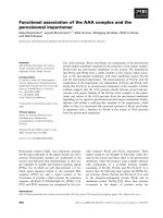
were used as markers for cell cycle synchronization. In
agreement with previous studies, cyclin B was
expressed prominently in the G2⁄ M phase, whereas
cyclin E expression was high in S and G1 phase, but
low in G2 ⁄ M phase (Fig. 1) [39].
In order to understand the dynamics of MLL1, we
performed immunofluorescence staining of the syn-
chronized HeLa cells with anti-MLL1 serum, and
visualized its localization using fluorescence micros-
copy at different stages of the cell cycle. In agree-
ment with our previous studies, we found that MLL1
was localized inside the euchromatic region [less
intense 4¢,6-diamidino-2-phenylindole (DAPI)-stained
region] of the nucleus at the G1 phase of the cells
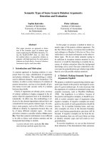
(G1 phase, panels 1–3, Fig. 2) [12]. However, as the
cell entered into mitosis and chromatin was con-
densed, most of the MLL1 protein was dissociated
from the chromatin and spread into the cytoplasm,
generating a distinct footstep (gap) for condensed
chromatin (see metaphase, anaphase and early telo-
phase stages, panels 1–3, Fig. 2). Notably, the spread-
ing of MLL1 protein into the cytoplasm coincided
with the disappearance of the nuclear membrane at
Unsynchr
onized
0 2.5 5 7.5 10 12.5 15 17.5 20
Time after synchronization (h)
S G2/M G1
Actin
Cyclin E
Cyclin B
Fig. 1. Synchronization of cells. HeLa cells were synchronized
using double thymidine treatment and released into the G1 ⁄ S
boundary, as described previously. Cyclins B and E were used as
markers for cell cycle synchronization. Proteins at different phases
of the cell cycle were analyzed by western blotting using anti-
cyclin E and B sera. Actin was used as loading control.
MLL and H3K4 methylations during cell cycle B. P. Mishra et al.
1630 FEBS Journal 276 (2009) 1629–1640 ª 2009 The Authors Journal compilation ª 2009 FEBS
the beginning of mitosis (Fig. S1, see Supporting
information). Interestingly, at early telophase, when
the cells were completely divided but the nuclei of
the nascent daughter cells were yet to relax into
euchromatin, MLL1 was present in the cytoplasm
(early telophase, panels 1–3, Fig. 2). However, at
later stages, MLL1 returned to the condensed chro-
matin, probably marking the initiation of chromatin
relaxation (euchromatin formation) (late telophase,
panels 1–3, Fig. 2).
Recently, Liu et al. [40] performed immunostaining
experiments with anti-MLL1 serum using asynchro-
nous HeLa cells. In contrast with our observations,
they reported that MLL1 remains associated with
condensed chromatins even during mitosis, but is
degraded at late M (mitosis) and S phases. To address
this apparent contradictory MLL1 distribution pattern
in mitotic cells, we performed further immunostaining
experiments with several MLL1-interacting proteins,
such as CGBP, Ash2, Rbbp5, etc., using synchronized
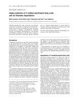
HeLa cells. Interestingly, each of these MLL-interact-
ing proteins (CGBP, Ash2 and Rbbp5) was dissociated
from mitotic chromatin, leaving a distinct gap in the
mitotic cells in a very similar fashion to the MLL1 dis-
tribution (Fig. 3). Notably, in our studies, we also
found the presence of these distinct gaps for MLL1
and interacting proteins in mitotic cells in a population
of asynchronous cells (data not shown). These results
indicate that MLL1 and its interacting proteins dissoci-
ate from mitotic chromatins, spread into the cytoplasm
and coordinate in a similar fashion during the cell
cycle.
1234
DAPI Merge1
G1 phase
Prophase
Metaphase
Anaphase
Telophase
Late
Early
10 µm
MLL1 Merge2
Fig. 2. Dynamics of MLL1 during the cell
cycle. Synchronized HeLa cells (at different
stages) were subjected to immunofluores-
cence staining with anti-MLL1 serum and
visualized by immunostaining with FITC
(green) conjugated secondary antibodies.
Cells were costained with DAPI to visualize
the DNA. Merge 1 shows the merge
between DAPI and MLL1 images. Merge 2
shows the merge between DAPI and differ-
ential interference contrast images of the
same cell.
B. P. Mishra et al. MLL and H3K4 methylations during cell cycle
FEBS Journal 276 (2009) 1629–1640 ª 2009 The Authors Journal compilation ª 2009 FEBS 1631
H3K4 trimethylation marks are associated with
mitotic chromatins
In contrast with MLL1 and its interacting proteins,
H3K4 trimethylation marks behave differently during
the cell cycle. Notably, like MLL1, H3K4 trimethyla-
tion is well known to be associated with transcription-
ally active euchromatin [12,41]. Therefore, MLL1 and
H3K4 trimethylation have been shown (by our labora-
tory and others) to be colocalized in the euchromatic
regions of the nucleus, and this is probably because of
their involvement in active gene expression [12,41].
Herein, in order to understand the dynamic association
of H3K4 trimethylation with chromatin during the cell
cycle, we performed immunofluorescence staining of
HeLa cells with anti-H3K4 trimethyl serum at different
stages of the cell cycle. The cell nucleus was counter-
stained and visualized using DAPI staining. As
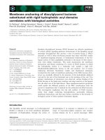
expected, in the G1 phase, H3K4 trimethylation marks
were localized in the less intense DAPI-stained regions
in the nucleus (representing less condensed euchroma-
tin), leaving gaps in the more intense DAPI-stained
regions (representing more condensed heterochromatin)
(G1 phase, panels 1–3, Fig. 4). However, in contrast
with MLL1, as the cells entered into mitosis and DNA
was condensed, H3K4 trimethylation marks still
remained associated with condensed chromatin and
remained so throughout the cell cycle (panels 1 and 2,
Fig. 4). As H3K4 trimethylation is well recognized as a
mark for active chromatins, the existence of these
marks, even in the highly condensed mitotic chromatin,
was unanticipated. The contradictory association of
MLL1 and H3K4 trimethylation marks indicates at
least two different possibilities. H3K4 trimethylation
marks that are introduced into transcriptionally active
euchromatins at the G1 phase are not removed from
Mitotic cell
MLL1
CGBP
Ash2
Rbbp5
DAPI FITC
Merge
10 µm
Fig. 3. Dynamics of MLL-interacting pro-
teins. Synchronized HeLa cells at meta-
phase stage (mitosis) were subjected to
immunofluorescence staining with anti-
MLL1, anti-CGBP, anti-Ash2 and anti-Rbbp5
sera, and visualized by immunostaining with
FITC (green) conjugated secondary anti-
bodies. Cells were costained with DAPI to
visualize the DNA. The merge panel shows
the overlay between DAPI and FITC images.
MLL and H3K4 methylations during cell cycle B. P. Mishra et al.
1632 FEBS Journal 276 (2009) 1629–1640 ª 2009 The Authors Journal compilation ª 2009 FEBS
histones and are carried over throughout the cell cycle.
Secondly, even in condensed chromatin during mitosis,
some genes remain transcriptionally active and these
are marked by H3K4 trimethylation. Notably, the asso-
ciation of H3K4 trimethylation marks with mitotic
chromatin has been observed previously by Valls et al.
[42]. We analyzed H3K9 dimethylation as the mark of
heterochromatin and, as expected, H3K9 methylation
marks were found to be associated with heterochroma-
tin throughout the cell cycle (panels 4–6, Fig. 4).
MLL1 and H3K4 trimethylation levels remain
unaffected whereas Hox genes are differentially
expressed during the cell cycle
As MLL1 and H3K4 trimethylation show distinct
dynamics during cell cycle progression, we analyzed
the expression profiles of MLL1, CGBP, Ash2 and
Rbbp5, together with cyclins E and B, as a function of
the cell cycle. Western blot analysis of the whole-cell
extract and histones from different stages of the cell
cycle demonstrated that the overall levels of MLL1
and H3K4 trimethylation were unaffected throughout
the cell cycle (Fig. 5). Similarly, MLL-interacting pro-
teins, such as CGBP, Ash2 and Rbbp5, were unaf-
fected during the cell cycle (data not shown). Notably,
again, our observations showing the unaffected global
level of MLL1 (protein level) during the cell cycle con-
tradict the observations by Liu et al. [40], who demon-
strated that MLL1 proteins were degraded during late
M (mitosis) and S phases. However, in agreement with
Liu et al. [40], using RT-PCR analysis, we observed
that the expression of MLL1 at the mRNA level was
increased from G1 ⁄ S towards G2 ⁄ M (Fig. S2, see
Supporting information). Furthermore, to confirm cell
synchronization, we analyzed the changes in phosphor-
ylation level of H3Ser28, which is considered to be a
marker for mitotic cells. Indeed, in agreement with
DAPI
1
G1 phase
Prophase
Metaphase
Anaphase
Telophase
DAPI
10 µm
5234 6
H3K9-di
methylation
Merge Merge H3K4-tri-
methylation
Fig. 4. Dynamics of H3K4 trimethylation and H3K9 dimethylation during the cell cycle. Synchronized HeLa cells (at different stages) were
subjected to immunofluorescnce staining with H3K4 trimethyl and H3K9 dimethyl antibodies, and visualized by immunostaining with rhoda-
mine (red) conjugated secondary antibodies. Cells were costained with DAPI to visualize the DNA. Merge panels show the overlay between
DAPI- and rhodamine-stained images.
B. P. Mishra et al. MLL and H3K4 methylations during cell cycle
FEBS Journal 276 (2009) 1629–1640 ª 2009 The Authors Journal compilation ª 2009 FEBS 1633
previous studies, we found that H3Ser28 phosphoryla-
tion was only observed during mitosis, indicating cor-
rect cell cycle progression and synchronization (Fig. 5)
[43,44]. These observations further support the fact
that H3K4 trimethylation marks are maintained
throughout the cell cycle, even in mitotically condensed
chromatins. As the levels of MLL1 protein remained
unaffected, we conclude that MLL proteins are not
degraded during mitosis, but rather moved away from
condensed chromatin towards the cytoplasm, generat-
ing the MLL1 gaps present in mitotic chromatin.
In contrast with MLL1 and H3K4 trimethylation
levels, the MLL target Hox genes were differentially
expressed during the cell cycle. We analyzed the
expression profiles of three Hox genes, HoxA5, HoxA7
and HoxA10. HoxA5 is expressed at a low level at the
beginning of the S phase and increases by approxi-
mately eight-fold as the cell progresses from S to
G2 ⁄ M (0–10 h); it then decreases to almost the initial
level and remains so throughout mitosis and the
G1 phase (Fig. 6A,B). In contrast, HoxA7 expression
is low at the beginning (S phase) and increases gradu-
ally all the way from S to G2 ⁄ M to G1 phases
(0–20 h) (Fig. 6A,B). Interestingly, however, HoxA10
is only expressed in the beginning of S phase and shuts
down almost completely for the remaining phases of
the cell cycle (Fig. 6A,B). Cyclins B and E were used
as markers, and their expression patterns were in
agreement with previous studies and the results
presented in Fig. 1.
Recently, several studies have indicated that Hox
genes may also be involved in cell cycle progression.
For example, HoxA5 activates p53, which regulates
the expression of p21, an inhibitor of cyclin-dependent
kinases, which are critical for cell cycle progression.
Furthermore, Bromleigh and Freedman [45] showed
that HoxA10 directly upregulates the expression of
p21, leading to cell cycle arrest at the G1 phase. Both
p21 and p53 play a vital role in cell cycle regulation.
Thus, although further studies are needed to elucidate
the detailed functions of different Hox genes in cell
cycle regulation, our studies showing the differential
expression of HoxA5 , HoxA7 and HoxA10 at different
phases of the cell cycle indicate that these genes may
have critical roles in cell cycle checkpoint regulation,
probably via the involvement of p53 and p21.
MLL1 and H3K4 methylation are critical for Hox
gene regulation during the cell cycle
In order to understand the molecular mechanism of
the differential regulation of Hox gene expression, we
analyzed the changes in H3K4 methylation and
recruitment of MLL1 and RNA polymerase II (RNAP
II) at the Hox gene promoters at different phases of
the cell cycle using chromatin immunoprecipitation
(ChIP) assay [12]. We performed ChIP analysis using
anti-RNAP II, anti-MLL1 and anti-H3K4 trimethyl
sera at three different phases of the cell cycle [0 h (S),
10 h (G2 ⁄ M) and 20 h (G1)] after synchronized cells
were released at the S phase. In the case of HoxA5,
recruitment of RNAP II and MLL1, and the level of
H3K4 trimethylation in the promoter, were low at
S phase (0 h), increased by 1.7-fold at G2 ⁄ M (10 h)
and decreased again at G1 (20 h) (Fig. 6C,D). Nota-
bly, the enrichment of RNAP II, MLL1 and H3K4
trimethylation at the HoxA5 gene promoter at the
G2 ⁄ M phase was correlated with its expression profile
(as shown in Fig. 6A,B), indicating the importance of
MLL1 and H3K4 trimethylation in HoxA5 gene
regulation during cell cycle progression. The associa-
tion of a certain amount of RNAP II with the HoxA5
gene promoter at 20 h (although much lower in com-
parison with that at 10 h) indicates that a certain
amount of basal transcription still continues at this
stage of the cell cycle. Similar to HoxA5, the occu-
pancy of RNAP II, MLL1 and H3K4 trimethylation
Actin
MLL1
H3K4-Tri
methyl
H4 acetyl
H3Ser28P
Ash2
H3K9-di
methyl
Histone
(coomassie
staining)
S
Unsynch
ronized
Time after synchronization
(h)
02012.5
1510
5
17.5
7.5
2.5
G2/M
G1
Fig. 5. MLL1 expression and histone modifications during the cell
cycle. Synchronized HeLa cells were collected at 2.5 h intervals
after release at the G1 ⁄ S boundary and subjected to whole-cell pro-
tein extract and histone purification. The protein extracts were ana-
lyzed using western blotting with antibodies specific to MLL1,
Ash2 and CGBP. Actin was used as loading control. Histones were
probed with anti-H3K4 trimethyl, anti-H3K9 dimethyl, anti-H4 acety-
lation and anti-H3S28 phosphorylation sera. Cyclin B and E expres-
sion and H3S28 phosphorylation were used as markers for cell
synchronization. Coomassie stain for histone was used as loading
control.
MLL and H3K4 methylations during cell cycle B. P. Mishra et al.
1634 FEBS Journal 276 (2009) 1629–1640 ª 2009 The Authors Journal compilation ª 2009 FEBS
RNAP II
H3K4-Trimethyl
MLL1
EC
D
AB
lortn
oC
1
L
LM
esnesitnA
MLL1
HoxA5
HoxA7
HoxA10
Actin
tnemtiurcer evitaleR
0.3
0.6
0.9
1.2
1.5
Control
0 h
10 h
20 h
Control
0 h
10 h
20 h
Control
0 h
10 h
20 h
HoxA5 HoxA7 HoxA10
Actin
Cyclin B
HoxA5
HoxA7
HoxA10
Cyclin E
Time after synchronization (h)
0 2.5 5 7.5 10 12.5 15 17.5 20
Unsynchro
nized
S G2/M G1
n
o
iss
er
pxe evita
leR
Time after synchronization (h)
0
0.4
0.8
1.2
1.6
0 5 10 15 20
HoxA7
HoxA5
HoxA10
{
RNAP II
H3K4-tri
methyl
MLL1
Time (h)
Input
ChIP
Input
ChIP
Input
ChIP
{
{
HoxA5 HoxA7 HoxA10
C 0 10 20 C 0 10 20 C 0 10 20
Fig. 6. (A) Hox gene expression during the cell cycle. Total RNA was isolated from HeLa cells at different phases of the cell cycle and ana-
lyzed by RT-PCR using primers specific to cyclin E, cyclin B, HoxA5, HoxA7 and HoxA10. Actin was used as loading control. (B) PCR prod-
ucts of Hox genes in (A) were quantified and plotted. Experiments were repeated thrice and the bars indicate the standard errors of the
mean (SEMs). (C) ChIP experiments. HeLa cells were collected at S (0 h), M (10 h) and G1 (20 h) phases of the cell cycle (after synchroniza-
tion), fixed with formaldehyde, sonicated and analyzed by ChIP assay using antibodies against RNAP II, H3K4 trimethyl and MLL1. The
immunoprecipitated DNAs were PCR amplified using primers specific to the promoters of HoxA5, HoxA7 and HoxA10 genes. (D) The PCR
products in (C) were quantified and the fold increase in ChIP PCR products compared with the control (input) was plotted for the respective
Hox genes. Bars indicate SEMs. (E) Antisense-mediated knockdown of MLL1 and its effect on the expression of Hox genes. HeLa cells
were transfected with MLL1 antisense or scramble phosphorothioate antisense for 48 h, and RNAs from the transfected cells were analyzed
by RT-PCR using primers specific to MLL1, HoxA5, HoxA7 and HoxA10. Actin was used as loading control.
B. P. Mishra et al. MLL and H3K4 methylations during cell cycle
FEBS Journal 276 (2009) 1629–1640 ª 2009 The Authors Journal compilation ª 2009 FEBS 1635
in HoxA7 and HoxA10 gene promoters was also corre-
lated with their respective expression profiles (compare
Fig. 6A,B with Fig. 6C,D). In the case of the HoxA7
gene promoter, the recruitment of RNAP II and
MLL1 and the level of H3K4 trimethylation were low
at the beginning (S phase) and gradually increased as
the cell progressed from S to G2 ⁄ M to G1, reaching a
maximum at G1 (20 h) (Fig. 6C,D). In the case of the
HoxA10 gene, significantly higher levels of RNAP II
and MLL1 recruitment and H3K4 trimethylation
marks were observed at the beginning of the S phase
(0 h), and these marks were attenuated for the rest of
the cell cycle (10 and 20 h), correlating with the
expression of the gene (Fig. 6C,D). The correlation of
promoter occupancy of MLL1, H3K4 trimethylation
and RNAP II with Hox gene expression indicates the
critical roles of MLL1 and H3K4 trimethylation in dif-
ferential Hox gene expression during the cell cycle. To
further confirm the importance of MLL1 in the regula-
tion of HoxA5, HoxA7 and HoxA10 genes and cell
cycle progression, we knocked down MLL1 using a
specific antisense oligonucleotide and analyzed the
expression of Hox genes and cyclins. As shown in
Fig. 6E, the knockdown of MLL1 down-regulated the
expression of HoxA5, HoxA7 and HoxA10 genes.
Notably, HoxA5 expression was almost completely
abrogated, whereas HoxA7 and HoxA10 were only
partially down-regulated. The partial down-regulations
of HoxA7 and HoxA10 on knockdown of MLL1 indi-
cate that, in addition to MLL1, other alternative fac-
tors may regulate their expression. Notably, cyclins B
and E were also down-regulated in an MLL1 knocked
down environment (data not shown).
To confirm further the role of MLL1 in cell cycle
regulation, we examined the effects of knockdown of
MLL1 on cell cycle progression using flow cytometry
analysis. Briefly, HeLa cells (at 60% confluence) were
transfected with MLL1-specific antisense oligonucleo-
tide for 24 h, stained with propidium iodide and ana-
lyzed using a flow cytometry analyzer. Interestingly, as
shown in Fig. 7, on treatment with the MLL1 anti-
sense oligonucleotide, the cell population at the
G2 ⁄ M phase increased from 3.5% (control) to 19.7%
(antisense treated). Notably, application of the scram-
ble antisense oligonucleotide (with no homology to
MLL1) also led to a slight increase in the G2 ⁄ M cell
population (to 7%) in comparison with the control.
The MLL1 antisense-mediated increase in the cell pop-
ulation at the G2 ⁄ M phase indicated that knockdown
of MLL1 resulted in cell cycle arrest at the
G2 ⁄ M phase. These observations further confirmed the
significant role of MLL1 in cell cycle progression.
Our results demonstrate that MLL1 and H3K4
trimethylation show different dynamics during the cell
cycle. MLL1, which is well known for transcription
activation, remains associated with transcriptionally
active chromatin (euchromatin), dissociates from con-
densed mitotic chromatin and returns at the end of
telophase when the nucleus starts to relax. In contrast,
H3K4 trimethylation marks, which are marks for gene
activation, remain associated with euchromatin in the
G1 phase and even with condensed chromatin
throughout the cell cycle. The global levels of MLL1
protein and H3K4 trimethylation are not degraded or
removed from the cells during mitosis, but H3Ser28
phosphorylation is only observed during mitosis. How-
ever, the recruitment of MLL1 and the level of H3K4
trimethylation are modulated in the promoters of spe-
cific Hox genes as a function of their expression.
Importantly, as we observed that H3K4 trimethylation
fluctuates at specific gene promoters, we hypothesize
that the H3K4 trimethylation marks that are present
Apoptotic 1.3
G0-G1 71.6
S 13.3
G2-M 7.0
Apoptotic 1.6
G0-G1 64.2
S 12.2
G2-M 19.7
Apoptotic 1.4
G0-G1 74.3
S 15.7
G2-M 3.5
AB C
Fig. 7. Knockdown of MLL1 induces cell cycle arrest at G2 ⁄ M phase. HeLa cells were treated with MLL1 and scramble antisense sepa-
rately for 24 h, and subjected to flow cytometry analysis. (A) Control cells treated with no antisense. (B) Cells treated with phosphorothioate
scramble antisense (no homology to MLL1). (C) Cells treated with MLL1-specific antisense. The cell populations at different stages of the
cell cycle are shown inside the respective panels.
MLL and H3K4 methylations during cell cycle B. P. Mishra et al.
1636 FEBS Journal 276 (2009) 1629–1640 ª 2009 The Authors Journal compilation ª 2009 FEBS
in S phase may not be the same as the marks in other
phases of the cell cycle (as shown by immunofluores-
cence staining and western blotting); rather, old marks
are removed and new marks are introduced, at least in
some of the promoters. Furthermore, although we
observed distinct gaps for MLL1 (as well as its inter-
acting proteins) in immunofluorescence staining experi-
ments in the region of mitotically condensed
chromatin, ChIP experiments demonstrated that
MLL1 is still bound to the promoters of active Hox
genes even during mitosis. These observations indicate
that a certain amount of MLL1 protein is still associ-
ated with chromatin even during mitosis, although
most of the proteins migrate away from chromatin.
Our studies also demonstrate that Hox genes
(HoxA5, HoxA7 and HoxA10) are differentially regu-
lated during the cell cycle and MLL1 occupancy at the
Hox gene promoter fluctuates as a function of Hox
gene expression. Notably, HoxA5 has been shown to
activate p53, which regulates the expression of the
cyclin-dependent kinase inhibitor p21 [46]. Similarly,
HoxA10 is known to upregulate p21, leading to cell
cycle arrest at the G1 phase in both monocytic and
fibroblast cell lines [45]. Thus, it is possible that
HoxA5, similar to HoxA10, regulates the cell cycle via
p53 and p21 channels. Similar to the Hox gene, MLLs
have also been shown to interact with the E2F family
of proteins and to regulate cell cycle regulatory genes,
including cyclins [26]. Thus, our results and indepen-
dent observations from different laboratories indicate
that both MLL1 and Hox genes are critical players in
cell cycle progression. Although further studies are
needed to understand the detailed roles of MLLs and
different Hox genes in cell cycle regulation, our studies
demonstrate distinct dynamics and the importance of
MLL1, H3K4 methylation and selected Hox genes
during cell cycle progression.
Experimental procedures
Cell culture and synchronization
HeLa cells were grown in Dulbecco’s modified Eagle’s med-
ium (DMEM) supplemented with heat-inactivated fetal
bovine serum (10%), l-glutamine (1%) and penicillin ⁄ strep-
tomycin (0.1%), as described previously [12,47,48]. Cells
were synchronized at G1 ⁄ S phase using double thymidine
treatment, as described previously [38,49]. Briefly, cells were
grown in a 10 cm tissue culture plate up to 25% confluence,
treated with 10 mm thymidine (Sigma, New York, NY,
USA) for 18 h, released into fresh medium for 9 h and
blocked again by the addition of 10 mm thymidine for an
additional 17 h. Finally, the cells were released into fresh
medium at G
1
⁄ S phase and analyzed at 2.5 h intervals.
Preparation of whole-cell extract, histones and
western blotting
HeLa cells (10 cm plates) were harvested, incubated with
200 lL of whole-cell extract buffer (50 mm Tris ⁄ HCI,
pH 8.0, 150 mm NaCl, 5 mm EDTA, 0.05% NP-40, 0.2 m m
phenylmethanesulfonyl fluoride, 1· protease inhibitors) on
ice for 20 min and centrifuged (10 000 g for 10 min). The
supernatant was used as whole-cell extract and the pellet
was used for histone purification, as described previously
[49]. The whole-cell protein extracts and histones were
analyzed by western blotting using anti-MLL1 (Bethyl
Laboratories, Montgomery, TX, USA), anti-Set1 (Bethyl
Laboratories), anti-Ash2 (Bethyl Laboratories), anti-Rbbp5
(Bethyl Laboratories), anti-CGBP (IMGENEX, San Diego,
CA, USA), anti-cyclin B (Santa Cruz Biotechnology, Santa
Table 1. Nucleotide sequences of the primers used in PCR and ChIP analyses.
Transcript Forward primer (5¢-to3¢) Reverse primer (5¢-to3¢)
MLL1 GAG GAC CCC GGA TTA AAC AT GGA GCA AGA GGT TCA GCA TC
Ash2 CCT GAA GCA GAC TCC CCA TA AGC CCA TGT CAC TCA TAG GG
Rbbp5 GCA TCC ATT TCC AGT GGA GT TGG TGA CAT CCA CTT CCT CA
CGBP GCC ACA CGA CTA TTC TGT GA CAG TAA TGG CGA TTG CAC TG
Cyclin E TTTCAGGGTATCAGTGGTGCGACA ACA ACA TGG CTT TCT TTG CTC GGG
Cyclin B TTG ATA CTG CCT CTC CAA GCC CAA TTG GTC TGA CTG CTT GCT CTT CCT
HoxA5 GGC TAC AAT GGC ATG GAT CT GCT GGA GTT GCT TAG GGA GTT
HoxA7 TTC CAC TTC AAC CGC TAC CT TTC ATC ATC GTC CTC CTC GT
HoxA10 CCA TAG ACC TGT GGC TAG ACG GAG ACT TTG GGG CAT TTG TC
HoxA5 (P)
a
AGT AAG TCC CGA AGG GCA TC GAG AGA CTG GGC TCT GTT GG
HoxA7 (P)
a
GAG CCT CCA GGT CTT TTT CC ACA CCC CCA GAT TTA CAC CA
HoxA10 (P)
a
CTC CTG GCC CAT CAA TAC AG TAG CCC TTT CTG GCT GAC AT
Actin AGA GCT ACG AGC TGC CTG AC GTA CTT GCG CTC AGG AGG AG
a
Primer pairs specific to promoters of respective genes.
B. P. Mishra et al. MLL and H3K4 methylations during cell cycle
FEBS Journal 276 (2009) 1629–1640 ª 2009 The Authors Journal compilation ª 2009 FEBS 1637
Cruz, CA, USA), anti-cyclin E (Santa Cruz Biotechnology),
anti-H3K4 trimethyl (Upstate Biotech, Waltham, MA,
USA), anti-H3S28 phosphoryl (Upstate Biotech) and anti-
H3K9 dimethyl (Upstate Biotech) sera.
RNA purification and RT-PCR
For RNA purification, cells were resuspended in 200 lLof
diethylpyrocarbonate (DEPC)-treated buffer A (20 mm
Tris ⁄ HCl, pH 7.9, 1.5 mm MgCl
2
,10mm KCl, 0.5 mm
dithiothreitol, 0.2 mm phenylmethanesulfonyl fluoride),
incubated on ice (10 min) and centrifuged at 3500 g for
5 min. The supernatant (cytoplasmic extracts) was subjected
to phenol–chloroform extraction, followed by ethanol pre-
cipitation, to obtain cytoplasmic mRNAs. mRNA was
washed with DEPC-treated 70% ethanol, air dried, resus-
pended in DEPC-treated water, quantified and subjected to
RT-PCR. RT reactions were performed in a total volume of
25 lL containing 1 lg of total RNA, 2.4 lm of oligo-dT,
100 U of MMLV reverse transcriptase (Promega, Madison,
WI, USA), 1· first strand buffer (Promega), 100 lm dNTPs,
1mm dithiothreitol and 20 U of RNaseOut (Invitrogen,
Carlsbad, CA, USA). This cDNA (1 lL) was PCR ampli-
fied with the specific primer pairs listed in Table 1.
Immunofluorescence studies
HeLa cells were grown on cover slips, synchronized, fixed in
4% p-formaldehyde, permeabilized with 0.2% Triton-X100,
blocked with goat serum, incubated (1 h) with the respective
primary antibodies (MLL1, CGBP, Ash2, Rbbp5, H3K4
trimethyl and H3K9 dimethyl antibodies), washed and incu-
bated with fluorescein isothiocyanate (FITC) or rhodamine
(Jackson Immuno Research Laboratories, West Grove, PA,
USA) conjugated secondary antibodies. Nuclear counter-
staining was performed with DAPI. Immunostained cells
were mounted and observed under a fluorescence microscope
(Nikon Eclipse TE2000-U; Nikon, Melville, NY, USA).
Antisense-mediated knockdown of MLL1 and
ChIP assay
HeLa cells were transfected with MLL1-specific phosphoro-
thioate antisense oligonucleotide (5¢-TGCCAGTCGTTCC
TCTCCAC-3¢) using commercial Maxfect transfection
reagent, following the manufacturer’s instructions (Molecu-
lA, Columbia, MD, USA). A scramble antisense oligonucleo-
tide without any sequence homology with MLL1 (5¢-CGT
TTGTCCCTCCAGCATCT-3¢) was used as control. For
ChIP assay, HeLa cells (collected at 0, 10 and 20 h after
synchronization) were fixed with 1% formaldehyde, washed,
resuspended in lysis buffer (1% SDS, 10 mm EDTA, 50 mm
Tris ⁄ HCl, pH 8, 1· protease inhibitors and 0.2 mm
phenylmethanesulfonyl fluoride), sonicated until chromatin
was sheared to an average DNA fragment length of
0.2–0.5 kb and subjected to ChIP assay as described previ-
ously [12].
Flow cytometry analysis
HeLa cells were grown to 60% confluence and transfected
with MLL1 and scramble antisense oligonucleotides sepa-
rately using Maxfect transfection (MoleculA) reagents, and
incubated for 24 h. Control and transfected cells were
harvested, fixed in 70% ethanol for 2 h, washed twice with
1· NaCl ⁄ P
i
and stained with propidium iodide (final con-
centration, 0.5 lgÆ mL
)1
). The cells were analyzed by flow
cytommetry, using a Fusing Beckman Coulter (Fullerton,
CA, USA) Cytomics FC500 Flow Cytometry Analyzer.
Acknowledgements
We thank Saoni Mandal and Mandal laboratory mem-
bers for critical discussions. This work was supported
by grants from the Texas Advanced Research Program
(00365-0009-2006) and the American Heart Associa-
tion (SM 0765160Y).
References
1 Goldberg AD, Allis CD & Bernstein E (2007) Epigenet-
ics: a landscape takes shape. Cell 128, 635–638.
2 Martin C & Zhang Y (2005) The diverse functions of
histone lysine methylation. Nat Rev Mol Cell Biol 6,
838–849.
3 Sims RJ III, Mandal SS & Reinberg D (2004) Recent
highlights of RNA-polymerase-II-mediated transcrip-
tion. Curr Opin Cell Biol 16, 263–271.
4 Bannister AJ & Kouzarides T (2004) Histone methyla-
tion: recognizing the methyl mark. Methods Enzymol
376, 269–288.
5 Elgin SC & Grewal SI (2003) Heterochromatin: silence
is golden. Curr Biol 13, R895–R898.
6 Fischle W, Wang Y & Allis CD (2003) Binary switches
and modification cassettes in histone biology and
beyond. Nature 425, 475–479.
7 Jenuwein T & Allis CD (2001) Translating the histone
code. Science 293, 1074–1080.
8 Wysocka J, Milne TA & Allis CD (2005) Taking LSD 1
to a new high. Cell 122, 654–658.
9 Peterson CL & Laniel MA (2004) Histones and histone
modifications. Curr Biol 14, R546–R551.
10 Shi YJ, Matson C, Lan F, Iwase S, Baba T & Shi Y
(2005) Regulation of LSD1 histone demethylase activity
by its associated factors. Mol Cell 19, 857–864.
11 Dutnall RN (2003) Cracking the histone code: one,
two, three methyls, you’re out!. Mol Cell 12, 3–4.
MLL and H3K4 methylations during cell cycle B. P. Mishra et al.
1638 FEBS Journal 276 (2009) 1629–1640 ª 2009 The Authors Journal compilation ª 2009 FEBS
12 Ansari KI, Mishra BP & Mandal SS (2008) Human
CpG binding protein interacts with MLL1, MLL2 and
hSet1 and regulates Hox gene expression. Biochim Bio-
phys Acta 1779, 66–73.
13 Hess JL (2004) MLL: a histone methyltransferase dis-
rupted in leukemia. Trends Mol Med 10, 500–507.
14 Steward MM, Lee JS, O’Donovan A, Wyatt M, Bern-
stein BE & Shilatifard A (2006) Molecular regulation of
H3K4 trimethylation by ASH2L, a shared subunit of
MLL complexes. Nat Struct Mol Biol 13, 852–854.
15 Yokoyama A, Somervaille TC, Smith KS, Rozenblatt-
Rosen O, Meyerson M & Cleary ML (2005) The menin
tumor suppressor protein is an essential oncogenic
cofactor for MLL-associated leukemogenesis. Cell 123,
207–218.
16 Hughes CM, Rozenblatt-Rosen O, Milne TA, Copeland
TD, Levine SS, Lee JC, Hayes DN, Shanmugam KS,
Bhattacharjee A, Biondi CA et al. (2004) Menin associ-
ates with a trithorax family histone methyltransferase
complex and with the hoxc8 locus. Mol Cell 13, 587–
597.
17 Nakamura T, Mori T, Tada S, Krajewski W, Rozovs-
kaia T, Wassell R, Dubois G, Mazo A, Croce CM &
Canaani E (2002) ALL-1 is a histone methyltransferase
that assembles a supercomplex of proteins involved in
transcriptional regulation. Mol Cell 10, 1119–1128.
18 Yu BD, Hanson RD, Hess JL, Horning SE & Kors-
meyer SJ (1998) MLL, a mammalian trithorax-group
gene, functions as a transcriptional maintenance factor
in morphogenesis. Proc Natl Acad Sci USA 95, 10632–
10636.
19 Goo YH, Sohn YC, Kim DH, Kim SW, Kang MJ,
Jung DJ, Kwak E, Barlev NA, Berger SL, Chow VT
et al. (2003) Activating signal cointegrator 2 belongs to
a novel steady-state complex that contains a subset of
trithorax group proteins. Mol Cell Biol 23, 140–149.
20 Mo R, Rao SM & Zhu YJ (2006) Identification of the
MLL2 complex as a coactivator for estrogen receptor
alpha. J Biol Chem 281, 15714–15720.
21 Nakanishi S, Sanderson BW, Delventhal KM, Bradford
WD, Staehling-Hampton K & Shilatifard A (2008) A
comprehensive library of histone mutants identifies
nucleosomal residues required for H3K4 methylation.
Nat Struct Mol Biol 15, 881–888.
22 Ng HH, Robert F, Young RA & Struhl K (2003) Tar-
geted recruitment of Set1 histone methylase by elongat-
ing Pol II provides a localized mark and memory of
recent transcriptional activity. Mol Cell 11, 709–719.
23 Zhang K, Lin WC, Latham JA, Riefler GM, Schumach-
er JM, Chan C, Tatchell K, Hawke DH, Kobayashi R
& Dent SYR (2005) The set1 methyltransferase opposes
IpI1 aurora kinase functions in chromosome segrega-
tion. Cell 122, 723–734.
24 Dou Y, Milne TA, Ruthenburg AJ, Lee S, Lee JW,
Verdine GL, Allis CD & Roeder RG (2006) Regulation
of MLL1 H3K4 methyltransferase activity by its core
components. Nat Struct Mol Biol 13, 713–719.
25 Crawford BD & Hess JL (2006) MLL core components
give the green light to histone methylation. ACS Chem
Biol 1, 495–498.
26 Takeda S, Chen DY, Westergard TD, Fisher JK,
Rubens JA, Sasagawa S, Kan JT, Korsmeyer SJ, Cheng
EH & Hsieh JJ (2006) Proteolysis of MLL family
proteins is essential for taspase1-orchestrated cell cycle
progression. Genes Dev 20, 2397–2409.
27 Tenney K & Shilatifard A (2005) A COMPASS in
the voyage of defining the role of trithorax ⁄ MLL-con-
taining complexes: linking leukemogensis to covalent
modifications of chromatin. J Cell Biochem 95, 429–
436.
28 Glaser S, Schaft J, Lubitz S, Vintersten K, van der
Hoeven F, Tufteland KR, Aasland R, Anastassiadis K,
Ang SL & Stewart AF (2006) Multiple epigenetic main-
tenance factors implicated by the loss of Mll2 in mouse
development. Development 133, 1423–1432.
29 Cho YW, Hong T, Hong S, Guo H, Yu H, Kim D,
Guszczynski T, Dressler GR, Copeland TD, Kalkum M
et al. (2007) PTIP associates with MLL3- and MLL4-
containing histone H3 lysine 4 methyltransferase com-
plex. J Biol Chem 282, 20395–20406.
30 Issaeva I, Zonis Y, Rozovskaia T, Orlovsky K, Croce
CM, Nakamura T, Mazo A, Eisenbach L & Canaani E
(2007) Knockdown of ALR (MLL2) reveals ALR target
genes and leads to alterations in cell adhesion and
growth. Mol Cell Biol 27, 1889–1903.
31 Pavri R, Zhu B, Li G, Trojer P, Mandal S, Shilatifard
A & Reinberg D (2006) Histone H2B monoubiquitina-
tion functions cooperatively with FACT to regulate
elongation by RNA polymerase II. Cell 125, 703–717.
32 Lappin TR, Grier DG, Thompson A & Halliday HL
(2006) HOX genes: seductive science, mysterious mecha-
nisms. Ulster Med J 75, 23–31.
33 Guenther MG, Jenner RG, Chevalier B, Nakamura T,
Croce CM, Canaani E & Young RA (2005) Global and
Hox-specific roles for the MLL1 methyltransferase.
Proc Natl Acad Sci USA 102, 8603–8608.
34 Capotosti F, Hsieh JJ & Herr W (2007) Species selectiv-
ity of mixed-lineage leukemia ⁄ trithorax and HCF
proteolytic maturation pathways. Mol Cell Biol 27,
7063–7072.
35 Tyagi S, Chabes AL, Wysocka J & Herr W (2007) E2F
activation of S phase promoters via association with
HCF-1 and the MLL family of histone H3K4 meth-
yltransferases. Mol Cell 27, 107–119.
36 Karnik SK, Hughes CM, Gu X, Rozenblatt-Rosen O,
McLean GW, Xiong Y, Meyerson M & Kim SK (2005)
Menin regulates pancreatic islet growth by promoting
histone methylation and expression of genes encoding
p27Kip1 and p18INK4c. Proc Natl Acad Sci USA 102 ,
14659–14664.
B. P. Mishra et al. MLL and H3K4 methylations during cell cycle
FEBS Journal 276 (2009) 1629–1640 ª 2009 The Authors Journal compilation ª 2009 FEBS 1639
37 Milne TA, Hughes CM, Lloyd R, Yang Z, Rozenblatt-
Rosen O, Dou Y, Schnepp RW, Krankel C, Livolsi
VA, Gibbs D et al. (2005) Menin and MLL coopera-
tively regulate expression of cyclin-dependent kinase
inhibitors. Proc Natl Acad Sci USA 102 , 749–754.
38 Adolph KW & Phelps JP (1982) Role of non-histones
in chromosome structure. Cell cycle variations in
protein synthesis. J Biol Chem 257, 9086–9092.
39 Hayami R, Sato K, Wu W, Nishikawa T, Hiroi J,
Ohtani-Kaneko R, Fukuda M & Ohta T (2005) Down-
regulation of BRCA1-BARD1 ubiquitin ligase by
CDK2. Cancer Res 65, 6–10.
40 Liu H, Takeda S, Cheng EH & Hsieh JJ (2008) Bipha-
sic MLL takes helm at cell cycle control: implications in
human mixed lineage leukemia. Cell Cycle 7, 428–435.
41 Lee JH & Skalnik DG (2002) CpG-binding protein is a
nuclear matrix- and euchromatin-associated protein
localized to nuclear speckles containing human tritho-
rax. Identification of nuclear matrix targeting signals.
J Biol Chem 277, 42259–42267.
42 Valls E, Sanchez-Molina S & Martinez-Balbas MA
(2005) Role of histone modifications in marking and
activating genes through mitosis. J Biol Chem 280,
42592–42600.
43 Eberlin A, Grauffel C, Oulad-Abdelghani M, Robert F,
Torres-Padilla ME, Lambrot R, Spehner D, Ponce-
Perez L, Wurtz JM, Stote RH et al. (2008) Histone H3
tails containing dimethylated lysine and adjacent phos-
phorylated serine modifications adopt a specific confor-
mation during mitosis and meiosis. Mol Cell Biol 28,
1739–1754.
44 Goto H, Tomono Y, Ajiro K, Kosako H, Fujita M,
Sakurai M, Okawa K, Iwamatsu A, Okigaki T, Takah-
ashi T et al. (1999) Identification of a novel phosphory-
lation site on histone H3 coupled with mitotic
chromosome condensation. J Biol Chem 274, 25543–
25549.
45 Bromleigh VC & Freedman LP (2000) p21 is a tran-
scriptional target of HOXA10 in differentiating myelo-
monocytic cells. Genes Dev 14, 2581–2586.
46 Raman V, Martensen SA, Reisman D, Evron E,
Odenwald WF, Jaffee E, Marks J & Sukumar S (2000)
Compromised HOXA5 function can limit p53 expres-
sion in human breast tumours. Nature 405, 974–978.
47 Osugi ME, Zanoni MVB, Chenthamarakshan CR, de
Tacconi NR, Woldemariam GA, Mandal SS & Rajesh-
war K (2008) Toxicity assessment and degradation of
disperse azo dyes by photoelectrocatalytic oxidation on
Ti ⁄ TiO
2
nanotubular array electrodes. J Adv Oxid
Technol 11, 425–434.
48 Woldemariam GA & Mandal SS (2008) Iron(III)-salen
damages DNA and induces apoptosis in human cell via
mitochondrial pathway. J Inorg Biochem 102, 740–747.
49 Rice JC, Nishioka K, Sarma K, Steward R, Reinberg D
& Allis CD (2002) Mitotic-specific methylation of his-
tone H4 Lys 20 follows increased PR-Set7 expression
and its localization to mitotic chromosomes. Genes Dev
16, 2225–2230.
Supporting information
The following supplementary material is available:
Fig. S1. Localization of the nuclear membrane during
the cell cycle.
Fig. S2. RT-PCR analysis of MLL1 and associated
proteins in the cell cycle.
This supplementary material can be found in the
online version of this article.
Please note: Wiley-Blackwell is not responsible for
the content or functionality of any supplementary
materials supplied by the authors. Any queries (other
than missing material) should be directed to the corre-
sponding author for the article.
MLL and H3K4 methylations during cell cycle B. P. Mishra et al.
1640 FEBS Journal 276 (2009) 1629–1640 ª 2009 The Authors Journal compilation ª 2009 FEBS