Báo cáo khoa học: Characterization of inhibitory mechanism and antifungal activity between group-1 and group-2 phytocystatins from taro (Colocasia esculenta) pdf
Bạn đang xem bản rút gọn của tài liệu. Xem và tải ngay bản đầy đủ của tài liệu tại đây (374.83 KB, 10 trang )
Characterization of inhibitory mechanism and antifungal
activity between group-1 and group-2 phytocystatins
from taro (Colocasia esculenta)
Ke-Ming Wang
1
, Senthil Kumar
1
, Yi-Sheng Cheng
1,2
, Shripathi Venkatagiri
3
, Ai-Hwa Yang
4
and Kai-Wun Yeh
1
1 Institute of Plant Biology, National Taiwan University, Taipei, Taiwan
2 Department of Life Science, National Taiwan University, Taipei, Taiwan
3 Department of Botany, Karnatak University, Dharwad, India
4 Tainan District of Agricultural Improvement and Extension Station, Council of Agriculture, Tainan, Taiwan
Phytocystatins are a class of reversibly binding cyste-
ine proteinase inhibitors found in plants. These
cysteine proteinase inhibitors lack disulfide bridges
and possess a conserved N-terminal amino acid
sequence [L-A-R-[FY]-A-[VI]-X(3)-N] [1]. Although
the primary sequences of phytocystatins are more
similar to the type II cystatins of animals, they are
assigned to an independent family [1]. Phytocystatins
have been reported to contain three motifs that are
involved in the interaction with their target protein-
ases: (a) the active site motif QxVxG; (b) a G near
N-terminus; and (c) a W in the second half of the
protein [2,3]. However, according to molecular
weight, they have been divided into three distinct
groups. Most of the phytocystatins are included in
group-1, such as oryzacystatin (OC)-I from rice, and
Keywords
allosteric activation; anti-fungal activity;
cysteine proteinase inhibitor; inhibitory
kinetics; tarocystatin (CeCPI)
Correspondence
K W. Yeh, Institute of Plant Biology,
National Taiwan University, Taipei 106,
Taiwan
Fax: +886 2 23622703
Tel: +886 2 33662536
E-mail:
(Received 10 June 2008, revised 5 August
2008, accepted 7 August 2008)
doi:10.1111/j.1742-4658.2008.06631.x
Tarocystatin from Colocasia esculenta, a group-2 phytocystatin, is a
defense protein against phytopathogenic nematodes and fungi. It is com-
posed of a highly conserved N-terminal region, which is homological to
group-1 cystatin, and a repetitive peptide at the C-terminus. The purified
recombinant proteins of tarocystatin, such as full-length (FL), N-terminus
(Nt) and C-terminus (Ct) peptides, were produced and their inhibitory
activities against papain as well as their antifungal effects were investi-
gated. Kinetic analysis revealed that FL peptide exhibited mixed type inhi-
bition (K
ia
= 0.098 lm and K
ib
= 0.252 lm) and Nt peptide showed
competitive inhibition (K
i
= 0.057 lm), whereas Ct peptide possessed
weak papain activation properties. A shift in the inhibitory pattern from
competitive inhibition of Nt peptide alone to mixed type inhibition of FL
peptide implied that the Ct peptide has an regulatory effect on the func-
tion of FL peptide. Based on the inhibitory kinetics of FL (group-2) and
Nt (group-1) peptides on papain activity, an inhibitory mechanism of
group-2 phytocystatins and a regulatory mechanism of extended Ct pep-
tide have each been proposed. By contrast, the antifungal activity of Nt
peptide appeared to be greater than that of FL peptide, and the Ct pep-
tide showed no effect on antifungal activity, indicating that the antifungal
effect is not related to proteinase inhibitory activity. The results are valid
for most phytocystatins with respect to the inhibitory mechanism against
cysteine proteinase.
Abbreviations
BANA, N
a
-benzoyl-D,L-arginine b-naphthylamide hydrochloride; Ct, C-terminus; FL, full-length; GST, glutathione S -transferase; Nt, N-terminus;
OC, oryzacystatin.
4980 FEBS Journal 275 (2008) 4980–4989 ª 2008 The Authors Journal compilation ª 2008 FEBS
they are usually 12–16 kDa in size and show high
homology with chicken egg white cystatin [4]. The
group-2 phytocystatins are approximately or greater
than 23 kDa, such as those found in cabbage [5],
soybean [6], taro [7], sesame [8] and strawberry [9].
They have a highly conserved N-terminal region,
which is similar to that in group-1, and are tailed
by a repetitive peptide at the C-terminus, in which
variation is possibly caused by gene duplication [10].
The third group of phytocystatins, group-3, is found
in potato [11] and tomato [12], and includes an
80 kDa multi-cystatin with eight cystatin domains.
Phytocystatins show variable expression patterns
during plant development and defense responses to
biotic and abiotic stresses [13–15]. Although at least
two functions have been assigned to phytocystatins,
such as regulation of protein turnover and protection
of plants against insects and pathogens [16], their
physiological functions remain obscure.
The taro, Colocasia esculenta, is an important staple
food of Taiwan aborigines, and is widely cultivated in
local mountainous farms. This crop, especially
Kaohsiung No. 1, is popular for its high productivity
and lower susceptibility to pathogens. It might be
expected that such taro corms display the characteristic
mechanisms regulating protein turnover, as well as
defense barriers towards pathogens. In a preliminary
survey of proteinase inhibitors from taro tuber, copi-
ous amount of a cysteine proteinase inhibitor were
discovered [7]. Recently, we isolated a group-2 phyto-
cystatin from taro corms, named CeCPI, and demon-
strated its anti-papain activity as well as anti-fungal
activity [7]. In the alignment data, we also found that
the group-2 phytocystatin is like a group-1 phyto-
cystatin with the addition of a C-terminal extension.
Moreover, the C-terminal region of the group-2 phyto-
cystatin shares a high consensus sequence among the
discovered species [7]. The C-terminal part is probably
responsible for regulating inhibitory activity and target
specificity. To obtain a better understanding of the
structure and biochemical function of tarocystatin
CeCPI, we amplified separately the intact full length
(FL), N-terminal region (Nt) and C-terminal region
(Ct) peptides by PCR and studied their relationship by
in-gel anti-papain activity, inhibitory patterns and
anti-fungal activity. Based on a comparative study of
group-1 (Nt peptide) and group-2 (FL peptide), we
discuss the inhibitory mechanism of group-2 phytocyst-
atins and their evolutionary significance. In addition,
both the inhibitory characteristics of the ‘noncanoni-
cal’ binding mode of group-2 phytocystatins towards
papain and the ‘canonical’ binding mode of group-1
phytocystatins are addressed.
Results
Purification of recombinant proteins from
Escherichia coli and in-gel inhibitory activity assay
The FL peptide comprises of 205 amino acids,
including 98 amino acids of Nt peptide and 107
amino acids of Ct peptide. Expressed recombinant
FL, Nt and Ct peptides were further purified from
the E. coli and analyzed by 12.5% SDS ⁄ PAGE. Puri-
fied proteins of both FL and Nt peptides showed
two bands, each with the lower band corresponding
to a 27 kDa glutathione S-transferase (GST) protein,
with the upper band corresponding to 56 kDa for
GST-FL and 40 kDa for GST-Nt peptide fusion pro-
teins (Fig. 1A). The Ct peptide showed only one band
corresponding to 42 kDa (GST-Ct). The free recombi-
nant proteins of the three peptides (Fig. 1B) were
obtained by digesting off GST peptide and performing
chromatography [1] for further biochemical analysis.
The inhibitory activity of recombinant proteins was
assessed by an in-gel activity assay and can be visual-
ized by the clear zone of hydrolysis (Fig. 1C). By con-
trast, increasing the concentration of recombinant Ct
peptide acting on papain confirmed that the Ct peptide
enhanced its capacity (Fig. 1D).
Antifungal activity assay
A previous study showed that tarocystatin (i.e. FL
peptide) has effective activity on hyphal growth inhibi-
tion against several phytopathogenic fungi [7]. In an
attempt to compare the antifungal effect of different
peptides of tarocystatin, a bioassay on mycelial growth
of Sclerotium rolfsii was carried out. FL (group-2) and
Nt (group-1) peptides exhibited apparent antifungal
activity at a concentration > 3.4 nm, but no anti-
fungal activity was observed in the Ct peptide bioassay
(Fig. 2A). It appeared that Nt peptide (group-1) was
more effective than the FL peptide (group-2) (Fig. 2B).
Although antifungal activity of phytocystatins from
taro, strawberry and chestnut has been reported previ-
ously [7,9,17], the mechanism of inhibitory activity of
phytocystatins against phytopathogenic fungi remains
unclear. The presence of the Ct peptide in the FL
peptide appears to be the cause of the reduction in
antifungal activity. The hyphal morphology was also
observed under low and high magnification micros-
copy. The growth-retarded mycelium exhibited swell-
ing, less branches and blunt tips at an Nt peptide
concentration of 3.4 nm (Fig. 2C), and displayed swell-
ing, no branches, very short tips and fragmentation at
a concentration of 5.1 nm.
K M. Wang et al. Cysteine proteinase inhibitor
FEBS Journal 275 (2008) 4980–4989 ª 2008 The Authors Journal compilation ª 2008 FEBS 4981
Inhibitory kinetics of different segments of
tarocystatin on papain activity
Before inhibition analysis, the recombinant protein
was purified by being passed through affinity columns
and subsequently cleaved by thrombin and identified
by SDS ⁄ PAGE analysis. Electrophoresis of free recom-
binant protein of FL, Nt and Ct peptides, showed
maximum purity (Fig. 1B). To determine the inhibition
constant, N
a
-benzoyl-d,l-arginine b-naphthylamide
hydrochloride (BANA) was used as a substrate at a
concentration range of 20–260 lm for the assay with
equimolar (25 nmol) papain and inhibitor concentra-
tions (Fig. 3A). The Ct peptide curve appeared above
the control (Ck), indicating that the Ct peptide
enhances the enzyme activity, which is consistent with
the anti-papain activity determined by the in-gel assay
(Fig. 1C,D). Both FL and Nt peptides could inhibit
papain activity by 55% and 39%, respectively, whereas
Ct peptide activated papain by 18% (Table 1). There-
fore, FL peptide exhibited mixed inhibition, Nt peptide
exhibited competitive inhibition and Ct peptide exhib-
ited allosteric activation (Fig. 3B).
Further verification of the inhibition characteristics
was performed by repeating the experiment after
making a slight modification, with BANA at a concen-
tration in the range 60–240 lm, as well as varying the
inhibitor level in the assay. A Lineweaver–Burk plot of
the reactions with varied inhibitor levels again showed
competitive inhibition for Nt peptide and mixed inhibi-
tion for FL peptide (Fig. 4A,B). Thus, the presence of
two inhibition types was confirmed. The inhibition
constants (K
i
values) could be calculated from the
apparent K
m
and V
max
changes (Table 1). The K
i
value
of Nt peptide (group-1) inhibition on papain was
found to be 5.7 · 10
)8
m. This value is very close to
the K
i
of rice OC-I (3.0 · 10
)8
m) [18]. In addition,
comparison of inhibitory activity with other group-1
species showed that K
i
for Nt peptide of tarocystatin is
lower than those for rice OC-II (8.3 · 10
)7
m) [18],
Job’s tears cystatin (1.9 · 10
)7
m) [19] and soybean
cystatin L1 (1.9 · 10
)5
m) [20], but higher than those
for sesame (2.7 · 10
)8
m) [8] and maize CCI
(2.3 · 10
)8
m) [21]. Nt peptide inhibitory activity
appears to be intermediate among the group-1 phyto-
cystatin family.
Hypothetical structural model of group-2
tarocystatin and the inhibitory mechanism
In mixed inhibition, the K
i
value is separated into K
ia
and K
ib
. K
ia
is described as the dissociation of inhibitor
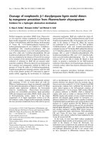
Fig. 1. Purification of recombinant proteins and their in-gel inhibitory activity assay. (A) SDS ⁄ PAGE analysis of purified recombinant GST-
fused proteins from bacterial extracts. Lane M, protein standard; FL lane, two bands corresponding to GST-FL (upper band) and GST (lower
band); Nt lane, GST-Nt peptide (upper band) and GST (lower band); Ct lane, only one band (GST-Ct peptide). (B) SDS ⁄ PAGE analysis of puri-
fied recombinant tarocystatin cleaved after thrombin digestion. (C) In-gel inhibitory activity assay for three different segment recombinant
proteins. The band brightness is proportioned to papain activity Samples containing FL or Nt peptides reduce the brightness on the gel, indi-
cating their inhibitory capacity. By contrast, the Ct peptide showed an enhancing capacity. (D) In-gel inhibitory activity assay for varied con-
centrations of the Ct peptide. The brightness of the band increased with increasing Ct peptide concentration, confirming its enhancing
capacity. Lane 8* indicates a subject containing only Ct peptide recombinant protein, and not containing any papain, where no digestion
occurred.
Cysteine proteinase inhibitor K M. Wang et al.
4982 FEBS Journal 275 (2008) 4980–4989 ª 2008 The Authors Journal compilation ª 2008 FEBS
from enzyme, whereas K
ib
is for that between the
inhibitor and enzyme–substrate complex. A prominent
characteristic of mixed inhibition compared to compet-
itive inhibition is that the mixed inhibitors bind to
enzymes as well as enzyme–substrate complexes, but
competitive inhibitors bind only enzymes. Thus, the Ct
peptide of tarocystatin may be able to dock onto the
papain structure when the active site is occupied by a
substrate. Furthermore, the occurrence of the K
ib
value
is always tailed with an unknown regulatory effect,
indicating that the Ct peptide functions to alter the
target protein conformation and prevent product for-
mation. The Nt peptide functions like the entire OC-I
and confers tarocystatin with an affinity for the
competing active site.
The 3D structural model of tarocystatin was pre-
dicted to infer the interaction between group-2 taro-
cystatin and papain. The Ct peptide sequence shares
48% identity and 68% similarity with taro Nt (1–97
amino acids), as solved by NMR spectroscopy [22].
Although there was no established template for Ct
peptide 3D structure prediction, it shares 13% identity
and 38% similarity to group-1 OC-I (Fig. 5). There-
fore, the Ct peptide structure was predicted using
secondary structure estimation and a folding pattern
simulation program with pseudo-energy minimization.
Subsequently, the entire tarocystatin 3D structure was
obtained by combining the structures of two segments.
Its conformation resembled an earphone comprising
two solid masses and a linear structure (Fig. 6). A
highly structural similarity between the Nt and Ct
peptides was found and, presumably, the Ct peptide
compete with the Nt peptide for binding to the active
site (Fig. 6). However, the assay using varied concen-
trations in the range 0–10 000 lm of Ct peptide to
compete with the Nt peptide at a concentration of
62.5 lm did not demonstrate that the Ct peptide
reduced the inhibitory capacity of the Nt peptide
(Fig. 7A). Instead, it revealed that the Ct peptide does
not act competitively.
To determine whether the connection between the
Nt and Ct peptides is important for inhibitory capacity
of the FL peptide, equal amounts of Nt and Ct pep-
tides were mixed and the inhibitory capacity of the
mixture was then compared with that of only the Nt
or FL peptides. The curve of the mixture of Nt and Ct
peptides did not tend to that of the FL peptide in the
retrieve test (Fig. 7B). The pattern of competitive inhi-
bition against papain by the Nt peptide of group-1 is
consistent with the previous findings obtained for
Fig. 2. Anti-fungal activity assay for recom-
binant proteins of different tarocystatin
segments. (A) Five pieces of sclerotia
cultured in the presence of recombinant
proteins of varied concentrations in a 1-cm
diameter glass tube. Inhibition efficacy is
proportional to the clarity of the medium.
Additional FL or Nt peptides in the sclerotia
culture caused an increase in clarity of the
medium, indicating their anti-fungal activity,
whereas Ct peptide did not. (B) The inhibi-
tion level was graded from high effective
(+++) to null (±) by visual quantification.
(C) The different inhibitory strengths of
varied FL peptide levels on mycelium
growth was observed under high and low
magnification. Mildly inhibited mycelium
exhibited swelling, less branching and blunt
tips. Fully inhibited mycelium exhibited more
swelling, no branches, very short tips and
fragmentation.
K M. Wang et al. Cysteine proteinase inhibitor
FEBS Journal 275 (2008) 4980–4989 ª 2008 The Authors Journal compilation ª 2008 FEBS 4983
many other group-1 phytocystatins [18,19], whereas
the mixed type inhibition against papain by FL pep-
tides of group-2 has not been reported to date.
Information about mixed inhibition by other phyto-
cystatins is scarce. A similar inhibition model, non-
competitive inhibition, was reported in strawberry
FaCPI-1 [9] and in soybean L1 and R1 [20]. Of these,
only FaCPI-1 belongs to the group-2 phytocystatins
and demonstrates a strong inhibitory activity
(1.9 · 10
)9
m). The FaCPI-1 amino acid sequence is
highly homologous with tarocystatin, but its mecha-
nism cannot show mixed inhibition. To unravel the
mechanism, a detailed investigation of the 3D struc-
tural interaction between group-2 phytocystatins and
papain is necessary.
Discussion
In the present study we are the first to show the inhibi-
tion difference between group-1 and group-2 phyto-
cystatins, and to examine the importance of the
extended C-terminal region (Ct peptide of tarocystatin)
with respect to interaction with anti-papain activity.
In the analysis of the primary structure of tarocysta-
tin, we found that the group-2 tarocystatin (FL pep-
tide) is a group-1 phytocystatin (Nt peptide) possessing
an additional Ct peptide. Moreover, the Ct peptide of
the group-2 phytocystatin shares a high consensus
sequence among the discovered species [7]. Both the
FL and Nt peptides exhibit a good inhibitory property
on papain activity, whereas the Ct peptide exhibited
papain activation that was also evident in an in-gel
inhibitory assay (Fig. 1C). The inhibition constant
demonstrated that the FL peptide exhibited mixed
inhibition, and the Nt peptide exhibited competitive
inhibition, suggesting a canonical binding mode as
with many other group-1 phytocystatin species previ-
ously reported (Table 2). The enhancement of the pro-
teinase activity by 18% (Table 1) implicates that the
interaction between papain and the Ct peptide pos-
sesses refolding in the conformation change of the
papain protein. The mixed type inhibition against
papain by the FL peptide might be due to the presence
Fig. 3. Analysis of inhibitory kinetics of different tarocystatin seg-
ments. (A) Plot of papain activity for a single inhibitor concentration
(0125 l
M) at various substrate concentrations. , Ck (water instead
of inhibitor); d, FL peptide; s, Nt peptide; h, Ct peptide. The y-axis
is the catalytic velocity of papain, expressed as the change in opti-
cal density per unit time. The x-axis is the substrate concentration
(m
M). Each point represents the mean value of three repeated
experiments, with the standard error shown as a bar. (B) Linewe-
aver–Burk plot for different tarocystatin segments, and also the
double reciprocal plot of (A). Ck line crosses lines of the FL, Nt and
Ct peptides in the second quadrant, y-axis and x-axis, respectively,
indicating that the FL peptide behaves with mixed inhibition, the Nt
peptide behaves with competitive inhibition and the Ct peptide
behaves as an allosteric activator.
Table 1. Inhibitory characteristics and K
i
values of diferent tarocyst-
atin segments.
Model
Average (%)
inhibitory
activity K
i
value (lM)
FL peptide (group-2)
mixed inhibition
55 K
ia
0.098, K
ib
0.252
Nt peptide (group-1)
competitive inhibition
39 K
i
0.057
Ct peptide allosteric
activation
)18 –
Cysteine proteinase inhibitor K M. Wang et al.
4984 FEBS Journal 275 (2008) 4980–4989 ª 2008 The Authors Journal compilation ª 2008 FEBS
of the Ct peptide, which plays an activation role on
papain when used alone. Therefore, the mechanism of
the Ct peptide with respect to enhancing papain activ-
ity presumably involves allosterically binding adjacent
to the active ⁄ substrate binding site and altering the
papain conformation to be more accessible for the sub-
strate, which is defined as the ‘noncanonical’ binding
mode, where these inhibitory characteristics are quite
different from the ‘canonical’ binding mode of group-1
phytocystatins, as noted previously [23]. This change
may also shift the orientation of the Nt peptide to
bind with competitive inhibition and result in blocking
the substrate from the approaching catalytic site.
When the Ct peptide was bound to papain and linked
with the Nt peptide, substrates still had the chance to
bind to the active cleft. Nevertheless, the Nt peptide
was so close to active cleft that allows Nt peptide
binding prior to any approaching substrates. Thus,
the enhancing effect of the Ct peptide was followed by
an immediate binding of the Nt peptide. In this case,
substrates still could reach the active cleft and be
trapped by some inner pulling force, but could not be
fixed in the catalytic site that the Nt peptide blocked.
This mechanism was like a noncompetitive inhibition,
where substrates could bind to the enzyme–inhibitor
complex but not to be turned to products. However, if
the Nt peptide bound to papain before the Ct peptide,
tarocystatin would simply exhibit competitive inhibi-
tion. The alternative binding pattern strongly supports
the idea that tarocystatin is a mixed type inhibitor,
and provides evidence for the difference between
group-1 and group-2 phytocystatins.
Phytocystatin has been known for its defense func-
tion against attack by insects and pathogens. These
proteins have received much attention from researchers
due to their potential utilization as bioinsectides in
agrobiotechnology [3,4]. To extend our previous study
on antifungal activity [7], a bioassay on mycelial
growth of S. rolfsii was performed, and revealed that
the FL and Nt peptides exhibited apparent antifungal
Fig. 4. Lineweaver–Burk plot for reactions
in the presence of two different concentra-
tions of Nt peptide (A) and FL peptide (B).
The inhibitor concentrations were 0.125 m
M
(s) and 0.0625 mM (d) in each case. Water
(h) was used as a control.
Fig. 5. Sequence alignment of the Nt and
Ct peptides of taro and OC-I (Protein Data
Bank: IEQK). The identical residues are
shown as a black box and the partially con-
served residues are in grey. OC-I shares
48% identity and 68% positives with the Nt
peptide of tarocystatin and 13% identity and
38% positives with the Ct peptide of taro-
cystatin.
K M. Wang et al. Cysteine proteinase inhibitor
FEBS Journal 275 (2008) 4980–4989 ª 2008 The Authors Journal compilation ª 2008 FEBS 4985
activity at a concentration above 3.4 nm, but no signif-
icant antifungal activity was observed for the Ct pep-
tide (Fig. 2A,B). Microscopic observations indicated
that the Nt peptide appeared to be stronger than FL
peptide (Fig. 2B). Because the Ct peptide alone does
not show any antifungal effect, this implies that the
antifungal activity might be connected to the Nt pep-
tide conformation. A reduction of antifungal activity
in vitro by FL peptide may be due to a molecular mass
difference. It has been speculated that the FL peptide
is larger than the Nt peptide, making it more ineffi-
cient to diffuse inside hyphal cells. The true mechanism
responsible for the antifungal activity of tarocystatin
still requires further investigation.
To date, the physiological significance of the Ct pep-
tide remains unknown. Accumulating evidence shows
that this repeated domain may originate from gene
duplication and be exploited for other functions [10].
Recent evidence also demonstrated that carboxy termi-
nus-extended PhyCys have the capacity to inhibit
human legumanin peptide due to the presence of the
conserved motif SNSL and act as a bifunctional inhibi-
tor [24]. Our findings focused on the Ct peptide on
cysteine protease, which may function with three roles:
(a) to endow the N-terminal domain with more speci-
ficity and inhibition to papain; (b) to prevent the
N-terminal domain from rapid digestion by endoge-
nous or exogenous peptidase; and (c) to enrich its
molecular size as an ideal storage protein. Due to these
beneficial characteristics, the Ct peptide could be
reserved under evolutionary selection. Further studies,
including mutagenesis and structural studies, are
required to better understand the molecular mecha-
nisms involved in the tarocystatin binding to papain
and to identify the regulatory cleft involved in the inhi-
bition process [15].
Based on the characterization of inhibitory function
of group-1 and group-2 phytocystatins, we suggest that
Fig. 7. Competition and retrieve test of the Ct peptide to Nt pep-
tide. (A) Competition test: the y-axis is catalytic velocity of papain,
which was measured as the change in optical density over time.
Each reaction had the indicated amount of Ct and Nt peptides
that reacted with papain. An increasing Ct level did not reduce
the inhibitory capacity of the Nt peptide, but instead maintained a
steady intensity. (B) Retrieve test: the mixture of Nt and Ct pep-
tides of 625 l
M was compared with an equal amount of only FL
or Nt peptides in the reaction with varied substrate concentra-
tions. The curve of Nt plus Ct peptides highly overlapped that of
the Nt peptide, indicating that the Nt peptide cannot retrieve the
inhibition efficacy of the FL peptide when it disconnects from the
Ct peptide.
Fig. 6. Conjectural structure model of tarocystatin. Flat arrows and
helical ribbons represent b-sheets and a-helix structures, respec-
tively. The entire structure resembles an earphone comprising two
solid masses and a linear structure.
Cysteine proteinase inhibitor K M. Wang et al.
4986 FEBS Journal 275 (2008) 4980–4989 ª 2008 The Authors Journal compilation ª 2008 FEBS
group-1 and group-2 both evolved from a common
ancestor. The evolutionary direction from group-1
toward group-2 by gene duplication appears to be an
adaptation resulting from an evolutionary ‘arms race’
of rapid change in both interacting proteins.
Experimental procedures
Construction of three DNA regions of the CeCPI
gene
Three different segments of the CeCPI gene were amplified
by PCR (Pfu; Stratagene, La Jolla, CA, USA). These DNA
segments correspond to the coding regions of the FL, Nt and
Ct peptides. Two forward (F1 and F2) and two reverse (R1
and R2) primers were designed to amplify the genes: F1,
5¢-TT
GGATCCATGGCCTTGATGGGGGC-3¢; R1, 5¢-TT
GAATTCTTTCCAGAGTCTGAATGATC-3¢; F2, 5 ¢-TT
GGATCCTCGGTTACGCCAGCAGAT-3¢; R1, 5¢-TTGA
ATTCTTTCCAGAGTCTGAATGATC-3¢; F2, 5¢-TTGGA
TCCTCGGTTACGCCAGCAGAT-3¢; and R2, 5¢-TTGAA
TTCGAATCGCCAATGGGGCT -3¢.
The underlined bases in the primers indicate restriction
sites for BamHI (GGATCC) or EcoRI (GAATTC). The
primer combination of F1 and R1 was used for amplifica-
tion of the FL peptide; F1 and R2 was for the Nt peptide,
and F2 and R1 was for the Ct peptide. The PCR products
were digested with BamHI and EcoRI, and ligated to
pGEX-2TK vector (Amersham Biosciences, Piscataway,
NJ, USA) at the corresponding restriction sites.
Expression, purification and characterization of
recombinant tarocystatin
E. coli BL21 (DE3) cells containing the appropriate con-
struct were grown at 37 °C in 2YTA liquid medium until
D
600
of 1.5 was reached. The recombinant CeCPI expres-
sion was induced by addition of 1 mm isopropyl-b-d-thiog-
alactopyranoside. Two hours after induction, the
recombinant proteins were extracted from 250 mL of bacte-
rial culture by using B-PERÒ GST-fusion protein purifica-
tion kit (Pierce No. 78400; Pierce Biotechnology, Rockford,
IL, USA). For the assay of inhibitory kinetics of CeCPI
fragments, the GST fusion protein was cleaved with
20 units of thrombin for 16 h at room temperature. Finally,
the recombinant proteins were collected by passing the
extract through a glutathione Sepharose 4B affinity column
(Amersham Biosciences). The protein was quantitated with
a Bio-Rad protein assay kit (Bio-Rad, Hercules, CA, USA)
using BSA as a standard.
In-gel antipapain assay
Qualitative analysis of CeCPI protein was performed
according to Michaud et al. [25] on 12.5% SDS ⁄ PAGE
containing 1% gelatin. A mixture of CeCPI proteins and
papain was first incubated at 37 °C for 15 min in a mildly
denaturing buffer (62.5 mm Tris–HCl, pH 6.8; 2% SDS,
2% sucrose; 0.01% bromophenol blue), and then subjected
to electrophoresis using a Hoefer SE250 system (Hoefer,
Inc., Holliston, MA, USA). After migration, the gels were
transferred to a 2.5% v ⁄ v aqueous solution of Triton
X-100 for 30 min at room temperature to allow renatur-
ation followed by incubating in reactive buffer (100 mm
sodium phosphate, pH 6.8, containing 8 mm EDTA,
10 mml-cysteine and 0.2% Triton X-100) for 75 min at
37 °C. Subsequently, the gels were rinsed with water and
stained with Coomassie Brilliant Blue. Proteinase inhibitor
activity was visualized as clear zones on a blue background
and the intensity of the clear band is inversely related to
the inhibition level.
Inhibitory tests and determination of K
i
values
K
i
values of papain inhibition were determined from
Lineweaver–Burk plots, a double reciprocal plot of sub-
strate concentration versus velocity. The velocity was deter-
mined by measuring the A
540
of the chromophore, as
described by Pernas et al. [17]. Briefly, an appropriate
amount of inhibitor was pre-incubated with 1 lm of papain
in 100 lL of reaction mixture containing 0.1 m sodium
phosphate buffer (pH 6.5), 10 mm EDTA and 10 mm
2-mercaptoethanol at 37 °C for 10 min. The reaction was
started by the addition of 100 lL of a varied concentration
in the range 20–260 lm of BANA (Sigma, St Louis, MO,
USA) as substrate. The reaction mixture was incubated at
room temperature for 20 min and 300 lL of 2% HCl in
ethanol (w ⁄ v) was added to stop the reaction. The chromo-
phore was then developed by addition of 300 lL of 0.06%
p-dimethylaminocinnamaldehyde in ethanol followed by
incubation at room temperature for 15 min and measure-
ment of A
540
.
Table 2. K
i
values comparison among published group-1 phytocyst-
atins. Both strawberry and soybean are noncompetitive type. The
rest are competitive type.
Species K
i
value Reference
Strawberry 1.9 · 10
)9
Martinez et al. [9]
Maize CCI 2.3 · 10
)8
Abe et al. [21]
Sesame 2.7 · 10
)8
Shyu et al. [8]
Oryza OC-I 3.0 · 10
)8
Kondo et al. [18]
Nt peptide
(group-1 phytocystatin)
5.9 · 10
)8
Present study
Soybean 1.9 · 10
)7
Misaka et al. [6]
Job’s tear 1.9 · 10
)7
Yaza et al. [19]
nTaMDC 1 5.8 · 10
)7
Christova et al. [32]
Oryza OC-II 8.3 · 10
)7
Kondo et al. [18]
WC5 10
)6
Corre-Menguy et al. [33]
K M. Wang et al. Cysteine proteinase inhibitor
FEBS Journal 275 (2008) 4980–4989 ª 2008 The Authors Journal compilation ª 2008 FEBS 4987
The inhibitory activity was recorded as the inhibition
percentage (%) and the inhibition percentage (I%) of
papain was calculated using the equation:
I% ¼
T À T
Ã
T
 100
where, T and T* are the velocities in the absence and
presence of the inhibitor from reactions, respectively. The
average inhibitory activity was calculated from I% values
of varied substrate concentrations.
Antifungal activity assay of different regions of
tarocystatin
The fungal activity assay was performed as described previ-
ously [7]. Five pieces of sclerotia of phytopathogenic fungus
S. rolfsii were cultured in 1 mL of half strength potato
dextrose broth, which contained purified GST-tarocystatin
segment fusion proteins at concentrations of 1.7, 3.4, 5.1
and 6.8 nm in four separate sets. The fungi were cultured at
28 °C under continuous shaking (200 r.p.m.) on an orbital
shaker for 72 h. Hyphal growth inhibition by tarocystatin
segment proteins was observed directly, as well as under a
microscope.
Conjectural tarocystatin 3D structure simulation
The tarocystatin primary sequence (AAM88397) was sub-
jected to NCBI psi-blast with a threshold of 0.0001 for
searching for homologous sequences from various plants.
The sequence similarities of 18 amino acid sequences were
distributed with the highest identities of 66% and a positive
of 83% for soybean to the lowest identities of 55% and a
positive of 77% for tomato, excluding nonplant and multi-
domain cystatin homologs. These 18 sequences were aligned
using clustalw [26] and shaded with genedoc [27] soft-
ware. The secondary structure of these 18 sequences was
analyzed by two programs, psi-pred [28] and yaspin [29].
The results obtained by the two programs were consistent
with each other and showed that both Nt and Ct peptide
secondary structures were arranged in a similar pattern.
This was also verified by aligning the OC-I with the taro-
cystatin Nt and Ct peptide regions. Therefore, the stereo-
folding pattern of OC-I [22] can be taken as a template for
the CeCPI folding prediction by modeler 8.1 [30]. The two
structural conformations were merged after analysis by the
automatic docking system, zdock 2.3 [31], and then remod-
eled by modeler 8.1 [30].
Acknowledgements
The present study was supported by the National
Science Council, Taiwan, under project NSC-95-2317-
B-002-005 to Kai-Wun Yeh. We thank Dr Michael
Conrad (University of North Carolina at Chapel Hill)
for critically reading the manuscript and for his helpful
suggestions.
References
1 Margis R, Reis EM & Villeret V (1998) Structural and
phylogenetic relationships among plant and animal cyst-
atins. Arch Biochem Biophys 359, 24–30.
2 Machleidt W, Thiele M, Laber B, Assfalg-Machleid I,
Esterl A, Wiegand G, Kos J, Turk V & Bode W (1989)
Mechanism of inhibition of papain by chicken egg white
cystatin. FEBS Lett 243, 234–238.
3 Arai S, Watanabe H, Kondo H, Emori Y & Abe K
(1991) Papain-inhibitory activity of oryzacystatin, a rice
seed cysteine proteinase inhibitor, depends on the cen-
tral Gln-Val-Val-Ala-Gly region conserved among cyst-
atin superfamily members. J Biochem (Tokyo) 109,
294–298.
4 Abe K, Emori Y, Kondo H, Suzuki K & Arai S (1987)
Molecular cloning of a cysteine proteinase inhibitor of
rice (oryzacystatin). Homology with animal cystatins
and transient expression in the ripening process of rice
seeds. J Biol Chem 262, 16793–16797.
5 Lim CO, Lee SI, Chung WS, Park SH, Hwang I & Cho
MJ (1996) Characterization of a cDNA encoding a cys-
teine proteinase inhibitor from chinese cabbage (Bras-
sica campestris L. ssp. pekinensis) flower buds. Plant
Mol Biol 30, 373–379.
6 Misaka T, Kuroda M, Iwabuchi K, Abe K & Arai S
(1996) Soyacystatin, a novel cysteine proteinase inhibi-
tor in soybean, is distinct in protein structure and gene
organization from other cystatins of animal and plant
origin. Eur J Biochem 240, 609–614.
7 Yang AH & Yeh KW (2005) Molecular cloning, recom-
binant gene expression, and antifungal activity of cysta-
tin from taro (Colocasia esculenta cv. Kaohsiung No.1).
Planta 221, 493–501.
8 Shyu DJH, Chou WM, Yiu TJ, Lin CPC & Tzen JTC
(2004) Cloning, functional expression, and characteriza-
tion of cystatin in sesame seed. J Agric Food Chem 52,
1350–1356.
9 Martinez M, Abraham Z, Gambardella M, Echaide M,
Carbonero P & Diaz I (2005) The strawberry gene Cyf1
encodes a phytocystatin with antifungal properties.
J Exp Bot 56, 1821–1829.
10 Christeller JT (2005) Evolutionary mechanism acting on
proteinase inhibitor variability. FEBS J, 272, 5710–
5722.
11 Waldron C, Wegrich LM, Merlo PAO & Walsh TA
(1993) Characterization of a genomic sequence coding
for potato multicystatin, an eight-domain cysteine pro-
teinase inhibitor. Plant Mol Biol 23, 801–812.
12 Wu J & Haard NF (2000) Purification and charac-
terization of a cystatin form the leaves of methyl
Cysteine proteinase inhibitor K M. Wang et al.
4988 FEBS Journal 275 (2008) 4980–4989 ª 2008 The Authors Journal compilation ª 2008 FEBS
jasmonate-treated tomato plants. Comp Biochem Physiol
C Toxicol Pharmacol 127, 209–220.
13 Felton GW & Korth KL (2000) Trade-offs between
pathogen and herbivore resistance. Curr Opin Plant Biol
3, 309–314.
14 Botella MA, Xu Y, Prabbha TN, Zhao Y, Narasimhan
ML, Wilson KA, Nielsen SS, Bressan RA & Hasegawa
PM (1996) Differential expression of soybean cysteine
proteinase inhibitor genes during development and in
response to wounding and jasmonate. Plant Physiol
112, 1201–1210.
15 Solomon M, Belenghi B, Delledonne M, Menachem E
& Levine A (1999) The involvement of cysteine prote-
ases and protease inhibitor genes in the regulation of
programmed cell death in plants. Plant Cell 11, 431–
443.
16 Turk V & Bode W (1991) The cystatins: protein inhibi-
tors of cysteine proteinase. FEBS Lett 285, 213–219.
17 Pernas M, Sa
´
nchez-Monge R, Go
´
mez L & Salcedo G
(1998) A chestnut seed cystatin differentially effective
against cysteine proteinases from closely related pests.
Plant Mol Biol 38 , 1235–1242.
18 Kondo H, Abe K, Nishimura I, Watanabe H, Emori Y
& Arai S (1990) Two distinct cystatin species in rice
seeds with different specificities against cysteine protein-
ases. J Biol Chem 265, 15832–15837.
19 Yoza K, Nakamura S, Yaguchi M, Haraguchi K &
Ohtsubo K (2002) Molecular cloning and functional
expression of cDNA encoding a cysteine proteinase
inhibitor, cystatin, from Job’s tears (Coix lacryma-jobi
L. var Ma-yuen Stapf). Biosci Biotechnol Biochem 66,
2287–2291.
20 Zhao Y, Botella MA, Subramanian L, Niu X, Nielsen
SS, Bressan RA & Hasegawa PM (1996) Two wound-
inducible soybean cysteine proteinases have greater
insect digestive proteinase inhibitory activities than a
constitutive homolog. Plant Physiol 111, 1299–1306.
21 Abe M, Abe K, Iwabuchi K, Domoto C & Arai S
(1994) Corn cystatin I expressed in Escherichia coli:
investigation of its inhibitory profile and occurrence in
corn kernels. J Biochem (Tokyo) 116, 488–492.
22 Nagara N, Kudo K, Abe S, Arai M & Tanokura X
(2000) Three dimensional solution structure of oryza
cystatin-I, a cysteine proteinase of the rice, Oryza sativa
L japonica. Biochemistry 39, 14753–14760.
23 Bode W & Huber R (2000) Structural basis of the endo-
proteinase-protein inhibitor interaction. Biochim Bio-
phys Acta 1477, 241–252.
24 Martinez M, Diaz-Mendoza M, Carrillo L & Diaz I
(2007) Carboxy terminal extended phytocystation are
bifunctional inhibitors of papain and legumanin cyste-
ines proteinases. FEBS Lett 581, 2914–2918.
25 Michaud D, Cantin L, Raworth DA & Vrain TC
(1996) Assessing the stability of cystatin ⁄ cysteine
proteinase complexes using mildly-denaturing gelatin
polyacrylamide gel electrophoresis. Electrophoresis 17,
14–19.
26 Thompson JD, Higgins DG & Gibson TJ (1994)
CLUSTAL W: improving the sensitivity of progressive
multiple sequence alignment through sequence weight-
ing, position-specific gap penalties and weight matrix
choice. Nucleic Acids Res 11, 4673–4680.
27 Nicholas KB, Nicholas HB Jr & Deerfield DW II
(1997) GeneDoc: analysis and visualization of genetic
variation. EMBNEW News
4, 14.
28 McGuffin LJ, Bryson K & Jones DT (2000) The PSI-
PRED protein structure prediction server. Bioinformat-
ics 16, 404–405.
29 Lin K, Simossis VA, Taylor WR & Heringa J (2005) A
simple and fast secondary structure prediction algo-
rithm using hidden neural networks. Bioinformatics 21,
152–159.
30 Sali A & Blundell TL (1993) Comparative protein mod-
elling by satisfaction of spatial restraints. J Mol Biol
234, 779–815.
31 Chen R, Li L & Weng Z (2003) ZDOCK: an initial-
stage protein-docking algorithm. Proteins 52, 80–87.
32 Christova PK, Christov NK & Imai R (2006) A cold
inducible multidomain cystatin from winter wheat inhi-
bits growth of the snow mold fungus, Microdochium
nivale. Planta 223, 1207–1218.
33 Corr-Menguy F, Cejudo FJ, Mazubert C, Vidal J,
Lelandais-Brie
`
re C, Torres G, Rode A & Hartmann C
(2002) Characterization of the expression of a wheat
cystatin gene during caryopsis development. Plant Mol
Biol 50, 687–698.
K M. Wang et al. Cysteine proteinase inhibitor
FEBS Journal 275 (2008) 4980–4989 ª 2008 The Authors Journal compilation ª 2008 FEBS 4989