Báo cáo khoa học: Molecular responses of Campylobacter jejuni to cadmium stress ppt
Bạn đang xem bản rút gọn của tài liệu. Xem và tải ngay bản đầy đủ của tài liệu tại đây (261.13 KB, 13 trang )
Molecular responses of Campylobacter jejuni to
cadmium stress
Nadeem O. Kaakoush
1
, Mark Raftery
2
and George L. Mendz
3
1 School of Medical Sciences, University of New South Wales, Sydney, Australia
2 Biological Mass Spectrometry Facility, University of New South Wales, Sydney, Australia
3 School of Medicine Sydney, University of Notre Dame Australia, Sydney, Australia
Cadmium ions (Cd
2+
) are a potent carcinogen in
animals, and cadmium is a toxic metal of significant
environmental and occupational importance for
humans [1–5]. Cadmium ions are very toxic even at
low concentrations, but the basis for their toxicity is
not fully understood. Cadmium is not a redox-active
metal and does not participate in Fenton-type reac-
tions. Moreover, it does not bind to DNA or interact
with DNA in a stable manner [1,2].
Several mechanisms have been proposed to explain
how bacteria and lower eukaryotes protect themselves
against cadmium toxicity. These include accumulation
of intracellular Zn
2+
, reduction of Cd
2+
uptake,
enhanced expression of the low-molecular weight cys-
teine-rich protein metallothionein that sequesters
cadmium, binding of cadmium ions by other heavy
metal-associated proteins, and an increase in intracellu-
lar disulfide content that contributes to effective bind-
ing of cadmium [6].
Disulfide reductases are responsible for the modula-
tion of intracellular disulfide concentrations. They are
essential enzymes in the antioxidant mechanisms of
Keywords
cadmium detoxification;
Campylobacter jejuni; citrate cycle;
glutathione; thioredoxin reductase
Correspondence
G. L. Mendz, School of Medicine Sydney,
University of Notre Dame Australia, Sydney,
NSW 2010, Australia
Fax: +61 293577680
Tel: +61 282044457
E-mail:
(Received 30 May 2008, revised 9 July
2008, accepted 11 August 2008)
doi:10.1111/j.1742-4658.2008.06636.x
Cadmium ions are a potent carcinogen in animals, and cadmium is a toxic
metal of significant environmental importance for humans. Response
curves were used to investigate the effects of cadmium chloride on the
growth of Camplyobacter jejuni. In vitro, the bacterium showed reduced
growth in the presence of 0.1 mm cadmium chloride, and the metal ions
were lethal at 1 mm concentration. Two-dimensional gel electrophoresis
combined with tandem mass spectrometry analysis enabled identification of
67 proteins differentially expressed in cells grown without and with 0.1 mm
cadmium chloride. Cellular processes and pathways regulated under cad-
mium stress included fatty acid biosynthesis, protein biosynthesis, chemo-
taxis and mobility, the tricarboxylic acid cycle, protein modification, redox
processes and the heat-shock response. Disulfide reductases and their sub-
strates play many roles in cellular processes, including protection against
reactive oxygen species and detoxification of xenobiotics, such as cadmium.
The effects of cadmium on thioredoxin reductase and disulfide reductases
using glutathione as a substrate were studied in bacterial lysates by spectro-
photometry and nuclear magnetic resonance spectroscopy, respectively.
The presence of 0.1 mm cadmium ions modulated the activities of both
enzymes. The interactions of cadmium ions with oxidized glutathione and
reduced glutathione were investigated using nuclear magnetic resonance
spectroscopy. The data suggested that, unlike other organisms, C. jejuni
downregulates thioredoxin reductase and upregulates other disulfide reduc-
tases involved in metal detoxification in the presence of cadmium.
Abbreviations
GSH, reduced glutathione; GSSG, oxidized glutathione; MTA, 5¢-methylthioadenosine; SAH, S-adenosylhomocysteine; TCA, tricarboxylic acid.
FEBS Journal 275 (2008) 5021–5033 ª 2008 The Authors Journal compilation ª 2008 FEBS 5021
many bacteria, and also play a role in protection
against the toxic effects of heavy metals [7–9]. CXXC
motifs and CXXC-derived motifs are present in the
active sites of disulfide reductases [10], and are capable
of metal coordination and metal detoxification. Clus-
ters of cysteinyls capable of coordinating zinc atoms
are known as ‘zinc knuckles’ or ‘zinc fingers’ [10,11].
Glutathione reductase is an enzyme that is responsi-
ble principally for maintaining intracellular levels of
reduced glutathione (GSH, c-Glu-Cys-Gly) by recy-
cling the oxidized tripeptide (GSSG) to its reduced
form at the expense of oxidizing a molecule of
NAD(P)H. GSH has many roles in cellular processes,
including protection against reactive oxygen species
(ROS) and detoxification of xenobiotic compounds
[12]. GSH is therefore an essential metabolite in the
antioxidant mechanisms of many bacteria, and protects
them from the toxic effects of heavy metals [13,14].
For example, glutathione reductase was found to be
upregulated under cadmium stress in Lemna polyrrhiza
[15].
Cadmium has multiple molecular effects in various
organisms. In Chlamydomonas reinhardtii, exposure to
cadmium resulted in the downregulation of central
metabolism pathways such as fatty acid biosynthesis,
the tricarboxylic acid (TCA) cycle, and amino acid and
protein biosynthesis [16]. In contrast, proteins involved
in glutathione synthesis, ATP metabolism, response to
oxidative stress and protein folding were upregulated
in the presence of cadmium [16]. The effect of cad-
mium on protein expression in Rhodobacter capsulatus
B10 involved upregulation of heat-shock proteins
GroEL and 70 kDa heat shock protein (DnaK),
S-adenosylmethionine synthetase, ribosomal protein
S1, aspartate aminotransferase and phosphoglycerate
kinase [17]. An interesting study in Escherichia coli
found that cadmium-stressed cells recovered more rap-
idly than unexposed cells when subsequently subjected
to other stresses such as ethanol, osmotic, heat shock
or nalidixic acid treatment [18]. In Saccharomyces cere-
visiae, cells exposed to cadmium showed increased syn-
thesis of glutathione and proteins with antioxidant
properties [19]. A proteomic evaluation of cadmium
toxicity on Chironomus riparius Meigen larvae showed
downregulation of energy production, nucleotide bio-
synthesis, cell division, transport and binding of ions,
signal transduction regulating citrate ⁄ malate metabo-
lism, and fatty acid and phospholipid metabolism [20].
Campylobacter jejuni belongs to an important group
of gastrointestinal spiral bacteria that have natural res-
ervoirs in many animals and birds that are in contact
with humans [21]; most human diseases caused by
organisms of the genus Campylobacter are due to
Campylobacter jejuni [21]. Little is known about the
detoxification defenses against metals in this micro-
aerophilic bacterium, which lives in habitats that are
subject to continual change. In the human gut, this
pathogen experiences turnover of the proliferative
intestinal epithelium and is exposed to the ever-chang-
ing chemical environment of the gastric tract that
results from the variety and combinations of food
ingested by higher animals. In addition, the bacterium
may encounter environments with diverse chemical
compositions before transmission to the host.
The inhibition of C. jejuni growth by cadmium ions
[22] and the reduction of inhibition by ferrous sulfate
[23] have been reported. Campylobacter isolates from
meat samples were shown to have higher tolerance to
Cd
2+
than clinical isolates [22], providing evidence
that strains with different habitats vary in their physi-
ologies. An important observation is that the genome
of C. jejuni NCTC 11168 does not contain genes
orthologous to those encoding glutathione reductase
or enzymes of the c-glutamyl cycle that are involved in
the synthesis of glutathione in other organisms.
In this study, changes induced in the proteome of
C. jejuni cells subjected to cadmium stress in vitro were
determined using two-dimensional gel electrophoresis
and mass spectrometry. In particular, a better under-
standing of the cellular role of disulfide reduction in
this microaerophilic human pathogen was achieved by
investigating the inhibition of glutathione reduction by
Cd
2+
in situ and in vitro, and the interactions of these
ions with glutathione and glutathione reductase.
Results and Discussion
Effects of cadmium on the survival of
Campylobacter jejuni
The effects of cadmium ions on the growth of C. jejuni
were measured at Cd
2+
concentrations of 0.05, 0.1,
0.3, 0.5 and 1 mm. Two colony-forming unit
(cfuÆmL
)1
) counts were taken at 0 and 24 h from each
culture (n = 3). The bacteria grew approximately
1.5 log (cfuÆmL
)1
)at0mm Cd
2+
(Fig. 1). Inhibition
of C. jejuni growth increased with Cd
2+
concentration,
and the cation was lethal at 1 mm concentration
(Fig. 1); changes in C. jejuni growth were observed at
micromolar concentrations of cadmium (Fig. 1). These
effects were comparable to those observed in other
bacteria and yeast [16,17,19]. The results indicated that
cadmium is highly toxic to C. jejuni, as is the case for
other microorganisms.
The growth-inhibition data enabled determination of
the Cd
2+
concentration at which C. jejuni cells could
Campylobacter jejuni and cadmium stress N. O. Kaakoush et al.
5022 FEBS Journal 275 (2008) 5021–5033 ª 2008 The Authors Journal compilation ª 2008 FEBS
be subjected to cadmium stress with only partial inhi-
bition of cell growth. At 0.1 mm Cd
2+
, C. jejuni
growth was significantly decreased but the bacteria
remained viable.
Proteomic analyses of Campylobacter jejuni
under cadmium stress
The response of C. jejuni to 0.1 mm Cd
2+
in the
growth medium was analyzed using two-dimensional
gel electrophoresis to determine the changes in the pro-
teome of the bacterium (Fig. 2). Two-dimensional gel
electrophoresis was performed using proteins extracted
from pairs of bacterial cultures grown with and with-
out Cd
2+
, and included three independent biological
repeats and one technical repeat. The four pairs of gels
obtained from cultures under both conditions were
analyzed to identify spots corresponding to proteins
whose expression was regulated under cadmium stress;
these proteins were identified using tandem mass spec-
trometry analyses. Sixty-seven proteins were differen-
tially expressed, of which 38 were downregulated and
29 were upregulated in the presence of Cd
2+
(Tables 1
and 2).
Bioinformatics analyses on regulated proteins
Effects on central metabolic pathways
Applying the functional classifications available in the
Kyoto Encyclopedia of Genes and Genomes (KEGG)
to the downregulated proteins in Table 1, it was con-
cluded that fatty acid biosynthesis and the TCA cycle
were downregulated. The former pathway is downreg-
ulated by metal ions in both prokaryotes and eukary-
otes [24–26]. Previous studies have suggested that the
effect of metals on fatty acid biosynthesis is indirect,
arising from changes induced in other metabolic path-
ways such as carbohydrate metabolism [25,26]. None-
theless, the modulation of fatty acid biosynthesis in
C. jejuni subjected to cadmium stress was notable. The
enzymes CJ1290c responsible for conversion of
acetyl CoA to malonyl CoA, and CJ0116 and
CJ0442 responsible for conversion of acetyl CoA to
acetyl ACP and malonyl CoA to malonyl ACP, respec-
tively, were all downregulated. In addition, the
enzymes CJ0442 and CJ1400c responsible for produc-
ing hexadecanoyl ACP from acetyl ACP or malo-
nyl ACP were also downregulated, indicating extensive
downregulation of fatty acid biosynthesis.
Fatty acid biosynthesis is the first step in membrane
lipid biogenesis. The downregulation of CJ0858c,
which catalyses the first step of lipopolysaccharide
synthesis, indicates that the pathway is disrupted from
its beginning. Similarly, CJ1054c, which catalyzes the
Fig. 1. Growth of C. jejuni NCTC 11168 in medium containing
CdCl
2
at various concentrations. Controls were cultures grown
without CdCl
2
. Bacteria were growth for 18 h in liquid cultures
under microaerobic conditions at 37 °C.
4
p
I7
p
I74
Fig. 2. Two-dimensional pI 4–7 protein pro-
files of C. jejuni NCTC 11168 grown without
CdCl
2
(left) and in the presence of 0.1 mM
CdCl
2
(right). Proteins differentially
expressed between the two growth
conditions are listed in Tables 1 and 2.
N. O. Kaakoush et al. Campylobacter jejuni and cadmium stress
FEBS Journal 275 (2008) 5021–5033 ª 2008 The Authors Journal compilation ª 2008 FEBS 5023
first step of peptidoglycan biosynthesis, was also down-
regulated, indicating disruption of this pathway also.
These effects, together with downregulation of the cell
division protein FtsA (CJ0695), could explain the
decreased cell growth observed in bacteria subjected to
cadmium stress.
An interesting finding was the downregulation of
CJ0117, which catalyzes the hydrolysis of 5¢-methyl-
thioadenosine (MTA) to 5¢-methylthioribose or S-ade-
nosylhomocysteine (SAH) to S-ribosylhomocysteine
and adenine in prokaryotes but not mammalian cells;
both MTA and SAH are potent inhibitors of impor-
tant cellular processes in prokaryotes, such as trans-
methylation [27,28]. The accumulation of these
intermediates in the bacterium could induce metabolic
changes responsible for inhibition of central metabolic
pathways in C. jejuni, such as the TCA cycle (Table 1).
It has been proposed that adenylated compounds alert
cells to the onset of stress, thus accumulation of the
adenylated compounds MTA and SAH could simply
be the result of onset of cadmium stress. This response
has been shown in Salmonella typhimurium and
Synechococcus spp. [29,30]. Moreover, phenylalanyl
and seryl tRNA synthetases are the only two synthetases
Table 1. C. jejuni NCTC 11168 proteins identified as downregulated in the presence of 0.1 mM CdCl
2
in three independent cultures (n = 3).
Proteins in spots were identified by LC-MS tandem mass spectrometry analyses. The ORF numbers correspond to those of the annotated
genome of C. jejuni strain NCTC 11168.
Functional category Protein Protein name Spot no.
Amino acid metabolism CJ0117 Probable MTA ⁄ SAH nucleosidase 1
CJ0402 Serine hydroxymethyl transferase 2
CJ0665c Argininosuccinate synthase 3
CJ0806 Dihydrodipicolinate synthase 4
CJ0858c UDP-N-acetyl glucosamine carboxyl transferase 5
CJ0897c Phenyl alanyl tRNA synthetase a subunit 6
CJ1054c UDP-N-acetylmuramate-
L-alanine ligase 7
CJ1681c CysQ protein homolog 8
Cell division CJ0695 Cell division protein ftsA 9
Chemotaxis and mobility CJ0144 Methyl-accepting chemotaxis protein 10
CJ0262c Putative methyl-accepting chemotaxis protein 11
CJ1338c Flagellin B 12
CJ1339c Flagellin A 13
CJ1462 Flagellar P-ring protein precursor 14
Fatty acid biosynthesis CJ0116 Acyl carrier protein S-malonyltransferase 15
CJ0442 3-oxoacyl acyl carrier protein synthase II 16
CJ1290c Acetyl CoA carboxylase 17
CJ1400c Enoyl acyl carrier protein reductase 18
Glycolysis CJ0597 Fructose bis-phosphate aldolase 19
Nucleic acid metabolism CJ0146c Thioredoxin reductase 20
CJ0953c Bifunctional formyltransferase ⁄ IMP cyclohydrolase 21
Redox CJ0779 Probable thiol peroxidase 22
TCA cycle CJ0409 Fumarate reductase 23
CJ0531 Isocitrate dehydrogenase 24
CJ0533 Succinyl CoA synthetase b chain 25
CJ0835c Aconitase 26
CJ0933c Putative pyruvate carboxylase B subunit 27
CJ1287c Malate oxidoreductase 28
CJ1682c Citrate synthase 29
Transport ⁄ binding proteins CJ1443c KpsF protein 30
CJ1534c Possible bacterioferritin 31
CJ1663 Putative ABC transport system ATP-binding protein 32
Metabolism of vitamins CJ1046c Thiamine biosynthesis protein ThiF 33
Unknown CJ0172c Hypothetical protein 34
CJ0662c ATP-dependent protease ATP-binding subunit 35
CJ1024c Signal transduction regulatory protein 36
CJ1214c Hypothetical protein 37
CJ1725 Putative periplasmic protein 38
Campylobacter jejuni and cadmium stress N. O. Kaakoush et al.
5024 FEBS Journal 275 (2008) 5021–5033 ª 2008 The Authors Journal compilation ª 2008 FEBS
involved in the production of adenylated nucleotides
[31], and these two enzymes were found to be regu-
lated under cadmium stress.
Inhibitory effects of cadmium on the TCA cycle of
other organisms have been reported [26]. The presence
of Cd
2+
modulated expression of all the enzymes of
the TCA cycle in C. jejuni: seven were downregulated
and two (2-oxoglutarate oxidoreductase and fumarate
dehydratase) were upregulated. These data suggest that
operation of the TCA cycle was downregulated, and
that the upregulation of expression of 2-oxoglutarate
oxidoreductase and fumarate dehydratase was a
response to their other metabolic roles. Some bacteria
have developed metal detoxification pathways in which
the metal ion is first reduced by various c-type cyto-
chromes, hydrogenases and reduced ferredoxins, and
subsequently transported outside the cell [6,32]. 2-oxo-
glutarate oxidoreductase can reduce the low-redox-
potential protein ferredoxin, and its activity can lead
to higher intracellular concentrations of reduced ferre-
doxin than normal basal conditions. In the presence of
cadmium, the increased expression by C. jejuni of
2-oxoglutarate oxidoreductase, leading to elevated con-
centrations of reduced ferredoxin, and the upregulation
of a putative cytochrome c encoded by cj0037c are
important responses to cadmium ions that may act as
detoxification pathways in C. jejuni.
Downregulation of the expression of malate oxidore-
ductase and pyruvate decarboxylase decreases the
entry of pyruvate into the TCA cycle via malate or
oxaloacetate, respectively, and avoids futile cycling of
pyruvate driven by these two enzymes. Malate can still
be produced at normal concentrations from phospho-
enol pyruvate via oxaloacetate, and is converted to
aspartate through the activities of pyruvate dehydroge-
nase and aspartate lyase whose expression was upregu-
lated in the presence of cadmium ions. Similarly to
Helicobacter pylori [33], the dicarboxylic acid branch
of the TCA cycle of C. jejuni functions in the reductive
direction in the presence of excess malate converting it
Table 2. C. jejuni NCTC 11168 proteins identified as upregulated in the presence of 0.1 mM CdCl
2
in three independent cultures (n = 3).
Proteins in spots were identified by LC-MS tandem mass spectrometry analyses. The ORF numbers correspond to those of the annotated
genome of C. jejuni strain NCTC 11168.
Functional category Protein Protein name Spot no.
Amino acid metabolism CJ0087 Aspartate ammonia lyase 39
CJ0389 Seryl tRNA synthetase 40
CJ1096c S-adenosylmethionine synthetase 41
CJ1197c Aspartyl ⁄ glutamyl tRNA amidotransferase subunit B 42
CJ1604 pAMP ⁄ APP hydrolase 43
Cell division CJ0276 Homolog of E. coli rod shape-determining protein 44
Chaperones, heat shock CJ0759 Molecular chaperone DnaK 45
CJ1221 Heat-shock protein GroEL 46
Metabolism of vitamins CJ1045c Thiazole synthase 47
Oxidative phosphorylation CJ0107 ATP synthase subunit B 48
Protein translation and modification CJ0115 Peptidyl prolyl cis–trans isomerase 49
CJ0193c Trigger factor 50
CJ0239c NifU protein homolog 51
CJ0470 Elongation factor Tu 52
CJ0493 Elongation factor EF-G 53
Redox CJ0012c Rbo ⁄ Rbr-like protein 54
CJ0037c Putative cytochrome c 55
CJ0169 Superoxide dismutase 56
CJ0414 Putative oxidoreductase subunit 57
Signal transduction CJ0355c Two-component regulator 58
CJ0448c Putative MCP-type signal transduction protein 59
TCA cycle CJ0536 2-oxoglutarate ferredoxin oxidoreductase 60
CJ1364c Fumarate dehydratase 61
Transcription ⁄ replication CJ0440c Putative transcriptional regulator 62
CJ1071 Single-stranded DNA-binding protein 63
Transport ⁄ binding proteins CJ0612c Ferritin 64
CJ0734c Histidine-binding protein precursor 65
Unknown CJ1136 Putative galactosyl transferase 66
Virulence CJ0039c GTP-binding protein TypA homolog 67
N. O. Kaakoush et al. Campylobacter jejuni and cadmium stress
FEBS Journal 275 (2008) 5021–5033 ª 2008 The Authors Journal compilation ª 2008 FEBS 5025
to pyruvate and then to succinate; this last step is cata-
lyzed by pyruvate reductase. Expression of this enzyme
was downregulated under cadmium stress; as a result,
the pyruvate produced by pyruvate dehydrogenase
could be directed to the synthesis of aspartate.
Increased production of this amino acid could reduce
intracellular cadmium concentrations by chelating the
metal ions [34,35], and could remove free ammonium
by incorporating it into aspartate. Reduction of the
intracellular ammonium concentration could explain
the downregulation of expression of the urea cycle
enzyme argininosuccinate lyase, as reduced use of the
urea cycle is necessary to maintain homeostasis of
intracellular nitrogen levels.
At the same time, an increase in malate concentra-
tion in the cells could play an important role in the
solubilization of cadmium, which is a function of
malate and other organic acids, as shown in rhizo-
sphere soil [36]. The ability of malate to bind cadmium
[37] and to detoxify metals in other organisms [38,39]
suggests that it could be part of a cadmium detoxifica-
tion process used by C. jejuni.
Effects on amino acid biosynthesis
Effects of cadmium on amino acid biosynthesis have
been reported previously; for example, cadmium
inhibits or blocks the threonine pathway in E. coli
[40]. In C. jejuni, cadmium appeared to enhance the
synthesis of aspartate from pyruvate through upregu-
lation of the expression of aspartate ammonia lyase
(CJ0087). The upregulation of expression of pAM-
P ⁄ APP hydrolase encoded by cj1604 under cadmium
stress suggested an increase in purine and ⁄ or histidine
biosynthesis. Since expression of the last enzyme of
de novo purine biosynthesis, PurH (CJ0953c), is
downregulated, the results suggest that synthesis of
histidine, an amino acid with very high affinity for
metal ions, was upregulated. The downregulation of
PurH and dihydrodipicolinate synthase (CJ0806) sug-
gest a decrease in the synthesis of arginine and lysine
using aspartate as a precursor. The increased produc-
tion of aspartate and decreased utilization in synthetic
pathways could constitute another mechanism used
by the bacterium for cadmium ion detoxification. The
downregulation of serine hydroxymethyl transferase
(CJ0402) suggests inhibition of glycine synthesis, as
this is the only de novo glycine pathway that has been
identified in C. jejuni.
In summary, cadmium had an inhibitory effect on
central metabolic pathways of C. jejuni, and appeared
to enhance the production of metabolites that may be
utilized for detoxification.
Effects on protein repair and oxidoreduction systems
The expression of proteins involved in translation ⁄
modification and oxireduction and of chaperones was
upregulated. Cadmium is capable of displacing metal
ions in proteins and affecting their structure and fold-
ing [41]. The upregulation of protein translation and
modification and of expression of chaperones such as
heat-shock proteins in response to Cd
2+
stress has been
reported previously [17]. The elongation factors upregu-
lated in C. jejuni exposed to cadmium are required for
extending the polypeptide chain in protein translation,
and the heat-shock proteins are required for proper
protein folding. These findings indicate that the cells
are responding to the negative effects of cadmium on
protein synthesis. Further evidence is provided by the
downregulation of an ATP-dependent protease subunit
encoded by cj0662c that is capable of degrading heat-
shock proteins. The NifU protein homolog encoded by
cj0239c participates in iron–sulfur center assembly [42];
its upregulation may help to counter cadmium-induced
displacement of iron from proteins.
Removal of iron bound to various cellular compo-
nents can cause a cascade of reactions leading to an
increase in oxidative stress in the cells. Upregulation of
proteins involved in oxireduction reactions helps to
combat the toxic effects of oxidative stress. This
response is found in E. coli, in which cadmium upregu-
lated proteins of heat shock and oxidative stress regu-
lons [43]. Similarly, exposure of anterior gills of the
Chinese mitten crab Eriocheir sinensis to cadmium
upregulated the expression of several antioxidant
enzymes and chaperonins [44]. Metaproteomic analyses
of the response of bacterial communities to cadmium
indicated that oxidoreductases were differentially
expressed [45]. Finally, transcriptional analyses of Cau-
lobacter crescentus cells exposed to cadmium showed
that the principal response to this metal was protection
against oxidative stress [46]. These observations
support the view that induction of oxidative stress and
binding of sulfhydryl groups are mechanisms of
cadmium toxicity [44].
An important detoxification mechanism is the trans-
formation of metals into organometallic compounds
by methylation, and the synthesis of several organo-
cadmium compounds has been demonstrated [6,47].
Adenosylmethionine occupies a central metabolic
position in both eukaryotes and prokaryotes, serving
as a major methyl group donor in biological systems
[27]. The upregulation of S-adenosylmethionine
synthetase encoded by cj1096c in bacteria exposed to
cadmium could promote cadmium methylation, and
thus neutralize the toxic effects of the metal.
Campylobacter jejuni and cadmium stress N. O. Kaakoush et al.
5026 FEBS Journal 275 (2008) 5021–5033 ª 2008 The Authors Journal compilation ª 2008 FEBS
Effects on chemotaxis and motility
No chemotaxis or motility genes showed regulated
expression under cadmium stress in E. coli or C. cres-
centus [41,46], but heavy metal ions strongly affect Bor-
relia burgdorferi motility [48]. The downregulation of
five proteins involved in chemotaxis and motility in
C. jejuni exposed to cadmium stress (Table 1) suggested
a decrease in these functions of the bacterium. Bacterial
motion is driven by either a proton motive force or a
sodium motive force [49,50], and the presence of heavy
metal ions may interfere with this function, leading to
downregulation of genes involved in motility.
The N-terminal half of CJ1024c has a signal-receiver
domain (REC) for proteins such as the chemotaxis
protein (CheY), the outer membrane protein (OmpR),
the bacterial enhancer-binding protein (NtrC), and the
activator protein (PhoB), and in its middle segment
there is a r54 interaction domain [51]. Transcription
of the flaB gene encoding flagellin B is regulated by
sigma factor r54 [51]. Thus, downregulation of
CJ1024c in the presence of cadmium may result in
downregulation of signaling by chemotaxis proteins
and transcription of flagellin B.
Effects on metal uptake and storage
A putative ABC transport system ATP-binding protein
encoded by cj1663 and a hypothetical protein encoded
by cj0172c were downregulated. The STRING tool
[52] predicted that the gene cj0172c is in a network
with cj0173c, cj0174c and cj0175c, which encode an
iron uptake ABC transport system, and with cj0271,
which encodes a bacterioferritin conjugatory protein
homolog. The bacterioferritin CJ1534c, which contains
heme and is involved in iron uptake, was also down-
regulated. In contrast, the heme-free ferritin encoded
by cj0612c involved in intracellular iron storage was
upregulated. Ferritin is involved in the primary detoxi-
fication response to heavy metals including Cd
2+
in
Xenopus laevis cells [53]. The principal function of
ferritins is to store iron inside cells in the ferric form; a
secondary function could be detoxification of iron or
protection against O
2
and its reactive products. A
C. jejuni CJ0612c-deficient mutant was more suscepti-
ble to killing by oxidant agents than the parent strain,
thus demonstrating that this ferritin makes a signifi-
cant contribution to protection of the bacterium
against oxidative stress [54]. It has been hypothesized
that C. jejuni CJ0612c plays a role mainly in regulating
cellular iron homeostasis by storing and releasing iron
under iron-restricted conditions, whereas C. jejuni
CJ1534c contributes mainly to protection against
oxidative stress by sequestering cellular free iron to
prevent the generation of hydroxyl radicals [55]. This
bacterioferritin may have a greater involvement than
ferritin CJ0612c in protection against oxidative stress,
but it contains heme, whose synthesis might be affected
by cadmium ions. For instance, in Bradyrhizobi-
um japonicum, an engineered d-aminolevulinic acid
dehydratase that uses Zn
2+
for activity is inhibited by
Cd
2+
ions [56]. d-aminolevulinic acid dehydratase is an
enzyme of the heme synthesis pathway that exists in
C. jejuni. This may explain why expression of the heme-
free ferritin was upregulated and expression of the
heme-containing bacterioferritin was downregulated.
CJ0355c has 58% similarity with CzcR of Strepto-
coccus agalactiae, and was upregulated under cadmium
stress. Czc systems have been studied in detail in Alca-
ligenes eutrophus and Pseudomonas aeruginosa [57,58].
Induced mechanisms of bacterial resistance to heavy
metals increase the expression of the heavy metal efflux
pump CzcCBA and its cognate two-component regula-
tor CzcR–CzcS in A. eutrophus [57] and P. aeruginosa
[58]. Furthermore, the cadmium stress response of
C. crescentus also involved reduction of the intracellu-
lar cadmium concentration using multiple efflux pumps
[46].
Finally, the rubredoxin-like protein encoded by
cj0012c was upregulated. This type of protein is
sensitive to oxidative stress and capable of forming
complexes such as [Cd(CysS)
4
]
2
with metals [59]. The
upregulation of CJ0012c may be another mecha-
nism used by C. jejuni to protect itself against Cd
2+
toxicity.
In summary, these observations suggested that, in
the presence of cadmium, C. jejuni downregulates pro-
teins involved in metal uptake and upregulates proteins
that are capable of binding, storing and exporting met-
als. In addition, the upregulation of proteins involved
in iron storage is in agreement with the ability of
cadmium to displace iron from proteins.
Effects on other cellular processes
Expression of the proteins CJ0355c and CJ0448c,
which participate in signal transduction, and CJ0440c
and CJ1071, which are involved in transcription, was
upregulated. Signal transduction cascades are essential
for metal-inducible protein transcription [60]. The
upregulation of these four proteins suggests that
C. jejuni may contain a metal-responsive signal
transduction pathway.
The upregulated ATP synthase subunit B encoded
by cj0107 forms part of the oxidative phosphorylation
pathway responsible for the production of ATP; this
N. O. Kaakoush et al. Campylobacter jejuni and cadmium stress
FEBS Journal 275 (2008) 5021–5033 ª 2008 The Authors Journal compilation ª 2008 FEBS 5027
pathway is also upregulated in other organisms sub-
jected to metal stress [26]. Oxidative phosphorylation
generates high-energy ATP, and upregulation of the
expression of this synthase may serve to offset the
downregulation of expression of TCA cycle enzymes
under cadmium stress.
Finally, a TypA homolog encoded by cj0039c and a
rod shape-determining protein encoded by cj0276 were
upregulated. Homologs of both these proteins have
been associated with virulence in other organisms
[61,62]. Cadmium-stressed E. coli were found to
recover more rapidly during subsequent stress condi-
tions than unexposed cells [18]. Cadmium is capable of
upregulating proteins involved in the virulence pheno-
type of C. jejuni that possibly make the bacterium
more tolerant to stresses such as the oxidative
bursts by the host’s immune system, hence exposure of
C. jejuni to cadmium ions may enhance its virulence,
with significant consequences for the hosts.
Confirmation of changes in the proteome
Changes in the proteome of C. jejuni exposed to cad-
mium stress were confirmed by measuring enzyme
activities that reflect changes in protein levels. Many
studies use quantitative real-time PCR to verify the
results of proteomic analyses, but this method detects
regulation at the transcription level and is more suit-
able for confirmation of transcriptome data. The activ-
ities of several enzymes of the TCA cycle were
measured because they are involved in the central
metabolism of the cell, and previous studies have
shown that this pathway is commonly regulated under
cadmium stress. Thioredoxin reductase activity was
determined because this enzyme is involved in the
response of other organisms to cadmium; thus, the
downregulation of its expression by C. jejuni required
verification.
Upregulation of fumarate dehydratase and 2-oxo-
glutarate ferredoxin oxidoreductase activities was veri-
fied using proton nuclear magnetic resonance
spectroscopy (
1
H-NMR) spectroscopy. Fumarate dehy-
dratase activity was 1.4-fold higher in whole-cell
lysates of cells grown with 0.1 mm cadmium than in
lysates of cells grown without cadmium. The activity
of 2-oxoglutarate ferredoxin oxidoreductase was two-
fold higher in cell-free extracts of cells grown with cad-
mium than in extracts of cells grown without
cadmium. Downregulation of fumarate reductase and
thioredoxin reductase activities were confirmed using
1
H-NMR spectroscopy and spectrophotometry, respec-
tively. Their activities were 1.3-fold lower in lysates
and 1.5-fold lower in cell-free extracts of cells grown
with cadmium than in cells grown without cadmium,
respectively. The changes in the reduction rates of the
four enzymes were in agreement with the regulation of
protein expression observed in the proteomic analyses.
Disulfide reductases in cadmium detoxification
The involvement of disulfide reductases, including
thioredoxin reductase, in cadmium detoxification has
been demonstrated in several microorganisms. For
example, S. cerevisiae strains lacking thioredoxin and
thioredoxin reductase are hypersensitive to cadmium
[19,35]. The genomes of many species of Campylobac-
terales bacteria do not contain genes orthologous to
those in other organisms that encode glutathione
reductases or enzymes of the c-glutamyl cycle for syn-
thesis of glutathione [63], and the thioredoxin system is
the only disulfide redox system that is present in these
bacteria. The activity of the metalloenzyme thioredoxin
reductase is also required to supply reduced thior-
edoxin for the reduction of pyrimidine nucleotides by
ribonucleotide reductase. The downregulation of thior-
edoxin reductase in C. jejuni exposed to cadmium was
unexpected because of its unique roles in cellular
metabolism, but this result was confirmed by the
measurement of enzyme activity by spectrophotometric
analyses.
The absence of glutathione-specific metabolic
pathways in C. jejuni allowed use of GSSG as a non-
specific disulfide substrate. The presence of glutathione
reduction activities in C. jejuni was established by
observing the reduction of GSSG to GSH with con-
comitant oxidation of NADH using
1
H-NMR spec-
troscopy. This measurement of disulfide reduction was
validated using several controls described in Experi-
mental procedures. An increase of approximately
1.6-fold in the rate of GSSG reduction was observed
in cells grown with 0.1 mm Cd
2+
. This result indicates
that disulfide reductases capable of reducing GSSG are
involved in the response of C. jejuni to cadmium ions.
Glutathione reduction was investigated further by
determining the kinetic parameters of this activity in
lysate suspensions; the values calculated for the Micha-
elis constants and maximal velocities were 4.7 ±
0.4 mm and 43 ± 2 nmolÆmg
)1
Æmin
)1
for GSSG, and
2.7 ± 0.1 mm and 42 ± 2 nmolÆmg
)1
Æmin
)1
for
NADH. The presence of Cd
2+
inhibited GSSG
reduction activity. The inhibition constant of the
cadmium ions was determined by measuring enzyme
activities in the presence of various concentrations of
the metal, and the K
i
value was 6.2 ± 0.6 lm. Addi-
tion of GSH to the assay mixtures relaxed the inhibi-
tion imposed by CdCl
2
on glutathione reduction.
Campylobacter jejuni and cadmium stress N. O. Kaakoush et al.
5028 FEBS Journal 275 (2008) 5021–5033 ª 2008 The Authors Journal compilation ª 2008 FEBS
The results suggest several possible Cd
2+
detoxifica-
tion mechanisms in which the metal is bound by: (a)
GSSG, (b) the enzyme, and ⁄ or (c) GSH. To differenti-
ate between these alternatives, the interactions of Cd
2+
ions with GSSG, GSH and glutathione reductase were
investigated using
1
H-,
13
C- and
113
Cd-NMR spectros-
copy. The
1
H-NMR spectrum of 2 mm GSSG was not
affected by the presence of 2 mm CdCl
2
; under the
same experimental conditions, the b-CH
2
cysteinyl pro-
ton resonances of GSH were strongly broadened in the
presence of cadmium. The
13
C-NMR resonances of the
c-glutamate, cysteine and glycine residues of 50 mm
GSSG suspensions were slightly broadened by the addi-
tion of 10 mm CdCl
2
. At similar concentrations of the
cadmium salt, the resonances arising from the c-gluta-
mate and glycine residues of 50 mm GSH were slightly
broadened, but strong broadenings and upfield shifts
were observed in the C
a
and C
b
of the cysteine residues.
Moderate broadening and a small change in chemical
shift were observed for the
113
Cd-NMR resonance of
50 mm CdCl
2
solutions by adding 5 mm GSSG. How-
ever, strong broadening and a marked upfield shift
occurred for the
113
Cd-NMR resonance of CdCl
2
in the
presence of 5 mm GSH; a binding constant
K
b
=7±1lm was determined from these data
(Fig. 3). The NMR spectroscopy data suggest that
Cd
2+
ions interact weakly with the residues of oxidized
glutathione, but show strong and specific interactions
with the cysteinyl of reduced glutathione.
The interactions of Cd
2+
ions with bovine glutathi-
one reductase were studied by
113
Cd-NMR spectros-
copy by titrating 50 mm CdCl
2
solutions with the
purified protein. Bovine glutathione reductase was
utilized because it is commercially available and is able
to reduce GSSG. Addition of the enzyme to the CdCl
2
solutions produced downfield shifts in the
113
Cd-NMR
resonance that were a linear function of the protein
concentration. Thus, the NMR spectroscopy data
showed significant binding of Cd
2+
ions to glutathione
reductase and GSH, but not to GSSG.
These results could be explained by a simplified model
that considers three populations of Cd
2+
ions: (a) bound
to the enzyme, (b) bound to the reduced thiol, and (c) a
heterogenous ensemble of ions that are free in solution,
bound to cellular components, etc. The proportion of
Cd
2+
ions bound by the reduced thiol will increase with
time as more thiol is produced by the reaction. This will
induce redistribution of ions in the other two popula-
tions. In particular, removal of Cd
2+
cations that are
available to interact with the protein will decrease inhibi-
tion of the enzyme activity. The redistribution of ions
between the three populations will continue until it
reaches a new equilibrium, which depends on factors
such as total Cd
2+
ion concentration, substrate concen-
tration, maximal rates of enzyme activity, etc.
Conclusion
This study identified features in the response of C. jejuni
to cadmium stress that are unique to it as well as
others that are common with the responses of other
bacteria. The modulation of expression of enzymes of
fatty acid biosynthesis and the TCA cycle by C. jejuni
is similar to that reported previously for other organ-
isms [24,26]. On the other hand, the downregulation
by C. jejuni of thioredoxin reductase expression and
the upregulation of expression of a disulfide-reducing
system capable of reducing GSSG are demonstrated
here for the first time. Cadmium affected the central
metabolism of C. jejuni, and the bacterium responded
by downregulating proteins involved in metal uptake,
and upregulating proteins involved in metal storage
and xenobiotic detoxification. Further studies will
characterize the glutathione-reducing system of C. jeju-
ni that is modulated by the presence of Cd
2+
ions; 35
putative redox proteins have been identified in this
bacterium [63] that are potentially responsible for this
activity. Finally, similar GSSG reduction activities
have been observed in four genera belonging to two
families of the order Campylobacterales [63], suggest-
ing that these bacteria may have in common a novel
system that is capable of detoxification of metal ions.
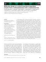
Fig. 3.
113
Cd-NMR resonances of 50 mM CdCl
2
in aqueous NaCl
(75 m
M), KCl (75 mM) buffer (bottom), and with 5 mM GSH added
to the solution (top). Instrument parameters are described in Exper-
imental procedures.
N. O. Kaakoush et al. Campylobacter jejuni and cadmium stress
FEBS Journal 275 (2008) 5021–5033 ª 2008 The Authors Journal compilation ª 2008 FEBS 5029
Experimental procedures
Materials
Blood agar base no. 2, brain heart infusion, defibrinated
horse blood and horse serum were obtained from Oxoid
(Heidelberg West, Australia). Amphotericin (Fungizone
Ò
),
bicinchoninic acid, BSA, chloramphenicol, copper II sul-
fate, dithiobis-2-nitrobenzoic acid, GSSG, GSH, NADH,
NADPH, polymixin B, trimethoprim, and bovine gluta-
thione reductase were obtained from Sigma (Castle Hill,
Australia). Vancomycin was obtained from Eli Lilly
(North Ryde, Australia), and Tris base was obtained from
Amersham Biosciences (Melbourne, Australia). All other
reagents were of analytical grade.
Bacterial strain and growth conditions
C. jejuni strain NCTC 11168 isolated from humans [51] was
grown at 37 °ConCampylobacter selective agar plates [64]
under microaerobic conditions (6% O
2
, 10% CO
2
). Liquid
cultures were grown in vented flasks using 50 mL brain
heart infusion supplemented with cadmium chloride (Sigma)
at concentrations of 0, 0.05, 0.1, 0.3, 0.5 and 1 mm. Cells
were tested for purity using phase-contrast microscopy.
Two-dimensional PAGE and mass spectrometry
identification of proteins
Preparation of cell-free protein extracts was performed as
described previously [65]. For the first dimension of two-
dimensional gel electrophoresis, samples were loaded onto
an 18 cm Immobiline DryStrip pH 4-7 (Amersham
Biosciences), and left to incubate sealed for 20 h at room
temperature. Isoelectric focusing was performed using a
flatbed Multiphor II unit (Amersham Biosciences). For the
second dimension, SDS–PAGE was performed on 11.5%
acrylamide gels using the Protean II system (Bio-Rad,
Sydney, Australia). The experimental conditions for two-
dimensional PAGE were as described previously [65]. Gels
were fixed individually in 0.2 L of fixing solution (50% v ⁄ v
methanol, 10% v ⁄ v acetic acid) for a minimum of 1 h, and
were subsequently stained using a sensitive ammoniacal
silver method. For comparative image analysis, statistical
data were acquired and analyzed using z3 compugen soft-
ware (Sunnyvale, CA, USA). Proteins were considered to
be regulated if the intensities of the corresponding spots on
test and control gels differed at least twofold.
The protocol to excise proteins from gels and digest
them, as well as the preparation of peptides for sequencing
by mass spectrometry, has been described previously [65].
Peptide identifications were performed using an API
QStar Pulsar I tandem MS instrument with the instrument
parameters used previously [65]. Protein searches were
performed on the National Center for Biotechnology Infor-
mation non-redundant database.
Bioinformatics
blastp searches were performed using the complete protein
sequences available at the NCBI database (http://
www.ncbi.nlm.nih.gov/). The Kyoto Encyclopedia of Genes
and Genomes (KEGG) available at />kegg was used to determine the biochemical pathways to
which the proteins were assigned. The Search Tool for the
Retrieval of Interacting Proteins (STRING), available at
which comprises known and pre-
dicted protein–protein interactions, was used to examine
predicted interactions between proteins.
Enzyme assays
Preparation of lysate fractions and cell-free protein extracts
for enzyme assays was carried out as previously described
[66]. Proton nuclear magnetic resonance (
1
H-NMR) spec-
troscopy was used to measure disulfide reduction. Free
induction decays were collected using a Bruker DMX-600
NMR spectrometer (Karlsruhe, Germany) operating in the
pulsed Fourier transform mode with quadrature detection
and the instrumental parameters used previously [66]. Disul-
fide reduction activities were measured in C. jejuni cell-free
extracts using GSSG and NADH as substrates. Chemical
reduction of GSSG in this system was ruled out because no
reduction was observed in the absence of cell-free extracts.
Negative controls showed that reduction of GSSG did not
take place if NADH was not present. The enzymatic origin
of the reactions was established by determining that no
activity was present in suspensions of cell-free extracts that
had been denatured by heating at 80 °C for 2 h.
Assays of fumarate reductase, fumarate dehydratase and
2-oxoglutarate ferredoxin oxidoreductase activities were
performed in whole-cell lysates as described previously [33].
Thioredoxin reductase activity was measured by dithiobis-
2-nitrobenzoic acid reduction in the presence of NADPH
using a Varian Cary-100 UV-visible spectrophotometer
(North Ryde, Australia) as described previously [63].
Effects of cadmium ions on enzyme activities
The effect of cadmium ions on glutathione reduction was
determined by measuring glutathione rates of reduction in
suspensions of whole bacterial lysates using
1
H-NMR spec-
troscopy. At substrate concentrations well below the K
m
, the
inhibition constant can be calculated from the expression
m
0
=m ¼ 1 þ I=K
i
where v
o
and v are the uninhibited and inhibited rates
of reduction, respectively, and I is the concentration of
inhibitor [67].
Campylobacter jejuni and cadmium stress N. O. Kaakoush et al.
5030 FEBS Journal 275 (2008) 5021–5033 ª 2008 The Authors Journal compilation ª 2008 FEBS
Interactions of cadmium ions with glutathione
and glutathione reductase
The interactions of oxidized and reduced glutathione with
Cd
2+
were studied using
1
H- and
13
C-NMR. Solutions of
GSSG or GSH were placed in 5 or 10 mm tubes at con-
centrations between 2 and 50 mm.
1
H-NMR free induction
decays were collected using a Bruker DMX-500 NMR
spectrometer, operating in the pulsed Fourier transform
mode with quadrature detection. Proton spectra were
acquired with presaturation of the water resonance. The
instrumental parameters were: operating frequency
500.13 MHz, spectral width 5000 Hz, memory size 16 K,
acquisition time 1.64 s, number of transients 64, pulse
angle 50° (3 ls), and relaxation delay with solvent presatu-
ration 1.4 s. Spectral resolution was enhanced by Gaussian
multiplication with line broadening of )0.6 Hz and Gauss-
ian broadening factor of 0.19. Chemical shifts are quoted
relative to sodium 4,4-dimethyl-4-silapentane-1-sulfonate
at 0 p.p.m.
One-dimensional natural-abundance
13
C-NMR spectra
were acquired with composite pulse proton decoupling in
an ACP-300 Bruker NMR spectrometer. The instrumental
parameters were: operating frequency 75.5 MHz, spectral
width 16129 Hz, memory size 16 K, acquisition time
0.508 s, number of transients 1600, and pulse angle 66°
(9 ls). Exponential filtering of 1 Hz was applied prior to
Fourier transformation. Chemical shifts are quoted with
respect to HCO
3
)
at 160 p.p.m.
The interactions of Cd
2+
with reduced or oxidized gluta-
thione and with bovine mucus glutathione reductase were
studied using
113
Cd-NMR spectroscopy. Solutions of CdCl
2
(50 mm) in buffer were placed in 10 mm tubes, and titrated
with either metabolite, the enzyme or both. The changes in
the spectral position and linewidth of the
113
Cd resonance
were measured from spectra of mixtures at various concen-
trations of GSSG, GSH or glutathione reductase.
113
Cd-NMR spectra were acquired using a Bruker DMX-
500 NMR spectrometer with composite pulse decoupling of
protons. The instrumental parameters for observing
113
Cd
were: operating frequency 110.9 MHz, spectral width
8865 Hz, memory size 16 K, acquisition time 0.92 s, relaxa-
tion delay 30 s, and pulse angle 90° (12 ls). The number of
transients was 128. Exponential filtering of 3 Hz was
applied prior to Fourier transformation. Chemical shifts
are quoted with respect to 0.1 m aqueous Cd(ClO
4
)
2
at
0 p.p.m.
Protein determination
Protein concentrations were determined using the bicinchoni-
nic acid method and a microtitre protocol (Pierce, Rockford,
IL, USA). Absorbances were measured using a Beckman
Du 7500 spectrophotometer (Gladesville, Australia).
Acknowledgement
This work was supported by the Australian Research
Council.
References
1 Bertin G & Averbeck D (2006) Cadmium: cellular
effects, modifications of biomolecules, modulation of
DNA repair and genotoxic consequences (a review).
Biochimie 88, 1549–1559.
2 Filipic
ˇ
M, Fatur T & Vudrag M (2006) Molecular
mechanisms of cadmium induced mutagenicity. Hum
Exp Toxicol 25, 67–77.
3 Navarro Silvera SA & Rohan TE (2007) Trace elements
and cancer risk: a review of the epidemiologic evidence.
Cancer Causes Control 18, 7–27.
4 Valko M, Rhodes CJ, Moncola J, Izakovic M & Maz-
ura M (2006) Free radicals, metals and antioxidants in
oxidative stress-induced cancer. Chem Biol Interact 160,
1–40.
5 Nauc
ˇ
ien
_
e Z, Mildazˇ ien
_
e V & Banien
_
e R (2002) Interac-
tions of cadmium and copper ions with complex I of
the respiratory chain in rat liver mitochondria.
Ekologija 2, 18–21.
6 Ron EZ, Minz D, Finkelstein NP & Rosenberg E
(1992) Interactions of bacteria with cadmium. Biodegra-
dation 3, 161–170.
7 Kwon SJ, Park JW & Kim K (1994) Inhibition of
metal-catalyzed oxidation systems by a yeast protector
protein in the presence of thiol. Biochem Mol Biol Int
32, 419–427.
8 Zegers I, Martins JC, Willem R, Wyns L & Messens J
(2001) Arsenate reductase from S. aureus plasmid pI258
is a phosphatase drafted for redox duty. Nat Struct Biol
8, 843–847.
9 Hayashi S, Abe M, Kimoto M, Furukawa S & Nakaza-
wa T (2000) The dsbA–dsbB disulfide bond formation
system of Burkholderia cepacia is involved in the pro-
duction of protease and alkaline phosphatase, motility,
metal resistance, and multi-drug resistance. Microbiol
Immunol 44, 41–50.
10 Rosato V, Pucello N & Giuliano G (2002) Evidence for
cysteine clustering in thermophilic proteomes. Trends
Genet 18, 278–281.
11 Maret W (2006) Zinc coordination environments in pro-
teins as redox sensors and signal transducers. Antioxid
Redox Signal 8, 1419–1441.
12 Masip L, Veeravalli K & Georgiou G (2006) The many
faces of glutathione in bacteria. Antioxid Redox Signal
8, 753–762.
13 Herbette S, Taconnat L, Hugouvieux V, Piette L,
Magniette M-LM, Cuine S, Auroy P, Richaud P,
N. O. Kaakoush et al. Campylobacter jejuni and cadmium stress
FEBS Journal 275 (2008) 5021–5033 ª 2008 The Authors Journal compilation ª 2008 FEBS 5031
Forestier C, Bourguignon J et al. (2006) Genome-wide
transcriptome profiling of the early cadmium response
of Arabidopsis roots and shoots. Biochimie 88, 1751–
1765.
14 Seib KL, Wu H-J, Kidd SP, Apicella MA, Jennings MP
& McEwan AG (2006) Defenses against oxidative stress
in Neisseria gonorrhoeae: a system tailored for a chal-
lenging environment. Microbiol Mol Biol Rev 70, 344–
361.
15 John R, Ahmad P, Gadgil K & Sharma S (2007)
Antioxidative response of Lemna polyrrhiza L. to
cadmium stress. J Environ Biol 28, 583–589.
16 Gillet S, Decottignies P, Chardonnet S & Le Mare
´
chal
P (2006) Cadmium response and redoxin targets in
Chlamydomonas reinhardtii: a proteomic approach.
Photosynth Res 89, 201–211.
17 El-Rab SMFG, Shoreit AA-F & Fukumori Y (2006)
Effects of cadmium stress on growth, morphology, and
protein expression in Rhodobacter capsulatus B10. Biosci
Biotechnol Biochem 70, 2394–2402.
18 Ferianc P, Farewell A & Nystro
¨
m T (1998) The cad-
mium-stress stimulon of Escherichia coli K-12. Microbi-
ology 144, 1045–1050.
19 Vido K, Spector D, Lagniel G, Lopez S, Toledano MB
& Labarre J (2001) A proteome analysis of the cad-
mium response in Saccharomyces cerevisiae. J Biol
Chem 276, 8469–8474.
20 Lee S-E, Yoo D-h, Son J & Cho K (2006) Proteomic
evaluation of cadmium toxicity on the midge Chirono-
mus riparius Meigen larvae. Proteomics 6, 945–957.
21 Moore JE, Corcoran D, Dooley JSG, Fanning S, Lucey
B, Matsuda M, McDowell DA, Me
´
graud F, Millar BC,
O’Mahony R et al. (2005) Campylobacter. Vet Res 36,
351–382.
22 Kazmi SU, Roberson BS & Stern NJ (1985) Cadmium
chloride susceptibility, a characteristic of Campylobacter
spp. J Clin Microbiol 21, 708–710.
23 Stern NJ, Kazmi SU, Roberson BS, Ono K & Juven BJ
(1988) Response of Campylobacter jejuni to combina-
tions of ferrous sulphate and cadmium chloride. J Appl
Bacteriol 64, 247–255.
24 Leal RS (1965) Effects of alkali-metal ions on phospho-
lipid and triglyceride synthesis in rat liver slices. J Lipid
Res 6, 80–83.
25 Pennanen T, Frostegard A
˚
, Fritze H & Baath E (1996)
Phospholipid fatty acid composition and heavy metal
tolerance of soil microbial communities along two
heavy metal-polluted gradients in coniferous forests.
Appl Environ Microbiol 62, 420–428.
26 Strydom C, Robinson C, Pretorius E, Whitcutt JM,
Marx J & Bornman MS (2006) The effect of selected
metals on the central metabolic pathways in biology: a
review. Water SA 32, 543–554.
27 Lu SC (2000) S-Adenosylmethionine. Int J Biochem Cell
Biol 32, 391–395.
28 Ueland PM (1982) Pharmacological and biochemical
aspects of S-adenosylhomocysteine and S-adenosylhom-
ocysteine hydrolase. Pharmacol Rev 34, 223–253.
29 Bochner BR, Lee PC, Wilson SW, Cutler CW & Ames
BN (1984) AppppA and related adenylylated nucleo-
tides are synthesized as a consequence of oxidation
stress. Cell 37, 225–232.
30 Pa
´
lfi Z, Sura
´
nyi G & Borbe
´
ly G (1991) Alterations in
the accumulation of adenylylated nucleotides in heavy-
metal-ion-stressed and heat-stressed Synechococcus sp.
strain PCC 6301, a cyanobacterium, in light and dark.
Biochem J 276, 487–491.
31 Pietrowska-Borek M, Stuible H-P, Kombrink E &
Guranowski A (2003) 4-Coumarate:coenzyme A ligase
has the catalytic capacity to synthesize and reuse vari-
ous (di)adenosine polyphosphates. Plant Physiol 131,
1401–1410.
32 Michel C, Giudici-Orticoni M-T, Baymann F & Bruschi
M (2003) Bioremediation of chromate by sulfate-reduc-
ing bacteria, cytochromes c
3
and hydrogenases. Water
Air Soil Pollut 3, 161–169.
33 Pitson SM, Mendz GL, Srinivasan S & Hazell SL
(1999) The tricarboxylic acid cycle of Helicobacter
pylori. Eur J Biochem 260, 258–267.
34 Gasque L, Berne
`
s S, Ferrari R & Mendoza-Dı
´
az G
(2002) Cadmium complexation by aspartate. NMR
studies and crystal structure of polymeric
Cd(AspH)NO3. Polyhedron 21, 935–941.
35 Rollin-Genetet F, Berthomieu C, Davin A-H & Queme-
neur E (2004) Escherichia coli thioredoxin inhibition by
cadmium: two mutually exclusive binding sites involving
Cys32 and Asp26. Eur J Biochem 271, 1299–1309.
36 Chiang PN, Wang MK, Chiu CY & Chou SY (2006)
Effects of cadmium amendments on low-molecular-
weight organic acid exudates in rhizosphere soils of
tobacco and sunflower. Environ Toxicol 21, 479–488.
37 Filella M, Town RM & Bugarin MG (1999) Cadmium
succinate and cadmium malate stability constants revis-
ited. J Chem Eng Data 44, 1009–1019.
38 Jo
´
csa
´
kI,Ve
´
gva
´
ri G & Droppa M (2005) Heavy metal
detoxication by organic acids in barley seedlings. Pro-
ceedings of the Eighth Hungarian Congress on Plant
Physiology and the Sixth Hungarian Conference on Pho-
tosynthesis, 2005 49, 99–101.
39 Ueno D, Ma JF, Iwashita T, Zhao F-J & McGrath SP
(2005) Identification of the form of Cd in the leaves of
a superior Cd-accumulating ecotype of Thlaspi caerules-
cens using
113
Cd-NMR. Planta 221, 928–936.
40 Chassagnole C, Quentin E, Fell DA, de Atauri P &
Mazat JP (2003) Dynamic simulation of pollutant
effects on the threonine pathway in Escherichia coli.
C R Biol 326, 501–508.
41 Wang A & Crowley DE (2005) Global gene expression
responses to cadmium toxicity in Escherichia coli.
J Bacteriol 187, 3259–3266.
Campylobacter jejuni and cadmium stress N. O. Kaakoush et al.
5032 FEBS Journal 275 (2008) 5021–5033 ª 2008 The Authors Journal compilation ª 2008 FEBS
42 Yuvaniyama P, Agar JN, Cash VL, Johnson MK &
Dean DR (2000) NifS-directed assembly of a transient
[2Fe–2S] cluster within the NifU protein. Proc Natl
Acad Sci USA 97, 599–604.
43 Van Bogelen RA, Kelley PM & Neidhart FC (1987)
Differential induction of heat shock, SOS, and oxida-
tion stress regulons and accumulation of nucleotides in
Escherichia coli. J Bacteriol 169, 26–32.
44 Silvestre F, Dierick JF, Dumont V, Dieu M, Raes M &
Devos P (2006) Differential protein expression profiles
in anterior gills of Eriocheir sinensis during acclimation
to cadmium. Aquat Toxicol 76, 46–58.
45 Lacerda CM, Choe LH & Reardon KF (2007) Meta-
proteomic analysis of a bacterial community response
to cadmium exposure. J Proteome Res 6, 1145–1152.
46 Hu P, Brodie EL, Suzuki Y, McAdams HH & Ander-
sen GL (2005) Whole-genome transcriptional analysis
of heavy metal stresses in Caulobacter crescentus. J Bac-
teriol 187, 8437–8449.
47 Jones PR & Desiot PJ (1978) The less familiar reactions
of organocadmium reagents. Chem Rev 78, 491–516.
48 Shi W, Yang Z, Geng Y, Wolinsky LE & Lovett MA
(1998) Chemotaxis in Borrelia burgdorferi. J Bacteriol
180, 231–235.
49 Maurer LM, Yohannes E, Bondurant SS, Radmacher
M & Slonczewski JL (2005) pH regulates genes for
flagellar motility, catabolism, and oxidative stress in
Escherichia coli K-12. J Bacteriol 187, 304–319.
50 Butler SM & Camilli A (2005) Going against the grain:
chemotaxis and infection in Vibrio cholerae. Nat Rev
Microbiol 3, 611–620.
51 Parkhill J, Wren BW, Mungall K, Ketley JM, Churcher
C, Basham D, Chillingworth T, Davies RM, Feltwell T,
Holroyd S et al. (2000) The genome sequence of the
food-borne pathogen Campylobacter jejuni reveals
hypervariable sequences. Nature 403, 665–668.
52 von Mering C, Jensen LJ, Kuhn M, Chaffron S, Doerks
T, Kruger B, Snel B & Bork P (2007) STRING 7 –
recent developments in the integration and prediction of
protein interactions. Nucleic Acids Res 35, D358–D362.
53 Muller JP, Vedel M, Monnot MJ, Touzet N & Wegnez
M (1991) Molecular cloning and expression of ferritin
mRNA in heavy metal-poisoned Xenopus laevis cells.
DNA Cell Biol 10, 571–579.
54 Wai SN, Nakayama K, Umene K, Moriya T & Amako
K (1996) Construction of a ferritin-deficient mutant of
Campylobacter jejuni: contribution of ferritin to iron
storage and protection against oxidative stress. Mol
Microbiol 20, 1127–1134.
55 Ishikawa T, Mizunoe Y, Kawabata S, Takade A,
Harada M, Wai SN & Yoshida S (2003) The
iron-binding protein Dps confers hydrogen peroxide
stress resistance to Campylobacter jejuni. J Bacteriol
185
, 1010–1017.
56 Chauhan S, Titus DE & O’Brian MR (1997) Metals
control activity and expression of the heme
biosynthesis enzyme delta-aminolevulinic acid dehydra-
tase in Bradyrhizobium japonicum. J Bacteriol 179,
5516–5520.
57 Grosse C, Grass G, Anton A, Franke S, Santos AN,
Lawley B, Brown NL & Nies DH (1999) Transcrip-
tional organization of the czc heavy-metal homeostasis
determinant from Alcaligenes eutrophus. J Bacteriol 181,
2385–2393.
58 Perron K, Caille O, Rossier C, Van Delden C, Dumas
JL & Kohler T (2004) CzcR–CzcS, a two-component
system involved in heavy metal and carbapenem resis-
tance in Pseudomonas aeruginosa. J Biol Chem 279,
8761–8768.
59 Henehan CJ, Pountney DL, Zerbe O & Vasak M (1993)
Identification of cysteine ligands in metalloproteins
using optical and NMR spectroscopy: cadmium-substi-
tuted rubredoxin as a model [Cd(CysS)(4)](2-) center.
Protein Sci 2, 1756–1764.
60 Adams TK, Saydam N, Steiner F, Schaffner W &
Freedman JH (2002) Activation of gene expression by
metal-responsive signal transduction pathways. Environ
Health Perspect 110, 813–817.
61 Scott K, Diggle MA & Clarke SC (2003) TypA is a
virulence regulator and is present in many pathogenic
bacteria. Br J Biomed Sci 60, 168–170.
62 Lin Y-F, Wu M-S, Chang C-C, Lin S-W, Lin J-T, Sun
Y-J, Chen D-S & Chow L-P (2006) Comparative immu-
noproteomics of identification and characterization of
virulence factors from Helicobacter pylori related to
gastric cancer. Mol Cell Proteomics 5 , 1484–1496.
63 Kaakoush NO, Sterzenbach T, Miller WG, Suerbaum S
& Mendz GL (2007) Identification of disulfide reducta-
ses in Campylobacterales: a bioinformatics investiga-
tion. Antonie Van Leeuwenhoek 92, 429–441.
64 Kaakoush NO, Miller WG, De Reuse H & Mendz GL
(2007) Oxygen requirement and tolerance of Campylo-
bacter jejuni. Res Microbiol 158, 644–650.
65 Fox EM, Raftery M, Goodchild A & Mendz GL (2007)
Campylobacter jejuni response to ox-bile stress. FEMS
Immunol Med Microbiol 49, 165–172.
66 Kaakoush NO & Mendz GL (2005) Helicobacter pylori
disulphide reductases: role in metronidazole reduction.
FEMS Immunol Med Microbiol 44, 137–142.
67 Schulz AR (1994) Enzyme Kinetics, from Diastase to
Multienzyme Systems. Cambridge University Press,
New York, NY.
N. O. Kaakoush et al. Campylobacter jejuni and cadmium stress
FEBS Journal 275 (2008) 5021–5033 ª 2008 The Authors Journal compilation ª 2008 FEBS 5033