Báo cáo khoa học: Acoustic microfluidic chip technology to facilitate automation of phage display selection doc
Bạn đang xem bản rút gọn của tài liệu. Xem và tải ngay bản đầy đủ của tài liệu tại đây (675.28 KB, 10 trang )
Acoustic microfluidic chip technology to facilitate
automation of phage display selection
Jonas Persson
1
, Per Augustsson
2
, Tomas Laurell
2
and Mats Ohlin
1
1 Department of Immunotechnology, Lund University, Sweden
2 Department of Electrical Measurements, Lund University, Sweden
The potential for recombinant antibodies in various
analytical and therapeutic applications has developed
substantially over recent years. With new therapeutic
targets emerging continuously for various diseases and
with the completion of the human genome sequencing
project [1,2], extensive efforts are now directed towards
understanding how complex sets of gene products are
responsible for the many different functions of living
cells with respect to both health and disease. The mul-
tiplex analysis approach, employing large arrays of
antibodies, is being used to expand our knowledge of
how proteins participate in such processes [3]. To carry
out such studies, there is a vast need for specific detec-
tion reagents. Indeed, several efforts are underway to
develop binders against large sets of proteins, such as
those produced by the human genome. The Human
Proteome Resource Center project is designed to raise
specific binders, mainly specific rabbit polyclonal anti-
bodies, targeting sequences with a unique potential
for essentially any human protein [4]. The Proteome-
Binders consortium ()
has been set up to establish an infrastructure to isolate
and use binding molecules (not necessarily antibodies)
targeting essentially every member of the human prote-
ome [5]. Similarly, the Antibody Factory (http://
www.antibody-factory.de) [6], the Sanger Institute’s
ATLAS of protein expression [7] and the US National
Cancer Institute proteome reagent program (http://
proteomics.cancer.gov) [8] have been organized to deli-
ver reagent resources to explore proteomes. Together,
these efforts are designed to raise specific binders, with
an origin in the antibody scaffold or other scaffolds
well suited for their intended applications.
Specific binders are raised in a relatively high-
throughput format by a number of approaches, such
as the development of rabbit polyclonal antibodies [9]
or murine monoclonal antibodies [10]. Subsequent to
its introduction as a tool for the isolation of specific
binders against essentially any target [11], phage dis-
play technology has evolved into a very efficient tool
Keywords
acoustic standing wave forces; antigen-
specific binding; microfluidic chip; phage
display; selection
Correspondence
M. Ohlin, Department of
Immunotechnology, Lund University, BMC
D13, SE-22184 Lund, Sweden
Fax: +46 4622 24200
Tel: +46 4622 24322
E-mail:
(Received 4 July 2008, revised 5 September
2008, accepted 18 September 2008)
doi:10.1111/j.1742-4658.2008.06691.x
Modern tools in proteomics require access to large arrays of specific bind-
ers for use in multiplex array formats, such as microarrays, to decipher
complex biological processes. Combinatorial protein libraries offer a solu-
tion to the generation of collections of specific binders, but unit operations
in the process to isolate binders from such libraries must be automatable
to ensure an efficient procedure. In the present study, we show how a
microfluidic concept that utilizes particle separation in an acoustic force
field can be used to efficiently separate antigen-bound from unbound mem-
bers of such libraries in a continuous flow format. Such a technology has
the hallmarks for incorporation in a fully automated selection system for
the isolation of specific binders.
Abbreviations
CMV, cytomegalovirus; gB, glycoprotein B; scFv, single chain antibody fragment; XG, xyloglucan.
FEBS Journal 275 (2008) 5657–5666 ª 2008 The Authors Journal compilation ª 2008 FEBS 5657
with a high utility for the very same purpose as the
development of polyclonal or monoclonal antibodies.
Many features of phage display and other display tech-
nologies make them amendable to automation, allow-
ing for the efficient development of the vast arrays of
specific binders that are required in proteome research
efforts [6,7].
Any process designed to develop specific binders
comprises a number of unit operations, each of which
has the goal to produce a product that is important
for a subsequent step in the process. To ensure high
throughput, a maximum level of automation is
required. For the development of specific binders using
phage display technology, several of these unit opera-
tions can be identified (Fig. 1A). Currently, large
collections of antigens are available that may serve as
sources of targets from a variety of species, including
Homo sapiens, Mus musculus and Saccharomyces cere-
visiae [4,9,12–15]. Efforts to use bioinformatics anti-
genic epitope analysis approaches and to produce new
antigens that are suitable for the development of
specific binders at a high rate are on-going [4,15,16].
Large collections of binders in the form of molecular
libraries intended for different selection procedures,
including phage display, are available [17–21]. Simi-
larly, automated screening systems are available that
can assess binding and specificity properties of large
number of selected clones [22–24]. Systems to produce
and purify specific binders at a high rate [9,25] and to
confirm their specificity properties [9,26] are being
established. The actual selection process and, specifi-
cally, the separation of unbound phage particles dis-
playing nonspecific antibodies is, however, still in need
of an automatable process. The selection quality gener-
ally depends on a number of washing and centrifuga-
tion steps to ensure the enrichment of rare phages
displaying binders specific for a given target from the
large bulk of other phages. Attempts to automate the
separation process by catching antigen-specific phages
on paramagnetic beads that are subsequently trapped
and washed on magnets have met with some success
[27,28]. Systems based on antigen-immobilized on
microtiter plates have also been utilized [23], but further
developments would facilitate this process and increase
throughput and yield. In an approach adapted to
selection from bacterial display libraries, Hu et al. [29]
developed a microfluidic system, based on dielectro-
phoretic forces, that could isolate rare species from such
entities. We now describe a highly flexible, fast and
continuous flow process, also based on microfluidics
and ultrasound-based focusing of particles (Fig. 1B–E),
for the efficient enrichment of phages displaying specific
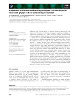
Fig. 1. Acoustic microfluidic chip technology in phage display. (A) Unit operations in a procedure to isolate antigen-specific binders by phage
display. The selection unit is further divided into tasks to define the placement of the herein-designed separation unit in the process.
(B) Specific antibody fragment-displaying phage particles bind to an antigen-coated bead as opposed to other phage particles. (C) Photograph
of the microfluidic separation device. (D) Schematic of the separation device (only one unit illustrated). A mixture of beads and phage parti-
cles (light gray) is flow-laminated along both sides of phage-free buffer in the channel center (upper). Beads are focused towards the center
of the flow under the influence of an ultrasonic standing wave field, whereas unbound phage particles, not being affected by the ultrasound,
remain in their flow-laminated position near the side walls (lower). (E) Illustration of trifurcation outlet collecting the bead-containing center
fraction (dark gray) of the flow while unbound phage-particles are effectively removed.
Acoustic microfluidic chip for phage selection J. Persson et al.
5658 FEBS Journal 275 (2008) 5657–5666 ª 2008 The Authors Journal compilation ª 2008 FEBS
binders from commonly used phage display libraries.
Beads carrying the target of interest are continuously
translated from a complex buffer solution (phage parti-
cle-containing mixture) into a clean carrier buffer lami-
nated in the center of a flow channel using acoustic
standing wave forces. This procedure has the hallmarks
of a process that lends itself to full automation. We
envisage that this technology will be used in high-
throughput operations for the development of a unit
operation involving the selection and separation of spe-
cific binders from large combinatorial libraries.
Results
System design
Using an artificial mixture of two different affinity
molecules [i.e. the carbohydrate-binding module
XG-34 that binds xyloglucan (XG) and the single
chain antibody fragment (scFv) GgB1 that binds cyto-
megalovirus glycoprotein B (CMV gB)] displayed on
the surface of phage particles, optimal conditions were
sought for enriching either of these two clones from a
1000-fold excess of the other clone using antigen
immobilized on microbeads. The separation of bound
and unbound phages was achieved using two serially
linked acoustic separation channels because the use of
a single channel device had proven insufficient. Gener-
ally, a 1000-fold enrichment factor of the phage dis-
playing the protein binding the immobilized target was
observed in a single round of selection (Fig. 2A).
Complex library selections
To validate the efficiency of the microchip-based sepa-
ration system and to compare it with the classic man-
ual separation method, parallel selections were
performed using a conventional antibody fragment-dis-
played library by selecting binders for one specific tar-
get, the grass pollen allergen Phl p 5. Titration of
input phagestocks and phagestocks made after selec-
tion and reinfection in Escherichia coli demonstrated
that the microchip-based separation system was at
least as efficient as conventional, manual separation in
producing a population enriched for specific phages
(Fig. 2B). After a single round of selection, 16 of 30
and nine of 30 randomly picked clones obtained after
microchip-based or manual separation, respectively,
Fig. 2. Performance of acoustic microfluidic chip separation in phage display. (A) Enrichment factor of antigen-specific phages using the
microchip-based washing principle. The results show the enrichment factor of CMV gB-specific antibody fragment GgB1 (experiments 1 and
2; duplicate experiments) and carbohydrate-binding module XG-34 (experiments 3 and 4; duplicate experiments) in the presence of a 1000-
fold excess of phages displaying the other protein. (B) Titration by antigen (Phl p 5)-specific ELISA of polyclonal phage stocks to illustrate the
enhanced recognition of allergen after enrichment of the antibody fragment library displayed on phage. Samples include a phage stock of
the original antibody fragment library population before selection (dashed line) and phage stocks made after one round of selection for
Phl p 5-specificity employing either a manual (closed symbols) or a microchip-based (open symbols) separation approach. (C) Antigen-speci-
ficity of selected binders. Representative clones of the five clonotypes (Fig. 3) identified after the use of microchip-based (clonotypes 16, 29,
35 and 38) and manual (clonotypes 29, 35 and 41) separation systems were assessed for specificity. Their binding to recombinant allergen
Phl p 5 (green) (the antigen used in selection) but not recombinant Phl p 2 (dark blue), Phl p 6 (orange), Phl p 7 (magenta), natural Phl p 4
(red) or streptavidin (light blue) demonstrated that selected clones were specific for the intended target.
J. Persson et al. Acoustic microfluidic chip for phage selection
FEBS Journal 275 (2008) 5657–5666 ª 2008 The Authors Journal compilation ª 2008 FEBS 5659
were specific for the target antigen, as determined by
ELISA. To assess the diversity of this selected popula-
tion, we performed sequencing of randomly picked
clones that produced antibody fragments specific for
the allergen. This procedure identified a diverse set of
sequences in both selected populations (Fig. 3) [30,31].
Because genes encoding the heavy chain variable
domain sequences of the library had been amplified
from the transcriptome encoding IgE, a population
restricted in the number of clonotypes that are con-
tained within it [30,32], several of the clones were simi-
lar, as expected. The obtained clones could be divided
into five groups based on their genetic resemblance.
Clones from four of the five groups were extracted
when using the microchip-based separation system,
whereas three of the five groups were identified among
the sequences found after the manual separation
method. The presence of different mutations and light
Fig. 3. Sequences of selected Phl p 5-specific scFv. Sequences of proteins selected by the microchip-based separation method (clones
denoted P5-AA and P5-AB) and the conventional manual wash procedure (clones denoted P5-MA and P5-MB). Clones are arranged accord-
ing to the separation method and their origin in a common clonotype as defined by Persson et al. [30] with the addition of clone P5-MA5
that represents a novel clonotype, number 41. All sequences, except P5-AB4 and P5-AB11, are unique. Complementarity determining
regions (CDR) of the heavy (H) and light (L) chains, as defined by ImMunoGeneTics nomenclature [31], are underlined (black line). The linker
region inbetween the H and L chain variable domains are underlined (gray line). Residues found in ‡ 50% of the sequences are boxed.
Acoustic microfluidic chip for phage selection J. Persson et al.
5660 FEBS Journal 275 (2008) 5657–5666 ª 2008 The Authors Journal compilation ª 2008 FEBS
chain variable domains nevertheless demonstrated that
many different sequences were selected in each group.
The microchip-based separation method thus did not
bias the selection to one or a few clones. In addition
to sequences similar to those that had been selected
previously [30,32], entirely new binders were selected,
one each from studies employing the two different sep-
aration methods (clones P5-AB5 and P5-MA5). The
specificity of representatives from the five groups for
the target antigen was investigated. It was shown that
binding to the target antigen was specific, demonstrat-
ing that the selection approaches were appropriate and
selected for specific binders (Fig. 2C). In conclusion,
the microchip-based separation method efficiently
enriched phages displaying specific antibody fragments
and retrieved a diverse population of specific sequence
variants.
Discussion
The aim of the present study was to develop an effi-
cient and easy-to-use separation method optimized for
high-throughput development of affinity binders
towards a multitude of targets, in order to cope with
the growing demand for such reagents in applications
such as global proteome analysis. These approaches
use large arrays of different specific binders such as
antibodies or antibody fragments towards the various
targets in a proteome. When aiming to generate large
enough numbers of antibodies, enormous pressure is
placed on the development and selection stages [33].
Several of the different steps in the process of
obtaining new antibodies through phage display,
a state-of-the-art source of specific binders, are already
automatable for high-throughput strategies. The actual
selection process and, specifically, the separation of
unbound phage particles displaying nonspecific anti-
bodies is, however, still in need of an automatable
process. We believe that the results presented in the
present study comprise a substantial step towards a
solution to this bottleneck in high-throughput phage
display selection. To this end, a chip-based microfluidic
wash system has been designed and tested because
such a system has the potential to be easily incorpo-
rated into an automated liquid handling system. Sub-
sequent to its introduction in 2001 [34,35], chip
integrated ultrasonic standing wave technology has
demonstrated important advancements in the precise
control of particles in microfluidic systems [36]. A
major development was the discovery that the induc-
tion of an acoustic standing wave in microchannels
orthogonal to the incident sound wave allowed for
acoustic force manipulation of cells and particles in
microfluidic networks [37]. Advanced acoustic micro-
chip particle separation approaches have subsequently
been successfully exploited in biomedicine and biotech-
nology [38–41]. Acoustic microfluidic chip technology
has recently also enabled noncontact particle and cell
trapping and manipulation for online bioassaying [42–
45]. The results of the present study now extend micro-
chip acoustic particle separation into selective targeting
of biomolecular entities, facilitating functional mole-
cular evolution by genetic engineering. The microscale
environment yields a low Reynolds number, and
ensures perfect laminar conditions in the flow system,
facilitating its separation efficiency. We have previ-
ously demonstrated the possibility of using acoustic
forces to extract particles from a contaminated envi-
ronment in a continuous flow format [41]. A system
for continuous flow phage library selection is now
proposed based on this concept. A detailed chip design
and fundamental microfluidic and acoustic perfor-
mances in conventional bioanalytical procedures evalu-
ation have recently been described (P. Augustsson,
J. Persson, S. Ekstro
¨
m, M. Ohlin & T. Laurell, unpub-
lished results). We now define optimum operation con-
ditions for the phage library selection performed in the
present study. The initial assessment of the system
indicated that it was capable of separating bound and
unbound phage particles and that it achieved an
enrichment factor in the order of 1000 in a single chip
comprising two serially coupled separation channels.
The exact level of enrichment will be dependent not
only on the separation approach itself, but also on
the specific character (level of display, affinity, etc.) of
the molecules displayed on the phage particles. The
achieved enrichment, therefore, does not define the
upper limit of enrichment but rather a realistic level.
Assessment of contamination of phages in an antigen-
free system indicated that the efficiency of separation
can be as high as 99.9999% for a double channel chip.
Efficiencies approaching an at least 1000-fold enrich-
ment may then be achievable depending on level and
nature of the displayed molecules. Importantly, the
separation step requires no manual intervention and it
is completed in approximately 8 min when applying a
500 lL sample, which is a volume typical of many
selection procedures, suggesting that even a single unit
can handle large numbers of samples in 1 day even
when considering the need for automated wash cycles
between different runs. Moreover, the throughput of
beads was approximately 5 · 10
4
s
)1
, which is consid-
erably high in a microfluidic chip context.
The usefulness of a unit operation in phage selection
depends not only on the speed, but also on its ability
to maintain diversity in the population of selected
J. Persson et al. Acoustic microfluidic chip for phage selection
FEBS Journal 275 (2008) 5657–5666 ª 2008 The Authors Journal compilation ª 2008 FEBS 5661
molecules. By assessing the diversity of clones obtained
after selection on Phl p 5, we determined that a variety
of clones could be obtained. It is evident that this sys-
tem is addressing a very similar antibody repertoire,
and certainly a no less diverse one, compared to the
manual wash system.
In conclusion, the chip-based microfluidic wash
system that separates bound and unbound phages, dis-
playing proteins with a specific binding property, is at
least as efficient as conventional separation approaches,
such as those involving washing of microtiter plates or
microbeads. However, it has several advantages, includ-
ing an automatable fluidic system approach and the
potential for high throughput. In addition, it has the
capacity to use a variety of beads and cells [39,46] as
antigen carriers because very different types of particles
can be focused by ultrasound. The system is thus highly
flexible and can be adopted to virtually any kind of
antigen carrier. Altogether, we foresee that the pro-
posed chip-based microfluidic wash system for antigen-
bound phage enrichment ⁄ extraction will be used as an
automated unit operation in approaches to isolate
binders specific for members of entire proteomes.
Experimental procedures
Proteins, genes, vectors and libraries
Recombinant CMV gB [47] and biotinylated XG [48] was
kindly provided by Sanofi-Pasteur (Marcy l’Etoile, France)
and H. Brumer (the Royal Institute of Technology, Stock-
holm, Sweden), respectively. Recombinant timothy allergens
(Phl p 2, Phl p 5, Phl p 6 and Phl p 7) were obtained from
BioMay (Vienna, Austria). The natural allergen Phl p 4
was kindly provided by J. Lidholm (Phadia AB, Uppsala,
Sweden). Recombinant gB and Phl p 5, biotinylated using
sulfo-NHS-biotin and sulfo-NHS-LC-biotin (Pierce, Rock-
ford, IL, USA), respectively, and extensively dialyzed
against NaCl ⁄ P
i
, were kindly provided by Fredrika
Axelsson and Kristina Lundberg (Lund University, Lund,
Sweden).
For the purpose of the present study, we used phagemid
vectors designed for display of proteins on protein 3 of fila-
mentous phage. These included a vector based on pAK100
[49] encoding chloramphenicol resistance, which encodes a
scFv, GgB1, specific for CMV gB (F. Axelsson, J. Persson,
E. Moreau, M. H. Coˆ te
´
, A. Lamarre & M. Ohlin, unpub-
lished data), and a vector based on a modified version of
pFab5c.His [50] encoding ampicillin resistance, which codes
for the carbohydrate-binding module XG-34 [48] specific
for XG.
A library [32] encoding scFv cloned into the pFab5c.His
vector was also used. The heavy chain variable domain-
encoding-sequences of this library had been amplified from
transcripts encoding IgE of an allergic donor. This library
has previously been used successfully to select a range of
scFv specific for a number of allergens [30,32].
Acoustic particle washing microchip
To create a chip for microbead separation, similar to that
relevant in a system designed to potentially enable auto-
mated selection from combinatorial protein libraries such
as those displayed on phage, we constructed a new micro-
fluidic washing device (Fig. 1C), based on previous work
(P. Augustsson, J. Persson, S. Ekstro
¨
m, M. Ohlin &
T. Laurell, unpublished results). The manufacturing of the
device was based on standard microfabrication techniques
that are accessible in most clean-room facilities. The basic
silicon processing scheme has been described in more detail
by Nilsson et al. [37]. Briefly, the separation channel was
etched in (100) silicon using standard KOH wet etch tech-
niques creating channels of rectangular cross section
(width = 375 lm, height = 160 lm). The channel width
was selected to match a k ⁄ 2 wavelength resonance criterion
in aqueous media. Borosilica glass was anodically bonded
to the silicon to enclose the flow structure and to allow for
optical surveillance. Particles passing along the channel
while actuated at 2 MHz will experience a primary acoustic
radiation force that will position them either in the center
of the channel or near the side walls. The magnitude and
direction of the force is dependent on the acoustic proper-
ties (density and compressibility) of the particles as well as
the suspending media. Most biological and fabricated parti-
cles are slightly denser than water, which makes them move
towards the center of the channel. Because the ultrasound
has little or no effect on the suspending media, it is possible
to utilize the force field to move particles from one media
to another by flow lamination of the two media in the
presence of an acoustic force field (Fig. 1D).
The separation chip was actuated using a 7 · 35 mm
piezoceramic (PZT 27; Ferroperm Piezoceramics A ⁄ S,
Kvistgard, Denmark) resonant at 2 MHz. The transducer
was glued to the upper side of the glass alongside to the
channel structure. A function generator (HP 3325A; Hew-
lett-Packard Inc, Palo Alto, CA, USA) coupled to a power
amplifier (Amplifier Research Model 50A15; Amplifier
Research, Souderton, PA, USA) fed the transducer with a
2 MHz sine wave. The net power (transmitted minus
reflected) was monitored using a wattmeter (43 Thruline
Wattmeter; Bird Electronic Corporation, Cleveland, OH,
USA).
Sample containing beads and unbound molecular mate-
rial entered the structure and was bifurcated to each side of
the first of two wash fluid inlets. The sample and wash fluid
did not mix due to the highly laminar flow condition in the
microchannels. The fluids passed a 2-cm long channel seg-
ment where the beads were acoustically focused towards
the center of the channel, whereas the unbound material
Acoustic microfluidic chip for phage selection J. Persson et al.
5662 FEBS Journal 275 (2008) 5657–5666 ª 2008 The Authors Journal compilation ª 2008 FEBS
remained in its flow-laminated position near the side walls.
By splitting the flow outlet in three, the undesired material
was separated from the beads that continued via yet
another bifurcation to a second identical wash step
(Fig. 1B–E).
Production of phage stocks
All phage stocks were produced by standard procedures.
Briefly, F-pili-carrying E. coli were grown in medium
containing 1% glucose and relevant antibiotics. When the
culture had reached exponential growth phase, the bacteria
were infected with VCS-M13 helper phages (Stratagene, La
Jolla, CA, USA) for 30 min at 37 °C. Phage stocks were
produced by culture in glucose-free medium containing
antibiotics and 0.25 mm isopropyl thio-b-d-galactoside at
30 °C overnight. In some cases, phages were precipitated by
the addition of 0.25 volumes of 20% PEG6000 ⁄ 2.5 m NaCl
and resuspended in NaCl ⁄ P
i
. Phage stock of the library
with an origin in IgE-encoding transcripts were used as
such, whereas phage stocks displaying XG-34 and GgB1
were mixed in ratios of approximately 1 : 1000 and 1000 : 1
to prepare model mini-libraries useful for evaluation of
phage purification efficiency.
Selection system
Biotinylated ligands, XG (20 lg), gB (5 lg) or Phl p 5
(20 lg), were added to 50 lL of streptavidin-coated M280
Dynabeads (Invitrogen, Carlsbad, CA, USA) and incubated
for 2 h on a rotator at room temperature. These beads were
washed three times with 3% BSA and 0.05% Tween-20 in
NaCl ⁄ P
i
(NaCl ⁄ P
i
-Tween) to remove excess ligand prior to
use. Phage populations, either artificial mixtures of those
displaying XG-34 and GgB1 or those displaying scFv with
an origin in the IgE-encoding population, were added in
NaCl ⁄ P
i
-Tween to beads ( 5 · 10
7
beadsÆmL
)1
in final
suspension) coated with the ligand. The mix was incubated
on a rotator for 1–2 h at room temperature.
Microchip-based wash procedure
Samples containing phages and antigen coated-microbeads
were aspirated into a 1 mL disposable syringe that was
inserted into a syringe pump (WPI SP210IWC; World Pre-
cision Instruments Inc., Sarasota, FL, USA) vertically and
above the microfluidic washing device. Wash fluid was
loaded into a pair of 10 mL glass syringes (1010 TLL;
Hamilton Bonaduz AG, Bonaduz, Switzerland) positioned
in a dual push–pull syringe pump (WPI SP2 60P; World
Precision Instruments Inc.) with an additional pair of syrin-
ges mounted reversely in the same pump for waste fluid
aspiration. TFE TeflonÔ Tubing (inner diameter 0.3 mm)
(Supelco, Bellefonte, PA, USA) was used for guiding fluids
in and out from the device. The sample outlet was open to
atmospheric pressure through a short piece of tubing ema-
nating in a sample collection test tube. The system was
primed with wash fluid (NaCl ⁄ P
i
-Tween) by compressing
the wash syringes until all air bubbles were completely
removed from the channel structure and all external tubing.
Prior to connecting the sample injection syringe, the wash
syringe pump was run for approximately 1 min to stabilize
flow in the system. The wash fluid flows were set to
120 lLÆmin
)1
into each washing chamber and the sample
injection and throughput flow was set to 60 lLÆmin
)1
. The
ultrasound was subsequently turned on at a frequency of
2 MHz delivering a net power of 1.1 W to the transducer.
Washed bead suspensions were collected from the device in
a continuously running process in fractions of 0.2 mL.
Manual wash procedure
Mixtures of antigen-coated beads and phage stocks were
washed five times with NaCl ⁄ P
i
-Tween and three times with
NaCl ⁄ P
i
using a magnet to retrieve the microbeads.
Bacterial infection procedure
A slurry of beads obtained after the manual wash proce-
dure or as the output from the outlet of the microchip
washing device was added to exponentially growing E. coli
carrying F-pili (Top10F¢) for 30 min at 37 °C (without
shaking). Dilutions of bacteria infected with artificial mix-
tures of phages displaying GgB1 and XG-34 were spread
on culture plates (LB agar) containing chloramphenicol
(25 lgÆmL
)1
) or ampicillin (100 lgÆmL
)1
). The relations
between the two clones in the libraries after the phage bind-
ing and subsequent wash procedure were determined from
the numbers of colonies on plates with the different antibi-
otics. The output after selection on Phl p 5 was grown on
plates containing ampicillin and 1% glucose. After culture
for 16 h at 37 °C, the number of colonies was counted.
Immunological analysis
To assess the quality of the output of selection of scFv on
the recombinant allergen, fifteen clones were picked from
each of four selections performed on Phl p 5 using the con-
ventional, manual washing approach (clones named with
prefixes P5-MA and MB) or the microchip-based washing
approach (clones named with prefixes P5-AA and AB).
Phage stocks for each of the 60 clones were analyzed in
ELISA to determine their ability to bind the antigen
Phl p 5 in addition to several other antigens from grass
pollen. Bound phages were detected with horseradish per-
oxidase-conjugated M13-specific monoclonal antibody
(GE Healthcare Biosciences Corp., NJ, USA) using
o-phenylenediamine as chromogen. Phage stock for the
J. Persson et al. Acoustic microfluidic chip for phage selection
FEBS Journal 275 (2008) 5657–5666 ª 2008 The Authors Journal compilation ª 2008 FEBS 5663
entire polyclonal outputs after selections (MA and MB, AA
and AB), in addition to the parental IgE-based library in
serial dilutions, was also analyzed by antigen-specific
ELISA to determine the accumulated specificity relative to
the parent library.
Sequencing and sequence analysis
Plasmids from the clones producing Phl p 5-specific scFv
were purified from bacterial cell pellets using QIAprep Spin
Miniprep Kit (Qiagen, Hilden, Germany) and subsequently
sequenced (MWG Biotech, Martinsried, Germany).
Sequences (GenBank Accession Numbers EF601881–
EF601896 and EU090053–EU090060) were compared with
previously selected clones from the library specific for
Phl p 5 [30,32]. Clones were named using the following
nomenclature: P5 (defining timothy group 5 allergen speci-
ficity), a letter combination denoting an origin in selections
employing either manual (MA and MB) or microchip-based
(AA and AB) washing approaches, and a clone number.
Acknowledgements
This study was supported by grants from BioInvent
International AB, the Swedish Research Council,
Crafoord Foundation, Carl Trygger Foundation, Cre-
ate Health, the Royal Physiographic Society and
ELFA Foundation.
References
1 Venter JC, Adams MD, Myers EW, Li PW, Mural RJ,
Sutton GG, Smith HO, Yandell M, Evans CA, Holt
RA et al. (2001) The sequence of the human genome.
Science 291, 1304–1351.
2 Lander ES, Linton LM, Birren B, Nusbaum C, Zody
MC, Baldwin J, Devon K, Dewar K, Doyle M, Fitz-
Hugh W et al. (2001) Initial sequencing and analysis of
the human genome. Nature 409 , 860–921.
3 Borrebaeck CAK (2006) Antibody microarray-based
oncoproteomics. Expert Opin Biol Ther 6, 833–838.
4 Uhle
´
n M, Bjo
¨
rling E, Agaton C, Szigyarto CA, Amini
B, Andersen E, Andersson AC, Angelidou P, Asplund
A, Asplund C et al. (2005) A human protein atlas for
normal and cancer tissues based on antibody proteo-
mics. Mol Cell Proteomics 4, 1920–1932.
5 Taussig MJ, Stoevesandt O, Borrebaeck CAK, Brad-
bury AR, Cahill D, Cambillau C, de Daruvar A, Du
¨
bel
S, Eichler J, Frank R et al. (2007) ProteomeBinders:
planning a European resource of affinity reagents for
analysis of the human proteome. Nat Methods 4, 13–17.
6 Konthur Z, Hust M & Du
¨
bel S (2005) Perspectives
for systematic in vitro antibody generation. Gene 364,
19–29.
7 Schofield DJ, Pope AR, Clementel V, Buckell J, Chap-
ple SDj, Clarke KF, Conquer JS, Crofts AM, Crowther
SR, Dyson MR et al. (2007) Application of phage dis-
play to high throughput antibody generation and char-
acterization. Genome Biol 8, R254.
8 Haab BB, Paulovich AG, Anderson NL, Clark AM,
Downing GJ, Hermjakob H, Labaer J & Uhle
´
nM
(2006) A reagent resource to identify proteins and pep-
tides of interest for the cancer community: a workshop
report. Mol Cell Proteomics 5, 1996–2007.
9 Nilsson P, Paavilainen L, Larsson K, Odling J, Sundberg
M, Andersson AC, Kampf C, Persson A, Al-Khalili
Szigyarto C, Ottosson J et al. (2005) Towards a human
proteome atlas: high-throughput generation of mono-
specific antibodies for tissue profiling. Proteomics 5,
4327–4337.
10 De Masi F, Chiarella P, Wilhelm H, Massimi M,
Bullard B, Ansorge W & Sawyer A (2005) High
throughput production of mouse monoclonal antibodies
using antigen microarrays. Proteomics 5, 4070–4081.
11 McCafferty J, Griffiths AD, Winter G & Chiswell DJ
(1990) Phage antibodies: filamentous phage displaying
antibody variable domains. Nature 348, 552–554.
12 Lueking A, Huber O, Wirths C, Schulte K, Stieler KM,
Blume-Peytavi U, Kowald A, Hensel-Wiegel K, Tauber
R, Lehrach H et al. (2005) Profiling of alopecia areata
autoantigens based on protein microarray technology.
Mol Cell Proteomics 4, 1382–1390.
13 Holz C, Lueking A, Bovekamp L, Gutjahr C, Bolotina
N, Lehrach H & Cahill DJ (2001) A human cDNA
expression library in yeast enriched for open reading
frames. Genome Res 11, 1730–1735.
14 Gutjahr C, Murphy D, Lueking A, Koenig A, Janitz
M, O’Brien J, Korn B, Horn S, Lehrach H & Cahill DJ
(2005) Mouse protein arrays from a TH1 cell cDNA
library for antibody screening and serum profiling.
Genomics 85, 285–296.
15 Zhu H, Bilgin M, Bangham R, Hall D, Casamayor A,
Bertone P, Lan N, Jansen R, Bidlingmaier S, Houfek T
et al. (2001) Global analysis of protein activities using
proteome chips. Science 293, 2101–2105.
16 Lindskog M, Rockberg J, Uhle
´
n M & Sterky F (2005)
Selection of protein epitopes for antibody production.
Biotechniques 38, 723–727.
17 So
¨
derlind E, Strandberg L, Jirholt P, Kobayashi N,
Alexeiva V, A
˚
berg AM, Nilsson A, Jansson B, Ohlin
M, Wingren C et al. (2000) Recombining germline-
derived CDR sequences for creating diverse single-
framework antibody libraries. Nat Biotechnol 18,
852–856.
18 Knappik A, Ge L, Honegger A, Pack P, Fischer M,
Wellnhofer G, Hoess A, Wo
¨
lle J, Plu
¨
ckthun A &
Virneka
¨
s B (2000) Fully synthetic human combinatorial
antibody libraries (HuCAL) based on modular
Acoustic microfluidic chip for phage selection J. Persson et al.
5664 FEBS Journal 275 (2008) 5657–5666 ª 2008 The Authors Journal compilation ª 2008 FEBS
consensus frameworks and CDRs randomized with tri-
nucleotides. J Mol Biol 296, 57–86.
19 Pini A, Viti F, Santucci A, Carnemolla B, Zardi L, Neri
P & Neri D (1998) Design and use of a phage display
library. Human antibodies with subnanomolar affinity
against a marker of angiogenesis eluted from a two-
dimensional gel. J Biol Chem 273, 21769–21776.
20 Vaughan TJ, Williams AJ, Pritchard K, Osbourn JK,
Pope AR, Earnshaw JC, McCafferty J, Hodits RA,
Wilton J & Johnson KS (1996) Human antibodies with
sub-nanomolar affinities isolated from a large non-
immunized phage display library. Nat Biotechnol 14,
309–314.
21 Sheets MD, Amersdorfer P, Finnern R, Sargent P,
Lindquist E, Schier R, Hemingsen G, Wong C, Gerhart
JC & Marks JD (1998) Efficient construction of a large
nonimmune phage antibody library: the production of
high-affinity human single-chain antibodies to protein
antigens. Proc Natl Acad Sci USA 95, 6157–6162.
22 Hallborn J & Carlsson R (2002) Automated screening
procedure for high-throughput generation of antibody
fragments. Biotechniques 33 (December supplement),
30–37.
23 Krebs B, Rauchenberger R, Reiffert S, Rothe C, Tesar
M, Thomassen E, Cao M, Dreier T, Fischer D, Ho
¨
ss A
et al. (2001) High-throughput generation and engineer-
ing of recombinant human antibodies. J Immunol
Methods 254, 67–84.
24 Ayriss J, Woods T, Bradbury A & Pavlik P (2007)
High-throughput screening of single-chain antibodies
using multiplexed flow cytometry. J Proteome Res 6,
1072–1082.
25 Bannister D, Wilson A, Prowse L, Walsh M, Holgate
R, Jermutus L & Wilkinson T (2006) Parallel, high-
throughput purification of recombinant antibodies for
in vivo cell assays. Biotechnol Bioeng 94, 931–937.
26 Michaud GA, Salcius M, Zhou F, Bangham R, Bonin
J, Guo H, Snyder M, Predki PF & Schweitzer BI (2003)
Analyzing antibody specificity with whole proteome
microarrays. Nat Biotechnol 21, 1509–1512.
27 Hoet R, Hoogenboom HRJM, Pieters H, Ladner RC,
Hogan S & Rookey K (2003) Method and apparatus
for washing magnetically responsive particles Patent
application WO03 ⁄ 049530.
28 Walter G, Konthur Z & Lehrach H (2001) High-
throughput screening of surface displayed gene prod-
ucts. Comb Chem High Throughput Screen 4, 193–205.
29 Hu X, Bessette PH, Qian J, Meinhart CD, Daugherty
PS & Soh HT (2005) Marker-specific sorting of rare
cells using dielectrophoresis. Proc Natl Acad Sci USA
102, 15757–15761.
30 Persson H, Karbalaei Sadegh M, Greiff L & Ohlin M
(2007) Delineating the specificity of an IgE-encoding
transcriptome. J Allergy Clin Immunol 120, 1186–1192.
31 Lefranc MP, Pommie
´
C, Ruiz M, Giudicelli V, Foulqu-
ier E, Truong L, Thouvenin-Contet V & Lefranc G
(2003) IMGT unique numbering for immunoglobulin
and T cell receptor variable domains and Ig superfamily
V-like domains. Dev Comp Immunol 27, 55–77.
32 Andre
´
asson U, Flicker S, Lindstedt M, Valenta R, Gre-
iff L, Korsgren M, Borrebaeck CAK & Ohlin M (2006)
The human IgE-encoding transcriptome to assess anti-
body repertoires and repertoire evolution. J Mol Biol
362, 212–227.
33 Hayhurst A & Georgiou G (2001) High-throughput
antibody isolation. Curr Opin Biol 6, 683–689.
34 Hawkes JJ & Coakley WT (2001) Force field particle fil-
ter, combining ultrasound standing waves and laminar
flow. Sensor Actuat B-Chem 75
, 213–222.
35 Harris NR, Hill M, Beeby S, Shen Y, White NM,
Hawkes JJ & Coakley WT (2003) A silicon microfluidic
ultrasonic separator. Sensor Actuat B-Chem 95, 425–
434.
36 Laurell T, Petersson F & Nilsson A (2007) Chip
integrated strategies for acoustic separation and
manipulation of cells and particles. Chem Soc Rev 36,
492–506.
37 Nilsson A, Petersson F, Jo
¨
nsson H & Laurell T (2004)
Acoustic control of suspended particles in micro fluidic
chips. Lab Chip 4, 131–135.
38 Jo
¨
nsson H, Holm C, Nilsson A, Petersson F & Laurell
T (2005) Separation by ultra-sonic standing waves can
ameliorate brain damage after cardiac surgery. Ann
Thorac Surg 78 , 1572–1578.
39 Petersson F, A
˚
berg L, Swa
¨
rd-Nilsson M & Laurell T
(2007) Free flow acoustophoresis: microfluidic-based
mode of particle and cell separation. Anal Chem 79,
5117–5123.
40 Petersson F, Nilsson A, Holm C, Jo
¨
nsson H & Laurell
T (2005) Continuous separation of lipid particles from
erythrocytes by means of laminar flow and acoustic
standing wave forces. Lab Chip 5, 20–22.
41 Petersson F, Nilsson A, Holm C, Jo
¨
nsson H & Laurell
T (2005) Carrier medium exchange through ultrasonic
particle switching in microfluidic channels. Anal Chem
77, 1216–1221.
42 Lilliehorn T, Nilsson M, Simu U, Johansson S, Almq-
vist M, Nilsson J & Laurell T (2005) Dynamic arraying
of microbeads for bioassays in microfluidic. Sens Actua-
tors B Chem 106, 851–858.
43 Wiklund M, Gu
¨
nther C, Lemor R, Ja
¨
ger M, Fuhr G &
Hertz HM (2006) Ultrasonic standing wave manipula-
tion technology integrated into a dielectrophoretic chip.
Lab Chip 6, 1537–1544.
44 Hawkes JJ, Barber RW, Emerson DR & Coakley WT
(2004) Continuous cell washing and mixing driven by
an ultrasound standing wave within a microfluidic chan-
nel. Lab Chip 4, 446–452.
J. Persson et al. Acoustic microfluidic chip for phage selection
FEBS Journal 275 (2008) 5657–5666 ª 2008 The Authors Journal compilation ª 2008 FEBS 5665
45 Neild A, Oberti S, Radziwill G & Dual J (2007)
Simultaneous positioning of cells into two-dimensional
arrays using ultrasound. Biotechnol Bioeng 97, 1335–
1339.
46 Petersson F, Nilsson A, Holm C, Jo
¨
nsson H & Laurell
T (2004) Separation of lipids from blood utilizing ultra-
sonic standing waves in microfluidic channels. Analyst
129, 938–943.
47 Spaete RR (1991) A recombinant subunit vaccine
approach to HCMV vaccine development. Transplant
Proc 23, 90–96.
48 Cicortas Gunnarsson L, Zhou Q, Montanier C, Nord-
berg Karlsson E, Brumer H III & Ohlin M (2006) Engi-
neered xyloglucan specificity in a carbohydrate-binding
module. Glycobiology 16, 1171–1180.
49 Krebber A, Bornhauser S, Burmester J, Honegger A,
Willuda J, Bosshard HR & Plu
¨
ckthun A (1997) Reliable
cloning of functional antibody variable domains from
hybridomas and spleen cell repertoires employing a
reengineered phage display system. J Immunol Methods
201, 35–55.
50 Engberg J, Andersen PS, Nielsen LK, Dziegiel M,
Johansen LK & Albrechtsen B (1995) Phage-display
libraries of murine and human antibody Fab fragments.
Methods Mol Biol 51, 355–376.
Acoustic microfluidic chip for phage selection J. Persson et al.
5666 FEBS Journal 275 (2008) 5657–5666 ª 2008 The Authors Journal compilation ª 2008 FEBS