Báo cáo khoa học: Using directed evolution to improve the solubility of the C-terminal domain of Escherichia coli aminopeptidase P Implications for metal binding and protein stability pptx
Bạn đang xem bản rút gọn của tài liệu. Xem và tải ngay bản đầy đủ của tài liệu tại đây (269.83 KB, 10 trang )
Using directed evolution to improve the solubility of the
C-terminal domain of Escherichia coli aminopeptidase P
Implications for metal binding and protein stability
Jian-Wei Liu
1
, Kieran S. Hadler
2
, Gerhard Schenk
2
and David Ollis
1
1 Research School of Chemistry, Australian National University, Canberra, Australia
2 School of Molecular and Microbial Sciences, University of Queensland, Brisbane, Australia
The Escherichia coli aminopeptidase P (AMPP) is a
protease with subunits that consist of two domains.
Solution studies have shown that the activity of AMPP
is manganese-dependent [1], and structural studies have
shown that its active site contains two metals that are
coordinated by residues from the C-terminal domain
[2]. AMPP has a structure that is similar to that of
prolidase and creatinase, but it is a tetramer, whereas
both prolidase and creatinase are dimers [3]. Creatinase
is a metal-independent enzyme that has an active site in
a similar location to that of AMPP, whereas prolidase
requires two metals that are coordinated to the protein
via residues homologous to those found in AMPP.
Methionine aminopeptidase is a monomeric protein
that consists of a single domain that has structural simi-
larity to the C-terminal domain of AMPP. Like pro-
lidase, methionine aminopeptidase requires two metals
that are coordinated via residues homologous to those
of AMPP. These observations indicate that the C-termi-
nal domain of AMPP, with its ‘pita-bread’ fold, is both
stable and capable of being utilized for a number of cat-
alytic functions. For this reason, we isolated the section
of the AMPP gene that codes for the C-terminal
domain and expressed it in E. coli. Surprisingly, this
catalytic domain proved to be insoluble. Initially, it was
thought that the change in solubility was due to the
Keywords
directed evolution; domain; fusion;
metalloprotein; protein solubility
Correspondence
J W. Liu, Research School of Chemistry,
Australian National University, Canberra,
ACT 2601, Australia
Fax: +61 2 6125 0750
Tel: +61 2 6125 5061
E-mail:
(Received 10 May 2007, revised 4 July
2007, accepted 11 July 2007)
doi:10.1111/j.1742-4658.2007.06022.x
There have been many approaches to solving problems associated with pro-
tein solubility. This article describes the application of directed evolution to
improving the solubility of the C-terminal metal-binding domain of amino-
peptidase P from Escherichia coli. During the course of experiments, the
domain boundary and sequence were allowed to vary. It was found that
extending the domain boundary resulted in aggregation with little improve-
ment in solubility, whereas two changes to the sequence of the domain
resulted in dramatic improvements in solubility. These latter changes
occurred in the active site and abolished the ability of the protein to bind
metals and hence catalyze its physiological reaction. The evidence presented
here has led to the proposal that metals bind to the intact protein after it
has folded and that the N-terminal domain is necessary to stabilize the
structure of the protein so that it is capable of binding metals. The acid
residues responsible for binding metals tend to repel one another ) in the
absence of the N-terminal domain, the C-terminal domain does not fold
properly and forms inclusion bodies. Evolution of the C-terminal domain
has removed the destabilizing effects of the metal ligands, but in so doing
it has reduced the capacity of the domain to bind metals. In this case,
directed evolution has identified active site residues that destabilize the
domain structure.
Abbreviations
AMPP, Escherichia coli aminopeptidase P; DHFR, dihydrofolate reductase; TMP, trimethoprim.
4742 FEBS Journal 274 (2007) 4742–4751 ª 2007 The Authors Journal compilation ª 2007 FEBS
exposure of hydrophobic residues that were covered in
the intact protein. It was reasoned that the domain
could be readily ‘solubilized’ using directed evolution.
That is, the residues responsible for the insolubility of
the domain could be altered using directed evolution so
that soluble mutants could be obtained.
There are a several methods available for evolving a
protein to make it more soluble. The method used in
this work will be described briefly here; a more detailed
account can be found elsewhere [4]. The method relies
on the fact that dihydrofolate reductase (DHFR) is
necessary for the survival of E. coli, and that low
concentrations of DHFR inhibitors (typically at
2 lgÆmL
)1
), such as trimethoprim (TMP), are lethal to
the organism [4]. However, DHFR is an extremely sol-
uble protein that can be easily expressed at much
higher levels of TMP than the normally lethal doses.
Overexpression of DHFR effectively renders E. coli
TMP-resistant. Thus, if a target protein is expressed as
a fusion protein with DHFR, its overexpression in sol-
uble form will lead to TMP resistance. However, if the
fusion construct is insoluble, E. coli will be susceptible
to the inhibitor. In order to increase solubility, the tar-
get gene is mutated ) using either error-prone PCR or
DNA shuffling [5] – and the genes in the resulting
mutant library are again fused to that of DHFR. The
resulting mutant fusion proteins can again be expressed
in E. coli, and TMP resistance can be monitored. The
genes of mutants that confer increased TMP resistance
are isolated and shuffled, and the new mutant library is
monitored for increasingly higher levels of TMP resis-
tance. After several rounds of evolution, the mutated
genes of the target protein that confer TMP resistance
are isolated and expressed to confirm that increased
solubility has been evolved. It should be noted that this
selection method does not prevent mutations that
result in a loss of functional activity.
The object of this study was to increase the solubil-
ity of the C-terminal domain of AMPP, and in so
doing to determine which residues are responsible for
its poor solubility. Mutations were to be mapped onto
the known structure so that possible reasons for poor
solubility could be determined. Does aggregation of
the AMPP C-terminal domain occur due to hydropho-
bic patches on the surface of the domain, or do specific
residues destabilize the domain? These are the types of
question that were to be addressed with the data that
we obtained.
Results
In this study, consideration was given to the starting
point of the AMPP C-terminal domain as well as its
sequence. The location of the domain boundary was
estimated by inspection of the structure, and this was
compared with fragment lengths obtained experimen-
tally. The experimental approach involved nuclease
digestion of the AMPP gene (pepP). The gene frag-
ments gave rise to a series of protein fragments that
were examined for their solubility by fusing them to
DHFR and monitoring the absence or presence of
TMP resistance. Several different-length fragments
were selected for further study. The genes for these
fragments were isolated and shuffled to produce a
mutant library, the members of which were then moni-
tored for their ability to confer increased TMP resis-
tance when fused to DHFR. The genes corresponding
to resistant fragments were sequenced. At this stage,
mutants of a single-length fragment were selected for a
further round of shuffling. Two further rounds of shuf-
fling were completed before a mutated fragment was
selected for expression, purification, and characteriza-
tion. At this stage, further refinement of the domain
size was carried out. The locations of mutations that
conferred increased solubility were noted.
Screening for the boundary of the C-terminal
AMPP domain
N-terminal deletions of AMPP were generated by exo-
nuclease III digestion of the pepP gene. A set of nested
truncated pepP genes was fused to that of DHFR in
the fusion vector pJWL1030folA and transformed into
competent E. coli cells. Two libraries of about 10 000
clones were screened against two concentrations of TMP:
2 lgÆmL
)1
and 20 lgÆmL
)1
. After 3–5 days of incuba-
tion at 37 °C, in comparison to plates without TMP,
about 5% of the colonies with the truncated AMPP
fragments appeared on the plates with 2 lgÆmL
)1
TMP, whereas none were visible on plates with
20 lgÆmL
)1
TMP. Thirty colonies were selected from
the plate with 2 lg ÆmL
)1
TMP. Plasmids were isolated,
and the genes corresponding to the truncated AMPP
were analyzed by restriction digestion and sequenced.
It was found that the deletions ranged in size from
201 bp to 636 bp. The predicted C–terminal boundary
of AMPP corresponded to a deletion of 522 bp or 174
amino acids, as judged by an inspection of the AMPP
crystal structure [2]. Most of the AMPP fragments that
were selected from the agar plate were close in size to
the C-terminal AMPP fragment predicted on the basis
of the structure. Two genes for truncated fragments
were isolated from the fusion vector and cloned into
the expression vector pJWL1030. These two fragments,
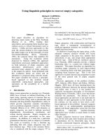
shown schematically in Fig. 1, corresponded to dele-
tions of 157 amino acids (AMPP#2) and 212 amino
J W. Liu et al. C-terminal domain of E. coli aminopeptidase P
FEBS Journal 274 (2007) 4742–4751 ª 2007 The Authors Journal compilation ª 2007 FEBS 4743
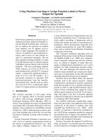
acids (AMPP#12). The truncated AMPP fragments
were expressed and assayed for solubility, and neither
gave rise to detectable levels of protein using the Gel-
Code Blue stain reagent as detector, as shown in
Fig. 2.
Improving solubility of the AMPP C-terminal
domain
The first round of shuffling was screened with
5 lgÆmL
)1
TMP and utilized the genes of the five most
common fragments found after screening for the
domain boundary. These fragments correspond to dele-
tions of 127, 143, 144, 157 and 212 amino acids, respec-
tively. The DNA for the AMPP fragments was isolated
from a number of resistant colonies and sequenced
(Table 1). As can be seen, after the second round of
DNA shuffling, all the chosen colonies gave fragments
of the same length ) all were derived from the
AMPP#2 fragment (Fig. 1). Most of the mutant genes
contained multiple mutations, two of which involved
metal-binding ligands. The D271N and E406G muta-
tions were expected to diminish or abolish the capacity
of AMPP to bind metals. The results of subsequent
rounds of evolution are also shown in Table 1. A num-
ber of mutations from round 1 disappeared in rounds 2
and 3, whereas the E406G mutation became common
to all the mutants that were selected for sequencing.
The G270V mutation appeared in the second round,
and was found in all but one mutant protein selected in
the third round. This latter mutation appeared to be
incompatible with the D271N mutation; however, its
close proximity to a metal-binding ligand suggested
that it could (like the D271N mutation) also reduce or
eliminate the capacity of the protein to bind metal. The
R166G mutation appeared in the first round of selec-
tion, increased in number in the second round, and was
present in all but one of the round 3 mutant proteins.
This mutation is close to the N-terminus of the frag-
ment ) it lies between the start of the fragment and
the predicted start of the domain (Fig. 1). From the
round 3 mutants, three were selected for further char-
acterization: AMPP#3-1, AMPP#3-22, and AMPP#3-
40. These fragments were subcloned so that they could
be expressed without DHFR. The AMPP#3-22 mutant
was clearly the most soluble (Fig. 2) and was chosen
for further study. It is likely that the reduced solubility
of the AMPP#3-40 mutant was due to the absence of
the R166G mutation, whereas the reduced solubility of
the AMPP#3-1 mutant could be attributed to a number
of changes (Table 1).
N-domain
C-domain
157
157
439
439
439
1
174
AMPP wt
AMPP #2
AMPP #3-22
172
439
AMPP #4-3
439
212
AMPP #12
R166G
G270V
E406G
G270V
E406G
Fig. 1. Schematic diagram of AMPP. Wild-type AMPP consists of
an N-terminal domain (1–174 amino acids) and a C-terminal domain
(174–439 amino acids). C-terminal domain AMPP#2 has a 157
amino acid deletion, AMPP#12 has a 212 amino acid deletion,
AMPP#3-22 has a 157 amino acid deletion, and AMPP#4-3 has
a 172 amino acid deletion. Mutations are R166G, G270V, and
E406G.
kDa
97.4
66.2
45.0
31.0
21.5
14.4
#2 #12 #3-22 #4-3 #2 #12 #3-22 #4-3
M S S S S P P P P
A
B
#3-1 #3-22 #3-40 #3-1 #3-22 #3-40
kDa
97.4
66.2
45.0
31.0
21.5
14.4
MSSSPPP
Fig. 2. Expression patterns of C-terminal AMPP domains. (A) An ali-
quot of supernatant (S) or pellet (P) from cells containing AMPP
domains (#2, #12, #3-22, or #4-3) was denatured and resolved by
15% SDS ⁄ PAGE. (B) An aliquot of supernatant (S) or pellet (P) from
cells containing AMPP domains (#3-1, #3-22, or #3-40) was dena-
tured and resolved by 15% SDS ⁄ PAGE. Overexpressed AMPP
domains are indicated by arrowheads. Low-range molecular mass
standards (M) from Bio-Rad.
C-terminal domain of E. coli aminopeptidase P J W. Liu et al.
4744 FEBS Journal 274 (2007) 4742–4751 ª 2007 The Authors Journal compilation ª 2007 FEBS
The AMPP#3-22 mutant has the three most com-
mon mutations found in round 3: R166G, G270V,
and E406G. The fragment was purified using two
chromatographic steps, Q-SepharoseHP and SOUR-
CE 15PHE. The purified fragment was then loaded
onto a size exclusion column, and eluted in two peaks
that corresponded to a monomer and a dimer of the
fragment (Table 2). The fragment and the wild-type
proteins were tested for enzymatic activity ) only the
wild-type protein displayed activity. Consistent with
this lack of activity, atomic absorption measurements
of the AMPP#3-22 mutant (as purified) gave no
detectable trace of metals, demonstrating the inability
of this mutant to bind metal ions. Furthermore, pro-
longed exposure of this fragment to high concen-
trations of divalent metal ions followed by dialysis
to remove excess metal ions gave preparations of
AMPP#3-22 that contain at most 0.15 ions per binu-
clear active site. This observation also argues for a
very low binding affinity of the mutant fragment for
metal ions. The residual metal ions ( £ 0.15) are adven-
titiously bound, as observed, for example, in other
binuclear metalloenzymes, such as purple acid phos-
phatases and methionyl aminopeptidases [6–8].
In vitro refolding
Wild-type AMPP and AMPP#3-22 were overexpressed
and purified. Subsequently, the purified proteins were
denatured with 6 m guanidine hydrochloride and rena-
tured by dialysis in the presence of EDTA or metals,
as described in Experimental procedures. Aggregated
proteins were removed by centrifugation, and the pro-
teins in the supernatant were analyzed by SDS ⁄ PAGE
electrophoresis. The AMPP#2 fragment was expressed
as an inclusion body and dissolved in 6 m guanidine
hydrochloride. The denaturant was removed in the
presence of EDTA or metals, and the soluble proteins
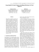
were subjected to SDS ⁄ PAGE analysis. The results of
these in vitro refolding attempts are shown in Fig. 3.
A previous study has shown that ZnCl
2
inhibits the
activity of AMPP [1]. Here, the presence of ZnCl
2
in
the dialysis buffer led to the precipitation of each of
the three proteins. Neither the intact protein nor the
Table 1. Sequence analysis of AMPP C-terminal domain mutants. The percentage of mutants containing a given mutation in each round is
indicated.
Domains(deletion) Mutations
#1-1(157 aa)
#1-9(157 aa) R166G
#1-21(157 aa) V169A E171G D271N E406G D407N V424M
#1-33(143 aa) Y209H H217R V326I P346L
#1-40(157 aa) C263Y E406G
%R1 2020202020 20 202020 402020
#2-1(157 aa) Y209H D271N P346L P376L E406G
#2-5(157 aa) R166G D271N E406G
#2-6(157 aa) V169A E171G G270V E406G
#2-13(157 aa) D271N E406G
#2-30(157 aa) R166G D271N E406G
%R2 40 20 20 20 80 2020100
#3-15(157 aa) R166G V169A E171G D271N E406G
#3-6(157 aa) R166G G270V E406G
#3-8(157 aa) R166G G270V E406G
#3-10(157 aa) R166G G270V E406G
#3-15(157 aa) R166G G270V E406G
#3-20(157 aa) R166G G270V E406G
#3-22(157 aa) R166G G270V E406G
#3-30(157 aa) R166G G270V E406G
#3-37(157 aa) R166G G270V E406G
#3-40(157 aa) Y226C G270V E406G
% R3 90 10 10 10 90 10 100
Table 2. Size exclusion chromatography of AMPP C-terminal
domains.
Peak I
(excluded)
Peak II
(dimer)
Peak III
(monomer)
AMPP#2 (refolded) > 99% – –
AMPP#3-22 – 28% 72%
AMPP#4-3 – – > 99%
J W. Liu et al. C-terminal domain of E. coli aminopeptidase P
FEBS Journal 274 (2007) 4742–4751 ª 2007 The Authors Journal compilation ª 2007 FEBS 4745
fragments required metals to produce soluble protein.
The wild-type and AMPP#3-22 proteins responded in
a similar (although not identical manner) to the vari-
ous metals. This observation, combined with the fact
that AMPP#3-22 did not appear to bind metals, sug-
gested that metals were not required for folding of
the native enzyme or the AMPP#3-22 fragment. The
response of the AMPP#2 fragment to metals differs
from that of the wild-type protein or the AMPP#3-22
fragment. In order to investigate this difference fur-
ther, the soluble AMPP#2 fragment (refolded with
EDTA or metals) was loaded onto a size exclusion col-
umn. The fragment was excluded from the resin pores,
suggesting that it had formed soluble microaggregates
of partially unfolded protein (Table 2).
Evolution of the AMPP#3-22 fragment –
optimizing the starting point
Exonuclease III digestion of the DNA corresponding
to the AMPP#3-22 fragment was used to generate a
library of N-terminal deletions of the fragment. This
library was screened with a higher concentration of
TMP than had been used in previous rounds of evolu-
tion. Several colonies were found to be resistant to
200 lgÆmL
)1
TMP. One of these colonies produced
a fragment designated AMPP#4-3. DNA sequencing
revealed that the size of the AMPP#4-3 fragment cor-
responded to a deletion of 172 amino acids from the
wild-type sequence ) this was very close to the bound-
ary position predicted from an inspection of the struc-
ture. The DNA for this fragment was isolated from
the fusion vector and cloned into the expression vector
pJWL1030. The AMPP#4-3 fragment was expressed
and assayed for solubility. From an inspection of
Fig. 2, it appeared that E. coli produced more soluble
AMPP#4-3 than AMPP#3-22. Whether AMPP#4-3
was more soluble than AMPP#3-22 was difficult to
ascertain from the gel shown in Fig. 2, as there
were background bands overlapping with that of the
AMPP#4-3 fragment. To address this question of solu-
bility, cells expressing AMPP#3-22 and AMPP#4-3
were grown on plates that contained TMP levels that
ranged from 20 to 200 lgÆmL
)1
. Both lines grew well
on all the plates, suggesting that the solubility of the
two fragments was similar. To ascertain the aggre-
gation state of the AMPP#4-3 fragment, it was puri-
fied and analyzed by size exclusion chromatography.
Unlike AMPP#3-22, AMPP#4-3 behaved as a mono-
mer (Table 2), with no dimer component evident.
Discussion
Two approaches were taken to produce a soluble
C-terminal domain of AMPP. Different-length domains
were tested, and mutations were made to the sequences
of these domains. It is known that the location of
domain boundaries is critical to the formation of sta-
ble, correctly folded, isolated domains [9,10]. Domain
boundaries can be predicted using sequence alignments
or bioinformatic tools [11–14]. In the case of AMPP, a
high-resolution structure is available, and it gives a
good indication of where the C-terminal domain starts
[2]. However, the expression of this domain based on
the predicted boundary resulted in the production of
inclusion bodies. This is not an uncommon problem,
as noted by Holland et al. [15] ) partitioning protein
structure into domains is not always easy and success-
ful. Two experimental approaches were considered as a
means of correctly locating the domain boundary.
First, consideration was given to limited proteolysis
coupled with amino acid sequencing and MS [16,17].
Second, gene truncation has also been been used to
obtain the soluble domains of multidomain proteins
[18] ) it is this method that was chosen for further
study. This latter approach requires the construction
of a truncation library and a method to screen for sol-
uble domains [19].
A library of nested N-terminal deletions of the
AMPP gene was created by exonuclease III digestion
and subsequent screening by fusing them to the DHFR
reporter gene and selecting with TMP. The initial
round of truncations gave a series of deletions that
allowed cells to survive on a minimal level of TMP.
These domains were shuffled and one, AMPP#2, could
be combined with mutations to produce a soluble
domain. The AMPP#2 fragment was expressed, but
gave rise to inclusion bodies ) no soluble protein was
detected. The fragment could be denatured, and it
remained soluble upon removal of the denaturant. A
sizing column revealed that the soluble form of the
fragment consisted of a very high molecular mass
AMPP #3-22
AMPP #2
AMPP wt
- Mn Zn Co Cu Fe
Fig. 3. In vitro refolding of AMPP and its C-terminal domains. Full-
length AMPP (wt) and C-terminal domains (#2, #3-22) were dena-
tured with 6
M guanidine hydrochloride and dialyzed overnight at
4 °C against 20 m
M Tris (pH 7.6), containing 1 mM EDTA (–) or
1m
M various metals (MnCl
2
, ZnCl
2
, CoCl
2
, CuCl
2
or FeCl
3
). The
precipitate was removed by centrifugation, and soluble proteins
were resolved on a 15% SDS ⁄ PAGE gel.
C-terminal domain of E. coli aminopeptidase P J W. Liu et al.
4746 FEBS Journal 274 (2007) 4742–4751 ª 2007 The Authors Journal compilation ª 2007 FEBS
aggregate (> 200 kDa). Soluble variants of this frag-
ment could be expressed in E. coli if suitable mutations
were made to the DNA coding for AMPP#2. One of
these variants, AMPP#3-22, was chosen for further
study. Analysis with size exclusion chromatography
revealed that AMPP#3-22 is a mixture of monomers
and dimers. Only three mutations (R166G, G270V,
and E406G) were required to convert the aggregated
AMPP#2 fragment into the soluble AMPP#3-22 frag-
ment. The first mutation (R166G) was removed in the
final round of mutations in which the fragment length
was varied to give the AMPP#4-3 fragment. This final
fragment ran as a monomer when applied to a sizing
column. This observation implicated the N-terminal
peptide and the R166G mutation in the monomer–
dimer equilibrium of AMPP#3-22. The AMPP#4-3
fragment has a length very close to that predicted for
the C-terminal domain, on the basis of an inspection
of the crystal structure (see above). Its amino acid
sequence differs from that of the corresponding wild-
type sequence at only two locations: positions 270 and
406. As noted in the previous section, E406 is a metal
ligand that coordinates both metals, whereas G270 is
adjacent to D271, which also coordinates both metals.
The G270V and E406G mutations are likely to be
responsible for the inability of the AMPP#3-22 frag-
ment to bind metals. From these results, it appears
that the solubility of the AMPP#4-3 fragment ) or at
least the ability to express this fragment in a soluble
form ) is connected with its inability to bind metals.
Metalloproteins can fold via metal-dependent or
metal-independent pathways [20,21]. They may bind
metal ions before polypeptide folding, after complete
protein folding, or after partial folding. Phosphoman-
nose isomerase is an example of a protein that requires
a metal to fold. It requires zinc ions for both in vivo
and in vitro folding [22]. The in vitro folding studies
described in this article suggest that AMPP and C-ter-
minal fragments fold in a metal-independent manner.
Denatured AMPP and AMPP#3-22 both fold in the
presence of EDTA, and both show similar folding pat-
terns when exposed to metals during renaturation
(Fig. 3). A plausible explanation for these observations
is that the protein must be folded before metals
bind ) the metal-binding ligands must be appropri-
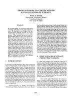
ately placed to coordinate the incoming metals. Four
acid residues coordinate the two divalent metal ions in
the active site of AMPP (Fig. 4). The positively
charged metals will neutralize the negatively charged
acids. In the absence of metals, the negatively charged
residues will tend to repel one another, thus destabiliz-
ing the protein. For the native protein, the presence of
the N-terminal domain and the oligomeric structure of
the protein may be necessary to maintain the structure
of the C-terminal domain in a conformation that
allows the metals to bind. Removing the N-terminal
domain results in a C-terminal domain in which the
acid residues of the active site repel one another, caus-
ing the protein to unfold (or to partially unfold). It is
this unfolded form of the protein that aggregates and
precipitates [23]. Mutations that abolish metal binding
allow the peptide to assume a conformation close to
that of the native protein ) a stable conformation that
results in soluble fragments that are incapable of bind-
ing metals.
The two rounds of evolution to optimize the starting
point of the AMPP domain had opposing effects ) the
first round extended the domain size, whereas the last
N
N
M n
O
O
O
O
O
O
O
O
M n
W 2
W 1
W 3
A s p 2 7 1
A s p 2 6 0
G l u 3 8 3
H i s 3 5 4
G l u 4 0 6
A
B
Fig. 4. The active site of AMPP. (A) Schematic diagram of the
AMPP metal-binding sites. Metal-binding ligands are Asp260,
Asp271, His354, Glu383, and Glu406. (B) Stereo view of the AMPP
active site. Two mutations (Glu270 and Glu406) are responsible for
improving the solubility of the C-terminal domain. The figure was
generated from published data [27].
J W. Liu et al. C-terminal domain of E. coli aminopeptidase P
FEBS Journal 274 (2007) 4742–4751 ª 2007 The Authors Journal compilation ª 2007 FEBS 4747
round moved the starting point close to that predicted
on the basis of an inspection of the structure. It would
appear that extending the domain boundary had the
effect of producing a slightly soluble aggregated form
of the protein. Subsequent changes to the amino acid
sequence were far more effective in improving the solu-
bility of the domain. In the case of the AMPP protein,
the boundary of the domain would have been better
determined from an inspection of the structure rather
than by the experimental methods that were used. The
reasons for this are related to the metal-binding prop-
erties of the domain, and these will not necessarily
affect studies with many other proteins. In the case of
a stable, soluble domain, the methods described in this
article should prove effective in locating the starting
point of the domain.
In summary, directed evolution has been used to
address the question of what causes the insolubility of
the C-terminal domain of AMPP. The answer is rela-
tively simple ) modifying two active site residues can
produce a soluble fragment. The E406G mutation con-
verts a metal-binding ligand to a residue that is unli-
kely to bind metal. The G270V residue is located next
to a metal-binding residue ) this mutation is likely to
cause a conformational change that is likely to further
reduce the capacity of the fragment to bind metals.
The conformational change could move E271 away
from the active site, hence stabilizing the structure of
the domain. In agreement with this interpretation,
metal ion analysis of AMPP#3-22 by atomic absorp-
tion spectroscopy demonstrates that this mutant frag-
ment has abolished the ability to bind metal ions.
Although these two mutations dominate the list of
mutations in round 3, it should be clear from the ear-
lier round of shuffling that the mutation rate is consid-
erably higher than two changes per round. Given the
size of the mutant libraries (150 000), it is evident that
the effects of all other mutations are significantly smal-
ler than those of E406G and G270V. This idea is sup-
ported by the data shown in Table 1. By round 3,
most of the mutations found in round 1 have been
lost. Normally, one would expect an increase in the
number of mutations per gene; however, we observed
a decrease in the number of mutations per gene. The
implication of this observation is that the effects of
most mutations are small compared with those of
G270V, E406G, and R166G. Changes at the surface
of the protein do not appear to be major contributors
to the solubility of the AMPP fragments. The AMPP
protein appears to have evolved so that the metal-
binding ligands are positioned optimally for the coor-
dination of incoming metals. Metal binding would
therefore stabilize the structure. One would expect that
proteolysis could be used to produce stable C-terminal
fragments, as these experiments could be conducted
once metals have been bound. However, fragments
identified in this manner may not fold when expressed
in E. coli. The results presented in this article may
explain the size of AMPP. It is a noncooperative tetra-
mer that is considerably larger than, for example, the
monomeric single-domain AMPM protein [3]. In the
case of AMPP, the N-terminal domain appears to have
a function in protein folding. Clearly, the single-
domain AMPM protein has found another solution to
this problem.
Experimental procedures
Chemicals and bacterial strains
All chemicals were purchased from Sigma-Aldrich (St Louis,
MO). Molecular biology reagents and enzyme were brought
from Roche (Basel, Switerland), New England Biolabs
(La Jolla, CA), Bio-Rad (Hercules, CA), Novagen (Kilsyth,
Australia), or GE Healthcare (Chalfont St Giles, UK).
Primers were obtained from GeneWork (Thebarton, Aus-
tralia). DNA purification kits (Qiagen, Doncaster, Australia)
were used for all DNA isolations and purifications.
The E. coli strain DH5a (supE44DlacU169 ø80 lacZDM15
hsd R17 recA1 endA1 gyrA96 thi-1 relA1) was used for all
aspects of the work. Cells were grown at 37 °C. Cell lines
were maintained on LB medium agar plates supplemented
with 50 lgÆmL
)1
kanamycin to maintain plasmids express-
ing recombinant E. coli AMPP and its domain variants.
Creating a library for truncated AMPP fragments
The 1.3 kb pepP gene encoding E. coli AMPP was PCR
amplified from plasmid pPL670 [2] using a forward pri-
mer (5¢-CCAAGCTTGTCGACGATGAGTGAGATATCC
CGG-3¢) and a reverse primer (5¢-CGGGAATTCCTG
CAGTTGCTTTCTCGCAGCAAC-3¢), and then cloned
between the SalI and PstI sites of the DHFR fusion vector
pJWL1030folA [4] to produce pJWL1030folA–pepP. N-ter-
minal deletions of AMPP were generated by partially
digesting the pepP gene with exonuclease III in a manner
similar to that described by Henikoff [24] and Ostermeier
et al. [25]. pJWL1030folA–pepP (1–5 lg) was cut (linear-
ized) at the 5¢-end of pepP with SalI. The SalI-digested
pJWL1030folA–pepP was digested with exonuclease III for
varying times to generate nested deletions [25]. The trun-
cated pepP fragments were then treated with Mung Bean
Nuclease to remove single-strand DNA tails, and Klenow
fragment DNA polymerase I was added to flush the DNA
ends. The truncated DNA fragments were released from
the pJWL1030folA vector by PstI digestion, and subse-
quently separated on an agarose gel. The pepP fragments
C-terminal domain of E. coli aminopeptidase P J W. Liu et al.
4748 FEBS Journal 274 (2007) 4742–4751 ª 2007 The Authors Journal compilation ª 2007 FEBS
with sizes between 0.9 kb and 1.3 kb were purified from the
agarose gel. The DHFR fusion vector pJWL1030folA was
digested with SalI, and then incubated with Klenow frag-
ment DNA polymerase I to produce blunt ends. The vector
was further digested with PstI. The truncated pepP frag-
ments were then ligated to the blunt end and PstI site of
pJWL1030folA. Finally, the ligation mixture was trans-
formed into DH5a cells by electroporation.
DNA shuffling
Random mutations were introduced into the pepP gene
using DNA shuffling as described by Stemmer [26]. The
shuffled pepP genes were ligated between the NdeI and PstI
sites of pJWL1030folA. The plasmid was then transformed
into cells by electroporation.
Selection for TMP resistance
The truncated pepP gene library was plated on Mueller–
Hinton agar (Difco, Becton Dickinson, Sparks, MD) plates
that were supplemented with 50 l gÆ mL
)1
kanamycin and 2
or 20 lgÆmL
)1
TMP. The TMP-resistant colonies appeared
after incubation at 37 °C for 3–5 days.
The transformed cells with shuffled pepP genes were pla-
ted on the Mueller–Hinton agar plates supplemented with
50 lgÆmL
)1
kanamycin and increasing concentrations of
TMP for the three rounds of evolution. For the first round,
5 lgÆmL
)1
TMP was used, and in the second and third
rounds, 10 and 20 lgÆmL
)1
TMP were used, respectively.
In each round, a library of 150 000 colonies was screened.
The DNA for the 10 mutant genes from round 1 was shuf-
fled for selection in round 2, and 18 genes were selected
from round 2 and shuffled for selection in round 3.
Protein expression and solubility assay
The intact AMPP as well as the C-terminal fragments of
AMPP were expressed in the same manner. The genes were
PCR amplified and cloned between the NdeI and EcoRI
sites of the pJWL1030 expression vector [4]. The plasmids
were then transformed into cells by electroporation. Cells
expressing each of these domains were grown overnight at
4 °C in LB medium containing 50 lgÆmL
)1
kanamycin.
Cells were harvested and lysed using the BugBuster deter-
gent (Novagen). Solubility assays were carried out using
SDS ⁄ PAGE gel electrophoresis and staining using the Gel-
Code Blue stain reagent (Pierce, Rockford, IL) as described
elsewhere [4].
Protein purification and activity assay
The wild-type AMPP as well as C-terminal domains of
AMPP were purified using a modified form of the protocol
used for AMPP [2]. Briefly, cells were harvested and resus-
pended in 20 mm Tris (pH 7.6), and then lysed using a
French press. The lysates were centrifuged at 30 000 g for
40 min at 4 °C (Sorvall RC5C, Thermo Electron, with
SS34 rotor), and the supernatants were applied to a
Q-SepharoseHP column (GM Healthcare) and eluted with
a gradient of 0–1 m NaCl in 20 mm Tris (pH 7.6). Pooled
fractions were combined with an equal volume of 20 mm
Tris (pH 7.6) and 3 m (NH
4
)
2
SO
4
. After centrifugation as
above, the supernatant was applied to a SOURCE 15PHE
column (GE Healthcare) and eluted with a gradient of
1.5–0 m (NH
4
)
2
SO
4
in 20 mm Tris (pH 7.6). The pooled
fractions were dialyzed against 20 mm Tris (pH 7.6), and
concentrated using Centriplus filter devices (YM-10; Milli-
pore, Bedford, MA). The enzymatic activities of intact and
C-terminal domains of AMPP were assayed using the
quenched fluorescent substrate Lys(Abz)-Pro-Pro-pNA
(Bachem, Bubendorf, Switzerland), as described elsewhere
[27].
In vitro refolding
The purified AMPP (wild-type) and AMPP#3-22 were
denatured with 6 m guanidine hydrochloride in the presence
of 1 mm EDTA or 1 mm various metals (MnCl
2
, ZnCl
2
,
CoCl
2
, CuCl
2
, or FeCl
3
). The denatured proteins were dia-
lyzed at 4 °C overnight against 20 mm Tris (pH 7.6) with
EDTA or metals. The inclusion bodies formed from
AMPP#2 were dissolved in 6 m guanidine hydrochloride,
and then dialyzed against 20 mm Tris (pH 7.6) with EDTA
or metals. After dialysis, the solutions containing AMPP,
AMPP#2 and AMPP#3-22 were centrifuged at 16 000 g for
10 min at 4 °C (Sorvall RC5C with SS34). The superna-
tants and pellets were separated. The pellets were mixed
with 20 mm Tris (pH 7.6) and vortexed to ensure that they
were resuspended. Equal volumes of the solutions contain-
ing the supernatants and the resuspended pellets were run
on a 15% SDS ⁄ PAGE gel and stained using the GelCode
Blue stain reagent.
Size exclusion chromatography
A gel filtration assay was carried out using a Superdex
200 HP 10 ⁄ 30 column (GM Healthcare). The column was
equilibrated with 20 mm Tris (pH 7.6) and 0.15 m NaCl,
and calibrated with a marker mix including aldolase
(158 kDa, GM Healthcare), phosphotriesterase (74 kDa)
[28] and dienelactone hydrolase (26 kDa) [29].
Metal ion analysis
Metal ion concentrations were determined in triplicate by
atomic absorption spectroscopy using a Varian SpectrAA
220FS instrument. Standard solutions for Fe
2+
,Mn
2+
,
J W. Liu et al. C-terminal domain of E. coli aminopeptidase P
FEBS Journal 274 (2007) 4742–4751 ª 2007 The Authors Journal compilation ª 2007 FEBS 4749
Zn
2+
and Co
2+
ranged from 20 p.p.b. to 200 p.p.b., and
were prepared from analytical stock solutions (Merck,
Kilsyth, Australia) using MilliQ water (produced by MilliQ
reagent water system; Millipore). Aliquots of purified pro-
tein samples were sufficiently diluted with MilliQ to obtain
metal ion concentrations in the range between 20 p.p.b.
and 200 p.p.b., assuming a full complement of two metals
per active site. The quantity of metal ions in MilliQ water
was below the detection limit of the instrument. The esti-
mated error for each measurement was less than 5%.
Acknowledgements
The authors thank Cameron McRae of the Bimolecu-
lar Resource Facility for DNA sequencing, and Profes-
sor Nick Dixon for providing plasmid pPL670.
References
1 Graham SC, Bond CS, Freeman HC & Guss JM
(2005) Structural and functional implications of metal
ion selection in aminopeptidase P, a metalloprotease
with a dinuclear metal center. Biochemistry 44,
13820–13836.
2 Wilce MC, Bond CS, Dixon NE, Freeman HC, Guss
JM, Lilley PE & Wilce JA (1998) Structure and mecha-
nism of a proline-specific aminopeptidase from Escheri-
chia coli. Proc Natl Acad Sci USA 95, 3472–3477.
3 Bazan JF, Weaver LH, Roderick SL, Huber R & Mat-
thews BW (1994) Sequence and structure comparison
suggest that methionine aminopeptidase, prolidase, ami-
nopeptidase P, and creatinase share a common fold.
Proc Natl Acad Sci USA 91, 2473–2477.
4 Liu JW, Boucher Y, Stokes HW & Ollis DL (2006)
Improving protein solubility: the use of the Escherichia
coli dihydrofolate reductase gene as a fusion reporter.
Protein Expr Purif 47 , 258–263.
5 Neylon C (2004) Chemical and biochemical strategies
for the randomization of protein encoding DNA
sequences: library construction methods for directed
evolution. Nucleic Acids Res 32, 1448–1459.
6 Schenk G, Boutchard CL, Carrington LE, Noble CJ,
Moubaraki B, Murray KS, de Jersey J, Hanson GR &
Hamilton S (2001) A purple acid phosphatase from
sweet potato contains an antiferromagnetically coupled
binuclear Fe–Mn center. J Biol Chem 276, 19084–19088.
7 Larrabee JA, Leung CH, Moore RL, Thamrong-
Nawasawat T & Wessler BS (2004) Magnetic circular
dichroism and cobalt(II) binding equilibrium studies of
Escherichia coli methionyl aminopeptidase. J Am Chem
Soc 126, 12316–12324.
8 Mitic N, Smith SJ, Neves A, Guddat LW, Gahan LR &
Schenk G (2006) The catalytic mechanisms of binuclear
metallohydrolases. Chem Rev 106, 3338–3363.
9 Xu Y, Wen D, Clancy P, Carr PD, Ollis DL & Vasud-
evan SG (2004) Expression, purification, crystallization,
and preliminary X-ray analysis of the N-terminal
domain of Escherichia coli adenylyl transferase. Protein
Expr Purif 34, 142–146.
10 Kerr ID, Berridge G, Linton KJ, Higgins CF &
Callaghan R (2003) Definition of the domain bound-
aries is critical to the expression of the nucleotide-
binding domains of P-glycoprotein. Eur Biophys J 32,
644–654.
11 Rigden DJ (2002) Use of covariance analysis for the
prediction of structural domain boundaries from mul-
tiple protein sequence alignments. Protein Eng 15,
65–77.
12 Dumontier M, Yao R, Feldman HJ & Hogue CW
(2005) Armadillo: domain boundary prediction by
amino acid composition. J Mol Biol 350, 1061–1073.
13 Liu J & Rost B (2004) Sequence-based prediction of
protein domains. Nucleic Acids Res 32, 3522–3530.
14 Galzitskaya OV & Melnik BS (2003) Prediction of pro-
tein domain boundaries from sequence alone. Protein
Sci 12, 696–701.
15 Holland TA, Veretnik S, Shindyalov IN & Bourne PE
(2006) Partitioning protein structures into domains: why
is it so difficult? J Mol Biol 361, 562–590.
16 Severinova E, Severinov K, Fenyo D, Marr M, Brody EN,
Roberts JW, Chait BT & Darst SA (1996) Domain orga-
nization of the Escherichia coli RNA polymerase sigma
70 subunit. J Mol Biol 263, 637–647.
17 Christ D & Winter G (2006) Identification of protein
domains by shotgun proteolysis. J Mol Biol 358,
364–371.
18 Hart DJ & Tarendeau F (2006) Combinatorial library
approaches for improving soluble protein expression in
Escherichia coli. Acta Crystallogr D Biol Crystallogr 62,
19–26.
19 Cornvik T, Dahlroth SL, Magnusdottir A, Flodin S,
Engvall B, Lieu V, Ekberg M & Nordlund P (2006) An
efficient and generic strategy for producing soluble
human proteins and domains in E. coli by screening
construct libraries. Proteins 65, 266–273.
20 Wittung-Stafshede P (2004) Role of cofactors in folding
of the blue-copper protein azurin. Inorg Chem 43,
7926–7933.
21 Wilson CJ, Apiyo D & Wittung-Stafshede P (2004) Role
of cofactors in metalloprotein folding. Q Rev Biophys
37, 285–314.
22 Proudfoot AE, Goffin L, Payton MA, Wells TN & Ber-
nard AR (1996) In vivo and in vitro folding of a recom-
binant metalloenzyme, phosphomannose isomerase.
Biochem J 318 (2), 437–442.
23 Villaverde A & Carrio MM (2003) Protein aggregation
in recombinant bacteria: biological role of inclusion
bodies. Biotechnol Lett 25, 1385–1395.
C-terminal domain of E. coli aminopeptidase P J W. Liu et al.
4750 FEBS Journal 274 (2007) 4742–4751 ª 2007 The Authors Journal compilation ª 2007 FEBS
24 Henikoff S (1987) Unidirectional digestion with exonu-
clease III in DNA sequence analysis. Methods Enzymol
155, 156–165.
25 Ostermeier M, Nixon AE, Shim JH & Benkovic SJ
(1999) Combinatorial protein engineering by incremen-
tal truncation. Proc Natl Acad Sci USA 96, 3562–3567.
26 Stemmer WP (1994) DNA shuffling by random frag-
mentation and reassembly: in vitro recombination for
molecular evolution. Proc Natl Acad Sci USA 91,
10747–10751.
27 Graham SC, Lilley PE, Lee M, Schaeffer PM, Kralicek AV,
Dixon NE & Guss JM (2006) Kinetic and crystallographic
analysis of mutant Escherichia coli aminopeptidase P:
insights into substrate recognition and the mechanism of
catalysis. Biochemistry 45, 964–975.
28 Yang H, Ca rr PD, McLoughlin SY, Liu JW, Horne I,
Qiu X, Jef fries CM, Russell RJ, Oakeshott JG & Ollis DL
(2003) Evolution of an organophosphate-degrading
enzyme: a comparison of natural and directed evolution.
Protein Eng 16, 135–145.
29 Kim HK, Liu JW, Carr PD & Ollis DL (2005) Follow-
ing directed evolution with crystallography: structural
changes observed in changing the substrate specificity of
dienelactone hydrolase. Acta Crystallogr D Biol Crystal-
logr 61, 920–931.
J W. Liu et al. C-terminal domain of E. coli aminopeptidase P
FEBS Journal 274 (2007) 4742–4751 ª 2007 The Authors Journal compilation ª 2007 FEBS 4751