Báo cáo khoa học: The ‘pair of sugar tongs’ site on the non-catalytic domain C of barley a-amylase participates in substrate binding and activity potx
Bạn đang xem bản rút gọn của tài liệu. Xem và tải ngay bản đầy đủ của tài liệu tại đây (422.56 KB, 13 trang )
The ‘pair of sugar tongs’ site on the non-catalytic domain C
of barley a -amylase participates in substrate binding and
activity
Sophie Bozonnet
1,2
, Morten T. Jensen
2
, Morten M. Nielsen
1
, Nushin Aghajari
3
, Malene H. Jensen
3
,
Birte Kramhøft
1,2
, Martin Willemoe
¨
s
1,2
, Samuel Tranier
3
, Richard Haser
3
and Birte Svensson
1,2
1 Enzyme and Protein Chemistry, BioCentrum-DTU, Technical University of Denmark, Kgs. Lyngby, Denmark
2 Carlsberg Laboratory, Valby, Denmark
3 Laboratoire de BioCristallographie, Institut de Biologie et Chimie des Prote
´
ines, Universite
´
de Lyon, France
a-amylases (EC 3.2.1.1) are endo-hydrolases acting on
a)1,4-glucosidic bonds in starch and related poly- and
oligosaccharides. They belong to the very large glyco-
side hydrolase family 13 (GH13) that, together with
GH70 and GH77, forms glycoside hydrolase clan
H (GH-H), representing about 30 enzyme specificities
(). Secondary carbohydrate-bind-
ing sites are found either on the surface of the catalytic
structural unit or on a separate carbohydrate-binding
module (CBM) in some of the GH-H members [1].
Keywords
barley a-amylase; crystal structures;
secondary carbohydrate-binding sites;
starch granules; surface plasmon resonance
Correspondence
B. Svensson, Enzyme and Protein
Chemistry, BioCentrum-DTU, Technical
University of Denmark, Søltofts Plads, Bldg
224, DK-2800 Kgs. Lyngby, Denmark
Fax: +45 45 88 63 07
Tel: +45 45 25 27 40
E-mail:
(Received 1 June 2007, revised 18 July
2007, accepted 1 August 2007)
doi:10.1111/j.1742-4658.2007.06024.x
Some starch-degrading enzymes accommodate carbohydrates at sites situ-
ated at a certain distance from the active site. In the crystal structure of
barley a-amylase 1, oligosaccharide is thus bound to the ‘sugar tongs’ site.
This site on the non-catalytic domain C in the C-terminal part of the mole-
cule contains a key residue, Tyr380, which has numerous contacts with the
oligosaccharide. The mutant enzymes Y380A and Y380M failed to bind to
b-cyclodextrin-Sepharose, a starch-mimic resin used for a-amylase affinity
purification. The K
d
for b-cyclodextrin binding to Y380A and Y380M was
1.4 mm compared to 0.20–0.25 mm for the wild-type, S378P and S378T
enzymes. The substitution in the S378P enzyme mimics Pro376 in the bar-
ley a-amylase 2 isozyme, which in spite of its conserved Tyr378 did not
bind oligosaccharide at the ‘sugar tongs’ in the structure. Crystal structures
of both wild-type and S378P enzymes, but not the Y380A enzyme, showed
binding of the pseudotetrasaccharide acarbose at the ‘sugar tongs’ site. The
‘sugar tongs’ site also contributed importantly to the adsorption to starch
granules, as K
d
¼ 0.47 mgÆmL
)1
for the wild-type enzyme increased to
5.9 mgÆmL
)1
for Y380A, which moreover catalyzed the release of soluble
oligosaccharides from starch granules with only 10% of the wild-type activ-
ity. b-cyclodextrin both inhibited binding to and suppressed activity on
starch granules for wild-type and S378P enzymes, but did not affect these
properties of Y380A, reflecting the functional role of Tyr380. In addition,
the Y380A enzyme hydrolyzed amylose with reduced multiple attack,
emphasizing that the ‘sugar tongs’ participates in multivalent binding of
polysaccharide substrates.
Abbreviations
AMY1 and AMY2, barley a-amylases 1 and 2; BASI, barley a-amylase ⁄ subtilisin inhibitor; b-CD, b-cyclodextrin; CBM, carbohydrate-binding
module; CBM20, carbohydrate-binding module family 20; Cl-pNPG
7
, 2-chloro-4-nitrophenyl b-D-maltoheptaoside; cv, column volume; DMA,
degree of multiple attack; DP, degree of polymerization; GH13, glycoside hydrolase family 13; GH-H, glycoside hydrolase clan H; iBS,
insoluble blue starch; RU, response unit; SBD, starch-binding domain; SPR, surface plasmon resonance; thio-DP4, methyl-4¢,4¢¢,4¢¢¢-
trithiomaltotetraoside.
FEBS Journal 274 (2007) 5055–5067 ª 2007 The Authors Journal compilation ª 2007 FEBS 5055
Plant a-amylases mobilize starch in plastids, tubers
and seeds, and barley isozyme 1 and 2 (AMY1 and
AMY2) are de novo synthesized in seed aleuron layers
at germination encoded by two multigene families
of $80% sequence identity and > 95% identity within
a subfamily. Only one AMY1 and two AMY2 iso-
forms were found in germinating seeds from a total of
10 barley a-amylase encoding genes; these three pro-
teins moreover underwent differential degradation dur-
ing germination [2]. AMY1 and AMY2 have virtually
identical three-dimensional structures composed of an
N-terminal catalytic (b ⁄ a)
8
-barrel (domain A), a
domain B, protruding between b-strand 3 and a-helix
3, and a C-terminal antiparallel b-sheet domain-C
[3,4]. The isozymes show functional and stability dif-
ferences and roles of selected amino acid residues were
characterized by mutational analysis [5–12]. The A and
B domains together form the active site [3,4]. Domain
B is also associated with effects of Ca
2+
on stability
and activity [5,13] and with the AMY2-specific sensi-
tivity to barley a-amylase ⁄ subtilisin inhibitor (BASI)
[5,14,15]. AMY1 furthermore binds substrates – starch
granules included – more tightly than does AMY2,
which shows a higher turn-over rate than AMY1
[16–18]. Domain-C is present in almost all GH-H
members and its functional role has not yet been
assigned. Remarkably, the ‘sugar tongs’ site defined
around Tyr380 in domain-C of AMY1 and binding
malto-oligosaccharide [4] was not occupied in the
structure of AMY2 [3] although this critical tyrosine is
conserved in AMY2.
AMY1, AMY2, and other GH-H enzymes possess
different secondary carbohydrate-binding sites that are
not part of the active site area but which are situated
on the surface of the catalytic domain or an inti-
mately associated domain rather than on a CBM, e.g.
a starch-binding domain (SBD) [1,3,4,19–22]. The role
of multivalent binding in enzymatic degradation of
polysaccharides is in general not clearly understood at
the molecular level. In amylolytic enzymes such sites
are thought to (a) ensure association with starch gran-
ules, (b) assist in disentangling of a-glucan chains,
(c) guide the substrate chain to the active site, and
(d) confer allosteric regulation. Multivalent binding is
also envisaged in the multiple attack mechanism
proposed in the late 1960s for amylose degradation
by a-amylase, in which an initial endo-attack was
followed by hydrolysis of more glucosidic bonds
before the enzyme–substrate complex dissociated [23].
Multiple attack was later described for cellulases,
chitinases, and pectinases and termed processivity [24].
Barley AMY1 hydrolyzes amylose with a degree of
multiple attack (DMA) of 2; thus, after the initial
cleavage, two substrate bonds were hydrolyzed with
release of shorter products [12]. Whereas DMA was
mostly reduced for AMY1 mutants in the substrate-
binding cleft, DMA values of 3.0 and 3.3 were found,
respectively, for an AMY1–SBD fusion [25] having an
SBD attached to the AMY1 C-terminus, and for the
AMY1 Y105A mutant at the high-affinity subsite )6
[12]. However, because maltoheptaose was the major
product released by wild-type AMY1 and all of the
different variants, it was suggested that amylose was
attached to the enzyme surface also outside the
substrate-binding cleft [12]. The ‘sugar tongs’ in
domain-C [4,21] seemed an obvious candidate for
such a binding site.
Tyr380 cOH moved 3.1 A
˚
when the ‘sugar tongs’
captured a ligand [4,21] and the engagement of Tyr380
in eight of 17 protein contacts with methyl-4¢,4¢¢,4¢¢¢-
trithiomaltotetraoside (thio-DP4) [4] underlines the
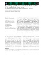
central role of Tyr380 (Fig. 1). Similarly, maltohepta-
ose in the inactive catalytic nucleophile mutant D180A
AMY1 curved with five visible rings around Tyr380.
Two adjacent rings, in a second maltoheptaose mole-
cule with five clearly defined rings, were stacked onto
the indole side chains of Trp278Trp279 on the surface
Fig. 1. Close-up view on the ‘pair of sugar tongs’ binding site in
the crystal structure of a-amylase 1 (AMY1) D180A, an inactive cat-
alytic nucleophile mutant, in complex with maltoheptaose [21].
Important residues defining this site have been highlighted. Ser378
and Tyr380 are mutated in the present work. As continuous elec-
tron density was only found for five sugar rings, a pentasaccharide
was modeled into the structure.
a-amylase ‘sugar tongs’ mutants S. Bozonnet et al.
5056 FEBS Journal 274 (2007) 5055–5067 ª 2007 The Authors Journal compilation ª 2007 FEBS
of domain A [21]. Seven rings in a third maltoheptaose
molecule occupied subsites )7 through )1 in the active
site [21]. Noticeably, AMY2 accommodated the
pseudotetrasaccharide inhibitor acarbose both at
Trp276Trp277 and at the active site, but not at the
‘sugar tongs’ [3]. Comparison of AMY1 and AMY2
structures [3,4] suggested that Pro376AMY2 – corre-
sponding to Ser378AMY1 – rigidified the loop carry-
ing Tyr378
AMY2
(Tyr380 in AMY1), hindering the
conformational shift needed in oligosaccharide binding
[4]. Different secondary carbohydrate-binding sites are
found in GH-H members, e.g. certain a-amylases
[3,4,22,26–28], cyclodextrin glucosyltransferase [29],
amylosucrase [30], amylomaltase [20], and Thermoacti-
nomyces vulgaris I amylase [31]. The Pseudomonas
maltotetraose-forming amylase structure closely resem-
bles that of AMY1 but has no tyrosine at the position
of Tyr380 [4]. Tyr380, however, is present in several
plant a-amylases [32–34], including AMY2, which did
not accommodate oligosaccharide at the ‘sugar tongs’
in the structure [3]. In the present work, the ‘sugar
tongs’ site was demonstrated by site-directed mutagen-
esis of Tyr380 to be involved in enzymatic activity and
confirmed to be particularly important for carbohy-
drate binding. However, mutating Ser378 in AMY1 to
proline to mimic AMY2 did not elicit lack of binding
as observed for the AMY2 structure [3]. The func-
tional analysis of the surface site furthermore indicated
a role in multivalent binding during polysaccharide
processing.
Results
Choice and production of AMY1 ‘sugar tongs’
mutants
Tyr380 in the ‘sugar tongs’ site on domain C of
AMY1 (Fig. 1) shifted 3.1 A
˚
when binding a malto-
oligosaccharide [4,21] and the Y380A, Y380M, and
Y380F enzymes were produced to investigate the
importance of the aromatic side chain, tryptophan
being omitted for steric reasons. The substituted methi-
onine also represented a bean a-amylase [35] (Fig. 2).
The lack of sugar binding at the conserved Tyr378 in
the AMY2 ⁄ acarbose structure [3] was proposed to be
due to lower mobility imposed by Pro376AMY2 (cor-
responding to Ser378AMY1, see Figs 1 and 2) on the
Arg377–Phe388AMY2 loop. Hence the AMY2 mimic,
AMY1 S378P, was constructed to check the impact of
proline; S378T represented rice and millet a-amylases
[33] (Fig. 2). The host Pichia pastoris secreted
10–44 mgÆL
)1
wild-type and AMY1 mutants as esti-
mated from specific activities against insoluble
blue starch (iBS) of the purified enzymes (Table 1).
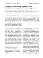
Fig. 2. Sequence alignment of domain-C of barley AMY1 and AMY2, four other cereal amylases, and a legume a-amylase. The secondary
structure of AMY1 is indicated above the alignment and mutated residues are highlighted in orange. Accession numbers are; wheat (AMY3):
P08117; maize: Q41770; millet: Q7Y1C3; rice (AMY3): P27933; kidney bean: Q9ZP43.
Table 1. Enzymatic properties of ‘sugar tongs’ mutants of barley a-amylase 1 (AMY1). U, one enzyme unit is the amount required to cause
an A
620
increase of 1.
Enzyme
iBS Amylose DP440 Cl-pNPG
7
Specific activity
(UÆmg
)1
)
k
cat
(s
)1
)
k
m
(mgÆmL
)1
)
k
cat
⁄ K
m
(s
)1
mL
)1
Æmg
)1
)
k
cat
(s
)1
)
K
m
(mM)
k
cat
⁄ K
m
(s
)1
mM
)1
)
Y380A 1400 95 ± 15 0.363 ± 0.023 261.7 19 ± 0.6 0.669 ± 0.046 28.4
Y380M 2000 149 ± 44 0.351 ± 0.083 424.5 34 ± 0.8 0.871 ± 0.027 39.0
Y380F 2790 162 ± 27 0.391 ± 0.146 414.3 56 ± 1.7 0.724 ± 0.123 77.3
S378P 2695 163 ± 36 0.203 ± 0.130 802.9 59 ± 0.6 0.861 ± 0.023 68.5
S378T 2705 144 ± 9 0.208 ± 0.058 692.3 48 ± 1.7 0.735 ± 0.087 65.3
AMY1 2500 185 ± 20 0.190 ± 0.010 973.7 52 ± 4.9 0.758 ± 0.112 68.6
AMY2 4000 721 ± 63 1.074 ± 0.283 671.3 86 ± 3.1 2.125 ± 0.180 40.5
S. Bozonnet et al. a-amylase ‘sugar tongs’ mutants
FEBS Journal 274 (2007) 5055–5067 ª 2007 The Authors Journal compilation ª 2007 FEBS 5057
Similarly to the AMY1 wild-type, S378P, S378T and
Y380F were obtained in $50% yield by affinity chro-
matography on b-cyclodextrin (b-CD)-Sepharose,
whereas Y380A and Y380M AMY1 did not bind to
the resin and were purified in $20% yield by ammo-
nium sulfate precipitation and ion exchange chroma-
tography (see Experimental procedures).
Enzymatic activity of ‘sugar tongs’ AMY1
mutants
Replacement of Tyr380 by alanine and methionine
caused 50–75% reduction in the activity of iBS (k
cat
),
amylose DP440 (k
cat
⁄ K
m
), and even the oligosaccha-
ride Cl-pNPG
7
(k
cat
⁄ K
m
) (Table 1). The mutations
reduced k
cat
for amylose and Cl-pNPG
7
and doubled
K
m
, whereas the conservative substitutions in Y380F,
S378P, and S378T had no effect on enzyme kinetic
parameters except for a twofold increase in K
m
for
Y380F against the amylose (Table 1). This probably
reflected that the mutant was unable to form the
hydrogen bond between Tyr380 cOH and O2 of glu-
cose as seen in the AMY1Æthio-DP4 complex [4]. Activ-
ity for iBS was routinely analyzed under saturating
conditions (i.e. 6.25 mgÆmL
)1
iBS), but in fact AMY1
showed a small and highly reproducible isozyme-char-
acteristic activity maximum near 2 mgÆmL
)1
iBS corre-
sponding to 115% of the activity at 6.25 mgÆmL
)1
iBS.
This property was lost in Y380A, suppressed for
Y380M, but retained by Y380F, S378P, and S378T
AMYl, and was missing for AMY2 (data not shown).
The earlier reported hydrolysis of the amylose of
DP440 in a multiple attack mechanism [12] was con-
firmed for AMY1, which showed a DMA of 1.9 as
determined from the ratio of rates of release of
reducing groups in the fraction of small (i.e. ethanol-
soluble) products over large (i.e. ethanol-precipitated)
products (see Experimental procedures and [12]). The
rates of product formation by the mutants (not
shown) agreed with the activity levels described in
Table 1. AMY1 Y380A had a DMA of 1.0 and thus
released fewer short products per enzyme–substrate
encounter than AMY1 wild-type, whereas AMY1
Y380M and S378P maintained a DMA of 2.0 and
2.2, respectively.
Binding of b-cyclodextrin to ‘sugar tongs’
mutants measured by surface plasmon resonance
analysis
Surface plasmon resonance (SPR) analysis was suitable
for measuring the affinity in the low millimolar range
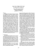
of b-CD for AMY1. SPR sensorgrams clearly illustrated
weaker binding to AMY1 Y380A than to wild-type
enzyme (Fig. 3) and K
d
was calculated to 1.40 mm for
both Y380A and Y380M, i.e. sevenfold higher than K
d
of AMY1 wild-type (Table 2). Y380F caused only a
slight reduction in affinity for b-CD and the binding to
S378P and S378T was essentially not affected by the
mutations. In comparison, the K
d
of AMY2 was three-
fold higher than that of AMY1 (Table 2).
Effects of ‘sugar tongs’ mutation on adsorption
to and hydrolysis of starch granules
Starch granules are the natural substrate for barley
a-amylases and it was hypothesized that the ‘sugar
tongs’ might play a role in interaction with this sub-
strate of giant size compared to the enzyme. The adsorp-
tion to barley starch granules of ‘sugar tongs’ mutants
was therefore examined. The K
d
was 0.47 mgÆmL
)1
for
AMY1 wild-type and very similar for S378P, but 13-fold
higher for AMY1 Y380A (Table 3). This indication of a
β
β
-cyclodextrin (mM)
01 34526
RU
0
100
200
300
400
Fig. 3. b-CD binding determined by SPR analysis. AMY1: d wild-
type, s Y380A. Response unit (RU) values are corrected for the
contribution given by a channel in the chip without bound enzyme
protein.
Table 2. Binding of b-cyclodextrin (b-CD) to ‘sugar tongs’ mutants
and wild-type AMY1 and AMY2 as determined by SPR. See Experi-
mental procedures for the SPR analytical procedure.
Enzyme K
d
(mM)
Y380A 1.40 ± 0.23
Y380M 1.39 ± 0.65
Y380F 0.36 ± 0.02
S378P 0.25 ± 0.03
S378T 0.23 ± 0.02
AMY1 0.20 ± 0.04
AMY2 0.63 ± 0.27
a-amylase ‘sugar tongs’ mutants S. Bozonnet et al.
5058 FEBS Journal 274 (2007) 5055–5067 ª 2007 The Authors Journal compilation ª 2007 FEBS
very important role of Tyr380 is in accordance with
b-CD having no impact on the apparent affinity of
AMY1 Y380A for starch granules, whereas the presence
of 0.5 mm b-CD increased the apparent K
d
four- to six-
fold for AMY1 wild-type and S378P and AMY2
(Table 3), confirming competition in binding to starch
granules. AMY2 showed threefold weaker affinity for
barley starch granules than did both the wild-type and
the AMY2 mimic, AMY1 S378P (Table 3).
The ‘sugar tongs’ substitution in AMY1 Y380A
greatly influenced the hydrolytic activity against granu-
lar starch, with release of soluble reducing sugars from
this substrate being strongly reduced (Fig. 4) to a
k
cat
⁄ K
m
value of $10% of that of AMY1 wild-type. In
contrast to wild-type, substrate saturation was not
achieved for AMY1 Y380A even at 400 mgÆmL
)1
of
starch granules and the shape of the corresponding
activity curve indicated that loss in substrate affinity
was a predominant factor in the reduced activity
(Fig. 4). For AMY1 S378P, k
cat
and K
m
were similar
to the wild-type values (Table 4), but AMY2 had infe-
rior affinity. The corresponding activity curve (Fig. 4)
allowed only estimation of kinetic parameters, the K
m
being considerably higher than in the case of AMY1,
whereas the k
cat
for AMY2 appeared higher than for
AMY1, as found in general for different substrates
(Table 1). The activity was reduced in the presence of
b-CD (Fig. 4; Table 4) due to competition with starch
granule binding. The low activity hampered analysis of
the effect of b-CD on AMY1 Y380A.
Table 3. Binding of ‘sugar tongs’ mutants, wild-type AMY1 and
AMY2 to barley starch granules. The binding was measured in the
range 0.01–40 mgÆmL
)1
starch granules (see Experimental proce-
dures and [29] for details). (A) no b-CD; (B) in the presence of
0.5 m
M b-CD.
Enzyme K
d
(mgÆmL
)1
)B
max
A
Y380A 5.90 ± 0.47 0.90 ± 0.05
S378P 0.57 ± 0.04 0.98 ± 0.01
AMY1 0.47 ± 0.06 1.03 ± 0.04
AMY2 1.27 ± 0.32 0.99 ± 0.03
B
Y380A 6.86 ± 0.55 0.81 ± 0.02
S378P 2.93 ± 0.36 0.95 ± 0.02
AMY1 2.85 ± 0.28 0.98 ± 0.02
AMY2 4.63 ± 0.37 0.87 ± 0.02
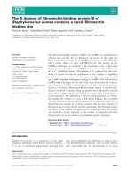
Fig. 4. Rates of release of soluble reducing products from barley starch granules as catalyzed by AMY1, AMY2, and Y380A and
S378P AMY1 in the absence (d), and in the presence (s) of 0.5 m
M b-CD.
S. Bozonnet et al. a-amylase ‘sugar tongs’ mutants
FEBS Journal 274 (2007) 5055–5067 ª 2007 The Authors Journal compilation ª 2007 FEBS 5059
Crystal structures of AMY1 ‘sugar tongs’ mutants
in complex with acarbose
The structures of AMY1 Y380A and S378P were com-
pared with the wild-type enzyme [21] both in free form
(not shown) and in complex with acarbose – a pseudo-
tetrasaccharide inhibitor whose rings A and B
correspond to the valienamine and 4-amino-4,6-dide-
oxy-a-d-glucose units in acarviosine, and rings C and
D (reducing end) constitute a maltose unit linked to
acarviosine (see Fig. 2 in [21]). Remarkably, compari-
son of the ‘sugar tongs’ region indicated no conforma-
tional differences between AMY1 wild-type, S378P
and Y380A acarbose complexes. The only obvious dif-
ference was found for the Ala211-Pro218 loop (Fig. 5)
that connects b5 and a5 of the catalytic (b ⁄ a)
8
-barrel
at the end of the aglycon-binding area of the active site
cleft. Earlier, significant deviation was found in this
region between the backbone conformation of AMY1
and AMY2 [36]. Thus Ca of Gly214 shifted 0.8, 1.2,
and 1.4 A
˚
relative to AMY1 ⁄ acarbose for the three
molecules A, B and C, respectively, present in the
asymmetric unit of S378P ⁄ acarbose (see supplementary
Table S1). At the ‘sugar tongs’ of S378P ⁄ acarbose
(molecule A) the electron density for rings B and C
was very clear (Fig. 6A) and almost entirely defined
for ring A; however, the density was badly defined for
ring D, which therefore was not inserted for refine-
ment. In molecule B of S378P ⁄ acarbose, rings B and C
were completely defined, ring D was better defined
than molecule A, and ring A was poorly defined. In
molecule C, rings A–C were defined very clearly,
whereas ring D lacked continuous electron density and
was omitted from the refinement. In spite of cocrystal-
lization, a hydrated calcium ion (Ca503) and not
acarbose was bound at the active site of AMY1 S378P
(not shown). Ca503 was also present in native S378P
(not shown) and it was previously observed in
Table 4. Hydrolysis of barley starch granules by ‘sugar tongs’
mutants, wild-type AMY1 and AMY2. (A) no b-CD; (B) in the pres-
ence of 0.5 m
M b-CD. NC, not calculated due poor affinity; ND, not
determined due to low activity. See also Fig. 4. See Experimental
procedures for details on the procedure.
A
Enzyme k
cat
(s
)1
) K
m
(mgÆmL
)1
)
k
cat
⁄ K
m
(s
)1
mLÆmg
)1
)
Y380A NC NC 0.151
S378P 149 ± 10 96 ± 23 1.547
AMY1 113 ± 12 73 ± 15 1.549
AMY2 251 ± 40 188 ± 26 1.338
B
k
cat
K
m,app
k
cat
⁄ K
m
Y380A ND ND ND
S378P 150 ± 40 248 ± 54 0.605
AMY1 122 ± 8 273 ± 62 0.449
AMY2 220 ± 28 275 ± 28 0.802
Fig. 5. Stereo view of the overall fold of the superimposed three-
dimensional structures of wild-type AMY1 (in red [21]), the S378P
(in blue) and the Y380A (in yellow) ‘sugar tongs’ AMY1 mutants in
complex with acarbose. The vertical arrow indicates the flexible
loop region, Ala211–Pro218.
Fig. 6. Close-up view on the ‘sugar tongs’ binding site. (A)
S378P ⁄ acarbose (molecule A), showing the bound sugar ligand
(rings A, B, and C) and (B) Y380A ⁄ acarbose, which has no ligand
bound at the ‘sugar tongs’ site.
a-amylase ‘sugar tongs’ mutants S. Bozonnet et al.
5060 FEBS Journal 274 (2007) 5055–5067 ª 2007 The Authors Journal compilation ª 2007 FEBS
AMY1Æthio-DP4 [4] as well as in AMY2ÆBASI (a pro-
teinaceous inhibitor complex) [37], but so far not in
native AMY1.
At the surface-binding site containing Trp278Trp279
on the (b ⁄ a)
8
-barrel [3,4,21], electron density studies
identified three sugar rings in AMY1 S378P ⁄ acarbose,
and in wild-type AMY1 ⁄ acarbose sugar binding
occurred at this site as well as to the active site cleft
and the ‘sugar tongs’ [21]. The three rings defined at
the Trp278Trp279 site corresponded to acarbose with
the reducing-end glucose cleaved off and with the same
orientation, but shifting position by one sugar unit
compared to the ligand in S378P ⁄ acarbose (mole-
cule A). Thus acarbose rings A and B stacked onto
Trp279 and Trp278, respectively, whereas ring C was
in the bulk solvent (not shown). In wild-type
AMY1 ⁄ acarbose, rings B and C stacked onto
Trp279Trp278. As a curiosity, rings A and B modeled
into the electron density on this surface site in
AMY2 ⁄ acarbose [3] were at the same position as in
the AMY2 mimic, S378P AMY1 ⁄ acarbose.
In the structure of AMY1 Y380A ⁄ acarbose (see sup-
plementary Table S1) only Trp278Trp279 and neither
the ‘sugar tongs’ nor the active site bound oligosaccha-
ride. Two rings were conjectured from the electron den-
sity; a third may be present, but due to poor definition,
water molecules were modeled into the electron densi-
ties. Thus the Y380A mutation in AMY1 destroyed
accommodation of oligosaccharide (Fig. 6B) at the
‘sugar tongs’, emphasizing the critical role of Tyr380.
Inspection of the active site region in AMY1 Y380A
suggested that neither oligosaccharide nor Ca503 was
present as opposed to the S378P ⁄ acarbose (this work)
and AMY1 ⁄ oligosaccharide structures [4,21]. Numer-
ous attempts at collecting data of improved quality for
AMY1 Y380A ⁄ acarbose failed and from the obtained
structure it cannot be excluded that trace amounts of
carbohydrate occupy the active site.
Discussion
Functional insight into amylolytic and related enzymes
is poor in regard to carbohydrate-binding surface sites
at a certain distance from the active site [3,4,20–22,
28–30] as opposed to sites residing on CBMs [1,39,40].
The discovery of oligosaccharide binding at the ‘sugar
tongs’ in the C-terminal domain in barley AMY1
[4,21] was therefore a welcome opportunity firstly to
investigate a surface site by mutational analysis cou-
pled with structure determination, carbohydrate bind-
ing and activity assays and, secondly, to learn more
about the role of domain-C in GH-H. Cereal a-amy-
lases do not hydrolyze b-CD, which thus can serve as
a molecular model in emulating protein–starch interac-
tions. Despite lack of binding to b-CD-Sepharose, the
SPR procedure developed in the present work enabled
analysis of b-CD affinity in the millimolar range for
AMY1 Y380A and Y380M. The K
d
of 1.40 mm for
b-CD was increased sevenfold, confirming the critical
functional role of Tyr380 in the ‘sugar tongs’. As
b-CD-Sepharose did not retain these mutants, their
still intact other surface site containing Trp278Trp279
was concluded to have very low affinity for b-CD.
b-CD accordingly was seen to bind at the ‘sugar
tongs’, and not at Trp278Trp279 in the structure of
AMY1 active site mutants (Tranier, Aghajari, Haser,
Mori and Svensson, unpublished). Crystallography on
AMY1 Y380A (present work) demonstrated that this
substitution destroyed acarbose binding to the ‘sugar
tongs’, whereas acarbose bound to Trp278Trp279.
Furthermore, acarbose occupied the ‘sugar tongs’ in
S378P ⁄ acarbose (the AMY2 mimic). Hence as
AMY1 S378P and wild-type also shared the same
affinity for b-CD, several properties of AMY1 S378P
did not confirm the earlier suggestion that Pro376
(AMY2-numbering) caused the lack of ligand binding
at the ‘sugar tongs’ in the AMY2 structure [3,4]. The
modest threefold weaker affinity seen for b-CD bind-
ing by AMY2 compared to AMY1 wild-type and
S378P, possibly combined with different crystallization
conditions for AMY1 and AMY2 [3,50,51], may have
prevented oligosaccharide binding in the AMY2 crystal
structure. Individual binding sites in multivalent pro-
tein–carbohydrate interactions are often of moderate
affinity and a rather small energy difference between
comparable binding events possibly elicits functional
differences of AMY1 and AMY2 in mobilization of
storage starch during germination.
The ‘sugar tongs’ site was critical for efficient bind-
ing to starch granules, as AMY1 Y380A showed a 13-
fold higher K
d
of 5.9 mgÆmL
)1
than did wild-type. The
a-amylase from azuki bean in which methionine corre-
sponds to AMY1 Tyr380, bound starch granules with
a K
d
similar to that of AMY1 Y380A [35] and oxidi-
zation to methionine sulfoxide further reduced the
affinity [41,42]. These findings confirmed that the func-
tional ‘sugar tongs’ of a biologically relevant binding
level of affinity was present in plant a-amylases. How-
ever, the precise natural role(s) of this site, for which
distinct variation in affinity has so far been demon-
strated for AMY1, AMY2 and the azuki bean enzyme,
is not yet disclosed.
Compared to AMY1, cyclodextrin glucosyltransfer-
ase from Bacillus circulans strain 251 having an SBD
of CBM20, showed a 16-fold lower affinity for starch
granules (K
d
¼ 7.6 mgÆmL
)1
), and its K
d
increased
S. Bozonnet et al. a-amylase ‘sugar tongs’ mutants
FEBS Journal 274 (2007) 5055–5067 ª 2007 The Authors Journal compilation ª 2007 FEBS 5061
only two- to threefold for SBD single and dual binding
site mutants [29]. The homologous SBD of Aspergillus
niger glucoamylase had K
d
values of 6.4 and 28 lm for
b-CD and of 0.95 and 17 lm for maltoheptaose for
each of the two binding sites, respectively [40]. Thus
AMY1 ‘sugar tongs’ and these SBDs show very differ-
ent ligand specificity, AMY1 having about 15-fold
higher and 30-fold lower affinity for starch granules
and b-CD, respectively, than does SBD. This is also
reflected in the lower B
max
values found for the cyclo-
dextrin glucosyltransferase [29].
AMY1 Y380A had only 10% hydrolytic activity of
wild-type against starch granules apparently due to
poor substrate binding. Furthermore, although b-CD
did not inhibit AMY1-catalyzed hydrolysis of amylose
[43], b-CD reduced the catalytic efficiency (k
cat
⁄ K
m
)on
starch granules, providing indirect support for the
‘sugar tongs’ being involved in degradation of storage
starch. Remarkably, the K
m
for hydrolysis of starch
granules was about two orders of magnitude higher
than the K
d
for binding of AMY1 wild-type, mutants
and AMY2. Even though activity and binding are
measured at 37 °C and 4 °C, respectively, this differ-
ence is very large and may reflect that only a few
a-glucan chains in the granules are readily hydrolyzed
or that a major fraction of the products remains asso-
ciated with the granules. The trend of an even slightly
larger difference between K
d
and K
m
for AMY1
Y380A compared with wild-type supported the role of
the ‘sugar tongs’ in activity, reflected also by a moder-
ately reduced k
cat
for hydrolysis of amylose by
AMY1 Y380A and Y380M and the unexpected
decrease in activity for Cl-pNPG
7
that covers only
seven to eight active site subsites [44]. This latter loss
in activity was speculated to stem from Cl-pNPG
7
binding to the ‘sugar tongs’, similarly to other oligo-
saccharides [21]. This binding may modulate activity,
as supported by the very detailed study of acarbose
inhibition kinetics of hydrolysis of amylose by barley
a-amylase, where acarbose was concluded to occupy at
least one secondary site in the productive enzyme–sub-
strate complex and, furthermore, that this binding al-
losterically enhanced activity [46]. As orientation of
maltoheptaose molecules bound to AMY1 D180A sug-
gested that three different, rather than the same, a-glu-
can chains were accommodated at the active site and
at the two surface sites [21], one cannot on a structural
basis, model interactions in the multiple attack mecha-
nism showing the substrate chain attached at the
‘sugar tongs’. Thus even though increased DMA of
the AMY1–SBD fusion suggested that enzyme–sub-
strate interactions at secondary binding sites were
favoring multiple attack [12,25], in agreement with the
reduced DMA of the AMY1 Y380A ‘sugar tongs’
mutant, these effects may stem from allosteric regula-
tion.
The mutational analysis of the ‘sugar tongs’ in barley
AMY1 explored the role of this so far unique carbohy-
drate-binding surface site from plant a-amylases. This
is the first demonstration of a function for a C domain
from the large GH clan-H. The work contributes to the
unraveling of the molecular basis of multivalent
enzyme–polysaccharide interactions. One future aim is
to extend this analysis to include different surface sites
in AMY1 and AMY2 to gain insight into the putative
cooperation among these sites and the active site.
Experimental procedures
Strains, plasmids and AMY2
Escherichia coli DH5a and P. pastoris GS115, transformed
with pPICZA (Invitrogen, Carlsbad, CA), were used for
standard cloning and expression. pPICZA-amy1D9 encoded
AMY1 (GenBank accession gi|113765) with a C-terminal
nonapeptide truncation [10], here referred to as AMY1.
AMY2 (gi|4699831) was purified from malt [45].
Site-directed mutagenesis
Standard cloning techniques were used [46]. Site-directed
mutagenesis was done by the mega-primer method [47]
using for S378P, 5¢-GATCGGG
CCCAGGTACGACGTC
GG-3¢; S378T, 5¢-GATCGGG
ACCAGGTACGACGTCG
G-3¢; Y380A, 5¢-GATCGGGTCCAGG
GCCGACGTC
-GG-3¢; Y380M, 5¢-GATCGGGTCCAGG
ATGGACGT
CGG-3¢; Y380F, 5¢-GATCGGGTCCAGG
TTCGAC
GTCGG-3¢ (underlined mutant codon) coding for the sense
strand, and 5¢-TTTGGTACCTCAGTTCTTCTCCCAGA
CGGCGTA-3¢ as antisense primer. Mutant cDNA was
amplified using 5¢-TTTGAATTCCATGGGGAAGAACG
GCAGC-3¢ as sense orientation primer and a purified mega-
primer. Pfu DNA polymerase (Stratagene, La Jolla, CA) was
used for PCR and products were cut by NarI and KpnI. The
700 bp fragments were purified (QIAquick gel extraction kit,
QIAGEN, Germantown, MD) and subcloned in NarI, KpnI-
linearized pPICZA-amy1D9. Plasmids were propagated in
E. coli DH5a [low-salt LB, 25 lgÆmL
)1
Zeocin
Ò
(Invitrogen,
Carlsbad, CA)], purified (Midiprep Plasmid extraction kit,
QIAGEN), sequenced (Big-Dye premix; ABI PRISM 310
Genetic Analyzer, Perkin Elmer Life Sciences, Waltham,
MA), and BglII-linearized prior to transformation of P. pas-
toris by electroporation [48]. Transformants were identified
on YPDS (1% yeast extract, 2% peptone, 2% glucose, 1 m
sorbitol, 2% agar, 100 lgÆmL
)1
Zeocin), transferred to meth-
anol ⁄ starch plates and selected for a-amylase secretion by
halos seen by exposure to I
2
[10].
a-amylase ‘sugar tongs’ mutants S. Bozonnet et al.
5062 FEBS Journal 274 (2007) 5055–5067 ª 2007 The Authors Journal compilation ª 2007 FEBS
Enzyme production and purification
Pichia pastoris transformants were grown in 1 L BMGY
(1% yeast extract, 2% peptone, 1% glycerol, 0.67% yeast
nitrogen base, 100 mm K phosphate, pH 6.0, 0.1 lgÆmL
)1
biotin) at 30 °C for 2 days in 5 L flasks to D
600
$15 and
the medium was changed for induction to 0.5 L BMMY (as
BMGY with 0.5% methanol replacing glycerol) followed by
24 h incubation [9,10]. Secreted activity was assayed using
insoluble blue starch (iBS). Cell harvest and induction were
repeated two to three times and combined supernatants were
concentrated to $300 mL (Pellicon, Millipore, Bedford,
MA). AMY1 S378P ⁄ T, Y380F, and wild-type were purified
on b-CD-Sepharose (diameter 2.6 cm; 0.2 mL resinÆmg
)1
a-amylase) [10]. As Y380A ⁄ M AMY1 were not retained
by b-CD-Sepharose, protein was precipitated from culture
supernatants using 85% saturated ammonium sulfate,
dissolved in 50 mm Na acetate, pH 5.5, 25 mm CaCl
2
,
and chromatographed on Hi Load 26 ⁄ 60 Superdex 75
(GE Healthcare, Uppsala, Sweden) at 2.5 mLÆmin
)1
. Eluate
with activity for iBS was dialyzed against 10 mm Hepes
pH 7.0, 1 mm CaCl
2
, applied to Resource Q (6 mL column)
equilibrated in buffer, and gradient-eluted [0–10%, 0.5 col-
umn volume (cv); 10–40%, 5 cv; 40–100%, 0.5 cv] at 1 mLÆ
min
)1
using buffer without and with 0.5 m NaCl (A
¨
KTA-
explorer, GE Healthcare). Two forms of differing pI were
resolved by anion exchange chromatography [10]. The first-
eluting and highly active form was dialyzed (10 mm Mes,
25 mm CaCl
2
, pH 6.8) and concentrated (Centriprep YM10,
Millipore), 0.02% (w ⁄ v) NaN
3
was added, and the form kept
at 4 °C, whereas the more acidic form containing gluta-
thionylated Cys95 [49] was discarded. All steps were carried
out at 4 °C. Proteins migrated as single bands in SDS ⁄ PAGE
and showed pI ¼ 4.8 by isoelectric focusing [9].
Enzyme activity
Insoluble blue starch
Enzyme was added (50 lL, final 1–12 nm) to 5 mg iBS
(Amersham Biosciences) in 20 mm Na acetate pH 5.5,
5mm CaCl
2
, 0.005% BSA (0.8 mL) and incubated at
37 °C. At 15 min, 0.5 m NaOH (200 lL) was added and
after centrifugation (10 000 g, 3 min) the absorbance of the
supernatants (300 lL, in duplicate) was measured at
620 nm in a microtiter plate reader (MRX-TC Revelation;
Dynex Technologies, Richfield, MN). One enzyme unit is
the amount causing an A
620
increase of 1.
Amylose
Initial rates of reducing power formation at six to nine con-
centrations (0.10–2.50 mgÆmL
)1
) of amylose DP440 (potato
type III, Sigma, St. Louis, MO) by 0.47–1.0 nm enzyme in
20 mm Na acetate, pH 5.5, 5 mm CaCl
2
, 4% dimethylsulf-
oxide (w ⁄ v), 0.005% BSA (w ⁄ v) at 37 °C [10] were deter-
mined using copper-bicinchoninate with maltose as
standard [10], and measured at A
540
in microtiter plates.
k
cat
and K
m
were obtained by fitting to the Michaelis-Men-
ten equation (curve expert version 1.3, http://curveexpert.
webhop.biz/).
2-Chloro-4-nitrophenyl b-D-maltoheptaoside
Initial rates of hydrolysis of Cl-pNPG
7
(Merck, Darmstadt,
Germany) at eight concentrations (0.25–10 mm)by
2.0–5.2 nm enzyme at 30 °Cin50mm phosphate pH 6.8,
50 mm KCl, 0.02% NaN
3
, 3167 nkatÆmL
)1
Saccharomyces
cerevisiae a-glucosidase, and 50 nkatÆmL
)1
almond b-gluco-
sidase (both Sigma) were measured at 405 nm in microtiter
plates using 4-nitrophenol as standard. k
cat
and K
m
were
obtained as above.
Starch granules
Enzyme (final concentration 4–7 nm) was added to barley
starch granules (Primalco, Helsinki, Finland) at 10 concen-
trations (0.8–400 mgÆmL
)1
)in20mm Na acetate, pH 5.5,
5mm CaCl
2
, 0.005% BSA (w ⁄ v) agitated (1000 r.p.m.) at
37 °C. Hydrolysis was measured over 25 min as reducing
power in supernatants of centrifuged (10 000 g, 5 min,
room temperature) aliquots. k
cat
and K
m
were obtained as
above. The effect of b-CD was determined in parallel.
Standard deviations
Standard deviations were calculated from triplicate experi-
ments.
Degree of multiple attack
The DMA was determined as described on amylose DP440
(final concentration in 1 mgÆmL
)1
) dissolved initially in
dimethylsulfoxide and diluted with 20 mm Na acetate,
5mm CaCl
2
, pH 5.5 to a final 2% dimethylsulfoxide [12].
Enzyme (final concentation 0.1–0.8 nm) was added to the
substrate and aliquots were removed at appropriate time
intervals guided by loss in iodine blue value [12]. DMA
(Eqn 1) was calculated as described:
DMA ¼ðRV
t
=RV
p
ÞÀ1 ðEqn 1Þ
where RV
t
and RV
p
are initial rates of reducing power
formation in the total digest and in the ethanol-insoluble
fraction, respectively [12]. The standard deviation was
calculated from at least triplicate experiments.
Surface plasmon resonance
Enzyme (0.9–1.1 nmol in 30–100 lL) was biotinylated and
immobilized on a streptavidin-coated chip, using BIAcore
S. Bozonnet et al. a-amylase ‘sugar tongs’ mutants
FEBS Journal 274 (2007) 5055–5067 ª 2007 The Authors Journal compilation ª 2007 FEBS 5063
3000 (BIAcore AB, Uppsala, Sweden) at $5ngÆmL
)1
in
running buffer [10 mm Mes, pH 6.5, 5 mm CaCl
2
, 0.005%
(v ⁄ v) surfactant P20] for 4 min at 10 lLÆmin
)1
[15] to reach
2000–3000 response units (RU). Sensorgrams (RU versus
time) were recorded of b-CD (12 concentrations,
15 lm)7mm) binding in running buffer at 30 lLÆmin
)1
and 25 °C for 3 min, followed by 3 min dissociation in
buffer. RU for a parallel flow cell without enzyme was
subtracted and K
d
was obtained by steady state affinity
fitting analysis (biaevaluation 3.1 software). Experiments
were carried out in triplicate.
Binding to starch granules
Enzyme (final 4–12 nm) was agitated 30 min with starch
granules at 10–13 concentrations (0.01–40 mgÆmL
)1
)in
20 mm Na acetate, pH 5.5, 5 mm CaCl
2
, 0.005% BSA
(w ⁄ v) at 4 °C (1000 r.p.m.), and centrifuged (10 000 g,
4 °C, 5 min). Activity on iBS was measured in the superna-
tant and K
d
(Eqn 2) was determined as for cyclodextrin
glucosyltransferase [29] where b is the bound enzyme frac-
tion, [S] the starch concentration, and B
max
the maximum
fraction of enzyme bound, which was derived by
b ¼
B
max
½S
½SþK
d
ðEqn 2Þ
fitting plots of b versus [S] to a hyperbola (Curve Expert).
The effect of b-CD on the binding was analyzed in parallel.
Experiments were done in triplicate.
Crystallization and data collection
Y380A and S378P AMY1 were crystallized at conditions
similar to AMY1 [50,51] and acarbose complexes were
obtained by soaking and cocrystallization, respectively (see
supplementary Table S1). Crystals, 0.5 · 0.02 · 0.01 mm
3
(Y380A ⁄ acarbose) and 0.3 · 0.02 · 0.01 mm
3
(S378P ⁄ acar-
bose), were cryo-protected by soaking a few seconds in
mother liquor made up to 10% (w ⁄ v) in ethylene glycol
and, for Y380A ⁄ acarbose, also 10 mm in acarbose. Data
were collected at beamline ID14-4 (European Synchrotron
Radiation Facility, Grenoble, France). Diffracted intensities
were integrated and scaled (xds program package [52]).
Crystal parameters and data collection statistics are given
in supplementary Table S1.
Structure determination and refinement
The S378P ⁄ acarbose structure was solved by molecular
replacement with AMY1 at 1.5 A
˚
resolution (Protein Data
Bank entry 1HT6) as search model [4], omitting water mole-
cules and calcium ions, and using data in the resolution range
15–3.5 A
˚
(cns software [53]). Initial rigid body refinement
included data to 3.5 A
˚
resolution; in the remaining refine-
ments a simulated annealing protocol was used extending
data up to 1.7 A
˚
combined with anisotropic B-factor refine-
ment. Due to crystal isomorphism with Y380A, wild-type
AMY1 [4] was used as the starting model in a difference Fou-
rier (water molecules and calcium ions were omitted) to solve
the structure of Y380A AMY1 ⁄ acarbose. Initial rigid body
refinement included data to 3.5 A
˚
resolution; in the remain-
ing refinements a simulated annealing protocol was used
including data to 2.2 A
˚
followed by an isotropic B-factor
refinement. Refinements (cns software [53]) were alternated
with visual electron density map examination and manual
building (graphics software turbo-frodo [54]). R- and
R-free factors [55] were monitored to avoid over-refinement;
R-free being calculated from a test set of 5% of the reflec-
tions randomly selected from all data. Based on inspection of
2F
o
-F
c
and F
o
-F
c
maps (contoured at 1 and 3 r , respectively),
calcium ions were inserted and water molecules were added,
respecting hydrogen-bonding distances and angles. Water
molecules at similar positions in the respective structures
have the same numbering. Acarbose was manually inserted
in the electron density. Model qualities were examined with
procheck [56] and whatcheck [57]. Refinement statistics
are summarized in supplementary Table S1.
Sequence alignment
Domain-C sequences from selected a-amylases were aligned
using clustalw [58]. Superimposition of secondary struc-
tures of AMY1 and rendering was done with the program
espript [59].
Acknowledgements
Sidsel Ehlers, Mette Hersom Bien, Lone Sørensen
(Carlsberg Laboratory) and Susanne Blume (Enzyme
and Protein Chemistry, BioCentrum-DTU) are grate-
fully acknowledged for excellent technical assistance,
and Peter K. Nielsen and Phaedria St. Hilaire for
advice on SPR analysis. Xavier Robert and Maher
Abou Hachem are thanked for stimulating discussions.
This work was supported by the European Union
Fourth Framework Program on Biotechnology (CT98-
0022, AGADE) and Fifth Framework Program ‘Qual-
ity of Life and Management of Living Resources’
(QLK3-2001–00149, CEGLYC), the Danish Natural
Science Research Council, the Carlsberg Foundation,
and a Ph.D. stipend from DTU (to MMN).
References
1 Coutinho PM & Henrissat B (1999) Carbohydrate-
active enzymes: an integrated database approach. In
Recent Advances in Carbohydrate Bioengineering (Gilbert
HJ, Davies G, Henrissat B & Svensson B, eds), pp.
3–12. The Royal Society of Chemistry, Cambridge, UK.
a-amylase ‘sugar tongs’ mutants S. Bozonnet et al.
5064 FEBS Journal 274 (2007) 5055–5067 ª 2007 The Authors Journal compilation ª 2007 FEBS
2 Bak-Jensen SK, Laugesen S, Østergaard O, Finnie C,
Roepstorff P & Svensson B (2007) Spatio-temporal pro-
filing and degradation of a-amylase isozymes during
barley seed germination. FEBS J 274, 2552–2565.
3 Kadziola A, Søgaard M, Svensson B & Haser R (1998)
Molecular structure of a barley a-amylase-inhibitor
complex: implications for starch binding and catalysis.
J Mol Biol 278, 205–217.
4 Robert X, Haser R, Gottschalk TE, Ratajczak F, Dri-
guez H, Svensson B & Aghajari N (2003) The structure
of barley a-amylase isozyme 1 reveals a novel role of
domain-C in substrate recognition and binding: a pair
of sugar tongs. Structure 11, 973–984.
5 Rodenburg KW, Valle
´
e F, Juge N, Aghajari N, Guo X,
Haser R & Svensson B (2000) Specific inhibition of barley
a-amylase 2 by barley a-amylase ⁄ subtilisin inhibitor
depends on charge interactions and can be conferred to
isozyme 1 by mutation. Eur J Biochem 267, 1019–1029.
6 Jensen MT, Gottschalk TE & Svensson B (2003) Differ-
ences in conformational stability of barley a-amylase
isozymes 1 and 2: Role of charged groups and isozyme
2 specific salt-bridges. J Cereal Sci 38, 289–300.
7 Søgaard M, Kadziola A, Haser R & Svensson B (1993)
Site-directed mutagenesis of histidine 93, aspartic acid
180, glutamic acid 205, histidine 290, and aspartic acid
291 at the active site and tryptophan 279 at the raw
starch binding site in barley a-amylase 1. J Biol Chem
268, 22480–22484.
8 Matsui I & Svensson B (1997) Improved activity and
modulated action pattern obtained by random mutagen-
esis at the fourth beta-alpha loop involved in substrate
binding to the catalytic (b ⁄ a)
8
-barrel domain of barley
a-amylase 1. J Biol Chem 272, 22456–22463.
9 Gottschalk TE, Tull D, Aghajari N, Haser R & Svens-
son B (2001) Specificity modulation of barley a-amylase
through biased random mutagenesis involving a con-
served tripeptide in b(r) a loop 7 of the catalytic (b ⁄ a)
8
-
barrel domain. Biochemistry 40, 12844–12854.
10 Mori H, Bak-Jensen KS, Gottschalk TE, Motawia MS,
Damager I, Møller BL & Svensson B (2001) Modula-
tion of activity and substrate binding modes by muta-
tion of single and double subsites +1 ⁄ +2 and – 5 ⁄ )6
of barley a-amylase 1. Eur J Biochem 268, 6545–6558.
11 Bak-Jensen KS, Andre
´
G, Gottschalk TE, Pae
¨
sG,
Tran V & Svensson B (2004) Tyrosine 105 and threo-
nine 212 at outermost substrate binding subsites )6
and +4 control substrate specificity, oligosaccharide
cleavage patterns, and multiple binding modes of
barley a-amylase 1. J Biol Chem 279, 10093–
10102.
12 Kramhøft B, Bak-Jensen KS, Mori H, Juge N, Nøhr J
& Svensson B (2005) Involvement of individual subsites
and secondary substrate binding sites in multiple attack
on amylose by barley a-amylase. Biochemistry 44,
1824–1832.
13 Abou Hachem M, Bozonnet S, Willemoe
¨
sM,
Kramhøft B, Fukuda K, Bønsager BC, Jensen MT,
Nøhr J, Tranier S, Juge N, et al. (2006) Calcium ions,
substrate binding surface sites, subsites and domains
involved in polysaccharide, oligosaccharide, and protein
inhibitor binding and activity of a-amylase. Biocatal
Biotransformation 24, 83–93.
14 Rodenburg KW, Juge N, Guo XJ, Søgaard M, Chaix
JC & Svensson B (1994) Domain B protruding at the
third beta strand of the a ⁄ b barrel in barley a-amylase
confers distinct isozyme-specific properties. Eur J
Biochem 221, 277–284.
15 Nielsen PK, Bønsager BC, Berland CR, Sigurskjold BW
& Svensson B (2003) Kinetics and energetics of the
binding between barley a-amylase ⁄ subtilisin inhibitor
and barley a-amylase 2 analyzed by surface plasmon
resonance and isothermal titration calorimetry. Bio-
chemistry 42, 1478–1487.
16 Søgaard M & Svensson B (1990) Expression of
cDNAs encoding barley a-amylase 1 and 2 in yeast
and characterization of the secreted proteins. Gene 94,
173–179.
17 Ajandouz EH, Abe J, Svensson B & Marchis-Mouren G
(1992) Barley malt a-amylase. Purification, action pat-
tern, and subsite mapping of isozyme 1 and two mem-
bers of the isozyme 2 subfamily using p-nitrophenylated
maltooligosaccharide substrates. Biochim Biophys Acta
1159, 193–202.
18 MacGregor AW & Balance DL (1980) Hydrolysis of
large and small starch granules from normal and waxy
barley cultivars by a-amylases from barley malt. Cereal
Chem 57, 397–402.
19 Knegtel RM, Strokopotov B, Penninga D, Faber OG,
Rozeboom HJ, Kalk KH, Dijkhuizen L & Dijkstra BW
(1995) Crystallographic studies of the interaction of
cyclodextrin glycosyltransferase from Bacillus circulans
strain 251 with natural substrates and products. J Biol
Chem 270, 29256–29264.
20 Przylas I, Tomoo K, Terada Y, Takaha T, Fujii K,
Saenger W & Straeter N (2000) Crystal structure of
amylomaltase from Thermus aquaticus, a glycosyltrans-
ferase catalysing the production of large cyclic glucans.
J Mol Biol 296, 873–886.
21 Robert X, Haser R, Mori H, Svensson B & Aghajari N
(2005) Oligosaccharide binding to barley a-amylase 1.
J Biol Chem 280, 32968–32978.
22 Abe A, Tonozuka T, Sakano Y & Kamitori S (2004)
Complex structures of Thermoactinomyces vulgaris R-47
a-amylase 1 with malto-oligosaccharides demonstrate
the role of domain N acting as a starch-binding domain.
J Mol Biol 335, 373–379.
23 Robyt JF & French D (1967) Multiple attack hypothesis
of a-amylase action: action of porcine pancreatic,
human salivary, and Aspergillus oryzae a-amylases. Arch
Biochem Biophys 122, 8–16.
S. Bozonnet et al. a-amylase ‘sugar tongs’ mutants
FEBS Journal 274 (2007) 5055–5067 ª 2007 The Authors Journal compilation ª 2007 FEBS 5065
24 Breyer WA & Matthews BW (2001) A structural basis
for processivity. Protein Sci 10, 1699–1711.
25 Juge N, Nøhr J, Le Gal-Coe
¨
ffet MF, Kramhøft B,
Furniss CS, Planchot V, Archer DB, Williamson G &
Svensson B (2006) The activity of barley a-amylase
on starch granules is enhanced by fusion of a starch
binding domain from Aspergillus niger glucoamylase.
Biochim Biophys Acta 1764, 275–284.
26 Larson SB, Greenwood A, Cascio D, Day J & McPher-
son A (1994) Refined molecular structure of pig pancre-
atic a-amylase at 2.1 A
˚
resolution. J Mol Biol 235,
1560–1584.
27 Brzozowski AM, Lawson DM, Turkenburg JP,
Bisga
˚
rd-Frantzen H, Svendsen A, Borchert TV,
Dauter Z, Wilson KS & Davies GJ (2000) Structural
analysis of a chimeric bacterial a-amylase. High-reso-
lution analysis of native and ligand complexes.
Biochemistry 39, 9099–9107.
28 Payan F & Qian M (2003) Crystal structure of the pig
pancreatic a-amylase complexed with malto-oligosaccha-
rides. J Protein Chem 22, 275–284.
29 Penninga D, van der Veen BA, Knegtel RM, van Hijum
SA, Rozeboom HJ, Kalk KH, Dijkstra BW & Dijkhui-
zen L (1996) The raw starch binding domain of cyclo-
dextrin glycosyltransferase from Bacillus circulans strain
251. J Biol Chem 271, 32777–32784.
30 Skov LK, Mirza O, Sprogøe D, Dar I, Remaud-Simeon
M, Albenne C, Monsan P & Gajhede M (2002) Oligo-
saccharide and sucrose complexes of amylosucrase.
Structural implications for the polymerase activity.
J Biol Chem 277, 47741–47747.
31 Abe A, Yoshida H, Tonozuka T, Sakano Y &
Kamitori S (2005) Complexes of Thermoactinomyces vul-
garis R-47 a-amylase 1 and pullulan model oligossacha-
rides provide new insight into the mechanism for
recognizing substrates with a-(1,6) glycosidic linkages.
FEBS J 272, 6145–6153.
32 Baulcombe DC, Huttly AK, Martienssen RA, Barker
RF & Jarvis MG (1987) A novel wheat a-amylase gene
(alpha-amy3). Mol General Genet 209, 33–40.
33 O’Neill SD, Kumagai MH, Majumdar A, Huang N,
Sutliff TD & Rodriguez RL (1990) The a-amylase genes
in Oryza sativa: characterization of cDNA clones and
mRNA expression during seed germination. Mol Gen
Genet 221, 235–244.
34 Young TE, DeMason DA & Close TJ (1994) Cloning
of an a-amylase cDNA from aleurone tissue of germi-
nating maize seed. Plant Physiol 105
, 759–760.
35 Mori H, Kobayashi T, Tonokawa T, Tatematsu A,
Matsui H, Kimura A & Chiba S (1998) Molecular clon-
ing of an a-amylase gene from germinating cotyledons
of kidney bean (Phaseolus vulgaris L. cv Toramame).
J Appl Glycosci 45, 261–267.
36 Robert X, Haser R, Svensson B & Aghajari N (2002)
Comparison of crystal structures of crystallographic
studies of barley a-amylase 1 and 2: implications for
isozyme differences in stability and activity. Biologia
57 ⁄ 11, 59–70.
37 Valle
´
e F, Kadziola A, Bourne Y, Juy M, Rodenburg
KW, Svensson B & Haser R (1998) Barley a-amylase
bound to its endogenous protein inhibitor BASI: crystal
structure of the complex at 1.9 A
˚
resolution. Structure
6, 649–659.
38 Janecek S, Svensson B & MacGregor EA (2003)
Relation between domain evolution, specificity, and
taxonomy of the a-amylase family members containing
a C-terminal starch-binding domain. Eur J Biochem
270, 635–645.
39 Rodriguez-Sanoja R, Oviedo N & Sanchez S (2005)
Microbial starch-binding domain. Curr Opin Microbiol
8, 260–267.
40 Giardina T, Gunning AP, Juge N, Faulds CB, Furniss
CS, Svensson B, Morris VJ & Williamson G (2001)
Both binding sites of the starch-binding domain of
Aspergillus niger glucoamylase are essential for inducing
a conformational change in amylose. J Mol Biol 313,
1149–1159.
41 Mori H (2006) Identification and manipulation of sub-
site structure and starch granule binding site in plant
a-amylase. J Appl Glycosci 53, 51–56.
42 Mar SS, Mori H, Lee JH, Fukuda K, Saburi W,
Fukuhara A, Okuyama M, Chiba S & Kimura A
(2003) Purification, characterization, and sequence
analysis of two a-amylase isoforms from azuki bean,
Vigna angularis, showing different affinity towards
b-cyclodextrin sepharose. Biosci Biotechnol Biochem
67, 1080–1093.
43 Oudjeriouat N, Moreau Y, Santimone M, Svensson B,
Marchis-Mouren G & Desseaux V (2003) On the
mechanism of a-amylase. Eur J Biochem 270, 3871–
3879.
44 Kandra L, Abou Hachem M, Gye
´
ma
´
nt G, Kramhøft B
& Svensson B (2006) Mapping of barley a-amylases and
outer subsite mutants reveals dynamic high-affinity sub-
sites and barriers in the long substrate binding cleft.
FEBS Lett 580 , 5049–5053.
45 Svensson B, Mundy J, Gibson R & Svendsen I (1985)
Partial amino acid sequences of a-amylase isozymes
from barley malt.
Carlsberg Res Commun 50, 15–22.
46 Sambrook J (1989) Molecular Cloning: A Laboratory
Manual. Cold Spring Harbor Laboratory Press, Cold
Spring Harbor, NY.
47 Datta AK (1995) Efficient amplification using ‘megapri-
mer’ by asymmetric polymerase chain reaction. Nucleic
Acids Res 23, 4530–4531.
48 Juge N, Andersen JS, Tull D, Roepstorff P &
Svensson B (1996) Overexpression, purification, and
characterization of recombinant barley a-amylases 1
and 2 secreted by the methylotrophic yeast Pichia pasto-
ris. Protein Expr Purif 8 , 204–214.
a-amylase ‘sugar tongs’ mutants S. Bozonnet et al.
5066 FEBS Journal 274 (2007) 5055–5067 ª 2007 The Authors Journal compilation ª 2007 FEBS
49 Søgaard M, Andersen JS, Roepstorff P & Svensson B
(1993) Electrospray mass spectrometry characterization
of post-translational modifications of barley a-amylase
1 produced in yeast. Biotechnology 11, 1162–1165.
50 Robert X, Gottschalk TE, Haser R, Svensson B &
Aghajari N (2002) Expression, purification and preli-
minary crystallographic studies of a-amylase isozyme 1
from barley seeds. Acta Crystallogr D Biol Crystallogr
58, 683–686.
51 Tranier S, Deville K, Robert X, Bozonnet S, Haser
R, Svensson B & Aghajari N (2005) Insights into the
‘pair of sugar tongs’ surface binding site in barley
a-amylase isozymes and crystallization of appropriate
sugar tongs mutants. Biologia (Bratisl) 60 ⁄ 16,
37–46.
52 Kabsch W (1993) Automatic processing of rotation dif-
fraction data from crystals of initially unknown symme-
try and cell constants. J Appl Cryst 26, 795–800.
53 Bru
¨
nger AT, Adams PD, Clore GM, DeLano WL,
Gros P, Grosse-Kunstleve RW, Jiang JS, Kuszewski J,
Nilges M, Pannu NS, et al. (1998) Crystallography &
NMR system: a new software suite for macromolecular
structure determination. Acta Crystallogr D Biol Crys-
tallogr 54, 905–921.
54 Roussel A & Cambillau C (1989) TURBO-FRODO. In
Silicon Graphics Geometry Partner Directory, pp. 77–78.
Silicon Graphics, Mountain View, CA.
55 Bru
¨
nger AT (1992) The Free R value: a Novel Statisti-
cal Quantity for Assessing the Accuracy of Crystal
Structures. Nature 355, 472–475.
56 Laskowski RA, MacArthur MW, Moss DS & Thornton
JM (1993) PROCHECK: a program to check the stereo-
chemical quality of protein structures. J Appl Cryst 26,
283–291.
57 Hooft RW, Vriend G, Sander C & Abola EE (1996)
Errors in protein structures. Nature 381, 272.
58 Thompson JD, Higgins DG & Gibson TJ (1994)
CLUSTAL W: improving the sensitivity of progressive
multiple sequence alignment through sequence
weighting, position-specific gap penalties and weight
matrix choice. Nucleic Acids Res 22, 4673–4680.
59 Gouet P, Courcelle E, Stuart DI & Metoz F (1999)
ESPript: analysis of multiple sequence alignments in
PostScript. Bioinformatics 15 , 305–308.
Supplementary material
The following supplementary material is available
online:
Table S1. Crystal data, data collection, and refinement
statistics for ‘sugar tongs’ AMY1 mutants in complex
with acarbose
This material is available as part of the online article
from
Please note: Blackwell Publishing is not responsible
for the content or functionality of any supplementary
materials supplied by the authors. Any queries (other
than missing material) should be directed to the corre-
sponding author for the article.
S. Bozonnet et al. a-amylase ‘sugar tongs’ mutants
FEBS Journal 274 (2007) 5055–5067 ª 2007 The Authors Journal compilation ª 2007 FEBS 5067