Báo cáo khoa học: Superoxide radical-scavenging effects from polymorphonuclear leukocytes and toxicity in human cell lines of newly synthesized organic selenium compounds potx
Bạn đang xem bản rút gọn của tài liệu. Xem và tải ngay bản đầy đủ của tài liệu tại đây (502.28 KB, 9 trang )
Superoxide radical-scavenging effects from
polymorphonuclear leukocytes and toxicity in human cell
lines of newly synthesized organic selenium compounds
Hiroyuki Tsukagoshi
1
, Mamoru Koketsu
2
, Masahiko Kato
1
, Masahiko Kurabayashi
3
,
Atsuyoshi Nishina
4
and Hirokazu Kimura
5
1 Gunma Prefectural Institute of Public Health and Environmental Sciences, Maebashi, Japan
2 Division of Instrumental Analysis, Life Science Research Center, Gifu University, Japan
3 Department of Medicine and Biological Science, Gunma University Graduate School of Medicine, Maebashi, Japan
4 Gunma Industrial Technology Center, Maebashi, Japan
5 Infectious Disease Surveillance Center, National Institute of Infectious Diseases, Tokyo, Japan
Keywords
antioxidant; O
2
–
scavenger; selenium;
selenourea; tertiary selenoamide
Correspondence
H. Kimura, Infectious Disease Surveillance
Center, National Institute of Infectious
Diseases, 4-7-1 Gakuen, Musashimurayama,
Tokyo 208-0011, Japan
Fax: +81 42 565 3315
Tel: +81 42 561 0771
E-mail:
(Received 2 August 2007, revised 1 October
2007, accepted 3 October 2007)
doi:10.1111/j.1742-4658.2007.06125.x
Synthetic organic selenium compounds such as 2-phenyl-1,2-benziso-
selenazol-3(2H)-one may show glutathione peroxidase-like antioxidant
activity. Recently, we synthesized new organic selenium compounds that are
thought to be effective antioxidants. To study their possible applications as
antioxidants, we evaluated two selenoureas, N,N-dimethylselenourea and
1-selenocarbamoylpyrrolidine, and two tertiary selenoamides, N-(phenylsele-
nocarbonyl)-piperidine and N,N-diethyl-4-chloroselenobenzamide, for their
superoxide radical (O
2
–
)-scavenging effects and toxicity. We measured
O
2
–
-scavenging effects in polymorphonuclear leukocytes (PMNs) with a spe-
cific, sensitive and real-time kinetic chemiluminescence method. Further-
more, the toxicity of these compounds was measured in some human cell
lines and PMNs using the tetrazolium method. Hydrogen peroxide was mea-
sured by a scopoletin method. Finally, translocation of an NADPH oxidase
component, p47 phagocyte oxidase, to the cell membrane was investigated
by confocal laser scanning microscopy. N,N-Dimethylselenourea and 1-sele-
nocarbamoylpyrrolidine effectively scavenged O
2
–
released from 4b-phorbol
12-myristate 13-acetate-stimulated PMNs, and the 50% inhibitory
concentrations were 6.8 ± 2.2 and 6.5 ± 2.5 lm, respectively. N-(Phenyl-
selenocarbonyl)-piperidine and N,N-diethyl-4-chloroselenobenzamide also
effectively scavenged O
2
–
from PMNs, and the 50% inhibitory concentra-
tions were 11.3 ± 4.8 and 20.3 ± 6.4 lm, respectively. Selenoureas showed
very low toxicity in human cell lines and PMNs, even at high concentrations,
whereas tertiary selenoamides were cytotoxic. These compounds did not pro-
duce significant amounts of hydrogen peroxide from 4b-phorbol 12-myristate
13-acetate-stimulated PMNs. None of the compounds significantly affected
the translocation of p47 phagocyte oxidase. Selenoureas acted as effective
antioxidants and showed low toxicity in some human cells. Thus, these
compounds might be new candidates as antioxidative substances.
Abbreviations
CI, confidence interval; CL, chemiluminescence; DAPI, 4,6-diamidinophenylindole; ebselen, 2-phenyl-1,2-benzisoselenazol-3(2H)-one;
GPX, glutathione peroxidase; HBSS, Hank’s balanced salt solution; IC
50
, 50% inhibitory concentration; LSM, laser scanning microscopy;
MCLA, 2-methyl-6-(p-methoxyphenyl)-3,7-dihydroimidazo-[1,2-a]-pyrazin-3-one; O
2
–
, superoxide radical; p47
phox
, p47 phagocyte oxidase;
PMA, 4b-phorbol 12-myristate 13-acetate; PMN, polymorphonuclear leukocyte; ROS, reactive oxygen species; selenoamide A,
N-(phenylselenocarbonyl)-piperidine; selenoamide B, N,N-diethyl-4-chloroselenobenzamide; selenourea A, N,N-dimethylselenourea;
selenourea B, 1-selenocarbamoylpyrrolidine; SOD, superoxide dismutase.
6046 FEBS Journal 274 (2007) 6046–6054 ª 2007 The Authors Journal compilation ª 2007 FEBS
Synthetic organic selenium compounds such as 2-phe-
nyl-1,2-benzisoselenazol-3(2H)-one (ebselen) may
mimic glutathione peroxidase (GPX; EC 1.11.1.9)
activity as antioxidants. Accumulating evidence indi-
cates that reactive oxygen species (ROS) act as oxida-
tive stressors in vivo [1]. ROS are associated with
degradation of biomolecules, such as DNA, proteins,
and lipids [2,3]. Excessive generation of ROS in vivo
triggers oxidative stress-related diseases such as can-
cers, atherosclerosis, and ageing [4,5]. Thus, it is
important to eliminate ROS in vivo [6–8].
Superoxide radical (O
2
–
) is a one-electron-reduced
oxygen molecule and acts as both a free radical and
anion [9]. A relatively large amount of O
2
–
is generated
in the cardiovascular system, in mitochondria, and
in phagocytes, such as polymorphonuclear leukocytes
(PMNs), macrophages ⁄ monocytes, eosinophils, mast
cells, and basophils [10]. O
2
–
reacts not only with bio-
molecules, but also with other ROS, such H
2
O
2
and
lipid hydroperoxides [10–12]. In addition, ROS derived
from leukocytes induce excessive inflammation, leading
to cell and tissue damage [13,14]. In extreme instances,
such as endotoxin shock, neutrophils kill the infected
host [15]. It is therefore important to eliminate ROS
from leukocytes at inflammatory sites [13,14].
Antioxidants, including antioxidative enzymes such
as superoxide dismutase (SOD; EC 1.15.1.1), gluta-
thione peroxidase (GPX, EC 1.11.1.9) and catalase
(EC 1.11.1.6), and low molecular weight antioxidants,
such as vitamins and various biological dyes, can reduce
ROS in vivo [16–19]. GPX is an important oxidative
enzyme and a selenoprotein, incorporating a seleno-
cysteine residue at the active site [20–22]. It catalyzes
the reduction of H
2
O
2
, and a variety of organic hydro-
peroxides, resulting in effective elimination of various
ROS in vivo and in vitro [23]. The selenium molecule in
GPX plays a crucial role in the metabolism of ROS,
and some organic selenium compounds mimicking
GPX have therefore been synthesized [24–28]. For
example, ebselen, a five-membered ring selenium-
containing heterocyclic compound, and diphenyl
diselenide, are synthetic organic selenium compounds
that are considered to be potential pharmacological
agents [29]. In fact, these compounds show antioxidant,
antinociceptive, neuroprotective and anti-inflammatory
properties in different experimental models [30–35]. Eb-
selen inhibits leukocyte infiltration and activation, lead-
ing to elimination of H
2
O
2
in vitro [27,28]. Hence, it
may be applicable as an anti-inflammatory drug for the
treatment of various inflammatory diseases [24–28]. It is
possible that other organic selenium compounds may
also become candidates as anti-inflammatory ⁄ antioxi-
dative drugs. Given this information, we have newly
synthesized various types of organic selenium com-
pounds, such as selenoureas, tertiary selenoamides, sele-
nocarbamates, and bis-(2-amino-5-selenazoyl) ketones
[36,37]. We have already evaluated the O
2
–
-scavenging
effects of these compounds using a hypoxanthine–
xanthine oxidase system, i.e. an enzymatic O
2
–
genera-
tion system, and have found that the selenoureas
N,N-dimethylselenourea (selenourea A) and 1-selenoc-
arbamoylpyrrolidine (selenourea B), and the tertiary
selenoamides, N-(phenylselenocarbonyl) piperidine
(selenoamide A) and N,N-diethyl-4-chloroselenobenza-
mide (selenoamide B), are effective O
2
–
scavengers
in vitro [38–40]. To investigate whether these com-
pounds can be applied as antioxidants in vivo, we evalu-
ated their scavenging effects on O
2
–
using PMNs. We
also assessed their cytotoxicity against some human cell
lines, such as human keratinocytes (HaCaT cells),
human embryo lung fibroblast cells (HEL cells), human
nasopharyngeal carcinoma cells (HEp-2 cells) and
PMNs in vitro.
Results
O
2
–
-scavenging effects of organic selenium
compounds in PMNs
We demonstrated O
2
–
-scavenging effects by testing
whether selenoureas and selenoamides scavenged O
2
–
from 4b-phorbol 12-myristate 13-acetate (PMA)-stim-
ulated PMNs using a specific, sensitive and real-time
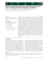
kinetic chemiluminescence (CL) method. Representa-
tive data showing inhibition of CL by one compound,
selenourea A, are shown in Fig. 1. This compound
incubated with PMNs significantly inhibited CL in a
dose-dependent manner. The other compounds gave
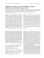
similar results to those for selenourea A. CL inhibi-
tion curves for the four organic selenium compounds
are shown in Fig. 2. The 50% inhibitory concentra-
tion (IC
50
) of each compound is shown in Table 1.
Selenourea A and selenourea B significantly inhibited
CL as compared with selenoamide A and seleno-
amide B (Fig. 2). The IC
50
values of selenourea A
and selenourea B were 6.8 ± 2.2 lm [95% confidence
interval (CI): 2.6–11.4] and 6.5 ± 2.5 lm (95% CI:
1.5–11.5), respectively. The IC
50
values of seleno-
amide A and selenoamide B were 11.3 ± 4.8 lm
(95% CI: 2.0–20.6) and 20.3 ± 6.4 lm (95% CI: 7.7–
32.9), respectively. O
2
–
decreased when selenoureas
and selenoamides were added at the peak of O
2
–
gen-
eration by PMA-stimulated PMNs (data not shown).
These results suggest that selenoureas and seleno-
amides effectively scavenged O
2
–
from PMA-stimu-
lated PMNs.
H. Tsukagoshi et al. O
2
–
-scavenging effects and toxicity of Se compounds
FEBS Journal 274 (2007) 6046–6054 ª 2007 The Authors Journal compilation ª 2007 FEBS 6047
Cytotoxicity of organic selenium compounds
in human cells
We examined the cytotoxic effects of selenoureas and
selenoamides using three cell lines, HaCaT, HEL, and
HEp-2, and PMNs, by the microtiter tetrazolium
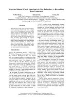
method [41–43]. The cytotoxicities of the compounds
in each cell line are shown in Fig. 3A–C. At 100 lm,a
relatively high concentration, both selenourea A and
selenourea B showed very low cytotoxic effects in
HaCaT, HEL and HEp-2 cells (Fig. 3A–C). In con-
trast, selenoamide B was relatively cytotoxic at 72 h
of coincubation: the viabilities of HaCaT, HEL and
HEp-2 cells were 9%, 25%, and 5%, respectively
(Fig. 3A–C). Similarly, another tertiary selenoamide,
selenoamide A, was relatively cytotoxic in HaCaT,
HEL and HEp-2 cells (Fig. 3A–C). At 100 lm, these
compounds did not significantly affect cell viabilities in
nonstimulated PMNs (Fig. 3D). These results suggest
that a relatively high concentration of selenoureas did
not affect cell viability in some human cells.
Measurement of H
2
O
2
production
As shown in Fig. 2, selenoureas and selenoamides were
effective scavengers of O
2
–
from PMA-stimulated
PMNs. We investigated whether or not selenoureas
and selenoamides produced H
2
O
2
from O
2
–
generated
by PMA-stimulated PMNs. After incubation with each
organic selenium compound or SOD for 5 min, the
PMNs were stimulated with PMA. As shown in Fig. 4,
1 lgÆmL
)1
SOD produced 82.0 ± 2.6 lm H
2
O
2
from
PMA-stimulated PMNs. At 100 lm, selenourea A
and selenourea B produced 8.3 ± 5.2 lm and
11.7 ± 1.7 lm H
2
O
2
, respectively, and selenoamide A
0
0 5 10 15 20
20
40
60
80
100
120
140
Incubation time (min)
Chemiluminescense intensity
(cpm x10
4
)
0 µM
1 µM
2 µM
5 µM
10
µM
MCLA
PMA
Fig. 1. Representative PMA-induced O
2
–
-scavenging effects of a
selenourea compound (selenourea A) determined using the CL
method. Detailed experimental procedures are described in Experi-
mental procedures. PMNs were preincubated with selenourea A
5 min before PMA treatment. Dose-dependent O
2
–
-scavenging
effects of the compound were found by eliminating MCLA-depen-
dent CL. Arrows indicate the time at which MCLA or PMA was
added. O
2
–
was recorded for 15 min with a luminescence reader.
0
0
110100
25
50
75
100
*
*
*
*
*
*
*
*
*
*
*
*
*
*
*
Inhibition (% of control)
Concentration (µM)
Selenourea A
Selenoamide A
Selenourea B
Selenoamide B
Fig. 2. Inhibition curves of four organic selenium compounds for
PMA-induced O
2
–
production from PMNs. m, selenourea A; j, sel-
enourea B; n, selenoamide A; h, selenoamide B. Detailed proce-
dures are described in the text. Results are expressed as
mean ± SEM from three independent experiments. *P<0.05.
Table 1. IC
50
values of organic selenium compounds: selenoureas
and tertiary selenoamides. PMNs were resuspended in Hank’s
balanced salt solution (HBSS) (pH 7.4) with 3 l
M MCLA and each
organic selenium compound at 37 °C for 5 min. After incubation,
PMNs were stimulated by 100 ngÆmL
)1
PMA. CL by O
2
–
was
recorded for 15 min with a luminescence reader at 37 °C. IC
50
values
are expressed relative to 0.1% dimethylsulfoxide as vehicle. Values
are presented as mean ± SEM from three independent experiments.
Entries Compounds IC
50
(lM)
Selenourea A
6.8 ± 2.2
Selenourea B
6.5 ± 2.5
Selenoamide A
11.3 ± 4.8
Selenoamide B
20.3 ± 6.4
O
2
–
-scavenging effects and toxicity of Se compounds H. Tsukagoshi et al.
6048 FEBS Journal 274 (2007) 6046–6054 ª 2007 The Authors Journal compilation ª 2007 FEBS
and selenoamide B produced 20.1 ± 2.5 and
41.6 ± 2.0 lm H
2
O
2
, respectively. These results sug-
gest that, as compared with the selenoamides, the sele-
noureas did not produce significant amounts of H
2
O
2
in PMA-stimulated PMNs.
Visualization of p47 phagocyte oxidase (p47
phox
)
with laser scanning microscopy (LSM)
The cytosolic component of NADPH oxidase complex,
p47
phox
, translocates from the cytosol to the cell mem-
brane upon activation. To examine the effects of sele-
noureas and selenoamides on O
2
–
production from
PMNs, we investigated the translocation of the mole-
cule to the cell membrane upon stimulation with
PMA. Figure 5 shows the red fluorescent phallodin
staining (F-actin) and the blue fluorescence of the
nuclei. Green fluorescence marks the p47
phox
translo-
cated from the cytosol to the cell membrane. After
incubation, about 70% of PMNs adhered to glass-
bottomed dishes in the presence or absence of organic
selenium compounds (data not shown). We observed
the translocation of p47
phox
only in adhered PMNs.
Even at the relatively high concentration of 100 lm,
p47
phox
translocation was not inhibited by organic
selenium compounds. These results suggest that sele-
noureas and selenoamides did not significantly affect
the translocation of p47
phox
to the cell membrane.
Discussion
We demonstrated the antioxidative effects and cytotox-
icity of four newly synthesized organic selenium
compounds: two selenoureas (selenourea A and sele-
nourea B) and two tertiary selenoamides (seleno-
amide A and selenoamide B). At relatively low
concentrations (IC
50
around 7 lm), both selenourea A
and selenourea B showed more potent O
2
–
-scavenging
effects without loss of NADPH oxidase activity in
PMA-stimulated PMNs than did selenoamide A and
selenoamide B. Furthermore, the two selenoureas
showed very low toxicity in PMNs and some cell lines,
even at high concentrations (100 lm). The results imply
that some types of selenourea, such as selenourea A
and selenourea B, may be new candidate antioxidants.
The generation of a low concentration of O
2
–
in the
human body generally plays a beneficial role in biolog-
ical defense and intercellular signal transduction [1].
Time (h)
Cell viability (% of control)
Selenourea A
Selenourea B
Selenoamide A
Selenoamide B
HaCaTcells
0
20
40
60
80
100
120
0244872
Time (h)
0
24 48
72
Time (h)
0
24 48
72
Time (h)
0244872
*
0
20
40
60
80
100
120
0
20
40
60
80
100
120
0
20
40
60
80
100
120
Cell viability (% of control)
*
*
*
*
*
*
*
*
*
*
*
*
*
*
*
Cell viability (% of control)Cell viability (% of control)
HEL cells
HEp-2 cells
PMNs
Selenourea A
Selenourea B
Selenoamide A
Selenoamide B
Selenourea A
Selenourea B
Selenoamide A
Selenoamide B
Selenourea A
Selenourea B
Selenoamide A
Selenoamide B
AB
CD
Fig. 3. Cytotoxicity of four organic selenium compounds in some human cell lines and PMNs. Cytotoxicity was determined by microtiter tetrazo-
lium assay. ‘Control’ contained cells plus cell culture medium supplemented with 2% fetal bovine serum. Results are expressed as mean ±
SEM from three independent experiments. (A) HaCaT cells were incubated with 100 l
M each organic selenium compound. Cell viability was
measured at intervals of 24 h. (B) Cell viability of HEL cells. (C) Cell viability of HEp-2 cells. (D) Cell viability of PMNs. PMNs maintained about
80% viability in control cultures after 48 h. Detailed procedures are described in the text. **P<0.01 versus control; *P<0.05 versus control.
H. Tsukagoshi et al. O
2
–
-scavenging effects and toxicity of Se compounds
FEBS Journal 274 (2007) 6046–6054 ª 2007 The Authors Journal compilation ª 2007 FEBS 6049
On the other hand, excessive O
2
–
production has a
detrimental role in the pathogenesis of a number of
disorders, including inflammation, rheumatoid arthri-
tis, and asthma [44,45]. Oxidative stress might be
defined as an imbalance between cellular production of
ROS and antioxidant defense mechanisms [1]. It is a
key component of inflammation and inflammatory dis-
orders. The processes associated with inflammatory
responses are complex and often involve ROS, includ-
ing O
2
–
. In this study, our results suggest that selenou-
reas acted as effective O
2
–
scavengers and showed very
low toxicity in human cells. Thus, the compounds may
eliminate excessive O
2
–
, leading to suppression of
inflammatory responses in sites overproducing ROS.
We also evaluated the toxicity of selenoureas and
selenoamides. The two selenoureas were simple struc-
tures and had low toxicity in PMNs and some cell
lines, even at high concentrations (100 lm). They do
not have a benzene ring or a chlorine bond, unlike
both selenoamides. Thus, selenoureas may not generate
toxic metabolites such as benzene compounds via met-
abolic enzymes (i.e. cytochrome P450).
We previously investigated the ROS-scavenging
effects of various organic selenium compounds, includ-
ing selenoureas and tertiary selenoamides, by using an
enzymatic O
2
–
generation system (hypoxanthine–xan-
thine oxidase system) [38–40]. Furthermore, we demon-
strated that these compounds do not significantly
inhibit xanthine oxidase activity [38–40]. In the present
study, we investigated the anti-inflammatory ⁄ antioxi-
dant potential of these compounds by examining their
scavenging effects on O
2
–
from PMA-stimulated
PMNs, with or without inhibition of NADPH oxidase
as an index of translocation of p47
phox
. The generation
of O
2
–
by PMNs is attributed to the activation of a
plasma membrane NADPH oxidase. The NADPH oxi-
dase multicomponent enzyme system catalyzes the pro-
duction of O
2
–
. A component of NADPH oxidase,
p47
phox
, translocates to the cell membrane and associ-
ates with cytochrome b
556
to form the active complex
that catalyzes the reduction of oxygen to O
2
–
at the
Selenourea A
100 µ
M
1% dimethylsulfoxideMedium alone
(no stimulated)
Selenoamide B
100 µ
M
Selenourea B
100 µ
M
Selenoamide A
100 µ
M
Control
A
B
C
D
E
Fig. 5. Translocation of p47
phox
in PMNs treated with four organic selenium compounds. Green fluorescence was induced by Alexa
Fluor 488 rabbit anti-(goat IgG) (p47
phox
). Blue fluorescence was induced by DAPI-stained nuclei. Red fluorescence was induced by Alexa
Fluor 532 phalloidin (F-actin). Control was a negative control of PMNs with no stimulation by PMA. PMNs were stimulated by PMA with 1%
dimethylsulfoxide as vehicle (A). The PMNs were stimulated by PMA with 100 l
M organic selenium compounds (B–E). Detailed procedures
are described in the text. Each upper figure of control and (A)–(E) is shown in green fluorescence alone.
0
25
50
75
100
dimethylsulfoxide
S
e
l
en
o
u
r
ea
A
S
e
l
e
n
o
u
re
a
B
S
e
l
e
n
o
a
m
i
d
e
A
S
e
l
e
n
o
a
m
i
d
e
B
S
O
D
% of SOD
* *
* *
* *
* *
Fig. 4. H
2
O
2
generation by four organic selenium compounds and
SOD following PMA-induced generation of O
2
–
from PMNs. PMNs
were preincubated with 100 l
M each organic selenium compound,
SOD or dimethylsulfoxide, 5 min before PMA treatment. H
2
O
2
was
measured after 15 min, using the scopoletin fluorescent method.
Detailed procedures are described in the text. Results are
expressed as mean ± SEM from three independent experiments.
**P < 0.01 versus SOD.
O
2
–
-scavenging effects and toxicity of Se compounds H. Tsukagoshi et al.
6050 FEBS Journal 274 (2007) 6046–6054 ª 2007 The Authors Journal compilation ª 2007 FEBS
expense of NADPH [46–48]. We examined the trans-
location of p47
phox
to the plasma membrane in PMA-
stimulated PMNs. In adherent PMNs, none of the
organic selenium compounds significantly affected the
translocation of p47
phox
to the plasma membrane.
Thus, we suggest that the O
2
–
-scavenging activity of
selenoureas and selenoamides was not due to the inhi-
bition of NADPH oxidase in PMNs.
We assessed the metabolism of O
2
–
by selenoureas
and selenoamides as an index of H
2
O
2
production.
Both selenourea A and selenourea B produced small
amounts of H
2
O
2
as compared with SOD. In this
study, however, we did not use the ESR method to
examine production of other ROS, such as the hydro-
xyl radical. Although it is not very likely that hydroxyl
radical can be directly produced via O
2
–
using seleno-
ureas and selenoamides, detailed additional studies
regarding the metabolism of ROS, using a physico-
chemical method such as ESR, may be required.
Some other organic selenium compounds such as eb-
selen have been developed [24–28]. At low concentra-
tions, ebselen inhibits a number of enzymes involved
in inflammation, such as lipoxygenases, nitric oxide
synthases, NADPH oxidase, protein kinase C, and
H
+
⁄ K
+
-ATPase [24]. Ebselen inhibits O
2
–
generation
from leukocytes through the inhibition of leukocytic
NADPH oxidase [24]. Moreover, it is possible that it
can decompose H
2
O
2
and produce H
2
O and O
2
. Ebse-
len is thought to act by mimicking an active center of
GPX in the cell [24]. By contrast, selenoureas and sele-
noamides had an O
2
–
-scavenging effect, and no signifi-
cant inhibition of NADPH oxidase activity was found.
Selenoureas and selenoamides, especially selenoureas,
seemed to scavenge O
2
–
directly to produce O
2
. Thus,
it is possible that reaction mechanisms affecting ROS
differ between selenoureas and selenoamides and ebse-
len. These differences may be attributed to structural
properties, although the precise mechanisms are not
fully understood at present.
In previous studies, using a hypoxanthine–xanthine
oxidase system as the O
2
–
generator, we measured IC
50
values of only selenourea B (mean of IC
50
values:
125 nm) and selenoamide A (mean of IC
50
values:
182 nm) [39,40]. In the present study, we assayed the
IC
50
values of the four compounds (selenourea A and
selenourea B, and selenoamide A and selenoamide B)
using about 30-fold amounts, as compared with previ-
ous studies, of O
2
–
-derived PMA-stimulated PMNs
[39,40]. As a result, the IC
50
means of selenourea B
and selenoamide A were 6.5 lm and 11.3 lm, respec-
tively. Thus, the differences in IC
50
values may be due
to differences in the experimental conditions between
the previous and present studies.
In conclusion, the results from this study may pro-
vide biological evidence that innovative organic sele-
nium compounds scavenge O
2
–
released from PMNs.
Furthermore, these compounds were not toxic in some
human cells and PMNs, indicating that they have the
potential to prevent inflammation caused by O
2
–
. The
next step should be to confirm the antioxidant effects
of these compounds, especially selenoureas, in animal
models such as mice.
Experimental procedures
Organic selenium compounds
In this study, we newly synthesized and used four types of
organic selenium compound: two types of selenoureas, sele-
nourea A selenourea B, and two types of tertiary seleno-
amides, selenoamide A and selenoamide B. The chemical
structures are shown in Table 1. Detailed synthetic proce-
dures for these compounds were as previously described, and
the chemical structures were confirmed by NMR analysis
[36,37]. The purity of these compounds was more than 97%
[36,37]. All compounds were dissolved in dimethylsulfoxide
at 10 mm and stored at ) 80 °C until required. When used
for the experiments, the compounds were diluted with HBSS.
Reagents
A Cypridina luciferin analog, 2-methyl-6-( p -methoxyphenyl)-
3,7-dihydroimidazo-[1,2-a]-pyrazin-3-one (MCLA), was pur-
chased from Tokyo Kasei Chemical Co. (Tokyo, Japan).
The compound was dissolved in double distilled water, and
stored at ) 80 °C until use. The concentration of MCLA
solution was determined by absorbance at 430 nm using an
absorbance coefficient value a
˚
of 9600 m
)1
Æcm
)1
, as previ-
ously described [49]. PMA and dimethylsulfoxide were pur-
chased from Sigma Aldrich Chemical Co. (St Louis, MO,
USA). The stock solution of PMA (5 mgÆmL
)1
) was
dissolved in dimethylsulfoxide, and stored at ) 80 °C until
use. 2-(2-Methoxy-4-nitrophenyl)-3-(4-nitrophenyl)-5-(2,4-
disulfophenyl)-2H-tetrazolium, monosodium salt (WST-8)
reagent was purchased from Dojindo Chemicals (Kumamot-
o, Japan) (Cell Counting Kit8). Alexa Fluor 532 phalloidin,
Alexa Fluor 488 rabbit anti-(goat IgG) (H + L) and 4,6-di-
amidino-phenylindole (DAPI) were obtained from Molecu-
lar Probes (Eugene, OR, USA). Antibody to p47
phox
was
purchased from Santa Cruz Biotechnology (Santa Cruz,
CA., USA). All other chemicals were of analytical grade and
used without further purification.
Isolation of PMNs
PMNs were isolated from heparinized peripheral blood of
healthy volunteers as previously described [50]. PMNs were
H. Tsukagoshi et al. O
2
–
-scavenging effects and toxicity of Se compounds
FEBS Journal 274 (2007) 6046–6054 ª 2007 The Authors Journal compilation ª 2007 FEBS 6051
isolated by using a gradient material, one-step Polymorpho-
prep (Accurate Chemical & Scientific Corp., Westbury,
NY, USA) following the procedure recommended by the
manufacturer. The purity of PMNs was > 98% as deter-
mined by Randolph’s stain.
Assay of effects on scavenging of O
2
–
from PMNs
Organic selenium compound solutions such as selenoureas
and tertiary selenoamides were diluted with HBSS. To ana-
lyze the real-time effects on scavenging of O
2
–
of these com-
pounds, we used a highly sensitive and specific CL method
with MCLA as a probe [51–53]. Briefly, 7 · 10
4
PMNs
were resuspended in HBSS (pH 7.4) with 3 lm MCLA and
0–100 lm (maximum dose used in this study) each organic
selenium compound at 37 °C for 5 min. After incubation,
PMNs were stimulated with 100 ngÆmL
)1
PMA [40]. CL by
O
2
–
was recorded for 15 min with a luminescence reader
(BLR-102; Aloka Co., Tokyo, Japan) with gentle agitation
at 37 °C [50]. The total volume of the assays was 2 mL
[50]. IC
50
values were determined using linear regression
analysis of the dose–response curves.
Cell culture and cytotoxicity assay
HaCaT cells, HEL cells and HEp-2 cells were cultured in
DMEM supplemented with 10% fetal bovine serum, l-gluta-
mine (0.6 mgÆmL
)1
) and 0.35% NaHCO
3
at 37 °Cinan
atmosphere of 95% air and 5% CO
2
. HaCaT cells were
kindly donated by N. E. Fusenig (German Cancer Research
Center, Heidelberg, Germany). We assayed the cytotoxicity
of the organic selenium compounds in HaCaT cells, HEL
cells, HEp-2 cells and PMNs using the microtiter tetrazolium
method [41]. Briefly, confluent cells were incubated in a
96-well microplate (Corning Costar, Cambridge, MA, USA)
with 100 l m each organic selenium compound in 100 lLof
DMEM containing 2% fetal bovine serum at 37 °Cinan
atmosphere of 95% air and 5% CO
2
. PMNs (2 · 10
4
) were
added to a 96-well microplate and were incubated with
100 lm each compound in 100 lL of RPMI-1640 medium
containing 2% fetal bovine serum at 37 °C, in an atmosphere
of 95% air and 5% CO
2
. After 24 h of incubation, we added
10 lL of WST-8 solution. Absorbance was measured at
450 nm using a microplate reader (Spectramax; Molecular
Devices, Sunnyvale, CA, USA) [42,43]. Cytotoxicity was cal-
culated as previously described [42,43].
Measurement of H
2
O
2
production
The production of H
2
O
2
was determined by the horseradish
peroxidase-catalyzed oxidation of fluorescent scopoletin
method with minor modifications [54–56]. Briefly,
2 · 10
5
PMNs were added to HBSS (pH 7.4) containing
1mm NaN
3
, and then incubated at 37 °C in an atmosphere
of 95% air and 5% CO
2
in the presence of each organic
selenium compound or SOD. After 5 min, PMNs were
stimulated with PMA at a final concentration of 1 lgÆ mL
)1
for 15 min. Samples were added to the reaction mixture
containing HBSS (pH 7.4), 10 lm scopoletin and
50 lgÆmL
)1
horseradish peroxidase in a 96-well plate. The
decrease in scopoletin fluorescence was measured using a
microplate fluorometer (1420 Multilabel counter, ARVO;
excitation and emission at 335 and 460 nm, respectively;
Wallac, Turuk, Finland). The concentration of the oxidant
in the samples was calculated using the standard curve,
which was made by adding known concentrations of the
authentic H
2
O
2
instead of the samples.
Visualization of p47
phox
by LSM
A suspension of 1 · 10
5
PMNs in phenol red ⁄ free RPMI-
1640 medium with 1 mm Hepes was incubated with or
without organic selenium compound for 5 min at 37 °Cin
35 mm glass-bottomed dishes (Matsunami, Osaka, Japan).
After incubation, PMA was added to a final concentration
of 100 ngÆmL
)1
. After 15 min at 37 °C, the mixture in each
dish was gently aspirated, and 2 mL of 3.7% formaldehyde
in NaCl ⁄ P
i
(v ⁄ v) was added carefully to each dish for
10 min at room temperature (25 °C) to fix the attached
cells. The cells were then washed gently with NaCl ⁄ P
i
three
times and permeabilized with 2 mL of 0.1% Triton X-100
in NaCl ⁄ P
i
in each dish for 10 min. After permeabilization,
cells were washed three times with NaCl ⁄ P
i
, and incubated
for 30 min in darkness at 4 °C with a primary antibody
(antibody to p47
phox
or goat IgG as a negative control; all
dilutions 1 : 500). The cells were then washed thoroughly
and incubated for 30 min in darkness at room temperature
with a mixture of Alexa Fluor 532 phalloidin at 1 unit per
dish and secondary antibody [Alexa Fluor 488 rabbit anti-
(goat IgG); dilution 1 : 1000]. Finally, after three washes
with NaCl ⁄ P
i
, DAPI was mounted on the samples and the
fluorescent signal was observed with a confocal LSM sys-
tem (MRC-1024; Bio-Rad, Hercules, CA, USA) as previ-
ously described, with minor modifications [57].
Statistical analysis
Raw values or normalized values from the indicated num-
ber of independent trials were averaged and expressed as
the mean ± SEM. Any significant differences between the
groups were determined using analysis of variance (anova)
followed by Dunnett’s test. Data with a P-value of less
than 0.05 were considered to be significant.
Acknowledgements
We thank Mr Takafumi Yamaguchi, Mr Taisei Ishioka,
Mr Masakazu Yoshizumi and Mrs Hitoe Takahashi
O
2
–
-scavenging effects and toxicity of Se compounds H. Tsukagoshi et al.
6052 FEBS Journal 274 (2007) 6046–6054 ª 2007 The Authors Journal compilation ª 2007 FEBS
for their help with the experiments. This work was
supported by Gunma prefecture (Maebashi, Japan).
References
1 Kimura H, Sawada T, Oshima S, Kozawa K, Ishioka T
& Kato M (2005) Toxicity and roles of reactive oxygen
species. Curr Drug Targets Inflamm Allergy 4, 489–495.
2 Valko M, Leibfritz D, Moncol J, Cronin MT, Mazur M
& Telser J (2007) Free radicals and antioxidants in nor-
mal physiological functions and human disease. Int J
Biochem Cell Biol 39, 44–84.
3 Flora SJ (2007) Role of free radicals and antioxidants
in health and disease. Cell Mol Biol 53, 1–2.
4 Valko M, Izakovic M, Mazur M, Rhodes CJ & Telser J
(2004) Role of oxygen radicals in DNA damage and
cancer incidence. Mol Cell Biochem 266, 37–56.
5 Knight JA (2000) The biochemistry of aging. Adv Clin
Chem 35, 1–62.
6 Bejma J & Ji LL (1999) Aging and acute exercise
enhance free radical generation in rat skeletal muscle.
J Appl Physiol 87, 465–470.
7 Bejma J, Ramires P & Ji LL (2000) Free radical genera-
tion and oxidative stress with ageing and exercise: dif-
ferential effects in the myocardium and liver. Acta
Physiol Scand 169, 343–351.
8 Møller P & Loft S (2006) Dietary antioxidants and ben-
eficial effect on oxidatively damaged DNA. Free Radic
Biol Med 41, 388–415.
9 Fridovich I (1983) Superoxide radical: an endogenous
toxicant. Annu Rev Pharmacol Toxicol 23, 239–257.
10 Balaban RS, Nemoto S & Finkel T (2005) Mitochon-
dria, oxidants, and aging. Cell 120, 483–495.
11 Fridovich I (1998) Oxygen toxicity: a radical explana-
tion. J Exp Biol 201, 1203–1209.
12 Meydani M, Evans W, Handelman G, Fielding RA,
Meydani SN, Fiatarone MA, Blumberg JB & Cannon
JG (1992) Antioxidant response to exercise-induced oxi-
dative stress and protection by vitamin E. Ann NY Acad
Sci 669, 363–364.
13 Curnutte JT, Whitten DM & Babior BM (1974) Defec-
tive superoxide production by granulocytes from
patients with chronic granulomatous disease. N Engl J
Med 290, 593–597.
14 Berridge MJ (1984) Inositol trisphosphate and diacyl-
glycerol as second messengers. Biochem J 220, 345–
360.
15 Ryter SW, Kim HP, Hoetzel A, Park JW, Nakahira K,
Wang X & Choi AM (2007) Mechanisms of cell death
in oxidative stress. Antioxid Redox Signal 9, 49–89.
16 Cerutti P, Ghosh R, Oya Y & Amstad P (1994) The
role of the cellular antioxidant defense in oxidant carci-
nogenesis. Environ Health Perspect 102, 123–129.
17 Wang X & Quinn PJ (1999) Vitamin E and its function
in membranes. Prog Lipid Res 38, 309–336.
18 Kojo S (2004) Vitamin C: basic metabolism and its
function as an index of oxidative stress. Curr Med Chem
11, 1041–1064.
19 Chu FF, Esworthy RS & Doroshow JH (2004) Role of
Se-dependent glutathione peroxidases in gastrointestinal
inflammation and cancer. Free Radic Biol Med 36
,
1481–1495.
20 Awasthi YC, Beutler E & Srivastava SK (1975) Purifica-
tion and properties of human erythrocyte glutathione
peroxidase. J Biol Chem 250, 5144–5149.
21 Arthur JR, Morrice PC, Nicol F, Beddows SE, Boyd R,
Hayes JD & Beckett GJ (1987) The effects of selenium
and copper deficiencies on glutathione S-transferase and
glutathione peroxidase in rat liver. Biochem J 248, 539–
544.
22 Brigelius-Flohe
´
R (2006) Glutathione peroxidases and
redox-regulated transcription factors. Biol Chem 387,
1329–1335.
23 Arthur JR (2000) The glutathione peroxidases. Cell Mol
Life Sci 57, 1825–1835.
24 Sies H (1993) Ebselen, a selenoorganic compound as
glutathione peroxidase mimic. Free Radic Biol Med 14,
313–323.
25 Muller A, Cadenas E, Graf P & Sies H (1984) A novel
biologically active seleno-organic compound ) I. Gluta-
thione peroxidase-like activity in vitro and antioxidant
capacity of PZ 51 (ebselen). Biochem Pharmacol 33,
3235–3239.
26 Ren X, Yang L, Liu J, Su D, You D, Liu C, Mu Y &
Shen J (2001) A novel glutathione peroxidase mimic
with antioxidant activity. Arch Biochem Biophys 387,
250–256.
27 Nakamura Y, Feng Q, Kumagai T, Torikai K, Ohigashi
H, Osawa T, Noguchi N, Niki E & Uchida K (2002)
Ebselen, a glutathione peroxidase mimetic seleno-
organic compound, as a multifunctional antioxidant
implication for inflammation-associated carcinogenesis.
J Biol Chem 277, 2687–2694.
28 Cotgreave IA, Duddy SK, Kass GE, Thompson D &
Moldeus P (1989) Studies on the anti-inflammatory
activity of ebselen. Ebselen interferes with granulocyte
oxidative burst by dual inhibition of NADPH oxidase
and protein kinase C? Biochem Pharmacol 38, 649–656.
29 Nogueira CW, Zeni G & Rocha JB (2004) Organosele-
nium and organotellurium compounds: toxicology and
pharmacology. Chem Rev 104, 6255–6286.
30 Meotti FC, Stangherlin EC, Zeni G, Nogueira CW & Ro-
cha JB (2004) Protective role of aryl and alkyl diselenides
on lipid peroxidation. Environ Res 94, 276–282.
31 Nogueira CW, Quinhones EB, Jung EA, Zeni G &
Rocha JB (2003) Anti-inflammatory and antinocicep-
tive activity of diphenyl diselenide. Inflamm Res 52,
56–63.
32 Porciu´ ncula LO, Rocha JB, Cimarosti H, Vinade
´
L,
Ghisleni G, Salbego CG & Souza DO (2003) Neuro-
H. Tsukagoshi et al. O
2
–
-scavenging effects and toxicity of Se compounds
FEBS Journal 274 (2007) 6046–6054 ª 2007 The Authors Journal compilation ª 2007 FEBS 6053
protective effect of ebselen on rat hippocampal slices
submitted to oxygen–glucose deprivation: correlation
with immunocontent of inducible nitric oxide synthase.
Neurosci Lett 346, 101–104.
33 Chander PN, Gealekman O, Brodsky SV, Elitok S,
Tojo A, Crabtree M, Gross SS & Goligorsky MS (2004)
Nephropathy in zucker diabetic fat rat is associated
with oxidative and nitrosative stress: prevention by
chronic therapy with a peroxynitrite scavenger ebselen.
J Am Soc Nephrol 15, 2391–2403.
34 Savegnago L, Jesse CR, Moro AV, Borges VC, Santos
FW, Rocha JB & Nogueira CW (2006) Bis selenide
alkene derivatives: a class of potential antioxidant and
antinociceptive agents. Pharmacol Biochem Behav 83,
221–229.
35 Savegnago L, Pinto LG, Jesse CR, Alves D, Rocha JB,
Nogueira CW & Zeni G (2007) Antinociceptive proper-
ties of diphenyl diselenide: evidences for the mechanism
of action. Eur J Pharmacol 555, 129–138.
36 Koketsu M, Fukuta Y & Ishihara H (2001) Preparation
of N,N-unsubstituted selenoureas and thioureas from
cyanamide. Tetrahedron Lett 42, 333–335.
37 Koketsu M, Okayama Y, Aoki H & Ishihara H (2002)
Facile synthesis of N,N-dialkylselenoamides from
amides. Heteroatom Chem 13, 195–198.
38 Takahashi H, Nishina A, Fukumoto RH, Kimura H,
Koketsu M & Ishihara H (2005) Selenocarbamates are
effective superoxide anion scavengers in vitro. Eur J
Pharm Sci 24, 291–295.
39 Takahashi H, Nishina A, Kimura H, Motoki K, Koke-
tsu M & Ishihara H (2004) Tertiary selenoamide com-
pounds are useful superoxide radical scavengers in vitro.
Eur J Pharm Sci 23, 207–211.
40 Takahashi H, Nishina A, Fukumoto RH, Kimura H,
Koketsu M & Ishihara H (2005) Selenoureas and
thioureas are effective superoxide radical scavengers
in vitro. Life Sci 76, 2185–2192.
41 Denizot F & Lang R (1986) Rapid colorimetric assay
for cell growth and survival modifications to the tetra-
zolium dye procedure giving improved sensitivity and
reliability. J Immunol Methods 89, 271–277.
42 Ishiyama M, Miyazono Y, Sasamoto K, Ohkura Y &
Ueno K (1997) A highly water-soluble disulfonated tet-
razolium salt as a chromogenic indicator for NADH as
well as cell viability. Talanta 44, 1299–1305.
43 Ishiyama M, Tominaga H, Shiga M, Sasamoto K,
Ohkura Y & Ueno K (1996) A combined assay of cell
viability and in vitro cytotoxicity with a highly water-
soluble tetrazolium salt, neutral red and crystal violet.
Biol Pharm Bull 19, 1518–1520.
44 Vignola AM, Chanez P, Campbell AM, Souques F, Le-
bel B, Enander I & Bousquet J (1998) Airway inflamma-
tion in mild intermittent and in persistent asthma. Am J
Respir Crit Care Med 157, 403–409.
45 Kato M, Hayashi Y & Kimura H (2005) Oxygen
radicals in inflammation and allergy related to viral
infections. Curr Drug Targets Inflamm Allergy 4, 497–
501.
46 DeLeo FR, Allen LA, Apicella M & Nauseef WM
(1999) NADPH oxidase activation and assembly during
phagocytosis. J Immunol 163, 6732–6740.
47 Park HS, Kim IS & Park JW (1999) Phosphorylation
induces conformational changes in the leukocyte
NADPH oxidase subunit p47
phox
. Biochem Biophys Res
Commun 259, 38–42.
48 Vignais PV (2002) The superoxide-generating NADPH
oxidase: structural aspects and activation mechanism.
Cell Mol Life Sci 59, 1428–1459.
49 Allen RC, Yevich SJ, Orth RW & Steele RH (1974)
The superoxide anion and singlet molecular oxygen:
their role in the microbicidal activity of the polymor-
phonuclear leukocyte. Biochem Biophys Res Commun
60, 909–917.
50 Kato M, Kimura H, Motegi Y, Tachibana A, Minaka-
mi H, Morikawa A & Kita H (2002) Platelet-activating
factor activates two distinct effector pathways in human
eosinophils. J Immunol 169, 5252–5259.
51 Kimura H & Nakano M (1988) Highly sensitive and
reliable chemiluminescence method for the assay of
superoxide dismutase in human erythrocytes. FEBS Lett
239, 347–350.
52 Nakano M, Kimura H, Hara M, Kuroiwa M, Kato M,
Totsune K & Yoshikawa T (1990) A highly sensitive
method for determining both Mn- and Cu-Zn superox-
ide dismutase activities in tissues and blood cells. Anal
Biochem 187, 277–280.
53 Nishida A, Kimura H, Nakano M & Goto T (1989) A
sensitive and specific chemiluminescence method for
estimating the ability of human granulocytes and
monocytes to generate O
À
2
. Clin Chim Acta 179, 177–
181.
54 Root RK, Metcalf J, Oshino N & Chance B (1975)
H
2
O
2
release from human granulocytes during phagocy-
tosis. I. Documentation, quantitation, and some regulat-
ing factors. J Clin Invest 55, 945–955.
55 Root RK & Metcalf JA (1977) H
2
O
2
release from
human granulocytes during phagocytosis. Relationship
to superoxide anion formation and cellular catabolism
of H
2
O
2
: studies with normal and cytochalasin B-treated
cells. J Clin Invest 60, 1266–1279.
56 Tsan MF, Douglass KH & McIntyre PA (1977) Hydro-
gen peroxide production and killing of Staphylococcus
aureus by human polymorphonuclear leukocytes. Blood
49, 437–444.
57 Suzuki M, Kato M, Hanaka H, Izumi T & Morikawa
A (2003) Actin assembly is a crucial factor for
superoxide anion generation from adherent human
eosinophils. J Allergy Clin Immunol 112, 126–133.
O
2
–
-scavenging effects and toxicity of Se compounds H. Tsukagoshi et al.
6054 FEBS Journal 274 (2007) 6046–6054 ª 2007 The Authors Journal compilation ª 2007 FEBS