Báo cáo khoa học: A novel factor XI missense mutation (Val371Ile) in the activation loop is responsible for a case of mild type II factor XI deficiency doc
Bạn đang xem bản rút gọn của tài liệu. Xem và tải ngay bản đầy đủ của tài liệu tại đây (396.61 KB, 11 trang )
A novel factor XI missense mutation (Val371Ile) in the
activation loop is responsible for a case of mild type II
factor XI deficiency
Cristina Bozzao
1
*, Valeria Rimoldi
1
*, Rosanna Asselta
1
, Meytal Landau
2
, Rossella Ghiotto
3
,
Maria L. Tenchini
1
, Raimondo De Cristofaro
4
, Giancarlo Castaman
3
and Stefano Duga
1
1 Department of Biology and Genetics for Medical Sciences, University of Milan, Italy
2 Department of Biochemistry, George S. Wise Faculty of Life Science, Tel Aviv University, Israel
3 Department of Hematology and Hemophilia and Thrombosis Center, San Bortolo Hospital, Vicenza, Italy
4 Haemostasis Research Centre, Catholic University School of Medicine, Rome, Italy
Coagulation factor XI (FXI) is the precursor of a tryp-
sin-like serine protease that catalyzes, upon activation,
the conversion of factor IX (FIX) to activated FIX
(FIXa) [1,2]. Human FXI, primarily produced by
hepatocytes, is a glycoprotein of 160 kDa circulating
in plasma in a noncovalent complex with high mole-
cular mass kininogen [3]. Structurally, FXI zymogen
comprises four N-terminal tandem repeats of about
90 residues, named apple domains (Ap1–4), followed
by a catalytic serine protease domain located at the
C-terminal end. Uniquely among coagulation serine
proteases, FXI is secreted as a homodimer composed
of two identical polypeptide chains linked by non-
covalent interactions and by a Cys321–Cys321 disulfide
bond between the Ap4 domains [4,5].
Among serine proteases that can activate FXI, i.e.
activated factor XII (FXIIa), FXIa, and thrombin, the
main physiologic activator is actually thrombin formed
on the surface of activated platelets [6–8]. Cleavage at
the Arg369–Ile370 bond in each monomer produces
both an N-terminal heavy chain, which binds FIX and
high molecular mass kininogen [9], and a C-terminal
Keywords
coagulation factor XI deficiency; functional
characterization; missense mutation;
mutational screening; type II defect
Correspondence
S. Duga, Department of Biology and
Genetics for Medical Sciences, University of
Milan, Via Viotti, 3 ⁄ 5-20133 Milan, Italy
Fax: +39 02 5031 5864
Tel: +39 02 5031 5823
E-mail:
*These authors contributed equally to this
study
(Received 21 May 2007, revised 11 Septem-
ber 2007, accepted 8 October 2007)
doi:10.1111/j.1742-4658.2007.06134.x
Coagulation factor XI (FXI) is the zymogen of a serine protease that,
when converted to its active form, contributes to blood coagulation
through proteolytic activation of factor IX. FXI deficiency is typically an
autosomal recessive disorder, characterized by bleeding symptoms mainly
associated with injury or surgery. Of the more than 100 FXI gene muta-
tions reported in FXI-deficient patients, most are associated with a propor-
tional decrease in FXI functional and immunologic levels (type I defects),
whereas only a few mutations leading to the presence of dysfunctional
molecules in plasma have been molecularly analyzed to date (type II defi-
ciencies). We report the functional and molecular characterization of a
missense mutation (Val371Ile) identified, in the heterozygous state, in a
25-year-old Italian male with mild FXI deficiency. Laboratory analysis
revealed reduced functional FXI levels (34%), but normal antigen levels
(102%), distinctive of a type II defect. Given the proximity of Val371 to
the FXI activation site, a possible interference with zymogen activation
was postulated. Expression experiments of the FXI–Val371Ile recombinant
protein, followed by activation assays, showed both a different time course
in FXI activation and a slight delay in factor IX activation by thrombin-
activated FXI.
Abbreviations
FIX, coagulation factor IX; FIXa, activated factor IX; FXI, coagulation factor XI; FXIa, activated factor XI; FXI:Ag, antigen FXI level;
FXI:C, functional FXI level; FXIIa, activated factor XII.
6128 FEBS Journal 274 (2007) 6128–6138 ª 2007 The Authors Journal compilation ª 2007 FEBS
light chain, containing the catalytic domain [10]. These
two chains are held together by three disulfide bonds
and both are essential for FIX activation [5]. FXI acti-
vation generates a new N-terminus of the catalytic
serine protease domain, which is called the activation
loop (residues 370–376). Following cleavage, the acti-
vation loop undergoes large movement towards the
activation pocket in the protease domain, stabilizing
the FXIa active site [11].
FXI deficiency is an autosomal recessive bleeding
disorder that is rare in most populations (prevalence
1:10
6
) but is particularly common among Ashkenazi
Jews, in whom a heterozygote frequency of 8% has
been reported [12]. This rare coagulopathy is charac-
terized by decreased FXI functional activity, usually
associated with low levels of FXI antigen (type I
defects). By contrast, type II defects, characterized by
the presence of dysfunctional molecules in plasma, are
rare [13,14]. Usually, homozygous and compound het-
erozygous patients have severe FXI deficiency (FXI
activity < 20%), whereas heterozygotes have mild ⁄ par-
tial FXI deficiency (20–50%) [15]. Bleeding tendency
in FXI-deficient patients seems to poorly correlate with
plasma FXI levels and hemorrhagic episodes are usu-
ally associated with injury or surgery, but may be so
severe as to demand replacement therapy [16].
The genetic basis of this rare coagulation disorder is
invariantly represented by mutations within the FXI
gene ( F11 ), which is located on chromosome 4q35.2
and consists of 15 exons spread over a genomic region
of $ 23 kb. To date, > 100 mutations responsible for
FXI deficiency have been described [13,17]. Among
Ashkenazi Jewish patients, two prevalent mutations
(Glu117stop and Phe283Leu, also called type II and
type III mutations) account for 95% of cases of FXI
deficiency [12]. However, in patients belonging to other
ethnic groups a significantly higher level of allelic het-
erogeneity has been reported. Remarkable exceptions
are represented by French Basques, French patients
from Nantes, and English patients, in whom different
prevalent ancestral mutations were found [18–20]. An
unusual dominant transmission of FXI deficiency has
been described in some families, in which four different
missense mutations exert a dominant-negative effect on
wild-type FXI secretion through intracellular hetero-
dimer formation [21,22].
The aim of this study was the molecular character-
ization of the F11 germline missense mutation
Val371Ile identified in the heterozygous state in an
Italian patient affected by mild FXI deficiency, who
had normal immunologic FXI levels associated with a
reduced activity of the factor, distinctive of a type II
defect.
Results
Patient data
The propositus was a male born in 1981, who was
referred in 2001 for the evaluation of a prolonged par-
tial thromboplastin time discovered prior to a surgical
procedure. An appendectomy, adenoidectomy, and
right-knee arthroscopy carried out previously had been
without mishap. Laboratory analysis revealed a
reduced functional FXI level (FXI:C ¼ 34%), although
the antigen FXI level was normal (FXI:Ag ¼ 102%),
suggestive of a mild type II FXI deficiency. His
mother, born in 1949, also had reduced FXI:C (43%)
associated with normal FXI:Ag (132%), whereas the
father, born in 1947, had both functional and antigen
FXI levels within the range of normality (96 and
134%, respectively). Both parents were asymptomatic.
Mutational screening
The entire coding region, including exon–intron
boundaries and $ 300 bp of the promoter region of
F11, was sequenced. Sequence analysis identified a het-
erozygous G fi A transition in exon 11 correspond-
ing to cDNA position 1165 (numbering according to
GenBank accession number NM_000128, starting from
the first nucleotide of the ATG start codon), which
causes a Val371Ile substitution (numbering omits the
signal peptide). The same mutation was found in the
heterozygous state in the proband’s mother. Amino
acid substitution involves the second residue of the
FXI light chain after the proteolytic cleavage that
leads to FXI activation. Residue Val371 is located one
residue following the cleavage site of FXI (P2¢ posi-
tion, according to the convention of numbering posi-
tion around the scissile bond), thus is part of the
activation loop.
One hundred haploid genomes from unrelated Ital-
ian control individuals were also analyzed by direct
sequencing and the Val371Ile mutation was absent in
all of them (data not shown).
Expression of wild-type and Val371Ile
recombinant FXI in COS-1 cells
To evaluate the pathogenic role of the Val371Ile muta-
tion, both the wild-type and mutant protein were
expressed in COS-1 cells. To this end, mutagenesis
was performed on the pCDNA3 ⁄ FXI plasmid to
produce the pCDNA3 ⁄ FXI–Val371Ile vector as des-
cribed in Experimental procedures. Following tran-
sient transfection with either pCDNA3 ⁄ FXI or
C. Bozzao et al. FXI–Val317Ile – a novel factor XI type II defect
FEBS Journal 274 (2007) 6128–6138 ª 2007 The Authors Journal compilation ª 2007 FEBS 6129
pCDNA3 ⁄ FXI–Val371Ile or with equimolar amounts
of both expression plasmids (to mimic the heterozy-
gous condition), serum-free conditioned media and cell
extracts were analyzed for the presence of FXI antigen
using ELISA. FXI antigen levels, measured in both
conditioned media and lysates of cells expressing the
mutant protein (in either the heterozygous or homozy-
gous state), were not significantly different from those
measured in wild-type samples (Fig. 1A). In particular,
in media conditioned by cells expressing either wild-
type or mutant FXI, antigen levels ranged from 300 to
500 ngÆmL
)1
, whereas, levels of immunoreactive FXI
were between 20 and 40 ngÆmL
)1
in the corresponding
lysates.
FXI specific activity was measured in conditioned
media as the ratio between FXI:C and FXI:Ag levels.
The specific activity of the FXI–Val371Ile protein was
significantly reduced when compared with the wild-
type one ($ 30 and 80% for the heterozygous and the
homozygous condition, respectively; Fig. 1B).
Our results are consistent with the FXI:C and
FXI:Ag measured in the patient’s plasma, further sup-
porting the hypothesis that the Val371Ile mutation is a
type II defect leading to the production of a defective
FXI molecule.
FXI activation
Three biologically relevant proteases can activate FXI
(FXIIa, FXIa, and thrombin) [8,23], all of which
cleave FXI at the Arg369–Ile370 bond and expose the
active-site catalytic triad. Given the proximity of
Val371 to the FXI activation site, a possible interfer-
ence on FXI activation was hypothesized. To this end,
equal amounts of wild-type and mutant recombinant
FXI molecules from conditioned media were directly
activated by either thrombin or FXIIa.
As shown in Fig. 2, time-course experiments of FXI
activation by either thrombin or FXIIa were per-
formed. Both proteases were able to cut wild-type and
FXI–Val371Ile precursors into two fragments corre-
sponding to the heavy (48 kDa) and light (32 kDa)
chains. The Val371Ile substitution causes a delay in
FXI activation time following both types of activation
protocols, as is clearly appreciable in Fig. 2A,B.
Indeed, after 16 h digestion with FXIIa at 37 °C,
0
0
1
02
0
3
04
05
06
07
08
0
9
001
0
1
1
0
21
031
sn
sn
dn
dn
sn
s
n
CAIDEMDENOITIDNOCSETASYLLLE
% of wild-type
YTIVITCA
CIF
ICEPS
LE
VE
L
N
EG
I
T
N
A
AB
el
I173la
Vs
uog
yz
o
r
etehepyt-dli
w k
c
o
m
0
01
0
2
03
04
05
06
0
7
08
09
001
011
***
*
**
dn
A
I
D
E
MD
ENOITIDNOC
% of wild-type
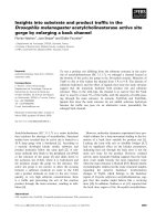
Fig. 1. Transient expression of wild-type and mutant FXI protein in COS-1 cells. pCDNA3 ⁄ FXI, pCDNA3 ⁄ FXI–Val371Ile or equimolar amounts
of both plasmids (heterozygous condition) were transiently transfected in COS-1 cells. Equal numbers of cells and equal amounts of plas-
mids were used in transfection experiments, as described in Experimental procedures. (A) Antigen levels of recombinant FXI were measured
in both conditioned media and the corresponding cell lysates using an ELISA assay. Bars represent relative concentrations of protein in
media and cell lysates compared with the mean antigen level measured in the wild-type. Results are given as mean ± SD. (B) The specific
activities of recombinant proteins were determined by calculating the ratio between FXI activity (measured using a one-stage method based
on a modified partial thromboplastin time) and FXI antigen levels. Bars represent mean ± SD of four independent experiments, each per-
formed in duplicate. The mean value of wild-type FXI was set as 100%. The results were analyzed by unpaired t-test (*P<0.05;
**P<0.01; ***P<0.001), ns, not significant; nd, not determined.
FXI–Val317Ile – a novel factor XI type II defect C. Bozzao et al.
6130 FEBS Journal 274 (2007) 6128–6138 ª 2007 The Authors Journal compilation ª 2007 FEBS
about half of the total mutated protein remains uncut,
while the wild-type FXI is almost entirely activated
(Fig. 2A).
In order to give a more accurate quantitative
description of the FXI activation process, a chromo-
genic assay was used to compare cleavage of the sub-
strate S-2366 by the wild-type and mutant FXI,
previously activated by thrombin in absence of dextran
sulfate. Activation of both proteins by human
a-thrombin followed pseudo-first-order kinetics, as
shown in Fig. 3. This was in agreement with a stochas-
tic model of FXI activation by thrombin, whereby the
latter cleaves either of the two chains of zymogen FXI
independently according to simple first-order kinetics.
If this were not the case, a double exponential or a
sigmoidal curve would have been observed in the acti-
vation kinetics. Under the experimental conditions
used in this study, after 2 h incubation $ 88% of wild-
type FXI and 60% of mutant FXI was activated by
thrombin. The k
cat
⁄ K
m
value of FXI activation was
9.8 ± 0.6 · 10
4
and 4.8 ± 0.8 · 10
4
m
)1
Æs
)1
for wild-
type and FXI–Val371Ile, respectively. These findings
showed that the Val371Ile mutation reduces by
approximately twofold the specificity of thrombin
interaction with the FXI–Val371Ile.
Activation of FIX by FXIa
The functional properties of activated FXI–Val371Ile
were explored both by a proteolytic assay using a com-
mercially available FIX and by measuring Michaelis
parameters of S-2366 hydrolysis. To this purpose,
wild-type FXIa and FXIa–Val371Ile, completely acti-
vated by thrombin (as described in Experimental pro-
cedures) were incubated for different periods with
commercial FIX. Upon FXI activation, FIX is cleaved
at two sites, releasing an activation peptide, and pro-
ducing the protease FIXa [10,24]. As shown in Fig. 4,
incubation of FIX with wild-type FXIa results in
almost complete activation after 30 min, whereas
FXIa–Val371Ile causes a dramatic reduction in the
uncleaved FIX form only after 60 min of incubation.
A possible effect of dextran sulfate on FIX activation
was ruled out by performing the same experiment in
the absence of FXI. No activation of FIX was detect-
able after 60 min of incubation (data not shown).
The observed delay in FIX activation may be due to
a decrease in the catalytic activity of mutant FXIa,
possibly caused by a perturbed conformational state of
FXIa linked to the Val371Ile mutation. A moderate
but significant reduction in the catalytic competence of
A
B
Fig. 2. Time course of wild-type and Val371Ile FXI activation. SDS ⁄ PAGE of wild-type FXI and FXI–Val371Ile (1.5 ng of protein) incubated
with FXIIa (1 lg) (A) or thrombin (0.5 U) (B). At various time points, indicated at the top of the panels, samples were stopped in reducing
sample buffer and eventually separated onto a 10% polyacrylamide gel. FXI activation was evaluated by western blotting using polyclonal
goat anti-human IgG recognizing both uncleaved FXI and FXI heavy and light chains. The estimated molecular masses of monomeric uncut
FXI (80 kDa), FXIa heavy chain (48 kDa), and FXIa light chain (32 kDa) are indicated.
C. Bozzao et al. FXI–Val317Ile – a novel factor XI type II defect
FEBS Journal 274 (2007) 6128–6138 ª 2007 The Authors Journal compilation ª 2007 FEBS 6131
Val371Ile FXIa was confirmed by investigating the
catalytic competence of FXIa towards the synthetic
substrate S-2366 (Fig. 5). The k
cat
and K
m
values of
S-2366 hydrolysis by wild-type FXI were 49.8 ± 3 s
)1
and 595 ± 63 lm, respectively, with k
cat
⁄ K
m
¼
8.37 · 10
4
m
)1
Æs
)1
. The same parameter values were
45 ± 4 s
)1
and 739 ± 100 lm for FXI–Val371Ile,
with k
cat
⁄ K
m
¼ 6.09 · 10
4
m
)1
Æs
)1
. The reduction in
the k
cat
⁄ K
m
value for S-2366 hydrolysis was significant,
but the effect of the mutation on FIX activation was
even more evident, as shown in Fig. 4. This suggests
that the mutation may alter molecular recognition
between FXIa and FIX, which necessarily involves, in
addition to the catalytic residues, a more extended sur-
face area of FXIa. The observed increase in K
m
for
S-2366 of the mutant FXI may arise from allosteric
effects, and thus may be generated from structural per-
turbations located far from the catalytic pocket.
Discussion
In this study, a novel missense mutation in F11 was
identified in a proband with mild type II FXI defi-
ciency. In vitro expression of the FXI–Val371Ile
recombinant protein, followed by activation assays,
showed slight differences in both FXI activation and
FIX activation by thrombin-activated FXI. The func-
tional defect evidenced by in vitro assays is compatible
with the deficiency observed in the two analyzed
Val371Ile carriers, even though the specific activity cal-
culated for the recombinant mutant protein is some-
what higher than expected. We cannot exclude that
differences in the dimerization and ⁄ or secretion effi-
ciency of mutant versus wild-type FXI might explain,
at least in part, this discrepancy.
Evolutionary conservation analysis of serine prote-
ase sequences shows that the position corresponding to
FXI–Val371 is highly conserved. For example, among
serine protease coagulation factors (i.e. FVII, FIX,
FX, FXII, plasminogen, and thrombin, showing an
overall amino acid sequence identity of 30–45%), this
position is occupied solely by a valine. This conserved
amino acid is replaced by isoleucine in the Val371Ile
FXI mutant. Interestingly, an isoleucine residue natu-
rally occupies the position corresponding to FXI
Val371 in some other serine proteases, such as vita-
min-K-dependent protein C, hepatocyte growth factor
activator, and neurotrypsin.
The Val371Ile mutation in FXI results in a relatively
mild physicochemical difference, because valine and
isoleucine are both highly hydrophobic, b-branched
XIF
06035150
IXFelI173laVIXFepyt-dliw
)nim()nim(06035150
aXIF
XIF
aXIF
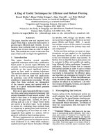
Fig. 4. Time course of FIX activation. Commercially available FIX (12.5 ng) was activated with 1.5 ng of recombinant FXI, either wild-type or
FXI–Val371Ile, both in turn activated by thrombin (0.5 U for 135 min; complete activation was assessed by western blot analysis). At differ-
ent time points (indicated at the top of each panel) digestions were stopped and proteins were resolved by Laemmli SDS ⁄ PAGE using 12%
(w ⁄ v) acrylamide gels.
021
00
1080604
0
20
01
8
6
4
2
0
emiT(nim)
FXIa (n
M
)
IXF-TW
IXF-I173V
Fig. 3. FXI activation by thrombin. Purified wild-type (d) and FXI–
Val371Ile (s) (10 n
M, final concentration) were activated by throm-
bin (3 n
M, final concentration). At different time points hirudin
(10 n
M, final concentration) was added to inhibit thrombin activity,
so that the chromogenic substrate S-2366 (500 l
M, final concentra-
tion) was hydrolyzed solely by activated FXI. The velocity of S-2236
hydrolysis by FXIa at each time point was converted into FXIa con-
centration by means of Eqn (2). Error bars indicate SEM.
FXI–Val317Ile – a novel factor XI type II defect C. Bozzao et al.
6132 FEBS Journal 274 (2007) 6128–6138 ª 2007 The Authors Journal compilation ª 2007 FEBS
amino acids (the b-carbon has two substitutions); nev-
ertheless, the mutation is not isosteric, isoleucine being
larger than valine, and having an additional methyl on
its side chain.
In the structure of the FXI zymogen [11], Val371 is
located on the linker region between the Ap4 and pro-
tease domains, and its surface area is 77% exposed to
the solvent. After activation of FXI, the activation
loop (residues 370–376), which is located at the new
N-terminus of the protease domain, undergoes a large
movement towards the activation pocket of FXIa. As
a result, in the structure of FXIa [25], the surface area
of Val371 is 92% buried within the protein, contacting
residues Arg144, Gly188, Asp189, Cys219, and Ala220.
Given that Val371 is buried in the structure of FXIa,
the introduction of a larger residue in this position
most likely causes some degree of structural change;
this is especially true in the case of the introduction of
an isoleucine, a b-branched amino acid that is not flex-
ible. In the active conformation, Val371 forms contacts
with neighboring residues that are important for stabi-
lizing the active state (e.g. Asp189, which is part of the
S1 pocket responsible for the binding specificity of the
substrate) [26]. Consequently, substitution of Val371 to
isoleucine might prevent the full development of the
active conformation. This hypothesis is further con-
firmed by the results of FIX proteolytic assays, which
showed a slight delay in FIX activation by FXIa acti-
vated by thrombin (Fig. 4); moreover the k
cat
and K
m
values of S-2366 hydrolysis showed that the Val371Ile
mutation has only minor conformational effects on the
geometry of the catalytic site of the enzyme (Fig. 5).
In contrast to the activated FXI, in the structure of
the FXI zymogen, Val371 is located on a loop region,
exposed to solvent, and does not form many contacts
with other residues (Fig. 6). Therefore, the additional
methyl in the Val371Ile mutant probably does not
disturb the structure and the domain rearrangement
in the zymogen FXI. Nevertheless, recombinant FXI–
Val371Ile activation was slower than that of the wild-
type protein (Fig. 2) suggesting a small activation
defect. This might be explained by the proximity of the
mutation to the cleavage site, probably resulting in a
small interference with the binding of the activator to
the FXI zymogen.
There are some examples of inherited coagulation
disorders in which one of the peptide linkages required
for the proteolytic zymogen activation cannot be
cleaved by the physiological activator. In most cases,
the mutated residue corresponds to the P1 site (i.e. the
C-terminal residue of the activation peptide) [27–33].
However, some mutations involving the P1¢ and P2¢
positions (i.e. the two first residues from the N-termi-
nal end of the catalytic domain) were previously
reported to cause mild to severe FIX deficiency either
6.12.18.04.00
04
03
02
0
1
0
m
M
)(6632-S
Velocity of hydrolysis (s
-1
)
IXF-TW
IXF-I173V
Fig. 5. Determination of Michaelis parameters of S-2366 hydrolysis.
Steady-state kinetics of S-2366 hydrolysis by wild-type (d) or FXI–
Val371Ile (s), under the conditions reported in Experimental proce-
dures. The continuous lines were drawn according to Michaelis
equation using the best fit parameters: (d) k
cat
¼ 49.8 ± 3 s
)1
,
K
m
¼ 595 ± 63 lM;(s) k
cat
¼ 45 ± 4 s
)1
, K
m
¼ 739 ± 100 lM.
Error bars indicate SEM.
Fig. 6. Structural consequences of the Val371Ile substitution. Rib-
bon representation of the superimposition between the structures
of the catalytic serine protease domain of the zymogen (red) and
activated (green) FXI. The Ile371 residue, in both structures, is dis-
played by space-filled atoms. The catalytic triad (blue space-filled
atoms) is also shown. The conformational movements of Ile371,
located in the activation loop at the N-terminus of the catalytic
domain, are notable. In the zymogen FXI, Ile371 is exposed to the
solvent, while in the activated FXI it is inserted into the protein.
The picture was drawn with
PYMOL (DeLano Scientific, San Carlos,
CA; ).
C. Bozzao et al. FXI–Val317Ile – a novel factor XI type II defect
FEBS Journal 274 (2007) 6128–6138 ª 2007 The Authors Journal compilation ª 2007 FEBS 6133
by altering the functional properties of FIXa or
by delaying its activation by FXIa. In particular,
four different amino acid substitutions (Val182Leu,
Val182Phe, Val182Ala, and Val182Gly) corresponding
to the here-reported Val371Ile in FXI, were found in
hemophilia B patients [34–37]. The phenotypic conse-
quences of these missense mutations were variable,
ranging from the complete loss of function of FIX
Kashihara (Val182Phe) to a residual 15% of procoagu-
lant activity of FIX Cardiff (Val182Leu) [37].
In conclusion, the Val371Ile mutation, identified and
characterized here, brings the number of naturally
occurring FXI variants responsible for type II deficien-
cies to seven [13]. Of these, three have been character-
ized in-depth, showing different mechanisms
underlying the pathologic phenotype, i.e. a reduction
in affinity for platelets (Ser248Asn) [38], a modest
reduction of FXI catalytic activity (Pro520Leu) [39],
and a greatly reduced rate of FIX activation associated
with resistance to antithrombin inhibition (Gly555Glu)
[40]. Uniquely, our mutation is associated with a defect
both in FXI activation (slower than normal), and in
FIX activation (slightly delayed), thus supporting the
role of residues neighboring the active site in influenc-
ing and stabilizing the enzyme active state.
Experimental procedures
Blood collection and genomic DNA extraction
This study was approved by the Institutional Review Board
of the University of Milan. All subjects signed an informed
consent according to the Declaration of Helsinki before
blood withdrawal. Peripheral venous blood was collected in
1 : 10 volume of 0.11 m trisodium citrate, pH 7.3. Genomic
DNA was extracted from whole blood using a standard
salting-out procedure.
Coagulation studies
Immediately after collection, citrated blood was centrifuged
at 2500 g for 15 min at room temperature. FXI activity was
performed by a one-stage method based on a modified par-
tial thromboplastin time, using FXI-deficient plasma as sub-
strate (Hemoliance, Salt Lake City, UT). FXI antigen was
measured by an ELISA based on a goat anti-human FXI
affinity purified IgG as capture antibody and a goat anti-
human FXI peroxidase-conjugated IgG as detecting anti-
body (Affinity Biological Inc., Hamilton, Ontario, Canada).
FXI levels were expressed in both tests as percentages of
pooled normal plasma from 30 normal male and female
individuals. The detection limits of the FXI functional and
immunologic assays were 1 and 0.1%, respectively.
PCR amplifications and DNA sequencing
PCR were performed on 50–100 ng of genomic DNA in a
25 lL volume, following standard procedures [41]. PCR
and sequencing primers were designed on the basis of the
known genomic sequence of F11 (GenBank accession num-
ber NM_000128). The primer couple used to amplify F11
exon 11 and to identify the Val371Ile mutation was FXI-
ex11-F 5¢-GTCAATTCCATTTTTCATGTGC-3¢ and FXI-
ex11-R 5¢-CGTTTTTTACCACTGAAGCAAT-3¢. All other
primer sequences, as well as the specific PCR condition for
each primer couple, are available on request. Sequencing
reactions were performed on both strands on PCR products
purified by MICROCON 100 columns (Millipore, Bedford,
MA). The BigDye Terminator Cycle Sequencing Kit ver-
sion 3.1 and an automated ABI-3100 DNA sequencer
(Applied Biosystems, Foster City, CA) were used.
Site-directed mutagenesis
The pCDNA3 ⁄ FXI expression plasmid, containing full-
length FXI complementary DNA (cDNA), was kindly
provided by A. Zivelin (Institute of Thrombosis and Hemo-
stasis, Chaim Sheba Medical Center, Tel Hashomer, Israel).
The identified missense mutation was introduced in
pCDNA3 ⁄ FXI by the QuikChange Site-Directed Muta-
genesis Kit (Stratagene, La Jolla, CA), according to the
manufacturer’s instructions. The mutant plasmid
pCDNA3 ⁄ FXI–Val371Ile was checked by sequencing the
whole FXI cDNA insert as well as 200 bp of flanking
DNA on both sides of the cloning site. Large-scale plasmid
preparations were obtained using the EndoFree Plasmid
Maxi Kit (Qiagen, Hilden, Germany).
Proteins and antibodies
Thrombin, FIX, FXI, and FXIIa were obtained from
Enzyme Research Laboratories (Swansea, UK). The sources
of the antibodies were as follows: rabbit anti-human FIX
(catalogue number F 0652) from Sigma (St Louis, MO), goat
anti-human FXI (catalogue number GAFXI-IG) from Affin-
ity Biologicals Inc., peroxidase-conjugated goat anti-rabbit
IgG from Pierce Biotechnology Inc. (Rockford, IL), and
peroxidase-conjugated donkey anti-goat IgG from Jackson
ImmunoResearch Laboratories Inc. (West Grove, PA).
Cell culture
African green monkey kidney COS-1 cells were cultured
in DMEM (EuroClone, Wetherby, UK) supplemented
with 10% fetal bovine serum (HyClone, South Logan,
UT), antibiotics (100 UÆmL
)1
penicillin and 100 lgÆmL
)1
streptomycin; EuroClone) and glutamine (2 mm; Euro-
Clone), and grown at 37 °C in a humidified atmosphere
FXI–Val317Ile – a novel factor XI type II defect C. Bozzao et al.
6134 FEBS Journal 274 (2007) 6128–6138 ª 2007 The Authors Journal compilation ª 2007 FEBS
of 5% CO
2
and 95% air, according to standard proce-
dures.
Expression of recombinant proteins
In each transfection experiment an equal number of cells
(400 000) were transiently transfected with the Lipofecta-
mine 2000 reagent (Invitrogen, Carlsbad, CA) in six-well
plates with 4 lg of plasmid DNA (pCDNA3 ⁄ FXI, or
pCDNA3 ⁄ FXI–Val371Ile, or equimolar amounts of both
plasmids), essentially as described by the manufacturer.
Twenty-nine hours after transfection, cells were washed
twice with NaCl ⁄ P
i
and cultured for additional 48 h in
1 mL of serum-free medium supplemented with glutamine,
antibiotics, and 5 mgÆmL
)1
BSA. For each experiment (per-
formed four times in duplicate) a mock sample, with the
empty pCDNA3 plasmid, was set up.
Conditioned media from each well were tested for both
FXI antigen and coagulation activity and used to prepare
FXIa for SDS ⁄ PAGE analysis.
FXI measurement in conditioned media and
in cell lysates
FXI antigen levels were evaluated by ELISA, as described
above, both in conditioned media and in cell lysates. Stan-
dard curves were constructed with reference plasma diluted
1 : 100 to 1 : 6400 in Tris-buffered saline (NaCl ⁄ Tris:
50 mm Tris, 150 mm NaCl, pH 7.5). Conditioned media
were collected in prechilled tubes containing a protease
inhibitor mixture (Complete; Roche, Basel, Switzerland),
centrifuged to remove cell debris, and stored at )80 °C until
use. To obtain cell lysates, cells were washed three times
with prechilled NaCl ⁄ P
i
and incubated for 1 h on ice with
1· NaCl ⁄ P
i
, 1.5% Triton X-100, and 1· Complete. Samples
were collected and centrifuged to remove cell debris.
FXI coagulant activity was measured in media (collected
without any protease inhibitor) as described above (see
‘Coagulation studies’).
Activation of FXI
FXI was activated either with FXIIa or with thrombin. For
each activation experiment, the exact amount of the recom-
binant protein was assessed by an ELISA assay, as
described above; on average, 2.5 lL of conditioned media
corresponded approximately to 1.5 ng of protein.
FXIIa (1 lg) and 1.5 ng of recombinant FXI, either
wild-type or mutant, were incubated in NaCl ⁄ Tris at 37 °C
for different periods. Each reaction was carried out in a
final volume of 20 lL. Samples were removed into reducing
SDS sample buffer and size-fractionated on 10% polyacryl-
amide SDS gels.
Because in vitro activation of FXI by thrombin is highly
enhanced in the presence of polyanions such as dextran
sulfate [42], 1.5 ng of recombinant FXI, either wild-type or
mutant, was activated with 0.5 U ($ 5nm) of human
thrombin in NaCl ⁄ TrisA (NaCl ⁄ Tris supplemented with
0.1 mgÆmL
)1
BSA) containing 1 lgÆmL
)1
dextran sulfate
(500 000 Da) at 37 °C for different periods. The concentra-
tion of dextran sulfate (1 lgÆmL
)1
) used in our experiments
was found to be optimal in previous studies [23,42,43].
Each reaction was carried out in a final volume of 20 lL.
Aliquots (each containing 1.5 ng of recombinant FXI) were
stopped by adding 10 lLof3· reducing Laemmli sample
buffer, and run on 10% SDS ⁄ PAGE.
Proteins were then transferred onto 0.45 lm pore-size
nitrocellulose membranes (Schleicher & Schuell, Brentford,
UK) and analyzed by western blotting, using a polyclonal
goat anti-human FXI IgG.
Activation of FIX by FXIa
Recombinant FXI, either wild-type or mutant, was acti-
vated with 0.5 U thrombin in NaCl ⁄ TrisA containing
1 lgÆmL
)1
dextran sulfate at 37 °C for 135 min in a total
reaction volume of 20 lL; complete activation was verified
by western blotting (see below). After that, 1.5 ng of FXIa
and 12.5 ng of FIX were incubated in NaCl ⁄ TrisA with
2.5 mm CaCl
2
at 37 °C for different periods. Each reaction
was carried out in a final volume of 30 lL. The ability of
residual thrombin and dextran sulfate in the buffer solution
to activate FIX was ruled out in preliminary experiments.
At different time points aliquots were removed into reduc-
ing SDS sample buffer, and size-fractionated on 12% poly-
acrylamide SDS gels.
Proteins were transferred onto nitrocellulose membranes
and analyzed by western blot using polyclonal rabbit anti-
human FIX IgG.
Western blotting analysis
Blots were incubated at room temperature for 1 h in
NaCl ⁄ Tris containing 0.1% Tween 20 and 5% (w ⁄ v)
skimmed milk. The membranes were then incubated for 2 h
with primary antibodies and subsequently for 1 h with
donkey anti-goat IgG or goat anti-rabbit IgG horseradish
peroxidase-conjugated secondary ones. When the anti-
human FIX IgG was used, dilutions were performed in
NaCl ⁄ Tris supplemented with 0.3% BSA at room tempera-
ture; all other incubations were done in NaCl ⁄ Tris contain-
ing 5% milk. Proteins were detected using Enhanced
Chemioluminescence, SuperSignal West Dura Extended
Duration Substrate (Pierce).
Assay of FXI activation
Before activation by thrombin, supernatants from cells
expressing recombinant FXI were concentrated approxi-
C. Bozzao et al. FXI–Val317Ile – a novel factor XI type II defect
FEBS Journal 274 (2007) 6128–6138 ª 2007 The Authors Journal compilation ª 2007 FEBS 6135
mately four- to fivefold by means of VivaSpin 30 concen-
trators (Sartorius Ltd., Epsom, UK). Activation of both
wild-type and FXI–Val371Ile (10 nm) by thrombin (3 nm),
purified as previously detailed [44], was measured by a
chromogenic assay, as follows. Incubations were carried
out in 100 lLof50mm Tris, 150 mm NaCl, pH 7.5, with
0.1% poly(ethylene glycol) 6000 at 25 °C. In the FXI acti-
vation by thrombin, dextran sulfate was omitted from the
reaction buffer to avoid any spurious effect on FXI auto-
activation. At various time intervals, 10 lL of recombinant
hirudin (Sigma) at a final concentration of 10 nm were
added to inhibit thrombin activity. Then 50 lL of 500 lm
(final concentration) S-2366 (pyroGlu-Pro-Arg-pNA; Chro-
mogenix, Mo
¨
lndal, Sweden) were added to the solution,
and the amount of free paranitroaniline released by FXIa
was determined by measuring the change in absorbance at
405 nm in a Benchmark II microplate reader (Bio-Rad
Laboratories, Hercules, CA). To eliminate any scattering
contribution, the absorbance at 620 nm was always sub-
tracted from the reading at 405 nm. The initial velocity of
S-2366 hydrolysis obtained at each time point was consid-
ered proportional to FXIa generated by thrombin. The
velocity of S-2366 hydrolysis was then analyzed as a func-
tion of time, to calculate the pseudo-first-order rate constant
of both wild-type and mutant FXI cleavage by thrombin.
Accordingly:
V
t
¼ V
1
Ãð1 À expðÀk à tÞÞ
where V
t
and V
¥
are the velocities of S-2366 hydrolysis by
formed FXIa at time t and ¥, respectively, and k is the
pseudo-first order rate constant of FXI activation by
thrombin. The best-fit value of k is thus independent from
the intrinsic catalytic activity of both wild-type and mutant
FXIa, but depends only on the specificity of thrombin–FXI
interaction. The only assumption made was that the value
of the asymptotic parameter V
¥
corresponds to the velocity
of the substrate hydrolysis by the FXIa concentration equal
to the nominal concentration of zymogen FXI present in
solution, assuming that the entire amount of zymogen FXI
was converted to FXIa at time ¥. The reaction was studied
at a concentration of FXI < K
m
of thrombin hydrolysis so
that the rate constant k was proportional to the value of
k
cat
⁄ K
m
of the activation, according to:
k ¼ T k
cat
=K
m
ð1Þ
where T is the thrombin concentration.
Measurement of Michaelis parameters of S-2366
hydrolysis by wild-type and FXI–Val371Ile
After 120 min of FXI activation by thrombin, $ 88%
(8.8 nm) of wild-type FXI and 63% (6.3 nm) of mutant
FXI were activated, according to [45]:
½FXIa
120
¼ V
120
=V
1
à FXI
T
ð2Þ
where V
120
is the velocity of S-2366 hydrolysis at 120 min
and FXI
T
is the total concentration of either wild-type or
mutant zymogen FXI present in the activation solution.
The validity of this approach was confirmed in the case of
the wild-type form, whose concentration, calculated by
Eqn (2) was in agreement within 10% error with that
obtained from a reference curve, where the catalytic acti-
vity of different concentrations of a purified FXIa prepara-
tion (Hematological Technologies Inc., Essex Junction, VT)
in the presence of 500 lm S-2366 were linearly correlated
to the nominal enzyme concentration (supplementary
Fig. S1).
At time 120 min, chosen to avoid instability or autohy-
drolytic damage of thrombin at longer incubation times, an
aliquot of the activation solution was taken to measure the
Michaelis parameters of S-2366 hydrolysis by FXIa. The
Michaelis parameters, k
cat
and K
m
, were calculated on
the basis of known concentration of wild-type and mutant
FXIa and using the program grafit (Erithacus Software
Ltd., Staines, UK).
Structural analysis
The structural analysis was conducted using the crystal
structure of the zymogen FXI (PDB code: 2F83) [11] and
FXIa (PDB code: 1XX9) [25]. The solvent-accessible area
for each residue in both structures was calculated using
the surfv program [46] with a probe sphere of radius
1.4 A
˚
and default parameters. The percentage of the
surface-exposure of each residue in the monomer was
calculated from the total solvent-accessible area on a
Gly-X-Gly tripeptide (where X represents each of the 20
amino acids).
Evolutionary conservation analysis
Evolutionary conservation analysis was carried out using
the ConSurf web-server [47] ( The
calculations were performed using the structure of FXIa
(PDB code: 1XX9) [25], based on an alignment of 200 ser-
ine protease sequences collected from the SWISSPROT
database [48] and default parameters.
Acknowledgements
The authors would like to thank Sofia H. Giacomelli
for excellent technical assistance. SD is a recipient of a
Bayer Hemophilia Early Career Investigator Award
2006. The financial support of PRIN (Programmi di
Ricerca Scientifica di Rilevante Interesse Nazionale,
Grant n. 2005058307-002) is gratefully acknowledged.
FXI–Val317Ile – a novel factor XI type II defect C. Bozzao et al.
6136 FEBS Journal 274 (2007) 6128–6138 ª 2007 The Authors Journal compilation ª 2007 FEBS
References
1 Davie EW, Fujikawa K, Kurachi K & Kisiel W (1979)
The role of serine proteases in the blood coagulation
cascade. Adv Enzymol Relat Areas Mol Biol 48, 277–318.
2 Fujikawa K, Legaz ME, Kato H & Davie EW (1974)
The mechanism of activation of bovine factor IX
(Christmas factor) by bovine factor XIa (activated
plasma thromboplastin antecedent). Biochemistry 13,
4508–4516.
3 Saito H (1977) Purification of high molecular weight
kininogen and the role of this agent in blood coagula-
tion. J Clin Invest 60, 584–594.
4 Dorfman R & Walsh PN (2001) Noncovalent interactions
of the Apple 4 domain that mediate coagulation factor
XI homodimerization. J Biol Chem 276, 6429–6438.
5 McMullen BA, Fujikawa K & Davie EW (1991) Loca-
tion of the disulfide bonds in human coagulation factor
XI: the presence of tandem apple domains. Biochemistry
30, 2056–2060.
6 Gailani D, Ho D, Sun MF, Cheng Q & Walsh PN
(2001) Model for a factor IX activation complex on
blood platelets: dimeric conformation of factor XIa is
essential. Blood 97, 3117–3122.
7 Yun TH, Baglia FA, Myles T, Navaneetham D, Lopez
JA, Walsh PN & Leung LL (2003) Thrombin activation
of factor XI on activated platelets requires the interac-
tion of factor XI and platelet glycoprotein Ib alpha with
thrombin anion-binding exosites I and II, respectively.
J Biol Chem 278, 48112–48119.
8 Gailani D & Broze GJ Jr (1991) Factor XI activation in
a revised model of blood coagulation. Science 253, 909–
912.
9 Baglia FA, Sinha D & Walsh PN (1989) Functional
domains in the heavy-chain region of factor XI: a high
molecular weight kininogen-binding site and a sub-
strate-binding site for factor IX. Blood 74, 244–251.
10 Bouma BN & Griffin JH (1977) Human blood coagula-
tion factor XI. Purification, properties, and mechanism
of activation by activated factor XII. J Biol Chem 252,
6432–6437.
11 Papagrigoriou E, McEwan PA, Walsh PN & Emsley J
(2006) Crystal structure of the factor XI zymogen
reveals a pathway for transactivation. Nat Struct Mol
Biol 13, 557–558.
12 Shpilberg O, Peretz H, Zivelin A, Yatuv R, Chetrit A,
Kulka T, Stern C, Weiss E & Seligsohn U (1995) One
of the two common mutations causing factor XI defi-
ciency in Ashkenazi Jews (type II) is also prevalent in
Iraqi Jews, who represent the ancient gene pool of Jews.
Blood 8, 429–432.
13 Saunders RE, O’Connell NM, Lee CA, Perry DJ &
Perkins SJ (2005) Factor XI deficiency database: an
interactive web database of mutations, phenotypes, and
structural analysis tools. Hum Mutat 26, 192–198.
14 Salomon O & Seligsohn U (2004) New observations on
factor XI deficiency. Haemophilia 10 (Suppl. 4), 184–187.
15 Ragni MV, Sinha D, Seaman F, Lewis JH, Spero JA &
Walsh PN (1985) Comparison of bleeding tendency,
factor XI coagulant activity, and factor XI antigen in 25
factor XI-deficient kindreds. Blood 65, 719–724.
16 Peyvandi F, Lak M & Mannucci PM (2002) Factor XI
deficiency in Iranians: its clinical manifestations in com-
parison with those of classic hemophilia. Haematologica
87, 512–514.
17 Quelin F, Francois D, d’Oiron R, Guillet B, de Rau-
court E & de Mazancourt P (2005) Factor XI defi-
ciency: identification of six novel missense mutations
(P23L, P69T, C92G, E243D, W497C and E547K).
Haematologica 90, 1149–1150.
18 Zivelin A, Bauduer F, Ducout L, Peretz H, Rosenberg
N, Yatuv R & Seligsohn U (2002) Factor XI deficiency
in French Basques is caused predominantly by an ances-
tral Cys38Arg mutation in the factor XI gene. Blood 99,
2448–2454.
19 Quelin F, Trossaert M, Sigaud M, Mazancourt PD &
Fressinaud E (2004) Molecular basis of severe factor XI
deficiency in seven families from the west of France.
Seven novel mutations, including an ancient Q88X
mutation. J Thromb Haemost 2, 71–76.
20 Bolton-Maggs PH, Peretz H, Butler R, Mountford R,
Keeney S, Zacharski L, Zivelin A & Seligsohn U (2004)
A common ancestral mutation (C128X) occurring in 11
non-Jewish families from the UK with factor XI defi-
ciency. J Thromb Haemost 2, 918–924.
21 Kravtsov DV, Monahan PE & Gailani D (2005) A
classification system for cross-reactive material-negative
factor XI deficiency. Blood 105, 4671–4673.
22 Kravtsov DV, Wu W, Meijers JC, Sun MF, Blinder
MA, Dang TP, Wang H & Gailani D (2004) Dominant
factor XI deficiency caused by mutations in the factor
XI catalytic domain. Blood 104, 128–134.
23 Baglia FA & Walsh PN (1998) Prothrombin is a cofac-
tor for the binding of factor XI to the platelet surface
and for platelet-mediated factor XI activation by throm-
bin. Biochemistry 37, 2271–2281.
24 Di Scipio RG, Kurachi K & Davie EW (1978) Activa-
tion of human factor IX (Christmas factor). J Clin
Invest 61, 1528–1538.
25 Jin L, Pandey P, Babine RE, Gorga JC, Seidl KJ,
Gelfand E, Weaver DT, Abdel-Meguid SS &
Strickler JE (2005) Crystal structures of the FXIa
catalytic domain in complex with ecotin mutants
reveal substrate-like interactions. J Biol Chem 280,
4704–4712.
26 Perona JJ & Craik CS (1995) Structural basis of sub-
strate specificity in the serine proteases. Protein Sci 4,
337–360.
27 Hamaguchi M, Matsushita T, Tanimoto M, Takahashi
I, Yamamoto K, Sugiura I, Takamatsu J, Ogata K,
C. Bozzao et al. FXI–Val317Ile – a novel factor XI type II defect
FEBS Journal 274 (2007) 6128–6138 ª 2007 The Authors Journal compilation ª 2007 FEBS 6137
Kamiya T & Saito H (1991) Three distinct point muta-
tions in the factor IX gene of three Japanese CRM+
hemophilia B patients (factor IX BMNagoya 2, factor
IX Nagoya 3 and 4). Thromb Haemost 65, 514–520.
28 Rabiet MJ, Furie BC & Furie B (1986) Molecular
defect of prothrombin Barcelona. Substitution of cyste-
ine for arginine at residue 273. J Biol Chem 261,
15045–15048.
29 O’Marcaigh AS, Nichols WL, Hassinger NL, Mullins
JD, Mallouh AA, Gilchrist GS & Owen WG (1996)
Genetic analysis and functional characterization of pro-
thrombins Corpus Christi (Arg382–Cys), Dhahran
(Arg271–His), and hypoprothrombinemia. Blood 88,
2611–2618.
30 Solera J, Magallon M, Martin-Villar J & Coloma A
(1991) Identification of a new haemophilia BM case
produced by a mutation located at the carboxy terminal
cleavage site of activation peptide. Br J Haematol 78,
385–389.
31 Bertina RM, van der Linden IK, Mannucci PM,
Reinalda-Poot HH, Cupers R, Poort SR & Owen WG
(1990) Mutations in hemophilia Bm occur at the
Arg180–Val activation site or in the catalytic domain of
factor IX. J Biol Chem 265, 10876–10883.
32 Lin SW & Shen MC (1993) Genetic basis and carrier
detection of hemophilia B of Chinese origin. Thromb
Haemost 69, 247–252.
33 Sun WY, Burkart MC, Holahan JR & Degen SJ (2000)
Prothrombin San Antonio: a single amino acid substitu-
tion at a factor Xa activation site (Arg320 to His)
results in dysprothrombinemia. Blood 95, 711–714.
34 Chen SH, Schoof JM, Weinmann AF & Thompson AR
(1995) Heteroduplex screening for molecular defects in
factor IX genes from haemophilia B families. Br J
Haematol 89, 409–412.
35 Maekawa H, Sugo T, Yamashita N, Kamiya K, Umey-
ama H, Miura N, Naka H, Nishimura T, Yoshioka A
& Matsuda M (1993) Molecular defect in factor IX
Tokyo: substitution of valine-182 by alanine at position
P2¢ in the second cleavage site by factor XIa resulting in
impaired activation. Biochemistry 32, 6146–6151.
36 Sakai T, Yoshioka A, Yamamoto K, Niinomi K,
Fujimura Y, Fukui H, Miyata T & Iwanaga S (1989)
Blood clotting factor IX Kashihara: amino acid substi-
tution of valine-182 by phenylalanine. J Biochem
(Tokyo) 105, 756–759.
37 Taylor SA, Liddell MB, Peake IR, Bloom AL & Lillic-
rap DP (1990) A mutation adjacent to the beta cleavage
site of factor IX (valine 182 to leucine) results in mild
haemophilia Bm. Br J Haematol 75 , 217–221.
38 Sun MF, Baglia FA, Ho D, Martincic D, Ware RE, Walsh
PN & Gailani D (2001) Defective binding of factor XI-
N248 to activated human platelets. Blood 98, 125–129.
39 Gailani D, Schmidt A, Sun MF, Bolton-Maggs PH &
Bajaj SP (2007) A cross-reactive material positive vari-
ant of coagulation factor XI (FXI) with a catalytic
defect. J Thromb Haemost 5, 781–787.
40 Zivelin A, Ogawa T, Bulvik S, Landau M, Toomey JR,
Lane J, Seligsohn U & Gailani D (2004) Severe factor
XI deficiency caused by a Gly555 to Glu mutation (fac-
tor XI–Glu555): a cross-reactive material positive vari-
ant defective in factor IX activation. J Thromb Haemost
2, 1782–1789.
41 Zadra G, Asselta R, Malcovati M, Santagostino E,
Peyvandi F, Mannucci PM, Tenchini ML & Duga S
(2004) Molecular genetic analysis of severe coagulation
factor XI deficiency in six Italian patients. Haematolog-
ica 89, 1332–1340.
42 Naito K & Fujikawa K (1991) Activation of human
blood coagulation factor XI independent of factor XII.
Factor XI is activated by thrombin and factor XIa in
the presence of negatively charged surfaces. J Biol Chem
266, 7353–7358.
43 Baglia FA & Walsh PN (2000) Thrombin-mediated
feedback activation of factor XI on the activated
platelet surface is preferred over contact activation by
factor XIIa or factor XIa. J Biol Chem 275, 20514–
20519.
44 De Cristofaro R, Rocca B, Bizzi B & Landolfi R (1993)
The linkage between binding of the C-terminal domain
of hirudin and amidase activity in human alpha-throm-
bin. Biochem J 289, 475–480.
45 Baglia FA & Walsh PN (1996) A binding site for
thrombin in the apple 1 domain of factor XI. J Biol
Chem 271, 3652–3658.
46 Sridharan S, Nicholls A & Honig B (1992) A new vertex
algorithm to calculate solvent accessible surface area.
Biophys J 61, A174.
47 Landau M, Mayrose I, Rosenberg Y, Glaser F, Martz E,
Pupko T & Ben-Tal N (2005) ConSurf 2005: the projec-
tion of evolutionary conservation scores of residues on
protein structures. Nucleic Acids Res 33, W299–W302.
48 Bairoch A & Apweiler R (1999) The SWISS-PROT pro-
tein sequence data bank and its supplement TrEMBL in
1999. Nucleic Acids Res 27, 49–54.
Supplementary material
The following supplementary material is available
online:
Fig. S1. Comparison of the catalytic properties of
recombinant and plasma-derived FXIa.
This material is available as part of the online article
from .
Please note: Blackwell Publishing is not responsible
for the content or functionality of any supplementary
materials supplied by the authors. Any queries (other
than missing material) should be directed to the corre-
sponding author for the article.
FXI–Val317Ile – a novel factor XI type II defect C. Bozzao et al.
6138 FEBS Journal 274 (2007) 6128–6138 ª 2007 The Authors Journal compilation ª 2007 FEBS