Báo cáo khoa học: Human retinol dehydrogenase 13 (RDH13) is a mitochondrial short-chain dehydrogenase⁄reductase with a retinaldehyde reductase activity pdf
Bạn đang xem bản rút gọn của tài liệu. Xem và tải ngay bản đầy đủ của tài liệu tại đây (259.4 KB, 10 trang )
Human retinol dehydrogenase 13 (RDH13) is a
mitochondrial short-chain dehydrogenase
⁄
reductase
with a retinaldehyde reductase activity
Olga V. Belyaeva, Olga V. Korkina*, Anton V. Stetsenko
†
and Natalia Y. Kedishvili
Department of Biochemistry and Molecular Genetics, Schools of Medicine and Dentistry, University of Alabama at Birmingham, AL, USA
Short-chain dehydrogenases ⁄ reductases (SDRs) com-
prise a large family of functionally heterogeneous pro-
teins that participate in the metabolism of steroids,
prostaglandins, retinoids, aliphatic alcohols and xeno-
biotics [reviewed in refs. 1,2]. Members of the SDR
superfamily are found in the cytoplasm, mitochondria,
nuclei, peroxisomes and endoplasmic reticulum. Many
enzymes exhibit the same substrate and cofactor speci-
ficity, but different subcellular localization and tissue
distribution [reviewed in ref. 3].
To date, about 3000 primary structures from various
species have been annotated in sequence databases as
members of the SDR superfamily on the basis of SDR
signature features, such as the TGX
3
GXG motif of the
nucleotide binding region and the catalytically active
tetrad N-S-Y-K, which constitutes the active site [1].
At least 63 SDR genes have been identified in the
human genome database [1]. For many of these puta-
tive oxidoreductases, the cellular functions are yet to
be determined.
Keywords
dehydrogenase; mitochondria; reductase;
retinaldehyde; retinol
Correspondence
N. Y. Kedishvili, Division of Biochemistry
and Molecular Genetics, Schools of
Medicine and Dentistry, University of
Alabama at Birmingham, 720 20th Street
South, 440B Kaul Genetics Building,
Birmingham, AL 35294, USA
Fax: 205 934 0758
Tel: 205 996 4023
E-mail:
Present address
*Department of Biochemistry, Tufts Univer-
sity School of Medicine, Boston, MA, USA
†Abbott Vascular, Abbott Park, IL, USA
(Received 12 September 2007, revised 31
October 2007, accepted 7 November 2007)
doi:10.1111/j.1742-4658.2007.06184.x
Retinol dehydrogenase 13 (RDH13) is a recently identified short-chain
dehydrogenase ⁄ reductase related to microsomal retinoid oxidoreductase
RDH11. In this study, we examined the distribution of RDH13 in human
tissues, determined its subcellular localization and characterized the sub-
strate and cofactor specificity of purified RDH13 in order to better
understand its properties. The results of this study demonstrate that
RDH13 exhibits a wide tissue distribution and, by contrast with other
members of the RDH11-like group of short-chain dehydrogenases ⁄ reduc-
tases, is a mitochondrial rather than a microsomal protein. Protease pro-
tection assays suggest that RDH13 is localized on the outer side of the
inner mitochondrial membrane. Kinetic analysis of the purified protein
shows that RDH13 is catalytically active and recognizes retinoids as sub-
strates. Similar to the microsomal RDHs, RDH11, RDH12 and RDH14,
RDH13 exhibits a much lower K
m
value for NADPH than for NADH
and has a greater catalytic efficiency in the reductive than in the oxidative
direction. The localization of RDH13 at the entrance to the mitochon-
drial matrix suggests that it may function to protect mitochondria against
oxidative stress associated with the highly reactive retinaldehyde produced
from dietary b-carotene.
Abbreviations
DHPC, 1,2-diheptanoyl-sn-glycero-3-phosphocholine; HSD, hydroxysteroid dehydrogenase; RDH, retinol dehydrogenase; SDR, short-chain
dehydrogenase ⁄ reductase.
138 FEBS Journal 275 (2008) 138–147 ª 2007 The Authors Journal compilation ª 2007 FEBS
Retinol dehydrogenase 13 (RDH13) is a recently
identified member of the SDR superfamily of proteins
that shares sequence similarity with RDH11 (also
known as retinal reductase 1 [4–6]), RDH12 [6,7] and
RDH14 (previously known as PAN2 [8]) proteins.
RDH11, RDH12 and RDH14 have been characterized
and found to be microsomal proteins that recognize
retinoids [4–8] and medium-chain aldehydes [7] as sub-
strates, with NADP
+
⁄ NADPH as the preferred cofac-
tors. However, the substrate and cofactor specificity of
RDH13 remains unknown, as it failed to exhibit any
enzymatic activity under the conditions of previous
assays [6]. Thus, it is not clear whether RDH13 repre-
sents a catalytically active member of the SDR super-
family.
This study was undertaken in order to better under-
stand the properties of RDH13 and to identify its
potential substrates. We examined the distribution of
RDH13 in human tissues, determined its subcellular
localization, expressed and purified the recombinant
protein, and characterized its substrate and cofactor
specificity. The results of this study reveal significant
differences between RDH13 and the other members of
the RDH11–14 group of proteins, and offer an impor-
tant insight into the properties of this new member of
the SDR superfamily.
Results
Tissue distribution of RDH13
It has been shown that a protein recognized by anti-
RDH13 serum is present in the inner segments of rod
and cone photoreceptors [6]; however, the distribution
of RDH13 in extra-ocular tissues has not yet been
determined. Therefore, we examined the expression
pattern of RDH13 in eight human tissues using poly-
clonal antiserum raised against bacterially expressed
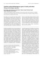
and purified RDH13. Western blot analysis revealed
that anti-RDH13 serum recognized a protein of the
expected size ( 36 kDa) in seven of the eight tissues
(Fig. 1). The intensity of immunostaining was stron-
gest in the kidney, heart and lung, but the corre-
sponding protein band was also detectable in the
prostate, testis and ovary. These results demonstrate
that RDH13 is a relatively widespread protein and
that its expression level varies considerably in differ-
ent tissues.
Subcellular localization of RDH13
RDH13 shares the greatest sequence similarity with
RDH11, RDH12 and RDH14, which are integral
membrane proteins of the endoplasmic reticulum. To
determine whether RDH13 is also targeted to the
endoplasmic reticulum, we analyzed its subcellular
localization in prostate cancer LNCaP cells, which
express endogenous RDH13 at high levels. LNCaP
cells were homogenized and the subcellular fractions
were resolved by discontinuous sucrose density gradi-
ent [9]. Equal aliquots of the gradient fractions were
subjected to denaturing SDS-PAGE and analyzed by
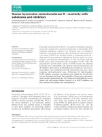
western blotting using anti-RDH13 serum. As shown
in Fig. 2, RDH13 was detected in fractions 3–7 of
the gradient. To identify the organelles present in
these fractions, we used antibodies against organelle-
specific marker proteins. Lamin, a nuclear protein,
was found only in the bottom two fractions (6 and
7), where nuclei, cell debris and unbroken cells were
Sk. muscle
Heart
Ovary
Spleen
Lung
Kidney
Testis
Prostate
Fig. 1. RDH13 expression in human tissues. Samples (100 lg) of
tissue homogenates were separated by SDS-PAGE and analyzed
by western blotting using anti-RDH13 serum, as described in
Experimental procedures. The arrow indicates the position of the
RDH13 protein. Sk. muscle, skeletal muscle.
RDH13
Porin
Lamin
Golgin
Calnexin
Top
Bottom
1 2 3 4 5 6 7
Fig. 2. Subcellular localization of RDH13 in LNCaP cells. Subcellular
fractions of LNCaP cells were separated by sucrose gradient, as
described in Experimental procedures, and analyzed by western
blotting using antibodies against RDH13 or specific marker proteins
of cellular organelles, as indicated. Fractions are numbered from
the top of the gradient.
O. V. Belyaeva et al. Human RDH13 is a mitochondrial retinal reductase
FEBS Journal 275 (2008) 138–147 ª 2007 The Authors Journal compilation ª 2007 FEBS 139
expected to be present. Golgin exhibited two peaks of
distribution, one at the 0.8 m ⁄ 1.2 m sucrose interface
(fractions 3 and 4), as expected, and also in the
unbroken cells area (fractions 6 and 7). Calnexin, the
marker for the endoplasmic reticulum, appeared to be
spread throughout the gradient, whereas porin, an
integral protein of the outer mitochondrial membrane,
was most abundant in fractions 3–7, similar to
RDH13. Thus, the flotation pattern of RDH13 coin-
cided best with that of porin, suggesting that, by con-
trast with the other members of the RDH11–14
cluster, RDH13 is a mitochondrial and not an endo-
plasmic reticulum protein.
Mitochondria have a highly compartmentalized
structure, which can influence the substrate and co-
factor availability for RDH13. To determine the sub-
mitochondrial localization of RDH13, freshly isolated
mitochondria were fractionated into the intermem-
brane space, outer membrane, matrix and inner mem-
brane, and the fractions were analyzed by western
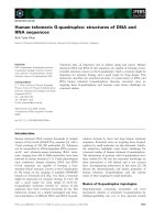
blotting using anti-RDH13 serum. RDH13 protein
was found to be most abundant in the fraction con-
taining the inner mitochondrial membranes (Fig. 3A),
suggesting that it is a membrane-bound protein. To
determine whether RDH13 is a peripheral or an
integral membrane protein, the inner mitochondrial
membranes or whole mitoplasts were treated with
NaCl ⁄ P
i
,1m NaCl, 100 mm Na
2
CO
3
or 1% Triton
X-100, as described in Experimental procedures. The
samples were centrifuged and the distribution of
RDH13 between the pellet and supernatant was ana-
lyzed by western blotting. As shown in Fig. 3B,
RDH13 protein remained associated with the mem-
branes after treatment with NaCl ⁄ P
i
or NaCl, but
was completely solubilized by Na
2
CO
3
and Triton
X-100 treatments. As integral membrane proteins can-
not be extracted by alkaline treatment [10], these
results indicate that RDH13 is a peripheral membrane
protein.
To determine whether RDH13 is localized on the
matrix side of the inner membrane or faces the inter-
membrane space, we carried out protease protection
assays. Mitochondria or mitoplasts (lacking the outer
membrane) were treated with increasing concentra-
tions of trypsin, and the stability of RDH13 protein
was analyzed by western blotting. RDH13 was com-
pletely resistant to trypsin digestion in intact mito-
chondria at all concentrations of trypsin. By contrast,
in mitoplasts, there was a progressive loss of RDH13
protein (Fig. 3C). This result indicates that the outer
membrane protects RDH13 from trypsin in intact
mitochondria, and the removal of the outer mem-
brane exposes RDH13 to trypsin. Thus, RDH13
appears to be localized on the outer side of the inner
mitochondrial membrane, facing the intermembrane
space.
Finally, we determined whether RDH13 contains
a cleavable mitochondrial targeting signal sequence.
Analysis of the primary structure of RDH13 using
the MitoProt II algorithm [11] suggested a potential
cleavage site at amino acid 62, with a probability of
export to mitochondria of 0.77. However, RDH13
produced by in vitro translation, using expression con-
struct under the T7 promoter in pCR4.2-TOPO and
the TNT Coupled Reticulocyte Lysate Transcrip-
tion ⁄ Translation System (Promega, Madison, WI,
USA), had the same size in SDS-PAGE as the fully
processed protein in LNCaP cells (data not shown),
indicating that RDH13 lacks a cleavable mitochon-
drial target sequence. This result is consistent with
the localization of RDH13 on the outer side of the
inner mitochondrial membrane.
NaCl/P
i
Triton
P S P S P S P S
mch
mpl
0 0.05 0.1 1 10 0 0.05 0.1 1 10
Mitochondria
IS OM MX IM
NaCl Na
2
CO
3
Mitoplasts
A
B
C
Fig. 3. Submitochondrial localization of RDH13. (A) Mitochondria
were fractionated into intermembrane space (IS), outer membranes
(OM), matrix (MX) and inner membranes (IM). One-fiftieth of each
fraction was separated by SDS-PAGE and the distribution of
RDH13 was determined by western blotting. (B) Mitoplasts (mpl)
were prepared by hypotonic or digitonin treatment of mitochondria
(mch) and incubated with NaCl ⁄ P
i
, NaCl, Na
2
CO
3
or Triton X-100
(Triton). Treated samples were centrifuged and the distribution of
RDH13 between soluble and insoluble fractions was analyzed by
western blotting. P, pellet; S, supernatant. The results were identi-
cal for digitonin- and hypotonically prepared mitoplasts. (C) Mito-
chondria or mitoplasts were incubated with the indicated amounts
of trypsin (lg) for 30 min on ice, followed by the addition of soy-
bean trypsin inhibitor. RDH13 protein stability was monitored by
western blotting.
Human RDH13 is a mitochondrial retinal reductase O. V. Belyaeva et al.
140 FEBS Journal 275 (2008) 138–147 ª 2007 The Authors Journal compilation ª 2007 FEBS
Substrate and cofactor specificity of purified
RDH13–His6
A previous study has examined RDH13 for activity
towards retinaldehyde in whole Sf9 cells [6]. This anal-
ysis failed to detect any increase in retinaldehyde
reduction by RDH13-expressing cells compared with
control cells. We re-examined the catalytic activity of
RDH13 by expressing the protein in Sf9 cells as a
fusion with the C-terminal His6 tag in order to purify
RDH13 to homogeneity and characterize its properties
under well-defined conditions. Similar to native
RDH13, recombinant RDH13–His6 was detected in
the mitochondrial fraction of Sf9 cells and exhibited
the same association with the inner mitochondrial
membrane as the native protein (data not shown).
Interestingly, the expression of RDH13 in Sf9 cells was
accompanied by the appearance of a weak retinalde-
hyde reductase activity in the mitochondrial fraction,
suggesting that RDH13 is active towards retinaldehyde
(data not shown).
To obtain further evidence to demonstrate that the
increase in mitochondrial retinaldehyde reductase
activity was associated with RDH13 expression, we
purified RDH13–His6 using Ni
2+
affinity chromato-
graphy. This single-step purification procedure pro-
duced an almost homogeneous protein (Fig. 4).
Activity assays showed that purified RDH13–His6 was
indeed active towards all-trans-retinaldehyde and
appeared to prefer NADPH to NADH as a cofactor,
because the conversion of 5 lm all-trans-retinaldehyde
in the presence of 1 mm NADPH was about 20-fold
greater than that in the presence of 1 mm NADH.
However, the specific activity of different RDH13–His6
preparations varied from 47 to 130 nmolÆ min
)1
Æmg
)1
.
In this respect, we observed that, if dithiothreitol was
omitted from the elution buffer during RDH13–His6
purification, the purified enzyme had a very low
activity, but could be reactivated by the addition of
dithiothreitol. A comparison of the more active and
less active preparations of RDH13 by gel electrophore-
sis revealed that, in the absence of dithiothreitol,
RDH13 appeared as two protein bands, one corre-
sponding to the monomeric form of the protein and
the other to the dimeric form (Fig. 5). After the addi-
tion of dithiothreitol, the dimer disappeared, shifting
to the faster moving monomeric form of RDH13–
His6. Glutathione (5 lm), which is the dominant
low-molecular-weight thiol in the cell, had the same
activating effect on RDH13 as dithiothreitol (data not
shown). These results indicate that reducing conditions
are essential for the maintenance of the active state of
RDH13, and that nonreducing conditions promote the
formation of inactive RDH13 dimers. In this respect,
RDH13 appears to be similar to another member of
the SDR superfamily, 11b-hydroxysteroid dehydroge-
nase type 2 (11b-HSD2) [12]. Like RDH13, 11b-HSD2
formed inactive dimers in the absence of 2-mercapto-
ethanol or dithiothreitol. The authors proposed that
the inactive dimers could represent a latent form of the
enzyme, and dimerization could serve as a mechanism
for modulating the enzyme’s activity [12]. RDH13
activity was also affected by the nature of the deter-
gent: the substitution of 1,2-diheptanoyl-sn-glycero-
3-phosphocholine (DHPC) for Tween-20 resulted in
complete inactivation of the enzyme. In addition,
RDH13 was sensitive to temperature, becoming par-
tially inactivated after 20 min of incubation in the
reaction buffer at 37 °C.
1 2 3 4 5 6 7
Fig. 4. Purification of RDH13–His6 from Sf9 cells. RDH13–His6
was purified by Ni
2+
affinity chromatography, and the fractions from
various stages of purification were analyzed by SDS-PAGE followed
by silver staining. Lane 1, homogenate; lane 2, wash with 10 m
M
imidazole; lanes 3–7, elution of RDH13–His6 with a stepwise imid-
azole gradient: 50 m
M (3), 100 mM (4), 200 mM (5), 300 mM (6),
400 m
M (7). Arrow indicates the position of RDH13–His6.
36
50
64
98
148
M
D
+
m
–
Fig. 5. Effect of dithiothreitol on oligomeric state of RDH13–His6.
RDH13 was purified and stored at )80 °C in the absence of reduc-
ing agents. Samples of this preparation were denatured in a boiling
water bath for 5 min using gel loading buffer with (+) or without ())
dithiothreitol and analyzed by SDS-PAGE. The positions of the
monomeric (M) and dimeric (D) forms of the protein are indicated
on the left. m, molecular mass markers.
O. V. Belyaeva et al. Human RDH13 is a mitochondrial retinal reductase
FEBS Journal 275 (2008) 138–147 ª 2007 The Authors Journal compilation ª 2007 FEBS 141
To determine the catalytic efficiency of RDH13,
we carried out kinetic characterization of the purified
enzyme (Table 1). This analysis showed that RDH13
reduced all-trans-retinaldehyde with an apparent K
m
value of 3.2 ± 0.7 lm and V
max
value of
230 ± 24 nmolÆmin
)1
Æmg
)1
. The apparent K
m
value
for all-trans-retinol ( 3 lm) appeared to be similar
to that for retinaldehyde; however, the rate of retinol
oxidation by RDH13 was extremely low ( 5 nmolÆ
min
)1
Æmg
)1
), which precluded an accurate determina-
tion of the kinetic constants. The apparent K
m
value
of RDH13–His6 for NADPH (1.5 ± 0.1 lm) was
three orders of magnitude lower than that for
NADH ( 6000 lm), consistent with its preference
for NADPH as a cofactor. Thus, kinetic analysis
reveals that RDH13 exhibits substrate and cofactor
specificity very similar to that of RDH11, RDH12
and RDH14.
RDH13–His6 was also tested for activity towards
17b-, 3a- and 11b-hydroxysteroids, and corresponding
ketosteroids, as described for other SDRs [13–15];
however, no significant conversion was observed.
Other compounds were examined as potential sub-
strates by evaluating their ability to inhibit the
RDH13-catalyzed reduction of all-trans-retinaldehyde.
These compounds included short-chain aldehydes, such
as nonanal, 6-cis-nonenal and 2-trans-nonenal, because
they have been shown to be good substrates for
RDH12 [7]. Glyceraldehyde and acetoacetyl-coenzyme
A were tested because they have been found to be
metabolized by another mitochondrial SDR,
17b-HSD10 [16]. In addition, we tested several com-
mercially available derivatives of cholesterol, such as
taurocholic acid, 25-hydroxycholesterol and 25-nor-
5-cholesten-3-ol-25b-one, as some steps of cholesterol
metabolism are catalyzed by cytochrome P450 enzymes
associated with the inner membrane of mitochondria.
No compound was inhibitory at a concentration of
50 lm, suggesting that they could not compete with
retinaldehyde and, most probably, were not substrates
for RDH13.
Thus, we have established that RDH13 is principally
different from related RDH11, RDH12 and RDH14 in
that it is targeted to the mitochondria, and is not an
integral but a peripheral membrane protein associated
with the inner mitochondrial membrane. Furthermore,
RDH13 is much more labile than RDH11 and related
microsomal proteins, and requires reducing conditions
to stay active. At the same time, RDH13 is very simi-
lar to the members of the RDH11–14 cluster of SDRs
in terms of its substrate and cofactor preferences.
Discussion
This study presents the first characterization of the tis-
sue distribution, subcellular localization and catalytic
activity of the recently discovered member of the SDR
superfamily, RDH13. Western blot analysis of RDH13
distribution in human tissues carried out in this study
shows that RDH13 is a widespread protein, being
expressed at some level in seven of the eight human tis-
sues examined. This protein expression pattern is in
agreement with the presence of RDH13 transcripts in
at least 32 adult tissues, as well as in embryonic and
cancer tissues, as reported in the Expressed Sequence
Tag GenBank database. Human RDH13 shares 83%
protein sequence identity with mouse RDH13 and
72% identity with frog RDH13, and the corresponding
genes have similar genomic organization [17], indicat-
ing that RDH13 is conserved across species. The high
degree of protein conservation and the ubiquitous
expression pattern suggest that RDH13 plays an
important metabolic role. However, until recently, no
enzymatic activity for RDH13 had been demonstrated.
RDH13 is most closely related to the NADP
+
-
dependent microsomal enzymes RDH11, RDH12 and
RDH14, which exhibit the highest activity as retinalde-
hyde reductases [4–8]. In this study, we have shown,
for the first time, that purified RDH13 exhibits an oxi-
doreductive activity towards retinoids, strongly prefers
NADPH over NADH as a cofactor, and has a much
greater catalytic efficiency as a reductase than as a
dehydrogenase. The catalytic efficiency of RDH13 as a
retinaldehyde reductase is significantly lower than that
of a related protein RDH11, primarily because of the
much higher K
m
value for retinaldehyde (3 lm versus
0.12 lm for RDH11 [5]). However, the k
cat
value of
RDH13 for retinaldehyde reduction (8.2 min
)1
)is
comparable with that of RDH11 (18 min
)1
), and the
K
m
values of the two enzymes for NADPH are also
very similar (1.5 and 0.47 lm for RDH13 and RDH11,
respectively [5]). Thus, consistent with its sequence sim-
ilarity to RDH11, RDH12 and RDH14, RDH13 acts
as an NADP
+
-dependent retinaldehyde reductase.
Table 1. Kinetic constants of purified RDH13.
Substrate ⁄ cofactor K
m
(lM)
V
max
(nmolÆmin
)1
Æmg
)1
)
All-trans-retinaldehyde 3.2 ± 0.7 230 ± 24
All-trans-retinol 3
a
5
NADPH 1.5 ± 0.1 230 ± 24
NADH 6000
a
25
a
a
The determination accuracy of kinetic constants for the oxidation
of retinol or the reduction of retinaldehyde in the presence of
NADH as cofactor was limited by the low reaction rates.
Human RDH13 is a mitochondrial retinal reductase O. V. Belyaeva et al.
142 FEBS Journal 275 (2008) 138–147 ª 2007 The Authors Journal compilation ª 2007 FEBS
The surprising finding of this study is that RDH13
is localized in the mitochondria rather than in the
endoplasmic reticulum, where the other members of
RDH11–14 group are localized. This finding is sup-
ported by the immunolocalization of RDH13 in whole
cells, as reported by Keller and Adamski [18] whilst
this manuscript was in preparation. It is possible that
mitochondrial RDH13 arose from the mistargeting of
microsomal RDH enzymes during evolution, as has
been suggested for mitochondrial P450s [19]. The exact
sequence targeting RDH13 to the mitochondria
remains to be established.
The analysis of the submitochondrial localization
of RDH13 carried out here shows that RDH13 is
associated with the inner mitochondrial membrane.
The primary structure of RDH13 contains two hydro-
phobic segments, 2–21 and 242–261, which are suffi-
ciently long to serve as transmembrane segments;
however, as shown in the present study, alkaline
extraction completely removes the protein from the
membrane, indicating that RDH13 is a peripheral
membrane protein [10]. The peripheral association of
RDH13 with the membrane further distinguishes this
protein from the microsomal retinaldehyde reductases,
which are integral membrane proteins that appear to
be anchored in the membrane via their N-terminal
hydrophobic segments [5].
The results of the protease protection assays carried
out in this study suggest that RDH13 is localized on
the outer side of the inner mitochondrial membrane,
facing the intermembrane space. This submitochon-
drial localization of RDH13 is consistent with the lack
of a cleavable N-terminal mitochondrial targeting pre-
sequence in the primary structure of RDH13, as shown
by the lack of size difference between the in vitro trans-
lated and fully processed native RDH13 protein. It is
well established that the mitochondrial targeting
sequence is cleaved by matrix proteases on transfer of
the protein across the inner mitochondrial membrane,
and that all proteins of the mitochondrial outer mem-
brane and some proteins of the intermembrane space
and the inner membrane are devoid of such signals
[20].
The association of RDH13 with the outer side of
the inner mitochondrial membrane suggests that it is
likely to be exposed to the cytosolic pool of sub-
strates and cofactors [21], because the outer mito-
chondrial membrane is highly permeable. This is
consistent with the function of RDH13 as a retinal-
dehyde reductase, as both retinaldehyde and
NADPH can diffuse through the outer mitochondrial
membrane. It should be noted that, with the excep-
tion of one study, which suggests that mitochondria
contain cellular retinoic acid binding protein [22],
mitochondria have not been previously considered to
play a role in retinoid metabolism. However,
recently, retinaldehyde has been implicated in the
impairment of mitochondrial function resulting from
increased consumption of b-carotene [23]. The anti-
oxidant properties of b-carotene have been explored
in smokers as part of intervention trials [23]. How-
ever, under the conditions of severe oxidative stress
existing in smokers’ lungs, b-carotene appears to act
as a pro-oxidant, causing a higher incidence of can-
cer. The primary product of the oxidative cleavage
of b-carotene is the highly reactive retinaldehyde,
which is formed in tissues by the widely expressed
b-carotene mono-oxygenase [24]. Numerous studies
have demonstrated that retinaldehyde is toxic for
mitochondria. For example, retinaldehyde has been
shown to inhibit adenine nucleotide translocase in a
concentration-dependent manner [23], uncouple oxi-
dative phosphorylation [25] and inhibit Na
+
⁄ K
+
-
ATPase activity more strongly than the endogenous
major lipid peroxidation product 4-hydroxynonenal
[26]. The incubation of mitochondria with retinalde-
hyde causes a dramatic decrease in the mitochondrial
content of glutathione and protein-SH and increases
the formation of highly toxic malonic dialdehyde,
promoting oxidative stress in the mitochondria [27].
However, by contrast with retinaldehyde, retinol has
been found to be protective against oxidative damage
[23]. It can be speculated that the localization of
detoxifying RDH13 retinaldehyde reductase at the
entrance to the mitochondrial matrix may serve as a
barrier protecting the mitochondria against the
highly reactive retinaldehyde. Retinaldehyde reducing
enzymes have been identified previously in the cyto-
plasm [28], endoplasmic reticulum [4–8] and peroxi-
somes [29]. This study expands the list of organelles
containing retinaldehyde reductases to include mito-
chondria, suggesting that protection against retinalde-
hyde is universally required.
The mitochondrial localization might imply that
RDH13 has other substrates in addition to retinalde-
hyde. However, none of the nonretinoid compounds
tested in this study have been demonstrated to be
utilized by RDH13. Nevertheless, the basic finding
that RDH13 has a catalytic activity that can be
tested using retinaldehyde provides a new opportu-
nity for screening multiple candidate compounds as
competitive inhibitors and potential substrates for
RDH13. Additional studies are necessary to
explore other potential functions for RDH13 in
mitochondria in addition to the reduction of retinal-
dehyde.
O. V. Belyaeva et al. Human RDH13 is a mitochondrial retinal reductase
FEBS Journal 275 (2008) 138–147 ª 2007 The Authors Journal compilation ª 2007 FEBS 143
Experimental procedures
DNA expression vectors
A full-length cDNA coding for RDH13 was obtained from
the American Type Culture Collection (Manassas, VA,
USA, IMAGE: 3687808 clone, ATCC No. 6111051). To
prepare RDH13 tagged with the C-terminal His6, RDH13
cDNA was cloned into the pET28a vector (Novagen, Madi-
son, WI, USA) between the NcoI and HindIII restriction
sites. Because RDH13 contains an endogenous NcoI site,
the coding sequence of RDH13 was PCR amplified starting
with the second codon using the forward primer 5¢-AG-
CCGCTACCTGCTGCCGCT-3¢ and the reverse primer
5¢-CCAGAAGCTTTCTGGGGAGGGGCTGCTCCCT-3¢
containing the HindIII restriction site (site in italic). The
first codon (ATG) for RDH13 was provided by pET28a
treated as follows. pET28a DNA was digested with NcoI
restriction endonuclease, blunt ended using T4 DNA poly-
merase (New England Biolabs, Inc., Beverly, MA, USA),
which created the ATG codon, and then digested with
HindIII to provide a sticky end for RDH13 ligation. The
PCR-amplified RDH13 lacking the ATG codon was gel
purified, digested with HindIII restriction endonuclease and
ligated in frame with the ATG codon supplied by the
pET28a vector via blunt end ⁄ sticky end ligation.
To create a construct encoding RDH13–His6 for expres-
sion in Sf9 cells, the RDH13 ⁄ pET28a vector was digested
with XbaI and NotI endonucleases to excise a fragment
containing the RDH13 coding sequence and a short portion
of the pET28a polylinker. This fragment was ligated in
frame with the His6 tag provided by the modified pVL1393
described previously [5]. Recombinant baculovirus was pro-
duced by cotransfection of Sf9 cells with the transfer vector
and the linearized Sapphire
TM
Baculovirus DNA (Orbigen
Inc., San Diego, CA, USA), according to the manufac-
turer’s instructions.
RDH13 expression construct in pCR4.2-TOPO was
obtained from P. Nelson (Fred Hutchinson Cancer
Research Center, Seattle, WA, USA) and used for in vitro
transcription ⁄ translation assay to determine the size of
unmodified protein, as described previously [15].
Preparation of antibodies and western blot
analysis
RDH13–His6 in pET28a vector was expressed in Escherichia
coli BL21(DE3) strain and purified using Ni
2+
-nitrilotri-
acetic acid metal affinity resin (Qiagen Inc., Valencia, CA,
USA), according to the manufacturer’s protocol. The
protein appeared to be inactive, but was obtained in
quantities sufficient for antiserum production. Rabbit
polyclonal antiserum against purified RDH13–His6 was
raised at Alpha Diagnostics International Inc. (San Antonio,
TX, USA).
For western blot analysis of RDH13 expression, samples
of human tissue obtained from the Anatomical Gift Foun-
dation (Laurel, MD, USA) were homogenized in 50 mm
Hepes, pH 6.8, 2 mm dithiothreitol, 1 m m benzamidine and
1mm EDTA, as described previously [14]. Proteins
were separated by 12% SDS-PAGE, and transferred to
Hybond
TM
-P membrane (Amersham Biosciences, Piscata-
way, NJ, USA). The membrane was blocked with a 5%
solution of BSA in Tris-buffered saline with 0.1% Tween-
20, rinsed and incubated with RDH13 antiserum in the
same buffer at a 1 : 4000 dilution.
Fractionation of LNCaP cells
Cells were harvested, washed with 10 mm Tris–HCl,
pH 7.4, 0.25 m sucrose with protease inhibitors, and dis-
rupted using a Dounce homogenizer. The homogenate was
adjusted to 1.4 m sucrose by the addition of 2 m sucrose in
10 mm Tris–HCl. The sample was layered over 2 mL of
1.6 m sucrose in a centrifuge tube, and sequentially overlaid
with 3 mL of 1.2 m, 1.5 mL of 0.8 m and 1 mL of 0.25 m
sucrose. The gradient was centrifuged for 3 h at 207 000 g.
in an SW41Ti Beckman rotor. One and half milliliter frac-
tions were harvested, starting from the top of the gradient
[9], and analyzed by western blotting using antiserum
against RDH13 and antibodies against porin, golgin
(Molecular Probes, Inc., Eugene, OR, USA), lamin (BD
Biosciences, Palo Alto, CA, USA) and calnexin (Stressgen
Biotechnologies, Victoria, BC, Canada), used at a 1 : 2000
dilution. The detection was performed using an enhanced
chemiluminescence western blotting analysis system (Amer-
sham Biosciences), according to the manufacturer’s recom-
mendations.
Isolation of mitochondria and submitochondrial
fractionation
LNCaP or Sf9 cells were collected, washed with NaCl ⁄ P
i
and resuspended in mitochondria isolation buffer (15 mm
Tris–HCl pH 7.4, 0.33 m sucrose, 0.025 mm EDTA) with
protease inhibitors. Cells were homogenized using a glass–
Teflon homogenizer. Unbroken cells, cell debris and nuclei
were removed by centrifugation at 1000 g for 10 min. The
supernatant was collected and centrifuged at 10 000 g for
10 min. Pellet representing the mitochondrial fraction was
resuspended in H medium (70 mm sucrose, 210 mm manni-
tol, 2 mm Hepes pH 7.4) with protease inhibitors. EDTA
was added to a final concentration of 1 mm.
Mitoplasts were prepared using French press, digitonin
or hypotonic treatment as indicated. The results obtained
with mitoplasts prepared by the three different methods
were essentially identical. French press treatment of mito-
chondria was carried out as described previously [30,31].
Mitoplasts were separated from the outer membranes and
Human RDH13 is a mitochondrial retinal reductase O. V. Belyaeva et al.
144 FEBS Journal 275 (2008) 138–147 ª 2007 The Authors Journal compilation ª 2007 FEBS
intermembrane space proteins by differential centrifugation
(10 min, 12 000 g). The 12 000 g pellet containing mito-
plasts was resuspended in one-half of the supernatant
volume and recentrifuged (10 min, 12 000 g). The 12 000 g
supernatants were combined and further fractionated into
the outer mitochondrial membranes and intermembrane
space proteins by centrifugation for 90 min at 144 000 g.
Purified mitoplasts were subjected to three cycles of freezing
and thawing and then centrifuged at 144 000 g for 90 min
to separate the matrix proteins from inner membrane pro-
teins [32]. The inner membrane fraction was washed three
times with NaCl ⁄ P
i
to remove residual soluble proteins.
The volumes of each mitochondrial fraction were recorded,
and one-fiftieth of each fraction was analyzed by western
blotting using anti-RDH13 serum.
The preparation of mitoplasts using digitonin was carried
out by the addition of digitonin to mitochondria to a final
concentration of 0.1% at a ratio of 0.125 mgÆ(mg protein)
)1
[33]. Samples were incubated on ice for 15 min, diluted with
H medium to 1 mL and centrifuged for 10 min at 10 000 g.
Pellets were washed with 1 mL of H medium, centrifuged
again for 10 min at 10 000 g and resuspended in the same
medium. For hypo-osmotic preparation of mitoplasts, a
mitochondrial suspension was diluted 20-fold with 2 mm
Hepes, pH 7.4, incubated on ice for 15 min and centrifuged
for 10 min at 10 000 g [34]. Pelleted mitoplasts were washed
and resuspended in H medium.
Alkaline and detergent extractions
Inner mitochondrial membranes or mitoplasts were treated
with 100 lL of one of the following buffers: NaCl ⁄ P
i
;1m
NaCl in 20 mm Tris–HCl, pH 7.4; 100 mm Na
2
CO
3
,
pH 11.5; or 1% Triton X-100 in NaCl ⁄ P
i
, pH 7.4. The
samples were incubated for 30 min on ice, loaded onto
100 lL cushions of 0.5 m sucrose prepared in the respective
treatment buffers and centrifuged for 1 h at 200 000 g.
Pellets and supernatants were processed as described previ-
ously [15], and analyzed by western blotting using
anti-RDH13 serum.
Purification of RDH13–His6 fusion protein from
Sf9 cells
The expression of RDH13–His6 in insect Sf9 cells was car-
ried out as described previously for RalR1 ⁄ RDH11 and
other microsomal SDRs [13–15]. Briefly, Sf9 cells were
infected with the recombinant virus at a virus to cell ratio of
10 : 1 and incubated at 28 °C for 3–4 days. The mitochon-
drial fraction was isolated as described above, and then sol-
ubilized with 15 mm DHPC (Avanti Polar Lipids,
Alabaster, AL, USA) in a buffer containing 100 mm potas-
sium phosphate, pH 7.4, 150 mm potassium chloride,
0.1 mm EDTA, 20% glycerol, 5 mm 2-mercaptoethanol,
5mm imidazole and protease inhibitors. Solubilization was
carried out for 30 min on ice with continuous vortexing. To
purify RDH13–His6, the extract was incubated with Ni
2+
-
nitrilotriacetic acid resin (Qiagen Inc.) in a batch mode for
30 min on ice. The resin was washed with 120–150 bed vol-
umes of buffer containing 40 mm potassium phosphate,
300 mm potassium chloride, 20% glycerol, 10 mm imidaz-
ole, 1 mm DHPC, 5 mm 2-mercaptoethanol and protease
inhibitors. RDH13–His6 was eluted with a stepwise gradient
of 50–500 mm imidazole in the same buffer, except that the
concentration of potassium chloride was 150 mm. Fractions
were analyzed by 12% SDS-PAGE. Purified RDH13–His6
preparations were stored at )80 °C. Some loss of enzymatic
activity was observed after several months of storage.
HPLC analysis of RDH13 activity
The catalytic activity of RDH13–His6 and the RDH13-con-
taining mitochondrial fraction was assayed as described
previously [7]. Retinoids were extracted twice with 2 mL of
hexane, separated in a hexane–tert-butyl-methyl ether
(96 : 4) mobile phase at a flow rate of 2 mLÆmin
)1
and ana-
lyzed using a Waters 2996 Photodiode Array Detector
(Waters Corp., Milford, MA, USA). The stationary phase
was a Waters Spherisorb S3W column (4.6 mm · 100 mm).
On a typical chromatogram, the elution times were as fol-
lows: 3.17 min for 9-cis-retinal, 4.38 min for all-trans- reti-
nal, 14.41 min for 9-cis-retinol and 15.59 min for all-trans-
retinol. Retinoids were quantified by comparing their peak
areas with a calibration curve constructed from the peak
areas of a series of standards.
Determination of kinetic constants
The apparent K
m
values for the reduction of retinaldehyde
were determined at 1 mm NADPH and five concentrations
of all-trans-retinaldehyde (0.4–6.4 lm). The apparent K
m
val-
ues for the oxidation of retinol were determined at 1 mm
NADP
+
and six concentrations of all-trans-retinol (0.4–
12.8 lm). The apparent K
m
values for reductive cofactors
were determined at 5 lm all-trans-retinaldehyde and five con-
centrations of NADPH (0.4–6.4 lm) or NADH (0.4–
6.4 mm). The reaction volume was varied between 0.5 and
1 mL and the reactions were incubated for 15 min. The con-
centration of purified RDH13–His6 in the reaction mixture
was varied between 0.2 and 0.5 lgÆmL
)1
, so that the amount
of product did not exceed 10% of the initial substrate
amount. The background value without cofactor was deter-
mined for each concentration of substrate and was sub-
tracted from each data point. Reaction rates were
determined on the basis of the percentage substrate conver-
sion, as described previously [7]. Initial velocities (nanomole
of product formed per minute per milligram of protein) were
obtained by nonlinear regression analysis. Kinetic constants
were calculated using grafit (Erithacus Software Ltd, Hor-
ley, UK) and expressed as the mean ± standard deviation.
O. V. Belyaeva et al. Human RDH13 is a mitochondrial retinal reductase
FEBS Journal 275 (2008) 138–147 ª 2007 The Authors Journal compilation ª 2007 FEBS 145
The results shown are representative of three to four experi-
ments.
The inhibitory effects of various compounds (at 50 lm)
on the retinal reductase activity of RDH13 were investi-
gated by adding the compounds to the reaction mixtures
with 5 lm retinaldehyde as a substrate. Nonanal, 6-cis-non-
enal, 2-trans-nonenal, 25-hydroxycholesterol (Sigma, St
Louis, MO, USA) and 25-nor-5-cholesten-3-ol-25b-one
(Steraloids, New Port, RI, USA) were added to the reaction
mixtures from ethanol stocks; glyceraldehyde, taurocholic
acid and acetoacetyl-coenzyme A were added from aqueous
stocks.
Acknowledgements
We are grateful to Dr Peter Nelson (Fred Hutchinson
Cancer Research Center, Seattle, WA, USA) for pro-
viding RDH13 cDNA in pCR4.2-TOPO plasmid. This
work was supported by the National Institute on Alco-
hol Abuse and Alcoholism (Grant AA12153).
References
1 Oppermann U, Filling C, Hult M, Shafqat N, Wu X,
Lindh M, Shafqat J, Nordling E, Kallberg Y, Persson B
et al. (2003) Short-chain dehydrogenases ⁄ reductases
(SDR): the 2002 update. Chem Biol Interact 143-144,
247–253.
2Jo
¨
rnvall H, Persson B, Krook M, Atrian S,
Gonzalez-Duarte R, Jeffery J & Ghosh D (1995) Short-
chain dehydrogenases ⁄ reductases (SDR). Biochemistry
34, 6003–6013.
3 Labrie F, Luu-The V, Lin SX, Labrie C, Simard J,
Breton R & Be
´
langer A (1997) The key role of 17
beta-hydroxysteroid dehydrogenases in sex steroid
biology. Steroids 62, 148–158.
4 Kedishvili NY, Chumakova OV, Chetyrkin SV, Belya-
eva OV, Lapshina EA, Lin DW, Matsumura M &
Nelson PS (2002) Evidence that the human gene for
prostate short-chain dehydrogenase ⁄ reductase (PSDR1)
encodes a novel retinal reductase (RalR1). J Biol Chem
277, 28909–28915.
5 Belyaeva OV, Stetsenko AV, Nelson P & Kedishvili NY
(2003) Properties of short-chain dehydrogenase ⁄ reductase
RalR1: characterization of purified enzyme, its orientation
in the microsomal membrane, and distribution in human
tissues and cell lines. Biochemistry 42, 14838–14845.
6 Haeseleer F, Jang G-F, Imanishi Y, Driessen CAGG,
Matsumura M, Nelson PS & Palczewski K (2002)
Dual-substrate specificity of short chain retinol
dehydrogenases from the vertebrate retina. J Biol Chem
277, 45537–45546.
7 Belyaeva OV, Korkina OV, Stetsenko AV, Kim T,
Nelson PS & Kedishvili NY (2005) Biochemical
properties of purified human retinol dehydrogenase 12
(RDH12): catalytic efficiency toward retinoids and C9
aldehydes and effects of cellular retinol-binding protein
type I (CRBPI) and cellular retinaldehyde-binding pro-
tein (CRALBP) on the oxidation and reduction of reti-
noids. Biochemistry 44, 7035–7047.
8 Belyaeva OV & Kedishvili NY (2002) Human pancreas
protein 2 (PAN2) has a retinal reductase activity and is
ubiquitously expressed in human tissues. FEBS Lett
531, 489–493.
9 Bonifacino JS, Dasso M, Harford JB, Lippincott-
Schwartz J & Yamada KM (2007) Current Protocols in
Cell Biology. Chapter 3: subcellular fractionation and
isolation of organelles. John Wiley & Sons, Inc.,
Hoboken, NJ.
10 Fujiki Y, Hubbard AL, Fowler S & Lazarow PB (1982)
Isolation of intracellular membranes by means of
sodium carbonate treatment: application to endoplasmic
reticulum. J Cell Biol 93, 97–102.
11 Claros MG & Vincens P (1996) Computational
method to predict mitochondrially imported proteins
and their targeting sequences. Eur J Biochem 241,
779–786.
12 Gomez-Sanchez EP, Ganjam V, Chen YJ, Liu Y, Clark
SA & Gomez-Sanchez CE (2001) The 11beta hydroxys-
teroid dehydrogenase 2 exists as an inactive dimer.
Steroids 66, 845–848.
13 Gough WH, VanOoteghem S, Sint T & Kedishvili NY
(1998) cDNA cloning and characterization of a new
human microsomal NAD
+
-dependent dehydrogenase
that oxidizes all-trans-retinol and 3alpha-hydroxyster-
oids. J Biol Chem 273, 19778–19785.
14 Chetyrkin SV, Hu J, Gough WH, Dumaual N &
Kedishvili NY (2001) Further characterization of
human microsomal 3a-hydroxysteroid dehydrogenase.
Arch Biochem Biophys 386, 1–10.
15 Chetyrkin SV, Belyaeva OV, Gough WH & Kedishvili
NY (2001) Characterization of a novel type of human
microsomal 3a-hydroxysteroid dehydrogenase: unique
tissue distribution and catalytic properties. J Biol Chem
276, 22278–22286.
16 He XY, Merz G, Yang YZ, Mehta P, Schulz H & Yang
SY (2001) Characterization and localization of human
type10 17beta-hydroxysteroid dehydrogenase. Eur J
Biochem 268, 4899–4907.
17 Kedishvili NY (2007) Retinoid-active short-chain dehy-
drogenases ⁄ reductases. In Enzymology and Molecular
Biology of Carbonyl Metabolism – 13 (Weiner H, Maser
E, Lindahl R & Plapp B, eds), pp. 217–223. Purdue
University Press, West Lafayette, IN.
18 Keller B & Adamski J (2007) RDH12, a retinol dehy-
drogenase causing Leber’s congenital amaurosis, is also
involved in steroid metabolism. J Steroid Biochem Mol
Biol 104, 190–194.
Human RDH13 is a mitochondrial retinal reductase O. V. Belyaeva et al.
146 FEBS Journal 275 (2008) 138–147 ª 2007 The Authors Journal compilation ª 2007 FEBS
19 Werck-Reichhart D & Feyereisen R (2000) Cytochromes
P450: a success story. Genome Biol 1, reviews3003.1–
reviews3003.9.
20 Diekert K, Kispal G, Guiard B & Lill R (1999) An
internal targeting signal directing proteins into the mito-
chondrial intermembrane space. Proc Natl Acad Sci
USA 96, 11752–11757.
21 Gordon DM, Dancis A & Pain D (2000) Mechanisms
of mitochondrial protein import. Essays Biochem 36,
61–73.
22 Ruff SJ & Ong DE (2000) Cellular retinoic acid binding
protein is associated with mitochondria. FEBS Lett 487 ,
282–286.
23 Siems W, Wiswedel I, Salerno C, Crifo
`
C, Augustin W,
Schild L, Langhans CD & Sommerburg O (2005) Beta-
carotene breakdown products may impair mitochondrial
functions – potential side effects of high-dose beta-caro-
tene supplementation. J Nutr Biochem 16, 385–397.
24 Lindqvist A & Andersson S (2002) Biochemical proper-
ties of purified recombinant human beta-carotene
15,15¢-monooxygenase. J Biol Chem 277, 23942–23948.
25 Stillwell W & Nahmias S (1983) Effect of retinol and
retinoic acid on P ⁄ O ratios of coupled mitochondria.
Biochem Int 6, 385–392.
26 Siems WG, Sommerburg O, Hurst JS & van Kuijk FJ
(2000) Carotenoid oxidative degradation products inhi-
bit Na
+
-K
+
-ATPase. Free Radic Res 33, 427–435.
27 Siems W, Sommerburg O, Schild L, Augustin W, Lan-
ghans CD & Wiswedel I (2002) Beta-carotene cleavage
products induce oxidative stress in vitro by impairing
mitochondrial respiration. FASEB J 16, 1289–1291.
28 Crosas B, Hyndman DJ, Gallego O, Martras S,
Pare
´
s X, Flynn TG & Farre
´
s J (2003) Human aldose
reductase and human small intestine aldose reductase
are efficient retinal reductases: consequences for retinoid
metabolism. Biochem J 373, 973–979.
29 Lei Z, Chen W, Zhang M & Napoli JL (2003) Reduc-
tion of all-trans-retinal in the mouse liver peroxisome
fraction by the short-chain dehydrogenase ⁄ reductase
RRD: induction by the PPAR alpha ligand clofibrate.
Biochemistry 42, 4190–4196.
30 Decker GL & Greenawalt JW (1977) Ultrastructural
and biochemical studies of mitoplasts and outer
membranes derived from French-pressed mitochondria.
J Ultrastr Res 59, 44–56.
31 Hoppel CL, Kerner J, Turkaly P, Turkaly J & Tandler
B (1998) The malonyl-CoA-sensitive form of carnitine
palmitoyltransferase is not localized exclusively in the
outer membrane of rat liver mitochondria. J Biol Chem
273, 23495–23503.
32 Okado-Matsumoto A & Fridovich I (2001) Subcellular
distribution of superoxide dismutases (SOD) in rat liver:
Cu,Zn-SOD in mitochondria. J Biol Chem 276, 38388–
38393.
33 Han D, Williams E & Cadenas E (2001) Mitochondrial
respiratory chain-dependent generation of superoxide
anion and its release into the intermembrane space.
Biochem J 353, 411–416.
34 Glick BS (1995) Pathways and energetics of mitochon-
drial protein import in Saccharomyces cerevisiae.
Methods Enzymol 260, 224–231.
O. V. Belyaeva et al. Human RDH13 is a mitochondrial retinal reductase
FEBS Journal 275 (2008) 138–147 ª 2007 The Authors Journal compilation ª 2007 FEBS 147