Báo cáo khoa học: dehydrogenase from Arabidopsis thaliana, a flavoprotein involved in vitamin C biosynthesis pot
Bạn đang xem bản rút gọn của tài liệu. Xem và tải ngay bản đầy đủ của tài liệu tại đây (593.69 KB, 14 trang )
L-Galactono-c-lactone dehydrogenase from
Arabidopsis thaliana, a flavoprotein involved in vitamin C
biosynthesis
Nicole G. H. Leferink, Willy A. M. van den Berg and Willem J. H. van Berkel
Laboratory of Biochemistry, Wageningen University, the Netherlands
l-Ascorbic acid (vitamin C) is an important antioxi-
dant, redox buffer and enzyme cofactor for many
organisms. Plants and most animals can synthesize
l-ascorbic acid to their own requirements, but humans
and other primates have lost this ability during evolu-
tion. l-Ascorbic acid is particularly abundant in plants
(mm concentrations) where it protects cells from oxida-
tive damage resulting from abiotic stresses and patho-
gens and is a cofactor for a number of enzymes [1].
Fruits and vegetables are the main dietary source of
vitamin C for humans.
l-Ascorbic acid and its fungal analogues, d-ery-
throascorbic acid and d-erythorbic acid, are produced
from hexose sugars. The final step in the biosynthesis
of these compounds is catalyzed by so-called sugar-
1,4-oxidoreductases or aldonolactone oxidoreductases.
Keywords
Arabidopsis thaliana; flavoprotein;
L-galactono-1,4-lactone dehydrogenase;
site-directed mutagenesis; vitamin C
biosynthesis
Correspondence
W. J. H. van Berkel, Laboratory of
Biochemistry, Wageningen University,
Dreijenlaan 3, 6703 HA Wageningen,
the Netherlands
Fax: +31 317 484801
Tel: +31 317 484468
E-mail:
Website:
(Received 10 September 2007, revised
14 November 2007, accepted 12 December
2007)
doi:10.1111/j.1742-4658.2007.06233.x
l-Galactono-1,4-lactone dehydrogenase (GALDH; ferricytochrome c oxi-
doreductase; EC 1.3.2.3) is a mitochondrial flavoenzyme that catalyzes the
final step in the biosynthesis of vitamin C (l-ascorbic acid) in plants. In the
present study, we report on the biochemical properties of recombinant
Arabidopsis thaliana GALDH (AtGALDH). AtGALDH oxidizes, in addi-
tion to l-galactono-1,4-lactone (K
m
= 0.17 mm, k
cat
= 134 s
)1
), l-gulono-
1,4-lactone (K
m
= 13.1 mm, k
cat
= 4.0 s
)1
) using cytochrome c as an
electron acceptor. Aerobic reduction of AtGALDH with the lactone sub-
strate generates the flavin hydroquinone. The two-electron reduced enzyme
reacts poorly with molecular oxygen (k
ox
=6· 10
2
m
)1
Æs
)1
). Unlike most
flavoprotein dehydrogenases, AtGALDH forms a flavin N5 sulfite adduct.
Anaerobic photoreduction involves the transient stabilization of the anionic
flavin semiquinone. Most aldonolactone oxidoreductases contain a histidyl-
FAD as a covalently bound prosthetic group. AtGALDH lacks the histi-
dine involved in covalent FAD binding, but contains a leucine instead
(Leu56). Leu56 replacements did not result in covalent flavinylation but
revealed the importance of Leu56 for both FAD-binding and catalysis. The
Leu56 variants showed remarkable differences in Michaelis constants for
both l-galactono-1,4-lactone and l-gulono-1,4-lactone and released their
FAD cofactor more easily than wild-type AtGALDH. The present study
provides the first biochemical characterization of AtGALDH and some
active site variants. The role of GALDH and the possible involvement of
other aldonolactone oxidoreductases in the biosynthesis of vitamin C in
A. thaliana are also discussed.
Abbreviations
ALO,
D-arabinono-1,4-lactone oxidase; AtGALDH, Arabidopsis thaliana L-galactono-1,4-lactone dehydrogenase; GALDH, L-galactono-1,4-
lactone dehydrogenase; GLO,
D-gluconolactone oxidase; GSH, reduced glutathione; GUDH, L-gulono-1,4-lactone dehydrogenase; GUO,
L-gulono-1,4-lactone oxidase; IPTG, isopropyl thio-b-D-galactoside; Ni-NTA, nickel nitrilotriacetic acid; VAO, vanillyl alcohol oxidase.
FEBS Journal 275 (2008) 713–726 ª 2008 The Authors Journal compilation ª 2008 FEBS 713
These enzymes contain a conserved FAD-binding
domain present in the vanillyl-alcohol oxidase (VAO)
family of flavoproteins [2].
In animals, microsomal l-gulono-c-lactone oxidase
(GUO) catalyzes the oxidation of l-gulono-1,4-lactone
into l-ascorbate [3]. Humans are deficient in GUO as
the guo gene is highly mutated; hence, ascorbate is a
vitamin for man [4]. In yeasts, d-arabinono-1,4-lactone
is converted to d-erythorbic acid by a mitochondrial
d-arabinono-c-lactone oxidase (ALO) [5] and, in fungi,
extracellular d-gluconolactone oxidase (GLO) pro-
duces d-erythroascorbic acid from d-gluconolactone
[6]. Recently, a mycobacterial gulonolactone dehydroge-
nase [7] and two aldonolactone oxidases from trypano-
some parasites [8,9] have been identified. The substrate
specificity of the aldonolactone oxidoreductases
varies considerably; for example, GUO and ALO can
both oxidize various aldonolactones [10,11], but
plant l-galactono-1,4-lactone dehydrogenase (GALDH;
ferricytochrome c oxidoreductase; EC 1.3.2.3) is highly
specific for l-galactono-1,4-lactone [12–14].
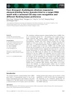
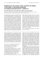
The biosynthesis of l-ascorbic acid in plants com-
prises multiple routes (Fig. 1), but not all of the
enzymes involved have yet been discovered. The
majority of the l-ascorbic acid pool is synthesized via
the so-called Smirnoff–Wheeler pathway [1]. Recently,
the final unknown enzyme from this pathway, respon-
sible for the conversion of GDP-l-galactose into
l-galactose-1-phosphate, has been identified [15]. Part
of the l-ascorbic acid pool is synthesized via d-galact-
uronic acid, a principal component of cell wall pectins
[16]. Furthermore, part of the ‘animal pathway’ with
l-gulono-1,4-lactone as the final precursor, appears to
be operating in plants, but the enzymes involved have
not yet been identified [17,18].
GALDH catalyzes the oxidation of l-galactono-1,4-
lactone to l-ascorbate with the concomitant reduction
of cytochrome c (Fig. 1). GALDH is presumed to be
an integral membrane protein of the innermitochondri-
al membrane where it shuttles electrons into the elec-
tron transport chain via cytochrome c [19]. GALDH
has been extracted from the mitochondria of a number
of plants, including cauliflower [20], sweet potato
[12,21], spinach [22] and tobacco [14]. GALDH from
cauliflower was expressed in yeast [13] and the enzyme
from tobacco has been produced in Escherichia coli
[14]. GALDH from Arabidopsis thaliana has been
expressed in E. coli as a b-galactosidase fusion protein,
but no characterization of the recombinant protein
was performed [23].
Most aldonolactone oxidoreductases contain a co-
valently bound FAD, whereas plant GALDH binds
the FAD cofactor in a noncovalent manner [14,21].
Recently, it was proposed that the aldonolactone oxi-
dase from Trypanosoma cruzi harbors a noncovalently
bound FMN as cofactor [9]. Although isolated from
various sources, aldonolactone oxidoreductases have
been poorly characterized. The molecular determinants
for the differences in cofactor binding and substrate
specificity between these enzymes are unclear, no infor-
mation is available about the nature of the active site,
and no 3D structure for this group of flavoenzymes is
Fig. 1. Proposed routes towards L-ascor-
bate biosynthesis in plants [43,44]. Oxido-
reductases involved: 1,
L-galactose
dehydrogenase; 2,
D-galacturonic acid reduc-
tase; 3, myo-inositol oxygenase; 4, GALDH;
5, GALDH or an unknown GUO ⁄ GUDH.
Galactonolactone dehydrogenase from Arabidopsis N. G. H. Leferink et al.
714 FEBS Journal 275 (2008) 713–726 ª 2008 The Authors Journal compilation ª 2008 FEBS
available. In the present study, mature GALDH from
A. thaliana (AtGALDH) was expressed in E. coli, and
its biochemical properties were investigated. Several
AtGALDH variants were constructed to address the
role of Leu56 in FAD binding.
Results
Sequence analysis
Genome analysis revealed that A. thaliana contains
one gene (At3g47930) coding for GALDH. The full-
length AtGALDH protein contains 610 amino acids
with a theoretical molecular mass of 68 496 Da. Multi-
ple sequence alignment showed that AtGALDH shares
approximately 80–90% sequence identity with
GALDH proteins from other plants. Less than 25%
sequence identity and approximately 30–40% sequence
similarity was found with other aldonolactone oxidore-
ductases. The highest degree of sequence conservation
was found in the FAD-binding domain (Fig. 2). From
the alignment, it is clear that GALDH in plants lacks
the histidine residue involved in covalent flavinylation
in GUO, ALO and GLO, but contains a leucine resi-
due instead (Leu56 in mature AtGALDH), indicating
that the flavin cofactor is noncovalently bound to the
protein.
Full-length AtGALDH contains a mitochondrial
target sequence with a putative FR ⁄ YA cleavage site
(Fig. 2). An identical cleavage site is present in the
sequences of GALDH from cauliflower, sweet potato
and tobacco [13,14,21]. N-terminal sequence analysis
of GALDH isolated from cauliflower mitochondria
showed that the mature protein starts exactly at the
tyrosine of the predicted cleavage site [13]. Although
plant GALDHs were previously identified as integral
membrane proteins of the inner mitochondrial mem-
brane [19,24], we did not find any transmembrane
regions in the sequence of mature AtGALDH.
Cloning and functional expression of AtGALDH
in E. coli
A 1.5 kb DNA fragment encoding mature
AtGALDH was PCR amplified from an A. thaliana
seedling cDNA library. The amplified fragment was
cloned into the pET23a vector under the control of
the strong T7 promoter. An in-frame fusion at the
3¢-end was made with a fragment encoding a His
6
-
tag on the vector. The resulting ORF encodes a
511-residue long polypeptide, comprising mature
AtGALDH, two extra residues (Leu and Glu) and
the His
6
-tag.
Mature AtGALDH-His
6
, with a predicted molecular
mass of 58 763 Da, was expressed in E. coli
BL21(DE3) cells as soluble cytoplasmic protein. High-
est levels of expression were found after 16 h of induc-
tion with 0.4 mm isopropyl thio-b-d-galactoside
(IPTG) at 37 °C. Expression of the recombinant His
6
-
tagged protein was confirmed by western blot analysis
with polyclonal rabbit anti-His
6
serum and by the
presence of GALDH activity in the cell extract of
IPTG-induced E. coli BL21(DE3): pET-AtGALDH-
His
6
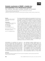
cells. The recombinant protein was purified to
apparent homogeneity by two successive chromato-
graphic steps (Fig. 3). Approximately 210 mg
of recombinant AtGALDH protein could be purified
from a 12 L batch culture containing 58 g of cells (wet
weight). The final preparation had a specific activity
of 76 UÆmg
)1
(Table 1). This ‘as isolated’ activity
increased by a factor of approximately 1.4 when
the enzyme was treated with 1 mm dithithreitol
(vide infra). Recombinant AtGALDH migrated in
SDS ⁄ PAGE as a single band with an apparent molecu-
lar mass of approximately 55 kDa (Fig. 3). This value
is in fair agreement with the calculated molecular mass
(58.8 kDa). The relative molecular mass of recombi-
nant AtGALDH was estimated to be 56 kDa by ana-
lytical size-exclusion chromatography, which indicates
a monomeric structure (data not shown).
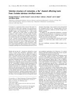
Spectral properties of AtGALDH
Recombinant AtGALDH showed a typical flavopro-
tein absorption spectrum with maxima at 276 nm,
375 nm and 450 nm and a shoulder at 475 nm
(Fig. 4A, solid line). The molar absorption coefficient
of the protein-bound flavin was determined to be
12.9 mm
)1
Æcm
)1
at 450 nm. The A
276
⁄ A
450
ratio of
the FAD-saturated protein preparation was 8.15.
The redox active flavin cofactor could be released
from the protein by boiling or acid treatment, confirm-
ing the noncovalent binding mode already predicted
from the amino acid sequence. The released cofactor
was identified as FAD by TLC.
Aerobic incubation of the protein with excess l-ga-
lactono-1,4-lactone resulted in a rapid bleaching of the
yellow color and a completely two-electron reduced
flavin spectrum, indicating that the FAD cofactor par-
ticipates in the electron-transfer reaction (Fig. 4A, dot-
ted line). Because cytochrome c is a one-electron
acceptor, the re-oxidation of AtGALDH by cyto-
chrome c involves two consecutive one-electron trans-
fer steps involving a flavin semiquinone intermediate.
In an attempt to identify the nature of this radical
species, the protein was artificially reduced by
N. G. H. Leferink et al. Galactonolactone dehydrogenase from Arabidopsis
FEBS Journal 275 (2008) 713–726 ª 2008 The Authors Journal compilation ª 2008 FEBS 715
Galactonolactone dehydrogenase from Arabidopsis N. G. H. Leferink et al.
716 FEBS Journal 275 (2008) 713–726 ª 2008 The Authors Journal compilation ª 2008 FEBS
photoreduction in the presence of EDTA and 5-deaza-
flavin (Fig. 4B). During the first part of the reduction,
an absorption peak appears at approximately 390 nm,
which is indicative for the formation of the red anionic
flavin semiquinone. Reduction proceeds until the fully
reduced flavin hydroquinone state is obtained. Expos-
ing the two-electron reduced protein to air readily
resulted in the re-appearance of the fully oxidized spec-
trum.
The stabilization of the red anionic form of the fla-
vin semiquinone intermediate together with the forma-
tion of a flavin N5 sulfite adduct are properties
commonly associated with flavoprotein oxidases, and
are indicative for the presence of a positive charge near
the flavin N1 locus [25,26]. The formation of such a
flavin-sulfite adduct results in bleaching of the yellow
color [27]. AtGALDH readily reacted with sodium sul-
fite with a dissociation constant (K
d
)of18lm for the
flavin–sulfite complex (Fig. 4C). Addition of excess
l-galactono-1,4-lactone (4 mm) to the AtGALDH–sul-
fite complex yielded the spectrum of the reduced
enzyme (cf. Fig. 4A), demonstrating that the reaction
with sulfite is reversible.
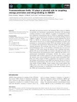
Catalytic properties of AtGALDH
Recombinant AtGALDH was highly active with its nat-
ural substrate l-galactono-1,4-lactone and its electron
acceptor cytochrome c (Table 2). The l-gulono-1,4-
lactone isomer was also oxidized at significant rate
(Table 2). AtGALDH was inhibited by the l-galactono-
1,4-lactone substrate at concentrations above 2 mm
(Fig. 5A; K
i
= 16.4 mm). No substrate inhibition
was found with l-gulono-1,4-lactone at concentrations
up to 100 mm (Fig. 5B). The substrate analogues
d-galactono-1,4-lactone, d-gulono-1,4-lactone, l-mann-
ono-1,4-lactone and d-galacturonic acid were no
substrates for AtGALDH and did not inhibit the oxida-
tion of l-galactono-1,4-lactone.
The product of the AtGALDH mediated oxidation
of l-galactono-1,4-lactone and l-gulono-1,4-lactone
was analyzed by HPLC. Because the presumed product
l-ascorbate can reduce cytochrome c, resulting in the
formation of dehydroascorbic acid, which is hydro-
lyzed to 2,3-diketo-l-gulonic acid at the pH of the
reaction, the reaction was performed without the addi-
tion of cytochrome c. Although the reaction with oxy-
gen occurs slowly, after several hours of incubation,
enough product was generated to perform the analysis.
The products of the reaction of AtGALDH with both
l-galactono-1,4-lactone and l-gulono-1,4-lactone
eluted with the same retention time as the l-ascorbic
acid reference and showed identical spectral properties
(results not shown).
AtGALDH was also active with the artificial elec-
tron acceptors phenazine methosulfate and 1,4-benzo-
quinone (Table 2). The reaction with molecular oxygen
(aerated buffer) proceeded very slowly with a bi-
molecular rate constant (k
ox
)of6· 10
2
m
)1
Æs
)1
.
Fig. 3. SDS ⁄ PAGE analysis of the purification of recombinant
AtGALDH. Lane A, low-molecular weight marker; lane B, cell
extract; lane C, Ni-NTA pool; lane D, Q-Sepharose pool.
Table 1. Purification of AtGALDH expressed in Escherichia coli.
Step
Protein
(mg)
Activity
(U)
Specific activity
(UÆmg
)1
)
Yield
(%)
Cell extract 3469 25947 7 100
Ni-NTA agarose 401 24878 62 96
Q-Sepharose 214 16365 76
a
63
a
As isolated.
Fig. 2. Multiple sequence alignment of the full length amino acid sequence of AtGALDH with several aldonolactone oxidoreductases. The
accession numbers (NCBI Entrez Protein Database) used for the multiple sequence alignment are: BoGALDH, cauliflower GALDH
(CAB09796); NtGALDH, tobacco GALDH (BAA87934); RnGUO, rat GUO (P10867); ScALO, Saccharomyces cerevisiae ALO (P54783); PgGLO,
Penicillium griseoroseum GLO (AAT80870); TbALO, Trypanosoma brucei ALO (AAX79383); MtGUDH, Mycobacterium tuberculosis GUDH
(CAB09342). Alignment was performed using
CLUSTAL W. Amino acid residue numbers are shown on the right. Identical residues are shaded
in black, similar residues are shaded in grey. The arrowhead (.) indicates the putative cleavage site of the mitochondrial targeting sequence
in plant GALDH (FR ⁄ YA). The asterisk (*) marks the histidine residue involved in covalent binding of the FAD cofactor in GUO, ALO and
GLO. The FAD-binding domain [2] is underlined.
N. G. H. Leferink et al. Galactonolactone dehydrogenase from Arabidopsis
FEBS Journal 275 (2008) 713–726 ª 2008 The Authors Journal compilation ª 2008 FEBS 717
2,6-Dichlorophenolindophenol and potassium ferri-
cyanide were no electron acceptors for recombinant
AtGALDH.
AtGALDH displayed a broad pH optimum for activ-
ity with cytochrome c between pH 8 and 9.5 with a
maximum around pH 8.8 (Fig. 5C). The activity of
AtGALDH with cytochrome c was highly dependent on
the ionic strength of the solution. Maximal activity was
at I =25mm and 75%, 30% and 10% of the maximal
activity was found at I =5mm, I = 100 mm and
I = 200 mm, respectively. No specific inhibition by
cations or anions was observed. The theoretical pI of
the recombinant AtGALDH-His
6
is 6.8. No interaction
between AtGALDH and cytochrome c (pI = 10–10.5)
was observed during analytical gel filtration at pH 8.8,
either in the absence or presence of l-galactono-1,4-
lactone (data not shown).
Recombinant AtGALDH appeared to be very
stable under storage conditions; long-term storage
(> 12 months) at )80 °C resulted in a 30–50% loss of
Fig. 4. Spectral properties of recombinant AtGALDH. (A) Aerobic
reduction with excess substrate. The reaction mixture contained
50 m
M sodium phosphate (pH 7.4), 20 lM AtGALDH and 1 mM
L
-galactono-1,4-lactone and was incubated at 25 °C. Spectra were
taken before (solid line) and after the addition of
L-galactono-1,4-lac-
tone. Complete reduction was achieved 4 min after the addition of
the substrate (dotted line). (B) Anaerobic photoreduction in the
presence of EDTA and 5-deazariboflavin. The reaction mixture con-
tained 50 m
M sodium phosphate (pH 7.4), 11 lM AtGALDH, 1 mM
EDTA and 7 lM 5-deazaflavin. Spectra were taken at regular inter-
vals before illumination (solid line), and at regular intervals during
illumination until complete reduction was achieved after 15 min
(dotted line). The dashed line and the dashed–dotted line represent
the intermediate spectra observed during the reduction after 1 min
and 2 min of illumination, respectively. Spectra were corrected for
5-deazaflavin absorption. (C) Titration of AtGALDH with sodium sul-
fite. The reaction was carried out with 10 l
M AtGALDH in 50 mM
sodium phosphate buffer (pH 7.4). Spectra are shown after the
addition of 0, 5, 10, 25, 49, 98 and 977 l
M sulfite (final concentra-
tions) until no further changes were observed. Spectra were
corrected for changes in the reaction volume during the experi-
ment. The inset shows the absorbance difference at 450 nm during
the titration, from which a dissociation constant (K
d
) for the
enzyme–sulfite complex of 18 l
M was calculated.
Table 2. Steady-state kinetic parameters of AtGALDH. Apparent
kinetic constants were determined at 25 °C in assay buffer (pH 8.8)
(I =25m
M). Substrate concentrations varied between 5 lM and
5m
M for L-galactono-1,4-lactone and between 0.5 and 100 mM for
L-gulono-1,4-lactone, with a constant cytochrome c concentration of
50 l
M. Values are presented as the mean ± SD of three experi-
ments. Electron acceptor concentrations varied between 1 l
M and
200 l
M for cytochrome c,1lM and 500 lM for phenazine metho-
sulfate and between 10 l
M and 2.3 mM for 1,4-benzoquinone, with
a constant
L-galactono-1,4-lactone concentration of 1 mM. Values
are the mean ± SD of two experiments.
K
m
(mM) k
cat
(s
)1
)
k
cat
⁄ K
m
(mM
)1
Æs
)1
)
Substrate
L-Galactono-1,4-lactone 0.17 ± 0.01 134 ± 5 7.7 · 10
2
L-Gulono-1,4-lactone 13.1 ± 2.8 4.0 ± 0.2 3.1 · 10
)1
Electron acceptor
Cytochrome c 0.034 ± 0.002 151 ± 1 4.4 · 10
3
Phenazine methosulfate 0.026 ± 0.004 64 ± 3 2.4 · 10
3
1,4-Benzoquinone 0.280 ± 0.05 108 ± 12 3.9 · 10
2
Galactonolactone dehydrogenase from Arabidopsis N. G. H. Leferink et al.
718 FEBS Journal 275 (2008) 713–726 ª 2008 The Authors Journal compilation ª 2008 FEBS
activity, which could be completely restored upon
incubation with the reducing agent dithiothreitol.
Recombinant AtGALDH was relatively stable when
incubated at elevated temperatures, with a half-life of
20 min at 52 °C. In the presence of excess FAD, the
half-life at 52 °C was increased to 115 min, suggesting
that the holo form of the enzyme is more thermostable
than the apo form. Both local and global unfolding
play a role in the thermoinactivation process. This is
concluded from the fact that, in both incubations,
10 ± 4% of enzyme activity was recovered at the end
of the heating process when excess FAD was included
in the assay mixture.
Properties AtGALDH Leu56 mutants
To determine more about the role of Leu56 in the
FAD binding site, several AtGALDH Leu56 variants
were constructed (see Experimental procedures). The
L56A, L56C and L56H variants were expressed and
purified in essentially the same way as wild-type
AtGALDH-His
6
with similar yields (see Experimental
procedures). The L56I and L56F variants were purified
in a single gravity-flow Ni-affinity chromatography
step with yields and purities comparable to the other
variants.
All AtGALDH Leu56 variants contained noncova-
lently bound FAD. The FAD cofactor was partially
released during the purification procedure, a phenome-
non hardly observed with the wild-type enzyme. The
holo forms of the Leu56 variants could easily be
reconstituted by the addition of FAD and their flavin
absorption properties were almost identical to the
wild-type enzyme.
The Leu56 variants showed interesting catalytic
properties. The L56I variant displayed a higher turn-
over rate with the l-galactono-1,4-lactone substrate
than wild-type AtGALDH and the L56F variant
(240 s
)1
versus 134 and 126 s
)1
, respectively). The other
Leu56 variants were all considerably less active than
the wild-type enzyme and showed remarkable differ-
ences in apparent Michaelis constants for the l-galac-
tono-1,4-lactone substrate (Table 3). L56H, as well as
L56I and L56F, showed a relatively low K
m
, which
was in the same range as wild-type AtGALDH,
whereas the L56C and L56A variants had rather high
K
m
values in the mm range. A similar trend in K
m
val-
ues was found for the l-gulono-1,4-lactone substrate.
As for wild-type AtGALDH, molecular oxygen could
not serve as efficient electron acceptor for the mutant
enzymes.
As noted above, the FAD cofactor is more loosely
bound in the Leu56 variants than in wild-type
AtGALDH. Cofactor binding was analyzed in more
detail by nickel-affinity chromatography [28]. Washing
the immobilized proteins with chaotropic salts resulted
in elution of the flavin for all Leu56 variants, but to a
lesser extent for wild-type AtGALDH as judged by the
presence of the yellow color. The (apo)proteins were
subsequently eluted from the column with buffer
Fig. 5. Activity of recombinant AtGALDH. (A) Michaelis–Menten kinetics of the AtGALDH-mediated oxidation of L-galactono-1,4-lactone.
(B) Michaelis–Menten kinetics of the AtGALDH-mediated oxidation of
L-gulono-1,4-lactone. (C) AtGALDH activity as a function of pH. Activi-
ties were measured in 25 m
M Hepes (pH 7–8), Taps (pH 8–9) and Ches (pH 9–9.5) buffers with a constant ionic strength of 25 mM adjusted
with NaCl containing 1 m
ML-galactono-1,4-lactone and 50 lM cytochrome c at 25 °C.
Table 3. Steady-state kinetic parameters of AtGALDH variants.
Apparent kinetic constants were determined at 25 °C in assay buf-
fer (pH 8.8) (I =25m
M) with L-galactono-1,4-lactone concentrations
varying between 10 l
M and 10 mM and a constant cytochrome c
concentration of 50 l
M. Values are the mean ± SD of at least two
experiments.
Enzyme K
m
(mM) k
cat
(s
)1
) k
cat
⁄ K
m
(mM
)1
Æs
)1
)
Wild-type 0.17 ± 0.01 134 ± 5 7.7 · 10
2
L56I 0.32 ± 0.01 240 ± 12 7.5 · 10
2
L56H 0.12 ± 0.01 32 ± 1 2.6 · 10
2
L56F 0.56 ± 0.02 126 ± 1 2.3 · 10
2
L56C 0.99 ± 0.05 76 ± 3 7.8 · 10
1
L56A 1.7 ± 0.05 45 ± 2 2.6 · 10
1
N. G. H. Leferink et al. Galactonolactone dehydrogenase from Arabidopsis
FEBS Journal 275 (2008) 713–726 ª 2008 The Authors Journal compilation ª 2008 FEBS 719
containing 300 m m imidazole and tested for activity.
In the absence of FAD in the assay mixtures, wild-type
AtGALDH and the L56F and L56I variants still
contained respectively 60%, 50% and 40% of their
original activity, whereas the other variants had lost
80–90% of their activity. All Leu56 variants regained
most of their activity (60–90%) in the presence of
FAD, whereas the activity of variant L56C was
restored to < 30%. The L56C variant is rather unsta-
ble without its cofactor bound, and irreversibly forms
aggregates after elution from the affinity column. It is
clear that, under the conditions applied, FAD is most
firmly bound in the wild-type enzyme and in the vari-
ants in which Leu56 is replaced by (large) hydrophobic
residues. Replacing Leu56 with a polar or less bulky
residue results in easier loss of FAD, indicating that
the interaction of Leu56 with the cofactor is of hydro-
phobic nature and may also involve a steric effect.
The thermal stability of variant L56H was examined
in more detail. This variant, with a half-life of 8 min
at 52 °C, appeared to be somewhat less thermostable
than wild-type AtGALDH. Addition of FAD during
the incubation increased the half-life of L56H at 52 °C
to 46 min.
Discussion
In the present study, we present for the first time a
detailed investigation of the biochemical properties of
recombinant AtGALDH and some active site variants.
By contrast with an earlier report [17], AtGALDH is
not strictly specific for l -galactono-1,4-lactone. The
enzyme oxidizes l-gulono-1,4-lactone at significant
rate, but the catalytic efficiency for the gulonolactone
isomer is relatively low. For GALDH from sweet
potato and tobacco, it was reported that these enzymes
also oxidize the gulonolactone isomer [12,14], but no
kinetic parameters were provided. From our results,
we conclude that AtGALDH shows a high enantiopre-
ference for l-galactono-1,4-lactone and that a differ-
ence in orientation of the 3-hydroxyl group of the
substrate is responsible for a 100-fold higher K
m
and
3000-fold lower catalytic efficiency.
The main precursor of l-ascorbate in plants is l-ga-
lactono-1,4-lactone [1]. It has been demonstrated that
plants can also produce l-ascorbate via l-gulono-1,4-
lactone, but the enzymes involved are unknown.
Arabidopsis cell suspensions can synthesize and
accumulate l-ascorbate from the precursor l-gulono-
1,4-lactone [18]. Furthermore, l-gulono-1,4-lactone
oxidase ⁄ dehydrogenase activity has been demonstrated
in hypocotyl homogenates of kidney beans [24] and in
cytosolic and mitochondrial fractions from Arabidop-
sis cell suspensions [18] and potato tubers [17]. These
data suggest the existence of differently localized iso-
zymes that can produce vitamin C from either l-galac-
tono- or l-gulono-1,4-lactone. Bartoli et al. [19]
predicted that GALDH from sweet potato tubers is an
integral membrane protein with three transmembrane
regions. We did not find any transmembrane regions
in the sequence of mature AtGALDH. In agreement
with this, the enzyme was expressed in soluble form in
E. coli. This leaves the possibility that the observed
gulonolactone oxidizing capability of AtGALDH is of
significance in vivo.
A recent study on the RNA interference silencing of
GALDH from tomato revealed the importance of
GALDH for plant and fruit growth. A severe reduc-
tion in GALDH activity can be lethal to the plant.
Interestingly, the total ascorbate content remained
unchanged in the GALDH silenced plants. As possible
explanations, the reduction in ascorbate turnover and
the activation of alternative ascorbate biosynthesis
pathways were proposed [29]. Although the gulonolac-
tone activity of AtGALDH might be of physiological
relevance, it cannot be excluded that other aldonolac-
tone oxidoreductases with different subcellular local-
izations are responsible for the observed gulonolactone
activity in vivo. It has been proposed that members of
a putative subfamily of VAO-like flavoproteins might
be responsible for the conversion of l-gulono-1,4-lac-
tone into l-ascorbate [17]. Sequence analysis of the
predicted gene products suggest that they are targeted
to different subcellular locations.
To date, no information was available about the
thermal stability of GALDH enzymes. AtGALDH
appeared to be a rather stable enzyme, although it
looses its FAD cofactor at elevated temperatures. The
strong increase in thermal stability in the presence of
excess FAD indicates that the cofactor protects the
enzyme from irreversible unfolding or aggregation.
Covalent flavinylation has also been associated with
improving flavoprotein stability, a covalent flavin–pro-
tein link is presumed to have a similar stabilizing effect
as a disulfide bridge [30]. Nevertheless, several aldono-
lactone oxidoreductases with a covalently bound
FAD are less stable than AtGALDH. ALO from
Candida albicans completely lost activity within 1 min
at 50 °C [10]. GLO from Pennicillium cyaneo-fulvum
(renamed Pennicillium griseoroseum) quickly lost its
activity above 45 °C [6] and, in addition, rat GUO
readily lost its activity at elevated temperatures;
90% of the activity was lost after 10 min incubation
at 49 °C [31]. The thermal stability of AtGALDH is
more comparable to that of GUDH from Glucono-
bacter oxydans [32] and Mycobacterium tuberculosis [7].
Galactonolactone dehydrogenase from Arabidopsis N. G. H. Leferink et al.
720 FEBS Journal 275 (2008) 713–726 ª 2008 The Authors Journal compilation ª 2008 FEBS
These enzymes lost approximately 50% of their activ-
ity after 5 min incubation at 55 and 60 °C, respec-
tively. The absence of a covalent flavin link could
provide GALDH with a greater conformational flexi-
bility which may be needed for cross-talk with cyto-
chrome c.
The mechanism of l-ascorbate production by At-
GALDH involves two half-reactions. In the reductive
half-reaction, the oxidized flavin cofactor is converted
to the hydroquinone state by the l-galactono-1,4-lac-
tone substrate. The two-electron reduced enzyme is
then re-oxidized in the oxidative half-reaction by cyto-
chrome c. This half-reaction involves two subsequent
one-electron steps and the formation of a flavin semi-
quinone radical. Spectral analysis revealed that At-
GALDH is able to form the red anionic flavin
semiquinone, which was visualized by artificial photo-
reduction of the protein and is characterized by
a strong absorbance at approximately 390 nm. At-
GALDH also readily reacted with sulfite, resulting in
the formation of a flavin N5 sulfite adduct, with a
K
d
of 18 lm for the enzyme–sulfite complex. The sta-
bilization of the red anionic semiquinone and the for-
mation of a flavin N5 sulfite adduct are properties
commonly associated with flavoprotein oxidases [27].
However, AtGALDH is not the only exception to this
rule. Flavocytochrome b
2
also stabilizes the red anionic
semiquinone and a flavin N5 sulfite adduct, and is
poorly active with oxygen [26,33]. In flavocyto-
chrome b
2
, an Arg residue is involved in both catalysis
and the stabilization of the N5 sulfite adduct [34]. A
similar situation is observed in adenosine-5¢-phopho-
sulfate reductase, another flavoprotein for which a
crystal structure of the enzyme–sulfite complex is
known [35]. Both flavocytochrome b
2
and adenosine-
5¢-phophosulfate reductase do bind a negatively
charged substrate. Therefore, it will be of interest to
determine whether a positively charged residue is pres-
ent in the active site of AtGALDH and related
enzymes.
Many aldonolactone oxidoreductases contain a
covalently bound FAD cofactor. The possible advanta-
ges of such a mode of flavin binding include saturation
of the active site with cofactor in flavin deficient envi-
ronments, anchoring of the isoalloxazine ring, and
modulating the redox properties [30,36]. AtGALDH
lacks the histidine involved in covalent attachment of
the FAD cofactor, but contains a leucine (Leu56)
at this position. Replacement of Leu56 into His in
AtGALDH revealed that the presence of a histidine at
this position does not initiate covalent binding of the
cofactor. Covalent coupling of the FAD cofactor
presumably is an autocatalytic process, requiring a
preorganized binding site [37]. Covalent flavinylation
commonly requires a base-assisted attack of the FAD
cofactor, resulting in a flavoquinone methide interme-
diate and subsequent formation of the covalent link
[30]. Mutagenesis studies in VAO revealed that the his-
tidine residue involved in covalent cofactor binding
(His422) is activated by a neighboring base (His61) for
attack of the C8a position of the isoalloxazine ring,
thus forming the covalent bond [37]. Covalent flaviny-
lation in the AtGALDH-L56H might thus require
nucleophilic activation of His56. The prediction of
such an activating base in the sequence of AtGALDH
is hampered by the lack of structural information for
GALDH and related aldonolactone oxidoreductases.
Leu56 replacements of AtGALDH established that
Leu56 plays an important role in binding of the non-
covalently bound FAD cofactor and in catalysis. Vari-
ants with a bulky hydrophobic residue at position 56
bind the cofactor more tightly than variants containing
small and ⁄ or polar residues. The catalytic and FAD-
binding properties of the Leu56 variants are not easily
explained but possibly reflect subtle changes in the
protein–FAD interaction rather than a direct inter-
action of residue 56 with the substrate.
In conclusion, we have described for the first time
the biochemical properties of recombinant AtGALDH
and some active site variants. The results obtained pro-
vide a good framework for further structure–function
relationship studies aimed at identifying important res-
idues involved in catalysis and flavin binding.
Experimental procedures
Chemicals
Nickel nitrilotriacetic acid (Ni-NTA) agarose was pur-
chased from Qiagen (Valencia, CA, USA) and Bio-Gel
P-6DG was from Bio-Rad (Hercules, CA, USA). HiLoad
26 ⁄ 10 Q-Sepharose HP, Superdex 200 HR 10 ⁄ 30, low-
molecular weight protein marker, prestained kaleidoscope
protein standards, and the reference proteins catalase
(232 kDa), aldolase (158 kDa), BSA (68 kDa) and
ovalbumin (43 kDa) were obtained from Pharmacia Biotech
(Uppsala, Sweden). l-Galactono-1,4-lactone, l-gulono-1,4-
lactone, d-gulono-1,4-lactone, l -mannono-1,4-lactone,
d-galacturonic acid, FAD, FMN, riboflavin, reduced
glutathione (GSH), nitroblue tetrazolium, 5-bromo-4-chlor-
3-indolylphosphate, bovine heart cytochrome c, 1,4-benzo-
quinone and phenazine methosulphate were from
Sigma-Aldrich (St Louis, MO, USA). d-Galactono-1,4-lac-
tone was from Koch-Light LTD (Haverhill, Suffolk, UK).
l-Ascorbic acid, glucose and 2,6-dichlorophenolindophenol
were from Merck (Darmstadt, Germany). IPTG and
N. G. H. Leferink et al. Galactonolactone dehydrogenase from Arabidopsis
FEBS Journal 275 (2008) 713–726 ª 2008 The Authors Journal compilation ª 2008 FEBS 721
dithiothreitol were obtained from MP Biomedicals (Irvine,
CA, USA). Secondary antibody conjugated to alkaline
phosphatase and DNaseI were from Boehringer Mannheim
GmbH (Mannheim, Gernamy). Restriction endonucleases,
T4-DNA ligase and dNTPs were purchased from Invitro-
gen (Carlsbad, CA, USA). Pwo DNA polymerase, glucose
oxidase and Pefabloc SC were obtained from Roche Diag-
nostics GmbH (Mannheim, Germany). Oligonucleotides
were synthesized by Eurogentec (Liege, Belgium). The
pET23a(+) expression vector and E. coli strain BL21(DE3)
were from Novagen (San Diego, CA, USA). All other
chemicals were from commercial sources and of the purest
grade available.
Sequence analysis
The genome of A. thaliana was analyzed for the presence
of GALDH and related aldonolactone oxidoreductase
sequences at . blast-p analysis
( was performed to
determine GALDH orthologs in other genomes [38]. Multi-
ple sequence alignments were made using clustal w
software [39]. targetp ( />TargetP) and psort () tools were used
to predict the subcellular localization of AtGALDH and
tmpred ( />form.html) was used to predict the presence of transmem-
brane regions in the sequence of AtGALDH.
Cloning of AtGALDH cDNA for expression in
E. coli
A 1.5 kb DNA fragment encoding mature AtGALDH
(amino acids 102–610) was PCR amplified from A. thaliana
(ecotype Columbia) seedling cDNA, using the oligo-
nucleotides AtGALDH_fw102 (5¢-GGAATTC
CATATG
TACGCTCCTTTACCTGAAG-3¢) and AtGALDH_rv
(5¢-CCG
CTCGAGAGCAGTGGTGGAGACTG-3¢), intro-
ducing NdeI and XhoI restriction sites (underlined),
respectively. The amplified fragment was cloned between the
NdeI and XhoI sites of the pET23a(+) expression vector
fused to a C-terminal His
6
-tag. The resulting construct (pET-
AtGALDH-His
6
) was verified by automated sequencing of
both strands and electroporated to E. coli BL21(DE3) cells
for recombinant expression.
Site-directed mutagenesis
The AtGALDH mutants L56A, L56C, L56F, L56H and
L56I were constructed using pET-AtGALDH-His
6
as tem-
plate with the QuikChange II method (Stratagene, La Jolla,
CA, USA). The oligonucleotides used are listed in Table 4,
changed nucleotides are underlined. Successful mutagenesis
was confirmed by automated sequencing of both strands.
The resulting constructs pET-AtGALDH_L56H-His
6
,
pET-AtGALDH_L56C-His
6
, pET-AtGALDH_L56A-His
6
,
pET-AtGALDH_L56I-His
6
and pET-AtGALDH_L56F-
His
6
were electroporated to E. coli BL21(DE3) cells for
recombinant expression.
Enzyme production and purification
The A
˚
kta explorer FPLC system (Pharmacia Biotech) was
used for all purification steps. For enzyme production,
E. coli BL21(DE3) cells, harboring a pET-AtGALDH plas-
mid, were grown in LB medium supplemented with
100 lgÆmL
)1
ampicillin until an attenuance of 0.7 at
D
600 nm
was reached. Expression was induced by the addi-
tion of 0.4 mm IPTG and the incubation was continued
for 16 h at 37 °C. Cells (58 g wet weight) were harvested by
centrifugation, resuspended in 60 mL of 100 mm potassium
phosphate, 1 mm Pefabloc SC and 5 mm GSH (pH 7.4)
and subsequently passed twice through a precooled French
Pressure cell (SLM Aminco, SLM Instruments, Urbana, IL,
USA) at 10 000 psi. The resulting homogenate was centri-
fuged at 25 000 g for 30 min at 4 °C to remove cell debris,
and the supernatant was applied onto a Ni-NTA agarose
column (16 · 50 mm) equilibrated with 50 mm sodium
phosphate, 300 mm NaCl and 5 mm GSH (pH 7.4). The
column was washed with two volumes of equilibration buf-
fer and two volumes of equilibration buffer containing
20 mm imidazole. The enzyme was eluted with 300 mm
imidazole in equilibration buffer. The active fraction was
dialyzed at 4 °C against 25 mm Tris–HCl, 0.1 mm EDTA,
5mm GSH and 200 lm FAD (pH 7.4). After removal of
insoluble material by centrifugation at 25 000 g for 30 min
at 4 °C, the soluble fraction was applied onto a Hi-
Load 26 ⁄ 10 Q-Sepharose HP column equilibrated with
25 mm Tris–HCl and 5 mm GSH (pH 7.4). After washing
with two column volumes of starting buffer, the protein
was eluted with a linear gradient of NaCl (0–0.2 m) in the
same buffer. Active fractions were pooled and concentrated
using the Ni-NTA agarose column (see above). The final
preparation was saturated with FAD; excess FAD was
removed by size-exclusion chromatography using a Bio-Gel
P-6DG column (15 · 130 mm) equilibrated with 20 mm
sodium phosphate and 0.1 mm dithiothreitol (pH 7.4) and
Table 4. Oligonucleotides used for the construction of AtGALDH
Leu56 variants. Only sense primers are shown, changed nucleo-
tides are underlined.
Variant Oligonucleotide sequence (5¢ to 3¢)
L56A CCCGTTGGATCGGGT
GCCTCGCCTAATGGGATTG
L56C CCCGTTGGATCGGGT
TGCTCGCCTAATGGGATTG
L56F CCCGTTGGATCGGGT
TTTTCGCCTAATGGGATTG
L56H CCCGTTGGATCGGGTC
ACTCGCCTAATGGGATTG
L56I CCCGTTGGATCGGGT
ATTTCGCCTAATGGGATTG
Galactonolactone dehydrogenase from Arabidopsis N. G. H. Leferink et al.
722 FEBS Journal 275 (2008) 713–726 ª 2008 The Authors Journal compilation ª 2008 FEBS
the enzyme was stored at )80 °C. Before analysis, the
enzyme was freshly incubated with 1 mm dithiothreitol.
Protein analysis
The SDS ⁄ PAGE was performed using 12.5% acrylamide
slab gels essentially as described by Laemmli [40]. Proteins
were stained using Coomassie Brilliant Blue R-250. For wes-
tern blot analysis, the gels were blotted onto a nitrocellulose
membrane (Optitran BA-S 85 Reinforced NC; Schleicher &
Schuell GmbH, Whatman group, Dassel, Germany) and
incubated with polyclonal rabbit anti-His
6
sera and a sec-
ondary antibody coupled to alkaline phosphatase. Proteins
were visualized using nitroblue tetrazolium and 5-bromo-4-
chlor-3-indolylphosphate as substrates for alkaline phospha-
tase detection. Total protein concentrations were estimated
using the Bradford protein assay from Bio-Rad with BSA as
standard [41]. Analytical gel filtration to investigate the
hydrodynamic properties of AtGALDH was performed on
a Superdex 200 HR 10 ⁄ 30 column running in 50 mm potas-
sium phosphate and 150 mm potassium chloride (pH 7.0),
coupled to the A
˚
kta explorer FPLC system. Gel filtration
experiments to study the interaction of AtGALDH with
bovine heart cytochrome c was performed using the same
column running in potassium pyrophosphate, pH 8.8,
I =25mm. Desalting or buffer exchange of small aliquots
of enzyme was performed with Bio-Gel P-6DG columns.
Spectral analysis
Absorption spectra were recorded at 25 °C on a Hewlett
Packard (Loveland, CO, USA) 8453 diode array spectro-
photometer in 50 mm sodium phosphate (pH 7.4). The
molar absorption coefficient of protein-bound FAD was
determined by recording the absorption spectrum of
AtGALDH in the presence and absence of 0.1% (w ⁄ v)
SDS, assuming a molar absorption coefficient for free
FAD of 11.3 mm
)1
Æcm
)1
at 450 nm. Purified enzyme con-
centrations were routinely determined by measuring the
absorbance at 450 nm using the molar absorption coeffi-
cient for protein-bound FAD. Spectra were collected
and analyzed using the UV-Visible chemstation software
package (Hewlett Packard).
Photoreduction of AtGALDH (11 lm) in the presence of
EDTA and 5-deazaflavin was performed in 50 mm sodium
phosphate (pH 7.4) as described previously [42]. Catalytic
amounts of glucose oxidase and 1.5 mm b-d-glucose were
added to scavenge final traces of oxygen and catalase was
added to remove hydogen peroxide formed during the reac-
tion. Solutions were made anaerobic by alternate evacua-
tion and flushing with oxygen-free argon. Illumination was
performed in a 25 °C water bath with a 375 W light source
(Philips, Eindhoven, the Netherlands) at a distance of
15 cm. Spectra were taken at regular intervals during illu-
mination until complete reduction was achieved.
Titration of AtGALDH (10 l m) with sodium sulfite was
carried out in 50 mm sodium phosphate buffer (pH 7.4). A
1 m sodium sulfite stock solution in 50 mm sodium phos-
phate buffer (pH 7.4) was freshly prepared before use, suit-
able dilutions were made in the same buffer before addition
to the enzyme solution. Spectra were taken until no further
change was observed before the next addition was carried
out. Final sodium sulfite concentrations were 0, 5, 10, 25,
49, 98, 196 and 977 lm. K
d
was calculated from the change
in the absorbance at 450 nm during the titration using a
direct nonlinear regression fit to the data with the igor pro
program (Wavemetrics, Lake Oswego, OR, USA):
DA450 ¼
DA450
max
 sulfite½
K
d
þ sulfite½
: ð1Þ
Cofactor determination
The flavin cofactor of AtGALDH was determined by TLC.
The cofactor was released from the protein by boiling for
30 min or acid treatment. The protein precipitate was
removed by centrifugation and the supernatant was applied
together with the reference compounds FAD, FMN and
riboflavin onto a TLC plate (Baker-flex Silica Gel 1B2;
JT Baker Inc., Phillipsburg, NY, USA). Butanol ⁄ acetic
acid ⁄ water (5 : 3 : 3) was used as the mobile phase.
Activity measurements
GALDH activity was routinely assayed by following the
reduction of cytochrome c at 550 nm at 25 °C on a Hewlett
Packard 8453 diode array spectrophotometer. Initial veloc-
ity values were calculated using a molar difference absorp-
tion coefficient (De)of21mm
)1
Æcm
)1
for reduced minus
oxidized cytochrome c. Because dithiothreitol interferes
with the reaction, it was removed from the enzyme solution
by Bio-Gel P-6DG gel filtration immediately prior to use.
For activity measurements enzyme preparations were
diluted in assay buffer containing 1 mgÆmL
)1
BSA. The
standard assay mixture (1 mL) contained assay buffer with
pH 8.8 and an ionic strength of 25 mm,1mml-galactono-
1,4-lactone and 50 lm cytochrome c; the reaction was
started by the addition of enzyme. One unit of enzyme
activity (U) is defined as the amount of enzyme that oxi-
dizes 1 lmol of l-galactono-1,4-lactone per min, which is
equivalent to the reduction of 2 lmol of cytochrome c [12].
The optimal pH for activity of AtGALDH was determined
using 25 mm Hepes, Taps and Ches buffers with vary-
ing pH (pH 7–9.5) and adjusted to an ionic strength of
25 mm with NaCl.
The kinetic parameters K
m
and V
max
were calculated
from multiple measurements with various substrate concen-
trations using a direct nonlinear regression fit to the data.
The activity of AtGALDH with l-gulono-1,4-lactone fol-
lowed Michaelis–Menten kinetics, K
m
and V
max
values of
N. G. H. Leferink et al. Galactonolactone dehydrogenase from Arabidopsis
FEBS Journal 275 (2008) 713–726 ª 2008 The Authors Journal compilation ª 2008 FEBS 723
wild-type GALDH for l-gulono-1,4-lactone were calculated
using the Michaelis–Menten equation. The K
m
and V
max
values of wild-type GALDH for l-galactono-1,4-lactone
were calculated using an equation which includes substrate
inhibition:
V ¼
V
app
½S
K
m
þ½Sþ
½S
2
K
i
ð2Þ
The turnover number (k
cat
,s
)1
) was calculated using:
k
cat
¼
V
max
 58:8kDa
60
ð3Þ
The activity of AtGALDH with other electron acceptors
was also determined from initial rate experiments. In all
cases, dithiothreitol can interfere with the reaction, so it
was removed from the enzyme stock solution by Bio-Gel
P-6DG gel filtration immediately prior to use. All reactions
were performed with 1 mml-galactono-1,4-lactone as sub-
strate in assay buffer with pH 8.8 and an ionic strength of
25 mm at 25 °C. The activity with 2,6-dichlorophenolindo-
phenol was measured at 600 nm (e
600
=22mm
)1
Æcm
)1
),
the activity with phenazine methosulfate was measured in
the presence of the mediator 2,6-dichlorophenolindophenol
at 600 nm, the activity with potassium ferricyanide was
measured at 420 nm (e
420
=1mm
)1
Æcm
)1
), and the activity
with 1,4-benzoquinone was measured at 290 nm
(e
290
= 2.3 mm
)1
Æcm
)1
). The reactivity with molecular oxy-
gen was determined with a polarographic oxygen uptake
assay using a Clark electrode.
Enzyme stability
The thermal stability of AtGALDH was determined
at 52 °C. Enzyme preparations were diluted in 50 mm
sodium phosphate (pH 7.4) to a final concentration of 1 lm
and incubated at the indicated temperatures in the presence
or absence of 10 lm FAD. The time-dependent loss of
activity was followed by the standard assay procedure;
aliquots were withdrawn from the incubation mixtures at
intervals and assayed for residual enzyme activity. To dis-
criminate between global and local unfolding, at the end of
the heating period, the enzyme activity was also measured
in the presence of 10 lm FAD.
Apoprotein preparation
The ease of cofactor release was examined by on-column
washing with chaotropic salts [28]. Accordingly, the His-
tagged proteins (approximately 2.5 mg) were bound to a
1 mL Ni-affinity gravity-flow column (HisGraviTrap;
GE Healthcare Bioscience AB, Uppsala, Sweden) in the
presence of 50 mm sodium phosphate, 300 mm NaCl
(pH 7.4); the column was washed with ten column volumes
of the same buffer containing 2 m KBr and subsequently
with buffer containing 2 m KBr and 2 m urea. (Apo)protein
was collected by elution with 300 mm imidazole in buffer.
The collected fractions were analyzed for GALDH activity
in the absence and presence of FAD.
Product analysis
To analyze the products of the enzymatic oxidation of
l-galactono-1,4-lactone and l-gulono-1,4-lactone by
AtGALDH, 1 mm solutions of these compounds were incu-
bated for 2 h at 25 °C with the recombinant enzyme
(10 lg) in air saturated assay buffer (0.24 mm O
2
). Before
analysis, the incubation mixtures were centrifuged for
10 min at maximal speed in a standard tabletop microfuge.
The supernatants were analyzed by HPLC using a Waters
(Milford, MA, USA) 600 controller with a Waters In Line
Degasser coupled to a Waters 996 photodiode array detec-
tor. Separation was performed at room temperature on a
Alltima C
18
column (150 · 4.6 mm, 5 lm particle size; All-
tech Associates, Deerfield, IL, USA). The column was
equilibrated with 0.1% trifluoroacetic acid, 5% acetonitrile
in water, elution was performed with a linear gradient
of 5–100% acetonitrile in 20 min. Chromatograms were
recorded at 254 nm. l-Ascorbic acid, l-galactono-1,4-lac-
tone and l-gulono-1,4-lactone served as references. System
control and data collection and analysis was performed
using the millenium
32
software package (Waters). Due to
its hydrophilic nature, l-ascorbate was not retarded on the
column under these conditions and eluted in the flow-
through with a retention time of approximately 2 min.
Acknowledgements
We are grateful to Yu Lu and Daan Binnewijzend for
experimental contributions. We thank Marco Fraaije
for critically reading the manuscript. This research was
supported by a grant from the Carbohydrate Research
Centre Wageningen (CRC-W).
References
1 Smirnoff N & Wheeler GL (2000) Ascorbic acid in
plants: biosynthesis and function. Crit Rev Biochem
Mol Biol 35, 291–314.
2 Fraaije MW, Van Berkel WJH, Benen JAE, Visser J &
Mattevi A (1998) A novel oxidoreductase family sharing
a conserved FAD-binding domain. Trends Biochem Sci
23, 206–207.
3 Linster CL & Van Schaftingen E (2007) Vitamin C.
Biosynthesis, recycling and degradation in mammals.
FEBS J 274, 1–22.
4 Nishikimi M, Fukuyama R, Minoshima S, Shimizu N
& Yagi K (1994) Cloning and chromosomal mapping of
Galactonolactone dehydrogenase from Arabidopsis N. G. H. Leferink et al.
724 FEBS Journal 275 (2008) 713–726 ª 2008 The Authors Journal compilation ª 2008 FEBS
the human nonfunctional gene for l-gulono-gamma-lac-
tone oxidase, the enzyme for l-ascorbic acid biosynthe-
sis missing in man. J Biol Chem 269, 13685–13688.
5 Huh WK, Lee BH, Kim ST, Kim YR, Rhie GE, Baek
YW, Hwang CS, Lee JS & Kang SO (1998) d-Ery-
throascorbic acid is an important antioxidant molecule
in Saccharomyces cerevisiae. Mol Microbiol 30, 895–903.
6 Salusja
¨
rvi T, Kalkkinen N & Miasnikov AN (2004)
Cloning and characterization of gluconolactone oxidase
of Penicillium cyaneo-fulvum ATCC 10431 and evalua-
tion of its use for production of d-erythorbic acid in
recombinant Pichia pastoris. Appl Environ Microbiol 70,
5503–5510.
7 Wolucka BA & Communi D (2006) Mycobacte-
rium tuberculosis possesses a functional enzyme for the
synthesis of vitamin C, l-gulono-1,4-lactone dehydroge-
nase. FEBS J 273, 4435–4445.
8 Wilkinson SR, Prathalingam SR, Taylor MC, Horn D
& Kelly JM (2005) Vitamin C biosynthesis in trypano-
somes: a role for the glycosome. Proc Natl Acad Sci
USA 102, 11645–11650.
9 Logan FJ, Taylor MC, Wilkinson SR, Kaur H & Kelly
JM (2007) The terminal step in vitamin C biosynthesis
in Trypanosoma cruzi is mediated by a FMN-dependent
galactonolactone oxidase. Biochem J 407, 419–426.
10 Huh WK, Kim ST, Yang KS, Seok YJ, Hah YC &
Kang SO (1994) Characterisation of d-arabinono-1,4-
lactone oxidase from Candida albicans ATCC 10231.
Eur J Biochem 225, 1073–1079.
11 Kiuchi K, Nishikimi M & Yagi K (1982) Purification
and characterization of l-gulonolactone oxidase from
chicken kidney microsomes. Biochemistry 21, 5076–
5082.
12 O
ˆ
ba K, Ishikawa S, Nishikawa M, Mizuno H &
Yamamoto T (1995) Purification and properties of
l-galactono-c-lactone dehydrogenase, a key enzyme for
ascorbic acid biosynthesis, from sweet potato roots.
J Biochem (Tokyo) 117, 120–124.
13 Østergaard J, Persiau G, Davey MW, Bauw G & Van
Montagu M (1997) Isolation of a cDNA coding for
l-galactono-c-lactone dehydrogenase, an enzyme
involved in the biosynthesis of ascorbic acid in plants.
Purification, characterization, cDNA cloning, and
expression in yeast. J Biol Chem 272, 30009–30016.
14 Yabuta Y, Yoshimura K, Takeda T & Shigeoka S
(2000) Molecular characterization of tobacco mitochon-
drial l-galactono-c-lactone dehydrogenase and its
expression in Escherichia coli. Plant Cell Physiol 41,
666–675.
15 Linster CL, Gomez TA, Christensen KC, Adler LN,
Young BD, Brenner C & Clarke SG (2007) Arabidopsis
VTC2 encodes a GDP-l-galactose phosphorylase, the
last unknown enzyme in the Smirnoff-Wheeler pathway
to ascorbic acid in plants. J Biol Chem 282, 18879–
18885.
16 Agius F, Gonza
´
lez-Lamothe R, Caballero JL,
Mun
˜
oz-Blanco J, Botella MA & Valpuesta V (2003)
Engineering increased vitamin C levels in plants by
overexpression of a d-galacturonic acid reductase. Nat
Biotechnol 21, 177–181.
17 Wolucka BA & Van Montagu M (2003) GDP-mannose
3’,5’-epimerase forms GDP-l-gulose, a putative interme-
diate for the de novo biosynthesis of vitamin C in
plants. J Biol Chem 278, 47483–47490.
18 Davey MW, Gilot C, Persiau G, Østergaard J, Han Y,
Bauw GC & Van Montagu MC (1999) Ascorbate bio-
synthesis in Arabidopsis cell suspension culture. Plant
Physiol 121, 535–543.
19 Bartoli CG, Pastori GM & Foyer CH (2000) Ascorbate
biosynthesis in mitochondria is linked to the electron
transport chain between complexes III and IV. Plant
Physiol 123, 335–344.
20 Mapson LW & Breslow E (1958) Biological synthesis of
ascorbic acid: l-galactono-c -lactone dehydrogenase.
Biochem J 68, 395–406.
21 Imai T, Karita S, Shiratori G, Hattori M, Nunome T,
O
ˆ
ba K & Hirai M (1998) l-galactono-c -lactone dehy-
drogenase from sweet potato: purification and cDNA
sequence analysis. Plant Cell Physiol 39, 1350–1358.
22 Mutsuda M, Ishikkawa T, Takeda T & Shigeoka S
(1995) Subcellular localization and properties of l-ga-
lactono-c-lactone dehydrogenase in spinach leaves.
Biosci Biotechnol Biochem 59, 1983–1984.
23 Tamaoki M, Mukai F, Asai N, Nakajima N, Kubo A,
Aono M & Saji H (2003) Light-controlled expression of
a gene encoding l-galactono-c-lactone dehydrogenase
which affects ascorbate pool size in Arabidopsis thali-
ana. Plant Sci
164, 1111–1117.
24 Siendones E, Gonza
´
lez-Reyes JA, Santos-Ocan
˜
aC,
Navas P & Co
´
rdoba F (1999) Biosynthesis of ascorbic
acid in kidney bean. l-galactono-c-lactone dehydroge-
nase is an intrinsic protein located at the mitochondrial
inner membrane. Plant Physiol 120, 907–912.
25 Fraaije MW & Mattevi A (2000) Flavoenzymes: diverse
catalysts with recurrent features. Trends Biochem Sci 25,
126–132.
26 Lederer F (1978) Sulfite binding to a flavodehydrogen-
ase, cytochrome b
2
from baker’s yeast. Eur J Biochem
88, 425–431.
27 Massey V, Mu
¨
ller F, Feldberg R, Schuman M, Sullivan
PA, Howell LG, Mayhew SG, Matthews RG & Foust
GP (1969) The reactivity of flavoproteins with sulfite.
Possible relevance to the problem of oxygen reactivity.
J Biol Chem 244, 3999–4006.
28 Hefti MH, Milder FJ, Boeren S, Vervoort J & van
Berkel WJH (2003) A His-tag based immobilization
method for the preparation and reconstitution of apo-
flavoproteins. Biochim Biophys Acta 1619, 139–143.
29 Alhagdow M, Mounet F, Gilbert L, Nunes-Nesi A,
Garcia V, Just D, Petit J, Beauvoit B, Fernie AR,
N. G. H. Leferink et al. Galactonolactone dehydrogenase from Arabidopsis
FEBS Journal 275 (2008) 713–726 ª 2008 The Authors Journal compilation ª 2008 FEBS 725
Rothan C et al. (2007) Silencing of the mitochondrial
ascorbate synthesizing enzyme l-galactono-1,4-lactone
dehydrogenase (l-GalLDH) affects plant and fruit
development in tomato. Plant Physiol 145, 1408–1422.
30 Mewies M, McIntire WS & Scrutton NS (1998) Cova-
lent attachment of flavin adenine dinucleotide (FAD)
and flavin mononucleotide (FMN) to enzymes: the cur-
rent state of affairs. Protein Sci 7, 7–20.
31 Eliceiri GL, Lai EK & McCay PB (1969) Gulonolac-
tone oxidase. Solubilization, properties, and partial
purification. J Biol Chem 244, 2641–2645.
32 Sugisawa T, Ojima S, Matzinger PK & Hoshino T
(1995) Isolation and characterization of a new vitamin
C producing enzyme (l-gulono-c-lactone dehydroge-
nase) of bacterial origin. Biosci Biotechnol Biochem 59,
190–195.
33 Ould Boubacar AK, Pethe S, Mahy JP & Lederer F
(2007) Flavocytochrome b2: reactivity of its flavin with
molecular oxygen. Biochemistry 46, 13080–13088.
34 Mowat CG, Beaudoin I, Durley RC, Barton JD, Pike
AD, Chen ZW, Reid GA, Chapman SK, Mathews FS
& Lederer F (2000) Kinetic and crystallographic studies
on the active site Arg289Lys mutant of flavocyto-
chrome b2 (yeast l-lactate dehydrogenase). Biochemistry
39, 3266–3275.
35 Schiffer A, Fritz G, Kroneck PM & Ermler U (2006)
Reaction mechanism of the iron-sulfur flavoenzyme
adenosine-5’-phosphosulfate reductase based on the
structural characterization of different enzymatic states.
Biochemistry 45, 2960–2967.
36 Fraaije MW, van den Heuvel RHH, van Berkel WJH &
Mattevi A (1999) Covalent flavinylation is essential for
efficient redox catalysis in vanillyl-alcohol oxidase.
J Biol Chem 274, 35514–35520.
37 Fraaije MW, van den Heuvel RHH, van Berkel WJH
& Mattevi A (2000) Structural analysis of flavinylation
in vanillyl-alcohol oxidase. J Biol Chem 275, 38654–
38658.
38 Altschul SF, Madden TL, Scha
¨
ffer AA, Zhang J, Zhang
Z, Miller W & Lipman DJ (1997) Gapped BLAST and
PSI-BLAST: a new generation of protein database
search programs. Nucleic Acids Res 25, 3389–3402.
39 Thompson JD, Higgins DG & Gibson TJ (1994)
CLUSTAL W: improving the sensitivity of progressive
multiple sequence alignment through sequence weight-
ing, position-specific gap penalties and weight matrix
choice. Nucleic Acids Res 22, 4673–4680.
40 Laemmli UK (1970) Cleavage of structural proteins
during the assembly of the head of bacteriophage T4.
Nature 227, 680–685.
41 Bradford MM (1976) A rapid and sensitive method for
the quantitation of microgram quantities of protein
utilizing the principle of protein-dye binding. Anal
Biochem 72, 248–254.
42 Macheroux P (1999) UV-visible spectroscopy as a tool
to study flavoproteins. In Flavoprotein Protocols (Chap-
man SK & Reid GA, eds), pp. 1–7. Humana Press,
Totowa.
43 Valpuesta V & Botella MA (2004) Biosynthesis of
l-ascorbic acid in plants: new pathways for an old anti-
oxidant. Trends Plant Sci 9
, 573–577.
44 Ishikawa T, Dowdle J & Smirnoff N (2006) Progress in
manipulating ascorbic acid biosynthesis and accumula-
tion in plants. Physiol Plant 126, 343–355.
Galactonolactone dehydrogenase from Arabidopsis N. G. H. Leferink et al.
726 FEBS Journal 275 (2008) 713–726 ª 2008 The Authors Journal compilation ª 2008 FEBS