Báo cáo khoa học: Epigenetics: the study of embryonic stem cells by restriction landmark genomic scanning pptx
Bạn đang xem bản rút gọn của tài liệu. Xem và tải ngay bản đầy đủ của tài liệu tại đây (141.93 KB, 7 trang )
MINIREVIEW
Epigenetics: the study of embryonic stem cells by
restriction landmark genomic scanning
Naka Hattori* and Kunio Shiota
Laboratory of Cellular Biochemistry, Animal Resource Sciences ⁄ Veterinary Medical Sciences, University of Tokyo, Japan
Differentiation of a specific cell type involves the
establishment of a precise epigenetic profile comprised
of genome-wide epigenetic modifications such as DNA
methylation and histone modification. Because epi-
genetic modifications in gene areas regulate transcrip-
tional activity, the epigenetic profile of the cell reflects
the transcriptome of the cell, at least partially. DNA
methylation is a major component of epigenetic modi-
fication in mammals [1,2]. The DNA methylation pro-
file at tissue-specific differentially methylated regions
(originally named tissue-dependent and differentially
methylated regions: T-DMRs) in one cell type is differ-
ent from others and represents a unique property of
the cell [3,4]. However, the precise mechanism behind
formation of the epigenetic profile, including the DNA
methylation profile during development, remains to be
elucidated.
A wide range of methods has been developed for
qualitative and quantitative DNA methylation assays
[5]. Although methods based on microarray technology
are undoubtedly useful and promising for analyzing
whole-genome profiles of DNA methylation, as well as
histone modifications [4], restriction landmark genomic
scanning (RLGS), which is based on 2D electrophore-
sis in combination with methylation-sensitive restric-
tion enzymes [6], is still a powerful method for DNA
Keywords
DNA methylation; DNA methylation profile;
Dnmt; epigenetics; ES cells; histone
methylase; histone modification; mammalian
development; RLGS; T-DMR
Correspondence
N. Hattori, Institute of Life Sciences,
Ajinomoto Co., Inc., 1-1 Suzuki-cho,
Kawasaki-ku, Kawasaki-shi 210-8681, Japan
Fax: +81 44 244 9617
Tel: +81 44 210 5959
E-mail:
*Present address
Institute of Life Sciences, Ajinomoto Co.,
Inc., Japan
(Received 30 November 2007, revised 25
January 2008, accepted 29 January 2008)
doi:10.1111/j.1742-4658.2008.06331.x
During mammalian development, it is essential that the proper epigenetic
state is established across the entire genome in each differentiated cell. To
date, little is known about the mechanism for establishing epigenetic modi-
fications of individual genes during the course of cellular differentiation.
Genome-wide DNA methylation analysis of embryonic stem cells by
restriction landmark genomic scanning provides information about cell
type- and tissue-specific DNA methylation profiles at tissue-specific methy-
lated regions associated with developmental processes. It also sheds light
on DNA methylation alterations following fetal exposure to chemical
agents. In addition, analysis of embryonic stem cells deficient in epigenetic
regulators will contribute to revealing the mechanism for establishing DNA
methylation profiles and the interplay between DNA methylation and other
epigenetic modifications.
Abbreviations
Dnmt, DNA methyltransferase; EB, embryoid body; ED, epigenetic distance; EG cell, embryonic germ cell; ES cell, embryonic stem cell;
RLGS, restriction landmark genomic scanning; T-DMR, tissue-specific differentially methylated region or tissue-dependent and differentially
methylated region; TS cell, trophoblast stem cell; Vi-RLGS, virtual image restriction landmark genomic scanning.
1624 FEBS Journal 275 (2008) 1624–1630 ª 2008 The Authors Journal compilation ª 2008 FEBS
methylation analysis. Although RLGS requires a larger
genomic sample than is necessary for microarray-based
methods, it has advantages for analyzing genome-wide
methylation states: (a) it is a highly reproducible quan-
titative method; (b) genomic DNA is not amplified,
thus limiting or avoiding detection bias; (c) it detects
unmethylated landmarks in the genome and keeps out
repeated sequences that are usually highly methylated;
and (d) it targets predominantly CpG islands by using
restriction enzymes that have recognition sites with
high CG contents, such as NotI. Moreover, virtual
image RLGS (Vi-RLGS), a recently developed soft-
ware simulating RLGS in silico using genomic
sequences, overcomes the difficulty in identifying
sequences of RLGS fragments [7].
One of the most important advances in develop-
mental biology and cell biology is the establishment
of embryonic stem (ES) cells, which maintain the
ability to form all types of cells in the body, and can
differentiate into a variety of cell types in vitro [8].
The use of ES cells in epigenetic studies enables us to
analyze how epigenetic profiles change during devel-
opmental processes and the effects on epigenetic
regulators of fetal exposure to chemical agents. In
addition, gene targeting of epigenetic regulators in ES
cells allows us to investigate the role of each epi-
genetic regulator in establishing the epigenetic profile,
and study the interplay between epigenetic modifica-
tions such as DNA methylation and histone modifica-
tion. In this minireview, we describe studies using
RLGS to analyze DNA methylation profiles in ES
cells.
Investigation of DNA methylation
profiles during mammalian
development using ES cells
In the mammalian genome, DNA methylation occurs
in T-DMRs according to cell- or tissue-type to
regulate the expression of neighboring genes [3]. By
comparing 10 different cell types and tissues, we
previously revealed that 247 T-DMRs existed among
1500 genomic loci, and that DNA methylation pro-
files comprise the methylation status of the T-DMRs
[9]. The DNA methylation profile of 247 T-DMRs
was identified as a unique code for the cell or tissue
[3,4]. Considering that there are more than 15 000
CpG islands in the mouse haploid genome, of which
RLGS can only sample a subset, and that there are
200 cell types in mammals, the number of identi-
fied T-DMRs is likely to expand in future studies,
exposing even more complex DNA methylation
profiles.
Differences in DNA methylation profiles between
ES and other stem cells
Comparing ES cells with other stem cells established
from developing embryos revealed the uniqueness of
the epigenetic profile in ES cells. In contrast to ES
cells, which maintain the ability to differentiate into all
cell types of the embryo proper [10], trophoblast stem
(TS) cells originate from the trophectoderm of blast-
ocysts and can differentiate only into placental cells
in vivo and in vitro [11]. Differentiation of cells from
the early blastomere stage to the blastocyst stage is
accompanied by a change in the epigenetic profile that
directs the differentiation pathway to either the
embryo proper or the placenta. Thus, a significant dif-
ference between ES and TS cells is likely to be
observed by comparing their epigenetic profiles. Analy-
sis by RLGS revealed that DNA methylation profiles
at T-DMRs are totally different between ES and TS
cells [9]. Compared with TS cells, 20 genomic loci were
methylated and 57 loci were demethylated in ES cells,
supporting the idea that a bifurcation of the epigenetic
profile exists before development of the blastocyst.
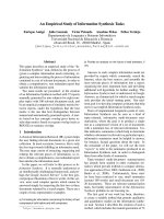
Embryonic germ (EG) cells are known to have simi-
lar characteristics to ES cells with respect to differenti-
ation and proliferation capabilities, despite their
different origins [12,13]. It was demonstrated that glo-
bal gene-expression profiles of ES and EG cells were
indistinguishable [14]. However, analysis of DNA-
methylation profiles by RLGS revealed a significant
difference between ES and EG cells [9]. Among 1500
genomic loci in the RLGS profile, 49 (3%) were found
to be methylated differentially in ES and EG cells,
indicating that ES and EG cells can be distinguished
from each other by the DNA methylation profiles. If
we defined ‘epigenetic distance’ (ED) as the number of
differentially methylated loci per 1500 genomic loci of
two given cell- or tissue types, the ED between ES and
EG cells (49) is less than that between ES and TS cells
(77), confirming the previous notion that EG cells are
more similar to ES cells than to TS cells (Fig. 1).
Change of DNA methylation profiles during the
developmental process
To examine how the DNA methylation profile changes
as the embryo develops, we utilized model differentia-
tion systems and analyzed the DNA methylation pro-
files of ES cells, embryoid bodies (EBs), teratomas
derived from the same ES cells, fetuses at E10.5 and
adult organs [15]. Teratomas are disorganized agglom-
erates with tissue or organ components derived from all
three germ layers. Teratomas, as well as fetuses, have
N. Hattori and K. Shiota Epigenetic study of embryonic stem cells
FEBS Journal 275 (2008) 1624–1630 ª 2008 The Authors Journal compilation ª 2008 FEBS 1625
DNA methylation profiles that are obtained from a
mixture of heterogeneous tissues or organs, meaning
that the methylation status at each locus in a profile
reflects average levels of DNA methylation of all cell
types analyzed. Thus, detectable alterations in the
DNA methylation profiles of teratomas or embryos
indicate common alterations that occurred in the whole
teratoma or embryo concurrent with the differentiation
of ES cells. Among the 259 T-DMRs, including the ori-
ginal 247 T-DMRs [9], the fraction of methylated loci,
which was 51.4% in ES cells, was lower in fetuses
(40.2%) and brain of adult mice (48.6%) but higher in
kidney (53.7%). A similar change was observed in the
in vitro differentiation system; methylation levels were
low (39.6%) in EBs and higher (41.3–44.4%) in three
independently developed teratomas derived from ES
cells or EBs. The number of methylated loci in the
profiles of teratomas was less than that of the somatic
tissues, probably because the teratomas still contained
a significant number of undifferentiated proliferating
cells, or all cells in teratomas were not fully differenti-
ated yet. Because the methylation status of T-DMRs
partially corresponds with the transcriptional status of
the neighboring gene, identifying differentially methyl-
ated genomic loci in ES cells, EBs and teratomas is
expected to provide information about genes that are
responsible for the developmental process.
Potential of ES cells in embryotoxicological
studies
Embryonic exposure to chemical agents or medicine
may have deleterious effects on proper embryogenesis,
especially during the early developmental stages. Such
agents may influence embryos at genetic, transcriptional
and protein levels. It is also conceivable that epigenetic
alterations occur with exposure of embryos to these
agents, because epigenetic profiles are established
actively in developing embryos. Differentiation of ES
cells into EBs has been studied as an in vitro model of
normal and abnormal mammalian development [16].
Because differentiation from ES cells to EBs is accom-
panied by changes in DNA methylation profiles at
T-DMRs [15], the in vitro differentiation model is
useful to assess the epigenetic effect of an agent on the
developmental process, and helps avoid the ethical issue
of embryotoxicological surveillance of ‘epimutagens’
[17]. In addition, it is necessary to assess the effects of
agents on the ES cell itself, for future therapeutic use in
regenerative medicine. For example, dimethyl sulfoxide,
an amphipathic molecule, is a commonly used cryopre-
servative for various cells, including ES cells, and a sol-
vent for water-insoluble substances in cytological and
cytotoxicological studies [18]. It has been reported that
exposure to dimethyl sulfoxide induced differentiation
in several types of cells [18], and that dimethyl sulfoxide
could improve the frequency of development to the
blastocyst stage after nuclear injection in mouse cloning
[19]. RLGS analysis revealed that dimethyl sulfoxide
treatment of ES cells differentiating into EBs, at con-
centrations lower than when used as a cryopreservative,
resulted in the alteration in the DNA methylation
profile [20]. Both hypo- and hypermethylation were
observed at T-DMRs depending on the genomic loci,
with hypermethylation occurring at minor satellite
repeats and endogenous C-type retroviruses. Among
epigenetic regulators, including DNA methyltransferas-
es (Dnmts) and histone modification enzymes, Dnmt3a
subtypes were upregulated both at the mRNA and pro-
tein level in dimethyl sulfoxide-treated cells, suggesting
that dimethyl sulfoxide might have a direct impact on
DNA methylation via up-regulation of Dnmt3a sub-
types, at least, at hypermethylated loci and repetitive
sequences.
Analysis of the DNA methylation profile for
therapeutic use of human ES cells in regenerative
medicine
The potential use of human ES cells in the field of
regenerative medicine has been discussed previously,
and differentiation of human ES cells into various tis-
sues has been investigated [8]. Several lines of human
ES cells were established, and differences between these
ES cell lines with respect to karyotypic stability [21]
and expression profiles [22] have been investigated. It
ICM
TE
PGC
TS cells
ES cells EG cells
4977
Placental cells Embr
y
onic cells
Fig. 1. Epigenetic distances between ES cells and other stem cells
derived from developing embryos. ES cells derived from the inner
cell mass (ICM) of blastocysts and EG cells derived from the pri-
mordial germ cells (PGCs) in developing genital ridges can develop
into cells of the embryo proper, after they are injected into blast-
ocysts to form chimeras. By contrast, TS cells derived from the
trophectoderm (TE) of blastocysts contribute only to placenta.
Although there is an apparent ED between ES cells and EG cells,
the ED of TS cells to ES cells (77) is greater than that of EG cells
to ES cells (49), confirming the similarity of EG cells to ES cells.
Epigenetic study of embryonic stem cells N. Hattori and K. Shiota
1626 FEBS Journal 275 (2008) 1624–1630 ª 2008 The Authors Journal compilation ª 2008 FEBS
has been demonstrated that mouse and human ES cells
have unique DNA methylation profiles compared with
other cell types, including EG cells, TS cells and sev-
eral adult stem cell populations [9,23]. Also, key regu-
lators of development such as Oct-4 and Nanog are
controlled by epigenetic mechanisms [24,25]. To ensure
the safe use of ES cells for regenerative medicine, it
will be necessary to evaluate the nature of differenti-
ated cells as thoroughly as possible. Accordingly, it is
also important to evaluate the epigenetic stability of
ES cell lines. Using RLGS, Allegrucci and co-workers
investigated the DNA methylation profiles of indepen-
dently isolated human ES cells after culture under vari-
ous conditions [26]. They demonstrated that variations
in DNA methylation profile existed between ES cell
lines, which could not be accounted for by genetic dif-
ferences of the source embryos. Although the number
of cell passages and culture conditions, such as the
existence of serum or feeder-layer, affected neither
morphology nor expression of cell markers, these
parameters changed the DNA methylation profile of
human ES cells. Considerable numbers of loci with
different DNA methylation status were also aberrantly
methylated in human tumor cells [27].
Investigation of epigenetic
mechanisms with ES cells deficient
in epigenetic regulators
Homologous recombination in ES cells enables us to
perform gene targeting at specific chromosomal loci
and to investigate gene function [28]. In addition,
knockout mice have been generated to study the devel-
opmental role of the gene by germline transmission of
a targeted allele. Genetic manipulations of many epige-
netic regulators, including Dnmts [29–33] and histone
methylases [34,35], have been reported. Genome-wide
DNA methylation analysis of ES cells deficient in epi-
genetic regulators will assist in revealing the mecha-
nism for maintaining DNA methylation in T-DMRs,
as well as the interplay between DNA methylation and
other epigenetic modifications.
Mechanism for maintaining DNA methylation
at T-DMRs
Based on studies regarding the properties of Dnmts, it is
widely accepted that Dnmt1 is a maintenance DNA
methyltransferase and Dnmt3a ⁄ 3b are de novo DNA
methyltransferases in vivo [36]. Dnmt3a and Dnmt3b
have no preference for hemimethylated DNA [37], and a
transgene of Dnmt3a, but not of Dnmt1, to Drosophila
exhibited de novo methylation activity [38], indicating
that Dnmt3a ⁄ 3b function in de novo DNA methylation,
but not in maintenance DNA methylation. However,
following these studies, it was still unclear how Dnmt1
and Dnmt3a ⁄ 3b are involved in DNA methylation
in T-DMRs, thereby establishing DNA methylation
profiles of cells, and whether Dnmt3a ⁄ 3b have any role
in maintenance DNA methylation in T-DMRs.
We demonstrated cooperation of Dnmt1 and either
Dnmt3a or Dnmt3b in the maintenance of DNA meth-
ylation in gene areas [39]. Using RLGS with Dnmt1-,
Dnmt3a- and ⁄ or Dnmt3b-deficient ES cells, we focused
on the involvement of Dnmts in the methylation of
CpG islands and CpG-rich regions near genes. Both
Dnmt1 single mutation and Dnmt3a ⁄ Dnmt3b double
mutation in ES cells resulted in the demethylation of
many loci. Surprisingly, target T-DMRs of Dnmt1 were
identical to those of Dnmt3a ⁄ Dnmt3b. Although a
single disruption of Dnmt3a or Dnmt3b resulted in no
change in DNA methylation at the same loci, it was
shown that maintaining DNA methylation at identified
loci requires both classes of Dnmts, Dnmt1 and either
Dnmt3a or Dnmt3b. Kinetic analysis of ES cells defi-
cient in Dnmts indicated that demethylation in repeat
sequences was progressive in Dnmt3a ⁄ 3b-deficient ES
cells, with notable demethylation during later stages of
cell culture, whereas demethylation in Dnmt1-deficient
ES cells was more rapid and greater during the initial
stages of culture [40]. This implies a predominant role
for Dnmt1 and supportive role for Dnmt3a and
Dnmt3b in maintaining DNA methylation at the repeat
sequences. By contrast, further analysis by bisulfite
sequencing of loci studied by RLGS determined that
extensive and almost complete demethylation occurred
at genes in Dnmt3a ⁄ 3b-deficient ES cells, whereas
demethylation was rather moderate in Dnmt1-deficient
ES cells [39]. It is probable that in Dnmt1-deficient ES
cells, Dnmt3a and Dnmt3b exert de novo DNA methyl-
ation activity at these genes, which are demethylated
through lack of maintenance activity because Dnmt1 is
absent. Consequently, Dnmt1-deficient ES cells seem to
have partial DNA methylation maintenance activity,
which is provided by the re-methylating actions of
Dnmt3a ⁄ Dnmt3b (Fig. 2). Dnmt3a and Dnmt3b
appear to function both as maintenance and as de novo
methyltransferases in gene areas, and thus are crucial
for the establishment of the DNA methylation profile
during development.
Analyzing the interplay between DNA
methylation and histone methylation
Chromatin structure, which is affected by DNA meth-
ylation and histone modification, is closely associated
N. Hattori and K. Shiota Epigenetic study of embryonic stem cells
FEBS Journal 275 (2008) 1624–1630 ª 2008 The Authors Journal compilation ª 2008 FEBS 1627
with the transcriptional activity of genes. During mam-
malian development, the epigenetic profile is not estab-
lished solely by one particular epigenetic regulator, but
rather by the interplay of epigenetic regulators [41,42].
The relationship between DNA methylation and other
epigenetic modifications can be examined by genome-
wide DNA methylation analysis using ES cells defi-
cient in epigenetic regulators. Growing evidence has
indicated that histone lysine methylation can direct
DNA methylation in many organisms [43]. G9a is a
euchromatin-localized histone methylase that catalyzes
the methylation of histone H3 at Lys9 and Lys27
(H3–K9 and H3–K27) [44], which are often found in
heterochromatic regions and in transcriptionally inac-
tive loci of the genome [45]. RLGS analysis of G9a-
deficient ES cells revealed a direct interaction between
DNA methylation and H3–K9 and H3–K27 methyla-
tion at T-DMRs during ES cell differentiation [46]. In
G9a-deficient ES cells, the levels of DNA methylation
decreased in some genomic loci, and Vi-RLGS
revealed the location of these loci in euchromatic
regions. Chromatin-immunoprecipitation confirmed
the demethylation of H3–K9 and H3–K27 at genomic
loci following G9a knockout, indicating that demethyl-
ation of H3–K9 and H3–K27 triggered the disruption
of maintenance DNA methylation. Restoration of G9a
activity by insertion of the transgene into G9a-deficient
ES cells resulted in full recovery of methylation
levels to almost all genomic loci. This suggests that
G9a also facilitates de novo DNA methylation of the
target loci. Because G9a does not have the cata-
lytic domain of Dnmts, G9a plays a role in DNA
methylation indirectly, possibly via methylation at
H3–K9 and ⁄ or H3–K27. This study also suggests the
potential to discover novel targets of an epigenetic
regulator that affects DNA methylation, by analyzing
alterations in DNA methylation in cells deficient in the
factor.
Conclusions
Genome-wide DNA methylation analysis of ES cells
has the potential to reveal the mechanisms used to
establish DNA methylation profiles, and the epigenetic
effects of fetal exposure to chemical agents during
mammalian development. An increased number of ES
cell lines deficient in epigenetic regulators will facilitate
investigations into the interplay between DNA methyl-
ation and other epigenetic modifications through
identification of DNA methylation profiles by RLGS
or other genome-wide analysis methods.
Acknowledgements
We thank M. Higgins for reviewing the original manu-
script. This work was supported by the Program for
Promotion of Basic Research Activities for Innovative
Biosciences (PROBRAIN).
References
1 Bird AP & Wolffe AP (1999) Methylation-induced
repression – belts, braces, and chromatin. Cell 99, 451–
454.
2 Bird A (2002) DNA methylation patterns and epigenetic
memory. Genes Dev 16, 6–21.
3 Shiota K (2004) DNA methylation profiles of CpG
islands for cellular differentiation and development in
mammals. Cytogenet Genome Res 105, 325–334.
4 Lieb JD, Beck S, Bulyk ML, Farnham P, Hattori N,
Henikoff S, Liu XS, Okumura K, Shiota K, Ushijima T
et al. (2006) Applying whole-genome studies of epige-
netic regulation to study human disease. Cytogenet
Genome Res 114, 1–15.
5 Fraga MF & Esteller M (2002) DNA methylation: a
profile of methods and applications. Biotechniques 33 ,
632, 634, 636–649.
6 Hayashizaki Y & Watanabe S (1997) Restriction
Landmark Genomic Scanning (RLGS). Springer, Tokyo.
7 Matsuyama T, Kimura MT, Koike K, Abe T, Nakano
T, Asami T, Ebisuzaki T, Held WA, Yoshida S &
Nagase H (2003) Global methylation screening in the
Arabidopsis thaliana and Mus musculus genome:
applications of virtual image restriction landmark
genomic scanning (Vi-RLGS). Nucleic Acids Res 31,
4490–4496.
RepeatRepeat GeneGene CpG island CpG island
Dnmt3a/3b
Dnmt3a/3b Dnmt3a/3b
Dnmt1
Dnmt3a/3b
Dnmt1
+
Maintenance
De novo (re-methylation)
Fig. 2. Involvement of Dnmts in establishing and maintaining DNA-
methylation profiles. Dnmt3a ⁄ 3b have an indispensable role for
maintaining DNA methylation in gene areas, which are constituents
of the DNA methylation profile, whereas in repeated sequences,
Dnmt3a ⁄ 3b have a supportive role for Dnmt1 [32,39,40].
Dnmt3a ⁄ 3b exert de novo DNA methylation activity at both gene
and repeat areas.
Epigenetic study of embryonic stem cells N. Hattori and K. Shiota
1628 FEBS Journal 275 (2008) 1624–1630 ª 2008 The Authors Journal compilation ª 2008 FEBS
8 Wobus AM & Boheler KR (2005) Embryonic stem
cells: prospects for developmental biology and cell ther-
apy. Physiol Rev 85 , 635–678.
9 Shiota K, Kogo Y, Ohgane J, Imamura T, Urano A,
Nishino K, Tanaka S & Hattori N (2002) Epigenetic
marks by DNA methylation specific to stem, germ and
somatic cells in mice. Genes Cells 7, 961–969.
10 Nagy A, Gocza E, Diaz EM, Prideaux VR, Ivanyi E,
Markkula M & Rossant J (1990) Embryonic stem cells
alone are able to support fetal development in the
mouse. Development 110, 815–821.
11 Tanaka S, Kunath T, Hadjantonakis AK, Nagy A &
Rossant J (1998) Promotion of trophoblast stem
cell proliferation by FGF4. Science 282, 2072–
2075.
12 Matsui Y, Zsebo K & Hogan BL (1992) Derivation of
pluripotential embryonic stem cells from murine pri-
mordial germ cells in culture. Cell 70, 841–847.
13 Resnick JL, Bixler LS, Cheng L & Donovan PJ (1992)
Long-term proliferation of mouse primordial germ cells
in culture. Nature 359, 550–551.
14 Sharova LV, Sharov AA, Piao Y, Shaik N, Sullivan T,
Stewart CL, Hogan BL & Ko MS (2007) Global gene
expression profiling reveals similarities and differences
among mouse pluripotent stem cells of different origins
and strains. Dev Biol 307, 446–459.
15 Kremenskoy M, Kremenska Y, Ohgane J, Hattori N,
Tanaka S, Hashizume K & Shiota K (2003) Genome-
wide analysis of DNA methylation status of CpG
islands in embryoid bodies, teratomas, and fetuses.
Biochem Biophys Res Commun 311, 884–890.
16 O’Shea KS (1999) Embryonic stem cell models of devel-
opment. Anat Rec 257, 32–41.
17 MacPhee DG (1998) Epigenetics and epimutagens:
some new perspectives on cancer, germ line effects
and endocrine disrupters. Mutat Res 400, 369–379.
18 Santos NC, Figueira-Coelho J, Martins-Silva J &
Saldanha C (2003) Multidisciplinary utilization of
dimethyl sulfoxide: pharmacological, cellular, and
molecular aspects. Biochem Pharmacol 65, 1035–1041.
19 Wakayama T & Yanagimachi R (2001) Effect of cyto-
kinesis inhibitors, DMSO and the timing of oocyte
activation on mouse cloning using cumulus cell nuclei.
Reproduction 122, 49–60.
20 Iwatani M, Ikegami K, Kremenska Y, Hattori N,
Tanaka S, Yagi S & Shiota K (2006) Dimethyl sulfox-
ide has an impact on epigenetic profile in mouse embry-
oid body. Stem Cells 24, 2549–2556.
21 Buzzard JJ, Gough NM, Crook JM & Colman A
(2004) Karyotype of human ES cells during extended
culture. Nat Biotechnol 22, 381–382; author reply 382.
22 Abeyta MJ, Clark AT, Rodriguez RT, Bodnar MS,
Pera RA & Firpo MT (2004) Unique gene expression
signatures of independently-derived human embryonic
stem cell lines. Hum Mol Genet 13, 601–608.
23 Bibikova M, Chudin E, Wu B, Zhou L, Garcia EW,
Liu Y, Shin S, Plaia TW, Auerbach JM, Arking DE
et al. (2006) Human embryonic stem cells have a unique
epigenetic signature. Genome Res 16, 1075–1083.
24 Hattori N, Nishino K, Ko YG, Hattori N, Ohgane J,
Tanaka S & Shiota K (2004) Epigenetic control of
mouse Oct-4 gene expression in embryonic stem cells
and trophoblast stem cells. J Biol Chem 279, 17063–
17069.
25 Hattori N, Imao Y, Nishino K, Hattori N, Ohgane J,
Yagi S, Tanaka S & Shiota K (2007) Epigenetic regula-
tion of Nanog
gene in embryonic stem and trophoblast
stem cells. Genes Cells 12, 387–396.
26 Allegrucci C, Wu YZ, Thurston A, Denning CN,
Priddle H, Mummery CL, Ward-van Oostwaard D,
Andrews PW, Stojkovic M, Smith N et al. (2007)
Restriction landmark genome scanning identifies cul-
ture-induced DNA methylation instability in the human
embryonic stem cell epigenome. Hum Mol Genet 16,
1253–1268.
27 Smiraglia DJ & Plass C (2002) The study of aberrant
methylation in cancer via restriction landmark genomic
scanning. Oncogene 21, 5414–5426.
28 Capecchi MR (1989) Altering the genome by homolo-
gous recombination. Science 244, 1288–1292.
29 Li E, Bestor TH & Jaenisch R (1992) Targeted muta-
tion of the DNA methyltransferase gene results in
embryonic lethality. Cell 69, 915–926.
30 Lei H, Oh SP, Okano M, Juttermann R, Goss KA, Jae-
nisch R & Li E (1996) De novo DNA cytosine methyl-
transferase activities in mouse embryonic stem cells.
Development 122, 3195–3205.
31 Okano M, Xie S & Li E (1998) Dnmt2 is not required
for de novo and maintenance methylation of viral DNA
in embryonic stem cells. Nucleic Acids Res 26, 2536–
2540.
32 Okano M, Bell DW, Haber DA & Li E (1999) DNA
methyltransferases Dnmt3a and Dnmt3b are essential
for de novo methylation and mammalian development.
Cell 99, 247–257.
33 Tsumura A, Hayakawa T, Kumaki Y, Takebayashi S,
Sakaue M, Matsuoka C, Shimotohno K, Ishikawa F,
Li E, Ueda HR et al. (2006) Maintenance of self-
renewal ability of mouse embryonic stem cells in the
absence of DNA methyltransferases Dnmt1, Dnmt3a
and Dnmt3b. Genes Cells 11, 805–814.
34 Peters AH, O’Carroll D, Scherthan H, Mechtler K, Sauer
S, Schofer C, Weipoltshammer K, Pagani M, Lachner
M, Kohlmaier A et al. (2001) Loss of the Suv39h histone
methyltransferases impairs mammalian heterochromatin
and genome stability. Cell 107, 323–337.
35 Tachibana M, Sugimoto K, Nozaki M, Ueda J, Ohta
T, Ohki M, Fukuda M, Takeda N, Niida H, Kato H
et al. (2002) G9a histone methyltransferase plays a
dominant role in euchromatic histone H3 lysine 9
N. Hattori and K. Shiota Epigenetic study of embryonic stem cells
FEBS Journal 275 (2008) 1624–1630 ª 2008 The Authors Journal compilation ª 2008 FEBS 1629
methylation and is essential for early embryogenesis.
Gene Dev 16, 1779–1791.
36 Bestor TH (2000) The DNA methyltransferases of
mammals. Hum Mol Genet 9, 2395–2402.
37 Okano M, Xie S & Li E (1998) Cloning and character-
ization of a family of novel mammalian DNA (cyto-
sine-5) methyltransferases. Nat Genet 19, 219–220.
38 Lyko F, Ramsahoye BH, Kashevsky H, Tudor M,
Mastrangelo MA, Orr-Weaver TL & Jaenisch R (1999)
Mammalian (cytosine-5) methyltransferases cause geno-
mic DNA methylation and lethality in Drosophila. Nat
Genet 23, 363–366.
39 Hattori N, Abe T, Hattori N, Suzuki M, Matsuyama
T, Yoshida S, Li E & Shiota K (2004) Preference
of DNA methyltransferases for CpG islands in
mouse embryonic stem cells. Genome Res 14, 1733–
1740.
40 Chen T, Ueda Y, Dodge JE, Wang Z & Li E (2003)
Establishment and maintenance of genomic methylation
patterns in mouse embryonic stem cells by Dnmt3a and
Dnmt3b. Mol Cell Biol 23, 5594–5605.
41 Li E (2002) Chromatin modification and epigenetic
reprogramming in mammalian development. Nat Rev
Genet 3, 662–673.
42 Morgan HD, Santos F, Green K, Dean W & Reik W
(2005) Epigenetic reprogramming in mammals. Hum
Mol Genet 14 (Spec No 1), R47–R58.
43 Lachner M & Jenuwein T (2002) The many faces of
histone lysine methylation. Curr Opin Cell Biol 14,
286–298.
44 Tachibana M, Sugimoto K, Fukushima T & Shinkai Y
(2001) Set domain-containing protein, G9a, is a novel
lysine-preferring mammalian histone methyltransferase
with hyperactivity and specific selectivity to lysines 9 and
27 of histone H3. J Biol Chem 276, 25309–25317.
45 Kouzarides T (2002) Histone methylation in transcrip-
tional control. Curr Opin Genet Dev 12, 198–209.
46 Ikegami K, Iwatani M, Suzuki M, Tachibana M, Shin-
kai Y, Tanaka S, Greally JM, Yagi S, Hattori N & Shi-
ota K (2007) Genome-wide and locus-specific DNA
hypomethylation in G9a deficient mouse embryonic
stem cells. Genes Cells 12, 1–11.
Epigenetic study of embryonic stem cells N. Hattori and K. Shiota
1630 FEBS Journal 275 (2008) 1624–1630 ª 2008 The Authors Journal compilation ª 2008 FEBS