Báo cáo khoa học: Order within a mosaic distribution of mitochondrial c-type cytochrome biogenesis systems? pptx
Bạn đang xem bản rút gọn của tài liệu. Xem và tải ngay bản đầy đủ của tài liệu tại đây (1.04 MB, 18 trang )
REVIEW ARTICLE
Order within a mosaic distribution of mitochondrial c-type
cytochrome biogenesis systems?
James W. A. Allen
1
, Andrew P. Jackson
2
, Daniel J. Rigden
3
, Antony C. Willis
4
, Stuart J. Ferguson
1
and Michael L. Ginger
5,6
1 Department of Biochemistry, University of Oxford, UK
2 Wellcome Trust Sanger Institute, Hinxton, Cambridgeshire, UK
3 School of Biological Sciences, University of Liverpool, UK
4 MRC Immunochemistry Unit, Department of Biochemistry, University of Oxford, UK
5 Sir William Dunn School of Pathology, University of Oxford, UK
6 Department of Biological Sciences, Lancaster University, UK
Keywords
bioinformatics; Ccm system; cytochrome c;
Diplonema papillatum; evolution; heme
lyase; lateral gene transfer; mitochondria;
post-translational modification; Trypanosoma
Correspondence
M. Ginger, Department of Biological
Sciences, Lancaster University, Lancaster
LA1 4YQ, UK
Fax: +44 1524 593192
Tel: +44 1524 593922
E-mail:
(Received 7 February 2008, revised 3 March
2008, accepted 5 March 2008)
doi:10.1111/j.1742-4658.2008.06380.x
Mitochondrial cytochromes c and c
1
are present in all eukaryotes that use
oxygen as the terminal electron acceptor in the respiratory chain. Matura-
tion of c-type cytochromes requires covalent attachment of the heme cofac-
tor to the protein, and there are at least five distinct biogenesis systems
that catalyze this post-translational modification in different organisms and
organelles. In this study, we use biochemical data, comparative genomic
and structural bioinformatics investigations to provide a holistic view of
mitochondrial c-type cytochrome biogenesis and its evolution. There are
three pathways for mitochondrial c-type cytochrome maturation, only one
of which is present in prokaryotes. We analyze the evolutionary distribu-
tion of these biogenesis systems, which include the Ccm system (System I)
and the enzyme heme lyase (System III). We conclude that heme lyase
evolved once and, in many lineages, replaced the multicomponent Ccm sys-
tem (present in the proto-mitochondrial endosymbiont), probably as a con-
sequence of lateral gene transfer. We find no evidence of a System III
precursor in prokaryotes, and argue that System III is incompatible with
multi-heme cytochromes common to bacteria, but absent from eukaryotes.
The evolution of the eukaryotic-specific protein heme lyase is strikingly
unusual, given that this protein provides a function (thioether bond forma-
tion) that is also ubiquitous in prokaryotes. The absence of any known
c-type cytochrome biogenesis system from the sequenced genomes of
various trypanosome species indicates the presence of a third distinct mito-
chondrial pathway. Interestingly, this system attaches heme to mitochon-
drial cytochromes c that contain only one cysteine residue, rather than the
usual two, within the heme-binding motif. The isolation of single-cysteine-
containing mitochondrial cytochromes c from free-living kinetoplastids,
Euglena and the marine flagellate Diplonema papillatum suggests that this
unique form of heme attachment is restricted to, but conserved throughout,
the protist phylum Euglenozoa.
Abbreviations
ccm, cytochrome c maturation; EF-1a, elongation factor-1a; EST, expressed sequence tag; IMS, intermembrane space; KH test, Kishino–
Hasegawa test; LGT, lateral gene transfer; ML, maximum likelihood; SOD, superoxide dismutase.
FEBS Journal 275 (2008) 2385–2402 ª 2008 The Authors Journal compilation ª 2008 FEBS 2385
Multiple pathways for heme cysteine
attachment
The c-type cytochromes are characterized by the cova-
lent attachment of heme to the apocytochrome through
thioether (carbon–sulfur) bonds (Fig. 1). Numerous
examples of distinct c-type cytochromes have been
described in Bacteria and, more recently, in some Ar-
chaea, where they typically function in electron transfer
or at the catalytic sites of certain enzymes [2–9] {In
agreement with the nomenclature proposed in [1], we
refer to the three domains of life as Bacteria (formerly
Eubacteria), Archaea (formerly Archaebacteria) and
Eucarya (the eukaryotes). When using the expression
‘Bacteria’, we therefore refer to the domain; when
using the term ‘bacteria’, we refer generically to non-
archaean prokaryotes}. However, the best known
examples from the c-type cytochrome family are mito-
chondrial cytochromes c and c
1
, which function as
essential electron transfer components of the respira-
tory chain [7,8,10].
The covalent attachment of two vinyl groups from
the heme cofactor to the thiols in the CXXCH heme-
binding motif of apocytochromes c is chemically far
from facile (X is any amino acid, except cysteine), and
there are multiple systems which catalyze this post-
translational modification in biology [2,4,11–15]. Sys-
tems I and II are modular and widely distributed
amongst bacteria [2,4,6,12,14]; they have been studied
using a combination of genetic and biochemical
approaches [2,4,14,16,17]. System I is understood best
in Escherichia coli, where it consists of eight dedicated
essential proteins, named CcmA–H (Fig. 2A), and a
number of accessory proteins. CcmA–H are all mem-
brane anchored or integral membrane proteins, and
collectively function in the periplasm. The biogenesis
of c-type cytochromes is a spatial and temporal prob-
lem; in bacteria, both heme and apoprotein are synthe-
sized in the cytoplasm and must be transported to the
periplasm, where heme attachment occurs. The apocy-
tochrome polypeptide is translocated by the general
type II secretion (Sec) proteins [18]. How heme is
transported remains an intriguing mystery.
CcmA and CcmB are reminiscent of an ATP-
dependent (ABC-type) transporter, and CcmA has
been shown to hydrolyze ATP [19]. However, no
transport substrate has yet been identified; heme has
been proposed, but much evidence weighs against
this possibility [19–22]. A more recent hypothesis is
that CcmA and CcmB are required to release heme
from the heme chaperone CcmE by coupling the free
energy gained from ATP hydrolysis [21]. CcmE is a
key player in the Ccm system; it binds heme cova-
lently as an intermediate in the cytochrome c biogen-
esis pathway [23]. This remarkable heme attachment
occurs between a histidine residue and a heme vinyl
group. Heme attachment to CcmE is dependent on
CcmC [24], an integral membrane protein with a
number of interesting phenotypes arising from muta-
tion in ccmC, some of which may be unrelated to
c-type cytochrome biogenesis [25]. CcmD is a very
small ( 60 amino acids) integral membrane protein
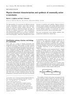
Fig. 1. Structures of (A) heme (Fe-protoporphyrin IX) and (B) heme bound to a polypeptide chain as in a typical c-type cytochrome, in which
the vinyl groups of the heme are saturated by the addition of cysteine thiols that occur in a Cys-Xxx-Xxx-Cys-His motif (only the sulfur atoms
of the cysteines are shown), forming covalent bonds between heme and protein. (C) Cartoon representation of heme attachment to protein
in mitochondrial cytochrome c. The porphyrin ring is shown in blue and the heme iron atom in brown. The cysteines of the CXXCH motif
form covalent bonds to the heme, and the histidine acts as a ligand to the heme iron atom via a nitrogen atom. The sixth ligand to the iron
atom is the sulfur of a methionine residue located distantly from the CXXCH motif in the primary structure of the protein. In bacterial c-type
cytochromes, histidine (rather than methionine) is often the sixth iron ligand, and there are examples with cysteine, an N-terminal amino
group, asparagine, lysine or a vacant coordination site. There are few restrictions on the nature of the Xxx-Xxx residues.
Evolution of mitochondrial cytochrome c maturation J. W. A. Allen et al.
2386 FEBS Journal 275 (2008) 2385–2402 ª 2008 The Authors Journal compilation ª 2008 FEBS
that mediates complex formation between CcmC and
CcmE [26]. CcmF and CcmH are implicated in the
transfer of heme from holo-CcmE to apocyto-
chrome c, including the covalent heme attachment
step to produce the product holocytochrome. E. coli
CcmH is a fusion protein which includes the proteins
known as CcmH and CcmI in many bacteria. CcmG
is a thioredoxin-like protein [27] that forms part of
an electron transfer chain. Electrons are transferred
from the cytoplasmic protein thioredoxin, via the
multidomain membrane protein DsbD, to CcmG,
and then to the apocytochrome to reduce a disulfide
bond that forms between the cysteines of the apocyto-
chrome CXXCH heme-binding motif; these thiols must
be reduced for heme attachment to occur (reviewed in
[2]). Such a reductive pathway is thought to be neces-
sary in E. coli, partly because the periplasm contains
the strong, indiscriminate, disulfide-oxidizing protein
DsbA.
System II (Fig. 2B) is less well understood than
System I at the molecular level, but it seems very likely
to consist of four proteins [28] {Note: The nomencla-
Fig. 2. Cytochrome c biogenesis systems found in bacteria. Each of these systems can mature a wide variety of c-type cytochromes,
including those with multiple hemes. (A) System I (the Ccm system) in Escherichia coli. Some uncertainties are designated with ‘?’; for
example, what, if anything, is transported by the ABC-type transporter CcmAB, and how is heme transported from its site of synthesis in
the cytoplasm to the periplasm? DsbD has two thiols amongst its eight transmembrane helices which are believed to accept reducing equiv-
alents from thioredoxin (TrxA). These thiols, in turn, pass on the reducing power to periplasmic C- and N-terminal domains. From there,
reductant passes to the c-type cytochrome biogenesis apparatus, tentatively by the route shown; CcmG has been shown in some schemes
to be the electron acceptor from CcmH but, although there is experimental evidence for this order, more evidence indicates the arrange-
ment shown in the figure. DsbA is a strong, non-specific disulfide bond-oxidizing protein found in the periplasm of E. coli. Ultimately, the
cysteine thiols of the apocytochrome CXXCH heme-binding motif become reduced to allow heme attachment. Heme becomes covalently
attached to the chaperone CcmE as an intermediate in the pathway. The specific covalent attachment of heme to apocytochrome c is
believed to involve CcmF and H. (B) Cytochrome c biogenesis System II in a Gram-negative bacterium. In some species, CcdA is replaced
by the protein DsbD shown in Fig. 2A. CcdA and ResA provide a pathway by which reductant is transferred to the apocytochrome to reduce
a disulfide bond in the CXXCH heme-binding motif. ResB and ResC provide the covalent heme attachment function to produce the product
holocytochrome c. Heme delivery to the periplasm from the cytoplasm may also occur through the ResBC complex, but this is presently not
certain. Other names are in common use for ResA ⁄ B ⁄ C (ResA = CcsX = HCF164; ResB = CcsB = Ccs1; ResC = CcsA; and CcdA = CcsC).
J. W. A. Allen et al. Evolution of mitochondrial cytochrome c maturation
FEBS Journal 275 (2008) 2385–2402 ª 2008 The Authors Journal compilation ª 2008 FEBS 2387
ture for System II c-type cytochrome biogenesis pro-
teins is somewhat inconsistent in the literature. Here,
we adopt the names used for the various biogenesis
proteins found in Bacillus subtilis, i.e. ResA (also
called CcsX or HCF164), ResB (also called CcsB or
Ccs1), ResC (also called CcsA) and CcdA (also called
CcsC)}. These include a thioredoxin-like protein called
ResA (similar in structure to CcmG) [29] and CcdA, a
functional analogue of DsbD, or DsbD itself (depend-
ing on the organism); together, these apparently form
a pathway analogous to that observed in System I for
reducing a disulfide bond in the apocytochrome
CXXCH motif. The heme attachment (and possibly
heme delivery) function of System II is catalyzed by
ResB and ResC. Indeed, a fusion protein cloned from
Helicobacter pylori containing elements of ResB and
ResC was sufficient to mature c-type cytochromes
when expressed in the periplasm of a ccm deletion
strain of E. coli [30].
Several recent studies have provided insight into the
flexible organization of prokaryotic c-type cytochrome
biogenesis pathways. For example, in the Archaea and
some bacteria, a divergent System I has recently been
described [3], and some bacteria contain components
of both System I and System II [3,4,6]. Although the
presence of multiple cytochrome c biogenesis systems
in a single bacterium might hint at possible redun-
dancy, additional c-type cytochrome maturation com-
ponents are sometimes required for heme attachment
to specific substrates. For example, in the e-proteobac-
terium Wolinella succinogenes, the ccsA1-encoded heme
lyase is required for thioether bond formation to
the remarkable CX
15
CH heme-binding motif of the
multi-heme c-type cytochrome MccA [31].
System III for cytochrome c maturation consists of
a single primary component, the enzyme heme lyase,
which is found only in the mitochondrial intermem-
brane space (IMS) of animals, fungi and some protists
[11,32] {The kingdom Protista refers to those eukary-
otes that cannot be classified as animals, plants or
fungi: it includes protozoa and algae. The protozoa [or
‘first (proto-) animals (zoa)’] are unicellular eukaryotes,
which lack the chitinous cell wall found in fungi}. At
least in fungi, heme lyase is supplemented by the flavo-
protein Cyc2, which is thought to provide reducing
equivalents for the heme attachment process [33]. The
biochemical study of heme lyase has proved challeng-
ing, and the molecular details of its enzymology are
still largely unclear.
Finally, a distinctive example of a biogenesis system
that is required for the dedicated maturation of a partic-
ular substrate is provided by the recent description of
System IV for cytochrome c maturation. Heme is
attached through a single thioether linkage to cyto-
chromes b
6
and b from the b
6
f and bc complexes of oxy-
genic phototrophs (cyanobacteria, plants, algae) and
certain Bacillus species, respectively [34,35]. The mecha-
nism by which covalent heme attachment to Bacillus
cytochrome b occurs is not yet known, but the identifi-
cation of gene products from the green alga Chlamydo-
monas reinhardtii that restore cytochrome b
6
formation
in four ccb mutants constitutes the initial step in the
characterization of System IV, which appears to be
conserved in all oxygenic phototrophs [36].
In species from the phylum Euglenozoa, which
includes Euglena gracilis and the medically relevant
trypanosomatids (Trypanosoma brucei, T. cruzi and
pathogenic Leishmania species), heme is uniquely
attached to the mitochondrial c-type cytochromes by a
single thioether bond within a F ⁄ AXXCH heme-bind-
ing motif [37–41]. In an earlier study, we determined
that, in the trypanosomatids, the occurrence of single-
cysteine-containing mitochondrial cytochromes c and
c
1
correlates with the absence from both nuclear and
mitochondrial genomes of genes encoding any compo-
nent of the known c-type cytochrome maturation
systems; we also provided experimental evidence
that, for the single-cysteine-containing T. brucei cyto-
chrome c, spontaneous (i.e. uncatalyzed) maturation is
unlikely [41]. These results indicate that at least one
further pathway for cytochrome c maturation awaits
discovery in the trypanosomatids.
In this article, we draw on the resources that are
provided through the availability of numerous com-
plete genome sequences and several ab initio modeling
programs. We consider in detail the evolutionary dis-
tribution of the machinery for mitochondrial cyto-
chrome c assembly throughout the Eucarya, and the
possible origins of heme lyase. Although the origin of
the exclusively eukaryotic heme lyase remains mysteri-
ous, replacement of a proto-mitochondrial System I
pathway for c-type cytochrome maturation occurred
multiple times during protist evolution. With rare
exceptions, these replacements probably occurred as a
result of eukaryote-to-eukaryote lateral gene transfer
(LGT) or endosymbiotic gene transfer of heme lyase.
We also approach defining the limits of the distribu-
tion of the single-cysteine heme-binding motif found in
some mitochondrial cytochromes c.
Mapping character traits onto a
consensus view of eukaryotic
phylogeny
The origin of the first eukaryotic cell has been debated
for many years; during the 1980s and early 1990s, the
Evolution of mitochondrial cytochrome c maturation J. W. A. Allen et al.
2388 FEBS Journal 275 (2008) 2385–2402 ª 2008 The Authors Journal compilation ª 2008 FEBS
available experimental evidence was generally consis-
tent with an evolutionary model (called the Archezoa
theory), which posited two early phases to eukaryotic
evolution: an ancestral phase, in which the hallmark
features of the eukaryotic cytoskeleton, endomembrane
system and nucleus were evolved, followed by the sec-
ond critical phase, which saw the acquisition of the
a-proteobacterial endosymbiont and the evolution of
the proto-mitochondrion. Although the results from
some phylogenetic analyses conflicted with the model
formulated by Cavalier-Smith (discussed in [42]), the
Archezoa theory generally received robust support in
phylogenetic trees derived from the analysis of small
subunit rRNA or translation elongation factor pro-
teins. Grouped at the base of many of these trees were
several eukaryotic lineages, including diplomonads
(represented by Giardia), the parabasalids (represented
by Trichomonas) and the Microsporidia [43,44] (and
reviewed recently in [45,46]). The distinctive ultrastruc-
ture of these organisms suggested that they apparently
possessed neither mitochondria nor other hallmark
eukaryotic organelles, such as peroxisomes and golgi,
and their status as Archezoa denoted that they were
believed to be ancestrally without these organelles. We
now know that this is not the case; more recent phylo-
genetic treatments have resulted in the repositioning of
at least some formerly basal or ‘primitive’ eukaryotes
elsewhere within the eukaryotic tree [46–48]. Further-
more, although the secondary loss of peroxisomes has
occurred numerous times in evolution, the aforemen-
tioned organisms crucially retain mitochondria, golgi
and other classically eukaryotic subcellular compart-
ments that have merely been remodeled beyond obvi-
ous or easy recognition [49–53]. Thus, there are no
known examples of contemporary eukaryotes that lack
double-membrane-bound organelles of mitochondrial
descent; indeed, although difficult to prove, a popular
current viewpoint is that the acquisition of the proto-
mitochondrial endosymbiont could have been coinci-
dent with eukaryotic origins (see, for example, [47,54]
for a further discussion).
Although the position of the root for eukaryotic
evolution remains a contentious issue – Cavalier-Smith
has argued that the last common ancestor of all extant
eukaryotes diverged with the unikont–bikont split
(Fig. 3) [55–57]; other results have suggested that it is
still not possible to discount a previously long-standing
view that the diplomonads and parabasalids belong to
the earliest diverging eukaryotic lineage [46,47,58] –
comparative interrogations of various morphological
and molecular character traits, as well as phylogenies
based on the analysis of multiple gene sets, have
resulted in a seemingly robust resolution of eukaryotic
diversity into six major groupings ([59] and reviewed in
[46,47,60,61]). The framework provided by this resolu-
tion is increasingly being used to inform on the evolu-
tion of various fundamental aspects of eukaryotic
biology, both within and between these major group-
ings [55–57,62–66]. It is this consensus view of eukary-
otic evolution on which the comparative analysis
described below is based.
A phylogeny for mitochondrial c-type
cytochrome maturation
Using the complete or draft nuclear and mitochondrial
genome sequences indicated in supplementary Doc S1,
we mapped the distribution of mitochondrial cyto-
chrome c maturation pathways onto a consensus view
of eukaryotic phylogeny (Fig. 3). Our aim was to
assess whether there was any obvious order to the
otherwise mosaic distribution of mitochondrial cyto-
chrome c biogenesis machineries that has previously
been hinted at [67,68].
The presence of the Ccm system in higher plants
and some unicellular eukaryotes [e.g. the deeply diver-
gent jakobid Reclinomonas americana, ciliates and the
rhodophyte (red alga) Cyanidioschyzon merolae] has
been described previously [69–74], whereas other
eukaryotes, such as the animals, the chlorophyte green
alga C. reinhardtii and the malarial parasite Plasmo-
dium falciparum (an apicomplexan) have heme lyase
for maturation of mitochondrial cytochromes c
[2,15,32,75–77]. The mitochondrial genome sequences
of various excavate, algal, plant and ciliate taxa very
clearly point to the presence of System I within the
a-proteobacterial endosymbiont from which mitochon-
dria evolved [69,70,72,78,79]. However, taking into
account the generally robust support for relationships
within and between the taxonomic groups shown in
Fig. 3, our comparative genomic analysis can be used
to provide new insight into the evolution of mitochon-
drial cytochrome c maturation. Observations that are
key to the discussion that follows in subsequent sec-
tions are: (a) there is no evidence for the occurrence of
heme lyase within the bikont supergroup Excavata; (b)
in the unikonts, heme lyase is the only c-type cyto-
chrome maturation system present; (c) there is a
mosaic distribution of the Ccm system and heme lyase
within the Chromoalveolata and Plantae; (d) wherever
the multicomponent Ccm system is used for mitochon-
drial cytochrome c maturation, it is always partially
encoded on the mitochondrial genome; this is perhaps
unsurprising given that CcmC and CcmF are mito-
chondrial integral membrane proteins containing mul-
tiple predicted transmembrane helices. Where a
J. W. A. Allen et al. Evolution of mitochondrial cytochrome c maturation
FEBS Journal 275 (2008) 2385–2402 ª 2008 The Authors Journal compilation ª 2008 FEBS 2389
mitochondrial genome sequence is complemented by
the availability of a complete or draft nuclear genome
sequence for the same organism, the following always
holds true: (i) if components of the Ccm system are
encoded in the mitochondrial genome, further dedi-
cated Ccm components are also encoded in the nuclear
genome; (ii) there are no examples of eukaryotes
possessing multiple systems for mitochondrial cyto-
chrome c maturation. Thus, even without the availabil-
ity of a sequenced nuclear genome, the absence from a
protozoan or algal mitochondrial genome of genes
encoding Ccm components almost certainly provides a
reliable indication that System I will not be used for
the maturation of mitochondrial cytochromes c and c
1
.
There are several green, red (rhodophyte) and chromist
algae (belonging to the Chromalveolata), plus other
Fig. 3. The phylogenetic distribution of the different pathways used for mitochondrial c-type cytochrome maturation in eukaryotes. (A) Rela-
tionships within and between five of the six eukaryotic supergroups – no relevant data for c-type cytochrome maturation in the sixth super-
group, Rhizaria, are currently available. The unikonts comprise the Amoebozoa (to which Dictyostelium discoideum and the human pathogen
Entamoeba histolytica belong) and the Opisthokonts (the animals, fungi and various protozoa). The unikonts differ from the bikonts (which
include the algae, land plants and many different protozoa) in that they possess (probably ancestrally [55]) only a single centriole (the barrel-
shaped structure from which flagellar basal bodies are derived and which, in many eukaryotes, is also involved in the organization of the mito-
tic spindle). The phylogeny reveals that, within some groups (e.g. Viridiplantae), some species contain System I, whereas others contain
System III; there were no examples of eukaryotes that contained multiple systems for the maturation of mitochondrial c-type cytochromes. A
more detailed overview of the distribution of mitochondrial c-type cytochrome maturation pathways in the Plantae is provided in (B). Lineages
belonging to the Streptophyta are highlighted by the grey background. The evolutionary relationships shown represent a consensus view of
published data. A complete list of species used to produce the phylogeny, including the databases searched, is provided in supplementary
Doc S1. Species for which the identification of the mitochondrial c-type cytochrome biogenesis apparatus is based on the interrogation of a
complete genome sequence are as follows: the choanoflagellate Monsiga brevicolis (System III); the amoebozoan Dictyostelium discoideum
(System III); the chlorophyte green algae Chlamydomonas reinhardtii, Volvox carteri, Ostreococcus lucimarinus and Ostreococcus tauri (all
System III); the red alga Cyanidioschyzon merolae; the ciliates Tetrahymena thermophila and Paramecium tetraurelia (System I); the Apicom-
plexans Plasmodium falciparum, Toxoplasma gondii and Theileria parva (System III); the dinoflagellate Perkinsus marinus (System III); the
oomycetes Phytophthora ramorum and Phytophthora sojae and the diatoms Thalassiosira pseudonana and Phaeodactylum tricomutum
(collectively belonging to a group known as the stramenopiles or chromists) (all System III); the Heterolobosean Naegleria gruberi (System I).
Entamoeba histolytica (Amoebozoa), Encephalitozoon cuniculi (Microsporidia), Cryptosporidium parvum (Apicomplexa) and the diplomonad
Giardia intestinalis all contain degenerate mitochondria known as mitosomes, and the parabasalid Trichomonas vaginalis possesses hydro-
genosomes; such degenerate forms of mitochondria lack a respiratory chain and therefore do not contain c -type cytochromes.
Evolution of mitochondrial cytochrome c maturation J. W. A. Allen et al.
2390 FEBS Journal 275 (2008) 2385–2402 ª 2008 The Authors Journal compilation ª 2008 FEBS
protozoan species (the amoebozoan Acanthamoeba cas-
tellanii), for which no nuclear genome sequence is
available, but there is an accessible or annotated [79]
mitochondrial genome sequence on which no compo-
nent of the Ccm system is encoded. Similarly, there is
extensive sequence coverage for the uniquely organized
(many small linear chromosomes of less than 8.3 kb in
length) mitochondrial genome of the ichthyosporean
Amoebidium parasiticum (belonging to the Opi-
sthokonta); from the sequence released thus far, this
genome also lacks genes encoding Ccm components
[80]. Assuming that none of these species contain
mitochondrial c-type cytochromes with atypical heme-
binding motifs, we suggest that it is likely that nuclear-
encoded heme lyase is used for the maturation of their
mitochondrial cytochromes c and c
1
.
We found a eukaryotic cytochrome c biogenesis Sys-
tem II only in those eukaryotes that contain chlorop-
lasts (data not shown), and we assume that, in these
cases, System II is used for the maturation of the
chloroplast c-type cytochromes, given the ancestral
relationship between chloroplasts and System II-con-
taining cyanobacteria [81,82]. Where System II was
observed, a second c-type cytochrome biogenesis appa-
ratus was always present and is presumed to be
responsible for maturing the mitochondrial cyto-
chromes c and c
1
(e.g. System I in Arabidopsis thaliana,
System III in C. reinhardtii and chromist algae). Simi-
larly, genes encoding the four chloroplast proteins
recently shown to be required for single-cysteine
attachment to cytochrome b
6
in Chlamydomonas – Sys-
tem IV for c-type cytochrome biogenesis – were also
only present in phototrophic eukaryotes [36]. The
absence of heme lyase from the excavates, the possible
origins of heme lyase and the molecular basis for the
mosaic distribution of Systems I and III in chromalve-
olates and the Plantae are the critical issues upon
which we focus in the remainder of this article.
Was heme lyase ever present in
the Excavata?
A number of important human pathogens, such as try-
panosomes, Giardia and Trichomonas, as well as a
diverse assortment of free-living protozoa, are included
in the supergroup Excavata. The validity of this classi-
fication was initially based on a number of shared
morphological features, but has more recently received
modest support from a variety of molecular phyloge-
nies [59,83–85]. Support for the monophyly of the
Excavata is, however, equivocal [86]; indeed, the possi-
bility that the earliest diverging eukaryote was an
ancestor of diplomonads (Giardia) and parabasalids
(Trichomonas) has not yet been entirely dismissed
[46,58]. Interestingly, if we accept the emerging evi-
dence that groups the Excavata together, a deep-
branching status for the supergroup can be inferred
from a variety of character traits. A prime example is
the distinctive mitochondrial genome of the jakobid
R. americana which, in terms of both gene content and
genome organization, more closely resembles an a-pro-
teobacterial genome than any other mitochondrial gen-
ome that has presently been sequenced [70,87]. Like
Reclinomonas, some of the other excavates currently
sampled (Naegleria and Malwimomonas) contain the
Ccm system for cytochrome c maturation (Fig. 3).
Others (Trichomonas vaginalis and Giardia intestinalis)
lack a capacity for respiration, and c-type cytochromes
are accordingly absent from their degenerate mito-
chondria, making it impossible to assess which system
for cytochrome c maturation would have been present
in their last aerobic ancestors. In trypanosomatids,
cytochromes c and c
1
are present, but there is no rec-
ognizable c-type cytochrome maturation system. Thus,
there is no evidence that heme lyase was ever present
within the excavate supergroup.
The recently described absence [41] of any known
cytochrome c biogenesis system from the various try-
panosomatids represents a particularly intriguing sce-
nario, as it correlates with the attachment of heme to
single-cysteine XXXCH mitochondrial cytochromes in
these organisms. Such single cysteine cytochromes are
also present in other kinetoplastids (the trypanosoma-
tid family evolved from a kinetoplastid ancestor) and
the euglenids Euglena gracilis and E. viridis [37–41].
All of these protists belong to the phylum Euglenozoa
(Fig. 4A), but, in addition to the euglenids and kine-
toplastids, the Euglenozoa includes a third major taxo-
nomic group, a family of mostly free-living marine
flagellates known as the diplonemids. Recent phyloge-
nies suggest that the diplonemids are likely to be a sis-
ter group to the Kinetoplastida [88]. Although there is
no genome project for a diplonemid, we have used the
relatively simple experiment of determining the type of
mitochondrial cytochromes present (either CXXCH
or XXXCH heme attachment) to look further at the
evolution of cytochrome c biogenesis in the Excavata.
From a combination of spectroscopic methods and
N-terminal sequencing (Fig. 4), Diplonema papillatum
unambiguously contains a single-cysteine c-type cyto-
chrome (AGQCH heme-binding motif). Thus, all three
major taxonomic groups of the Euglenozoa (diplone-
mids, kinetoplastids and euglenids) contain single-
cysteine mitochondrial cytochromes c, and hence it is
likely that they all contain the same, as yet unidenti-
fied, apparatus for maturation of cytochromes c, which
J. W. A. Allen et al. Evolution of mitochondrial cytochrome c maturation
FEBS Journal 275 (2008) 2385–2402 ª 2008 The Authors Journal compilation ª 2008 FEBS 2391
is distinct from that found in any other organisms.
Analysis of all the available genome sequences and all
publicly accessible expressed sequence tag (EST) collec-
tions (including ESTs for the excavates Malawimon-
as californiana, M. jakobiformis and R. americana)
using blast reveals that, strikingly, single-cyste-
ine attachment of heme to mitochondrial cyto-
chrome c remains a characteristic that is unique to
species from the phylum Euglenozoa. Crucially, these
analyses included the use of the draft nuclear genome
sequence for Naegleria gruberi, an amoeboflagellate
with an aerobic metabolism from the phylum Hetero-
lobosea, the eukaryotes with the closest evolution-
ary relationship to the Euglenozoa [47,59,85]. The
Fig. 4. Diplonema cytochrome c has only a single cysteine in its heme-binding motif. (A) Probable evolutionary relationships within the phy-
lum Euglenozoa, as suggested by taxon-rich small subunit rRNA phylogeny. (B) Absorption spectrum of semi-purified D. papillatum cyto-
chrome c, recorded at 25 °C with the protein in 50 m
M Tris ⁄ HCl (pH 8.0) containing a few grains of disodium dithionite to reduce the heme
iron. The protein was purified from a culture of D. papillatum strain ATCC50162 by SP-Sepharose chromatography. Absorption maxima were
at 419.5, 523.5 and 554.0 nm. Inset: reduced pyridine hemochrome spectrum of the same protein. Pyridine hemochrome analysis was con-
ducted according to Bartsch [133]: final concentrations of hydroxide and pyridine were 0.2
M and 30% (v ⁄ v), respectively, and a few grains
of dithionite were added. The a-band peak maximum at 553.0 nm (indicated by the vertical broken line) diagnostically indicates heme attach-
ment to the polypeptide via one cysteine residue [37,39,41,133–135]. Diplonema was cultured in artificial seawater as described previously
[136], and subjected to detergent extraction [41] prior to isolation of cytochrome c. (C) Sequence alignment of the N-terminal 40 amino acids
of Diplonema cytochrome c, as determined by Edman degradation, and the N-terminal regions of cytochromes c from other organisms: Cf,
Crithidia fasciculata; Dp, Diplonema papillatum; Eg, Euglena gracilis; iso, isoform; Sc, Saccharomyces cerevisiae; Tb, Trypanosoma brucei.
The c-type cytochrome heme-binding motif is highlighted in bold for each cytochrome. Underlined residues denote differences between the
major and minor isoforms of mitochondrial cytochrome c in D. papillatum: Dpiso1 is the major form (75% of the total protein) and Dpiso2 is
the minor form (25%). Cytochrome c as analyzed in (B) was further purified using a CM-Sepharose column before N-terminal sequencing.
Cysteine gives a blank (X) in the sequencing reaction unless appropriately alkylated [137]; thus X is what is expected and observed for cyste-
ine covalently bound to a heme in a c-type cytochrome. It is, however, clear that the first residue of the heme-binding motif of D. papillatum
cytochrome c is alanine not cysteine, and thus the cytochrome has a single cysteine heme-binding motif of the type found in other Eugleno-
zoaons, rather than CXXCH as observed in typical mitochondrial cytochromes c.
Evolution of mitochondrial cytochrome c maturation J. W. A. Allen et al.
2392 FEBS Journal 275 (2008) 2385–2402 ª 2008 The Authors Journal compilation ª 2008 FEBS
N. gruberi mitochondrial cytochromes c and c
1
contain
CAQCH and CSACH motifs, respectively; these
cytochromes are matured by cytochrome c biogenesis
System I.
We postulated previously [41] that the acquisition of a
novel mitochondrial cytochrome c biogenesis system in
the Euglenozoa provided not only a driving force for the
loss of a pre-existing maturation system, but also the
evolutionary pressure to move from CXXCH to
XXXCH cytochromes c. If increased taxon sampling
fails to detect the existence of an excavate heme lyase,
this is likely to influence which of the models discussed
below most parsimoniously explains the distribution of
cytochrome c maturation systems shown in Fig. 3.
Probing the origins of heme lyase
Heme lyase has, since its discovery [15], remained a
rather enigmatic enzyme: the origin of this eukaryotic-
specific protein is obscure and little is known about
the biochemistry of System III-dependent cyto-
chrome c maturation [13]. From the analysis shown in
Fig. 3, it is clear that, although animals and Dictyoste-
lium each encode a single form of heme lyase, two iso-
forms of heme lyase are found in other eukaryotes. At
least in Saccharomyces cerevisiae, the presence of two
lyases reflects the distinct substrate preferences of each
enzyme: either cytochrome c or c
1
, respectively
[15,32,75]. In order to obtain an insight into the origin
of heme lyase and to explore a molecular explanation
for its evolutionary distribution, we performed a phy-
logenetic analysis, and also applied a number of bioin-
formatics tools that can be used to detect remote
structural similarities between different proteins that
are undetectable even by sensitive iterative database
searches.
Assuming that the presence of multiple heme lyases
always reflects, as it does in yeast, the deployment of
one enzyme to catalyze the maturation of each mito-
chondrial c-type cytochrome, one aim with the phylog-
eny was to determine whether the transition from a
single heme lyase with broad substrate specificity to
dual enzymes, each with their own specificity for either
cytochrome c or cytochrome c
1
[15,32], was likely to
have occurred just once or on a number of occasions.
With the exception of their N-termini, which were lar-
gely unique to each taxonomic group, heme lyase pro-
tein sequences were reliably aligned. Following the
omission of sequences corresponding to putative heme
lyases from the choanoflagellate Monsiga brevicolis and
the dinoflagellate Perkinsus marinus, a bootstrapped
maximum likelihood (ML) phylogeny robustly resolved
distinct heme lyase clades for the metazoan, fungal,
algal and apicomplexan sequences. These clades were
supported by bootstrap values greater than 75 (Fig. 5).
However, the relationships between these clades were
not robust, and therefore could not be resolved satis-
factorily. Clearly, the arrangement of the basal nodes
towards the root of the phylogeny is crucial to an
understanding of the evolution of the c–c
1
heme lyase
distinction, and the number of origins in particular.
However, all heme lyases from Apicomplexa clustered
together with reasonable robustness (bootstrap value,
87), largely due to the distinct N-termini shared by
these proteins.
The monophyly of all apicomplexan heme lyases
points towards at least two origins of the c–c
1
distinction
amongst eukaryotes: one prior to the divergence of the
fungi and one affecting the alveolates [the group that
includes the ciliates, apicomplexans and dinoflagellates
(Fig. 3)]. Further origins of the c–c
1
distinction affecting
diatoms (Thalassiosira pseudonana and Phaeodacty-
lum tricomutum) and chlorophyte algae are possible, but
increased taxon sampling is necessary to allow the reso-
lution of these possibilities. In the example of Dictyoste-
lium, we cannot know whether the presence of a single
heme lyase represents an ancestral state or the reverse
transition of going from two distinct lyases to a single
lyase of broader substrate specificity. However, with
regard to the opisthokonts, the presence of a single heme
lyase in animals, but multiple lyases in the choanoflagel-
late Monsiga brevicolis (Fig. 3) and the fungi, points
either to multiple origins for the c–c
1
dichotomy or a
loss of a heme lyase isoform from animals with,
presumably, relaxation of the substrate specificity.
To determine whether the monophyly of the apicom-
plexan sequences was an artifact introduced by the
biased base composition common to apicomplexan
genomes, a neighbor-joining phylogeny was estimated
with logdet genetic distances [89], which correct for
base composition imbalance. Monophyly of apicom-
plexan sequences was still recovered after correction
for base composition. The result of the Kishino–Ha-
segawa (KH) test also corroborated the view that there
have been multiple origins for the c–c
1
distinction.
Here, to test whether the optimal topology obtained
from the ML and Bayesian inference (BI) trees was
significantly more likely than a ‘single-origin’ scenario,
the likelihood score of an alternative tree, in which all
c- and c
1
-type sequences were reciprocally monophy-
letic (i.e. one simulating a single origin for the c–c
1
dis-
tinction), was compared with the optimal ML estimate
using a KH test [90] and phylip v3.65 [91]. A signifi-
cant reduction in likelihood score when this constraint
was enforced demonstrated that a single origin of the
c–c
1
distinction could be rejected – the alternative ML
J. W. A. Allen et al. Evolution of mitochondrial cytochrome c maturation
FEBS Journal 275 (2008) 2385–2402 ª 2008 The Authors Journal compilation ª 2008 FEBS 2393
tree topology had a likelihood score of )24248.9,
which was significantly worse than the unconstrained,
optimal tree topology (Dln L = )54.2, P < 0.001).
To seek insight into the possible origin of System III
for cytochrome c maturation, we used a variety of bio-
informatics tools (as described in supplementary
Doc S1) to search for protein families distantly related
to heme lyase. The application of these approaches
served only to highlight further the enigmas that sur-
round this fundamentally important enzyme; however,
as cytochromes c and c
1
are matured within the mito-
chondrial intermembrane space (IMS), two possible
candidate proteins identified are nonetheless worthy of
mention. Thus, after the obvious match to the heme
lyase domain itself, the first HHPRED result initially
appeared interesting. A small portion, 34 residues, of
the heme lyase was matched to a region of a Pfam
entry for the Erv1 ⁄ Alr family of IMS proteins involved
in protein import into the IMS and export of mito-
chondrial Fe ⁄ S clusters into the cytoplasm [92–95].
However, the heme lyase secondary structure predic-
tion was not in good agreement with the four helical
bundle architecture of the Erv1 ⁄ Alr sulfhydryl oxidase,
and no other fold recognition method (below) flagged
up this putative relationship.
The best 3D-Jury consensus fold recognition scores
were obtained for the conserved domain of the human
heme lyase but, at up to 45, did not reach the bench-
mark significance cut-off of 50 [96]. Once again the
matched protein, superoxide dismutase (SOD), was
Fig. 5. An unrooted, maximum likelihood (ML) phylogeny of heme lyase protein sequences. A WAG substitution matrix was applied with
among-site rate heterogeneity described by a gamma distribution estimated from the data. Branch lengths are measured in substitutions per
site. Non-parametric bootstrap values from the ML analysis over 50, and their corresponding posterior probabilities from the Bayesian analy-
sis, are shown adjacent to the nodes. An asterisk denotes bootstrap values > 95 and posterior probabilities of 1.00. Full details of the meth-
ods used for phylogeny construction and in the predictive modeling of heme lyase are provided in supplementary Doc S1. Clades are color
coded by taxon: Fungi (red; c-type heme lyases are shaded lighter); Metazoa (yellow); Apicomplexa (blue; lighter and darker shading highlight
distinct subclades); algal ⁄ stramenophile (green; lighter and darker shading highlight distinct subclades).
Evolution of mitochondrial cytochrome c maturation J. W. A. Allen et al.
2394 FEBS Journal 275 (2008) 2385–2402 ª 2008 The Authors Journal compilation ª 2008 FEBS
imported into the mitochondrial IMS, and this time
the match between the predicted heme lyase secondary
structure and actual SOD secondary structure was rea-
sonable. However, SOD was not suggested as a good
match when other heme lyase sequences were submit-
ted, often being entirely absent from the list of top
3D-Jury hits, and the modeling of heme lyase based on
the SOD structure required the deletion of a complete
template helix. Furthermore, when sequence conserva-
tion in the heme lyase family was mapped onto the
model surface, conserved positions were distributed
over most of the protein, in contrast with the cluster-
ing around binding and catalytic sites that would be
expected.
Finally, programs for the ab initio modeling of small
proteins are starting to provide useful predictions (for
example [97]). We reasoned that a reliably ab initio
predicted fold with a detectable similarity to known
protein structures could therefore be indicative of a
distant relationship. Thus, rosetta was applied to the
conserved domain of a Candida albicans heme lyase
(accession code XP_722795.1 in the nr database [98]),
chosen as, at 162 residues, it was the shortest in our
set. The top 10 clusters were processed and analyzed
as described in supplementary Doc S1. In no case did
dali discover any significant structural relationship
between a model and a known structure. Nor did
profunc locate any matches to three-dimensional
structural motifs, the presence of which could have
increased confidence in the models. From the bioinfor-
matics analyses, therefore, it is clear that there is no
strong evidence to support the existence of distant rela-
tionships between heme lyase and other proteins of
known structure or function. Although ab initio model-
ing is not yet a mature technology, the sequence and
structure matching analyses represent the current state
of the art. Thus, this suggests that any relationship
between heme lyase in the taxa sampled thus far and
other characterized proteins must be exceedingly
distant: the origin of the exclusively eukaryotic heme
lyase therefore remains mysterious.
Eukaryote–eukaryote LGT events could
readily account for the observed
distribution of heme lyase
Although several state-of-the-art predictive computa-
tional tools failed to shed any light on how heme lyase
has evolved, two models can be invoked to explain the
observed phylogenetic distribution of Systems I (Ccm
system) and III (heme lyase) (Fig. 3).
The mitochondrial genome sequences from various
excavate, algal, plant and ciliate taxa very clearly
point to the presence of System I within the a-proteo-
bacterial endosymbiont from which mitochondria
evolved [69,70,72,78,79]. System I is the only c-type
cytochrome biogenesis apparatus identified to date in
a-proteobacteria [6]. Thus, was the eukaryotic-specific
enzyme heme lyase also present in the last common
ancestor of extant eukaryote taxa, or did heme lyase
evolve in a single eukaryote following the divergence
and radiation of the six eukaryotic supergroups
(Excavata, Plantae, Chromalveolata, Rhizaria,
Amoebozoa and Opisthokonts)? As sophisticated bio-
informatics approaches have failed to detect any
homology signature between heme lyase and any other
known protein, we consider it highly unlikely that this
enzyme, which is conserved between evolutionarily
diverse taxa, has evolved independently on multiple
occasions. If heme lyase was present within a common
ancestor of the unikont and bikont lineages, selective
loss of either the partially mitochondrially encoded
System I, or nuclear-encoded System III, would
explain the observed phylogenetic distribution
(model 1). Alternatively, if the origin of heme lyase
postdates the divergence of the six eukaryotic super-
groups, LGT of heme lyase on multiple occasions
(model 2) provides the explanation for the phyloge-
netic distribution shown in Fig. 3. With respect to the
LGT model (model 2), there are a number of other rel-
evant points. (a) The requirement in heme lyase-depen-
dent cytochrome c maturation for a single obligatory
protein component means that the System III pathway
is a realistic candidate for lateral transfer. (b) Given
the widespread conservation of mitochondrial targeting
sequences and protein import mechanisms [99–105],
there is a high probability that, in any recipient line-
age, the protein encoded by a heme lyase gene, later-
ally transferred from a eukaryotic donor, is targeted
correctly into the mitochondrial IMS. (c) Although it
appears that many eukaryotes use distinct lyases for
the maturation of cytochromes c and c
1
, respectively,
the phylogenetic analysis shown in Fig. 5 suggests that
the transition from using a single to two distinct iso-
forms of heme lyase has occurred multiple times, and,
even in S. cerevisiae, where distinct isoforms are pres-
ent, the cytochrome c heme lyase can also mature
cytochrome c
1
[32] – thus, the use of distinct lyases for
the maturation of each mitochondrial cytochrome only
necessitates lateral transfer of a single gene, followed
by a gene duplication. (d) As the molecular compo-
nents in the different c-type cytochrome maturation
pathways (i.e. Systems I and III) are completely non-
homologous, the case for LGT cannot be erroneously
enhanced as a consequence of a phylogenetic artifact
or the distribution of a misleading character trait, such
J. W. A. Allen et al. Evolution of mitochondrial cytochrome c maturation
FEBS Journal 275 (2008) 2385–2402 ª 2008 The Authors Journal compilation ª 2008 FEBS 2395
as amino acid insertions or deletions, within the sam-
pled proteins (e.g. as discussed in [46,106,107]). (e) No
eukaryote analyzed thus far contains more than one
system for mitochondrial cytochrome c maturation. (f)
Various phenomena, including the commonality of
phagotrophic feeding modes, the independent acquisi-
tion or even replacement of algal plastids through sec-
ondary and tertiary endosymbiosis on multiple
occasions [108–110], the variety of endosymbiotic asso-
ciations seen in distantly related protozoa [111–115],
and the ease with which stable transformation of many
protists can be achieved, all support the likelihood that
LGT, through a ‘you-are-what-you-eat’ gene ratchet
model [116], is a significant process in the evolution of
unicellular eukaryotes. Classically, prokaryotic–eukary-
otic LGT and, more recently, intertaxon eukaryote–
eukaryote LGT have been invoked as critical factors
in the metabolic adaptation of various protists – gener-
ally parasitic protozoa – to specific niche environments
[117–122]. However, there are also intriguing ‘punctate’
distributions for several nuclear-encoded genes that, at
first glance, are unlikely to confer niche adaptation
[e.g. alanyl-tRNA synthetase and elongation factor-1a
(EF-1a)-like GTPase, which is otherwise known as
EFL], and LGT has been invoked as a possible expla-
nation for these distributions [123–125]. If LGT cor-
rectly explains the distribution of heme lyase within
protists (including the green and red algae), the chal-
lenge is perhaps to also ask what selective advantage is
provided by the lateral transfer of an alternative path-
way for mitochondrial cytochrome c maturation.
A selective force for the evolution
of System III for cytochrome c
maturation?
Many bacteria mature a wide range of c-type cyto-
chromes with diverse functions and folds, and often
with multiple (sometimes numerous) heme groups;
these c-type cytochromes are matured using either bio-
genesis System I or System II [2,6]. In contrast, heme
lyase (System III) only has to mature the two mito-
chondrial c-type cytochromes c and c
1
, which are both
monoheme proteins sharing essentially the same fold.
The available evidence suggests that the substrate spec-
ificities of heme lyases are limited to mitochondrial cy-
tochromes c [126]; such strict specificity, in contrast
with the wide variety of substrates matured by the
modular biogenesis Systems I and II in bacteria, pro-
vides a plausible explanation for the absence, thus far,
of a prokaryotic System III. Moreover, the need to
mature only the two similar mitochondrial cyto-
chromes c would mean that the broad substrate speci-
ficity possessed by System I, the ancestral system in
mitochondria, was no longer required. The derived
and strict specificity of heme lyase for its mitochon-
drial cytochrome substrates provides a further argu-
ment in favor of a single evolutionary origin for
this eukaryotic-specific c-type cytochrome maturation
system.
Invoking biochemically significant LGT
in the Plantae and endosymbiotic gene
transfer of heme lyase
Interestingly, a dichotomy between the use of heme
lyase or the Ccm system is seen within the Plantae,
and, in that regard, the results reported here extend the
recently reported complex, mutually exclusive distribu-
tion of translation EF-1a and EFL in the green algae
[127]. Within chlorophyte green algae, there is evidence
for the use of heme lyase only. The placement of the
scaly green flagellate Mesostigma viride within the
Streptophyta (the groups highlighted by the grey back-
ground in Fig. 3) is equivocal [128]. However, if the
absence of mitochondrially encoded Ccm components
provides, as seems likely (see above), a reliable marker
that heme lyase will be used for the maturation of con-
ventional CXXCH-containing cytochromes c, there is
evidence from the published mitochondrial genomes of
three charophyte green algae [78,129,130] for the pres-
ence of both System III and System I in the algal
group from which higher plants evolved ([78]; Fig. 3).
The phylogenetic distributions of System I versus
System III and EF-1a versus EFL [127] are not identi-
cal. Within red algae (Rhodophyta), although the Ccm
system is found in the early diverging Cyanidiales,
which live in extremely acidic (pH 1–2), high-salt envi-
ronments, it is likely that species from other lineages
(e.g. potentially Chondrus crispus and Porphyra purpu-
rea) contain heme lyase. Phagotrophy is extremely rare
within extant green and red algae, and in contrast with
mixotrophic algae (i.e. capable of photosynthesis and
phagocytosis), with plastids of secondary or tertiary
endosymbiotic origin, evidence of substantial LGT in
the green or red algae is at best sparse – LGT has been
invoked to explain the phylogeny of some shikimate
pathway genes in the Plantae [131], but in a study of
nuclear-encoded plastid genes in C. reinhardtii no
evidence of LGT was found [132]. If the last common
ancestor of glaucophytes, red algae and the Viridiplan-
tae did not contain both the Ccm system and heme
lyase, the survey presented here provides persuasive
evidence for functionally significant LGT during algal
evolution. Of course, such speculation is only likely to
be informed further by continued mapping of charac-
Evolution of mitochondrial cytochrome c maturation J. W. A. Allen et al.
2396 FEBS Journal 275 (2008) 2385–2402 ª 2008 The Authors Journal compilation ª 2008 FEBS
ter traits, such as the pathways used for c-type cyto-
chrome maturation or the distribution of EF-1a and
EFL, onto algal phylogenies. Importantly, an insight
into the extent of LGT during early algal evolution
could have wider reaching implications for understand-
ing the origins of LGT candidates in other eukaryotes.
For example, the likely widespread occurrence of heme
lyase in green algae, including the early diverging chlo-
rophytes Ostreococcus lucimarinus and O. tauri, and
plausibly its occurrence in red algae too, suggests that
heme lyase is a candidate for endosymbiotic gene
transfer rather than eukaryote-to-eukaryote LGT,
within plastid-bearing chromalveolates, during the
window of gene transfer from the nucleus of the endo-
symbiont to the host cell nucleus.
Conclusions and wider perspectives
Obtaining a mechanistic understanding of how the
chemically far from facile process of heme attachment
to apocytochromes c is achieved by several very differ-
ently organized c-type cytochrome biogenesis machin-
eries represents a formidable biochemical challenge,
but one in which considerable progress is being made.
With regard to eukaryotes, the molecular diversity that
is apparent in the organization of mitochondrial cyto-
chrome c maturation contrasts with the strict co-occur-
rence of two c-type cytochrome biogenesis systems in
apparently all chloroplasts and cyanobacteria. In this
article, we have sought to illustrate how a variety of
predictive and comparative genomics approaches can
be used to analyze the evolution of mitochondrial
cytochrome c maturation. With the release of more
sequence data, the evolution of structural bioinformat-
ics tools and a resolution of eukaryotic phylogeny, the
hypotheses and models discussed here provide a useful
framework which can be interrogated further in the
years to come.
Acknowledgements
This work was funded by grants from the Royal Society
and the BBSRC (BB ⁄ C508118 ⁄ 1 to S. J. F., M. L. G.
and J. W. A. A., and BB ⁄ D019753 ⁄ 1 to J. W. A. A.).
J. W. A. A. is a BBSRC David Phillips Fellow and
M. L. G. is a Royal Society University Research
Fellow. A. P. J. is a Wellcome Trust Sanger Institute
Postdoctoral Fellow, and was supported for part of this
work by a Wellcome Trust Programme Grant to Keith
Gull (University of Oxford). We thank Dr Julius Lukes
ˇ
for supplying the Diplonema culture, and the various
genome consortia, as listed in supplementary Doc S1,
for access to the data sets used in the analysis.
References
1 Woese CR, Kandler O & Wheelis ML (1990) Towards
a natural system of organisms: proposal for the
domains Archaea, Bacteria, and Eucarya. Proc Natl
Acad Sci USA 87, 4576–4579.
2 Allen JW, Daltrop O, Stevens JM & Ferguson SJ
(2003) C-type cytochromes: diverse structures and bio-
genesis systems pose evolutionary problems. Philos
Trans R Soc London B: Biol Sci 358, 255–266.
3 Allen JW, Harvat EM, Stevens JM & Ferguson SJ
(2006) A variant System I for cytochrome c biogenesis
in archaea and some bacteria has a novel CcmE and
no CcmH. FEBS Lett 580, 4827–4834.
4 Stevens JM, Daltrop O, Allen JW & Ferguson SJ
(2004) C-type cytochrome formation: chemical and bio-
logical enigmas. Acc Chem Res 37, 999–1007.
5 Barker PD & Ferguson SJ (1999) Still a puzzle: why is
haem covalently attached in c-type cytochromes? Struc-
ture 7, R281–R290.
6 Bertini I, Cavallaro G & Rosato A (2007) Evolution of
mitochondrial-type cytochrome c domains and of the
protein machinery for their assembly. J Inorg Biochem
101, 1798–1811.
7 Pettigrew GW & Moore GR (1987) Cytochromes: Bio-
logical Aspects. Springer, New York, NY.
8 Moore GR & Pettigrew GW (1990) Cytochromes c:
Evolutionary, Structural, and Physiochemical Aspects.
Springer-Verlag, New York, NY.
9 Scott RA & Mauk AG (1995) Cytochrome c: A Multi-
disciplinary Approach. University Science Books, Mill
Valley, CA.
10 Nicholls DG & Ferguson SJ (2002) Bioenergetics 3.
Academic Press, London.
11 Pollock WB, Rosell FI, Twitchett MB, Dumont ME &
Mauk AG (1998) Bacterial expression of a mitochon-
drial cytochrome c. Trimethylation of lys72 in yeast
iso-1-cytochrome c and the alkaline conformational
transition. Biochemistry 37, 6124–6131.
12 Kranz RG, Beckett CS & Goldman BS (2002) Geno-
mic analyses of bacterial respiratory and cytochrome c
assembly systems: Bordetella as a model for the system
II cytochrome c biogenesis pathway. Res Microbiol
153, 1–6.
13 Steiner H, Kispal G, Zollner A, Haid A, Neupert W &
Lill R (1996) Heme binding to a conserved Cys-Pro-
Val motif is crucial for the catalytic function of mito-
chondrial heme lyases. J Biol Chem 271, 32605–32611.
14 Thony-Meyer L (2000) Haem–polypeptide interactions
during cytochrome c maturation. Biochim Biophys Acta
1459, 316–324.
15 Dumont ME, Ernst JF, Hampsey DM & Sherman F
(1987) Identification and sequence of the gene encoding
cytochrome c heme lyase in the yeast Saccharomyces
cerevisiae. EMBO J 6, 235–241.
J. W. A. Allen et al. Evolution of mitochondrial cytochrome c maturation
FEBS Journal 275 (2008) 2385–2402 ª 2008 The Authors Journal compilation ª 2008 FEBS 2397
16 Thony-Meyer L, Fischer F, Kunzler P, Ritz D & Hen-
necke H (1995) Escherichia coli genes required for cyto-
chrome c maturation. J Bacteriol 177, 4321–4326.
17 Arslan E, Schulz H, Zufferey R, Kunzler P & Thony-
Meyer L (1998) Overproduction of the Bradyrhizobium
japonicum c-type cytochrome subunits of the cbb
3
oxi-
dase in Escherichia coli. Biochem Biophys Res Commun
251, 744–747.
18 Thony-Meyer L & Kunzler P (1997) Translocation to
the periplasm and signal sequence cleavage of preapo-
cytochrome c depend on sec and lep, but not on the
ccm gene products. Eur J Biochem 246, 794–799.
19 Christensen O, Harvat EM, Thony-Meyer L, Ferguson
SJ & Stevens JM (2007) Loss of ATP hydrolysis activ-
ity by CcmAB results in loss of c-type cytochrome syn-
thesis and incomplete processing of CcmE. Febs J 274,
2322–2332.
20 Cook GM & Poole RK (2000) Oxidase and periplasmic
cytochrome assembly in Escherichia coli K-12: CydDC
and CcmAB are not required for haem–membrane
association. Microbiology 146, 527–536.
21 Feissner RE, Richard-Fogal CL, Frawley ER & Kranz
RG (2006) ABC transporter-mediated release of a
haem chaperone allows cytochrome c biogenesis. Mol
Microbiol 61, 219–231.
22 Page MD, Pearce DA, Norris HA & Ferguson SJ
(1997) The Paracoccus denitrificans ccmA, B and C
genes: cloning and sequencing, and analysis of the
potential of their products to form a haem or apo-
c-type cytochrome transporter. Microbiology 143,
563–576.
23 Schulz H, Hennecke H & Thony-Meyer L (1998) Pro-
totype of a heme chaperone essential for cytochrome c
maturation. Science 281, 1197–1200.
24 Schulz H, Fabianek RA, Pellicioli EC, Hennecke H &
Thony-Meyer L (1999) Heme transfer to the heme
chaperone CcmE during cytochrome c maturation
requires the CcmC protein, which may function inde-
pendently of the ABC-transporter CcmAB. Proc Natl
Acad Sci USA 96, 6462–6467.
25 Cianciotto NP, Cornelis P & Baysse C (2005) Impact
of the bacterial type I cytochrome c maturation system
on different biological processes. Mol Microbiol 56,
1408–1415.
26 Ahuja U & Thony-Meyer L (2005) CcmD is involved
in complex formation between CcmC and the heme
chaperone CcmE during cytochrome c maturation.
J Biol Chem 280, 236–243.
27 Edeling MA, Guddat LW, Fabianek RA, Thony-
Meyer L & Martin JL (2002) Structure of
CcmG ⁄ DsbE at 1.14 A
˚
resolution: high-fidelity reduc-
ing activity in an indiscriminately oxidizing environ-
ment. Structure 10, 973–979.
28 Feissner RE, Beckett CS, Loughman JA & Kranz RG
(2005) Mutations in cytochrome assembly and periplas-
mic redox pathways in Bordetella pertussis. J Bacteriol
187, 3941–3949.
29 Crow A, Acheson RM, Le Brun NE & Oubrie A
(2004) Structural basis of redox-coupled protein sub-
strate selection by the cytochrome c biosynthesis pro-
tein ResA. J Biol Chem 279, 23654–23660.
30 Feissner RE, Richard-Fogal CL, Frawley ER, Lough-
man JA, Earley KW & Kranz RG (2006) Recombinant
cytochromes c biogenesis systems I and II and analysis
of haem delivery pathways in Escherichia coli. Mol
Microbiol 60, 563–577.
31 Hartshorne RS, Kern M, Meyer B, Clarke TA, Karas
M, Richardson DJ & Simon J (2007) A dedicated
haem lyase is required for the maturation of a novel
bacterial cytochrome c with unconventional covalent
haem binding. Mol Microbiol 64, 1049–1060.
32 Bernard DG, Gabilly ST, Dujardin G, Merchant S &
Hamel PP (2003) Overlapping specificities of the mito-
chondrial cytochrome c and c
1
heme lyases. J Biol
Chem 278, 49732–49742.
33 Bernard DG, Quevillon-Cheruel S, Merchant S, Guiard
B & Hamel PP (2005) Cyc2p, a membrane-bound
flavoprotein involved in the maturation of mitochon-
drial c-type cytochromes. J Biol Chem 280, 39852–
39859.
34 Stroebel D, Choquet Y, Popot JL & Picot D (2003)
An atypical haem in the cytochrome b
(6)
f complex.
Nature 426, 413–418.
35 Yu J & Le Brun NE (1998) Studies of the cytochrome
subunits of menaquinone:cytochrome c reductase (bc
complex) of Bacillus subtilis. Evidence for the covalent
attachment of heme to the cytochrome b subunit.
J Biol Chem 273, 8860–8866.
36 Kuras R, Saint-Marcoux D, Wollman FA & de Vitry
C (2007) A specific c-type cytochrome maturation sys-
tem is required for oxygenic photosynthesis. Proc Natl
Acad Sci USA 104, 9906–9910.
37 Pettigrew GW, Aviram I & Schejter A (1975) Physico-
chemical properties of two atypical cytochromes c, Cri-
thidia cytochrome c-557 and Euglena cytochrome c-
558. Biochem J 149, 155–167.
38 Ambler RP, Kamen MD, Bartsch RG & Meyer TE
(1991) Amino acid sequences of Euglena viridis ferre-
doxin and cytochromes c. Biochem J 276, 47–52.
39 Priest JW & Hajduk SL (1992) Cytochrome c reductase
purified from Crithidia fasciculata contains an atypical
cytochrome c
1
. J Biol Chem 267, 20188–20195.
40 Mukai K, Yoshida M, Toyosaki H, Yao Y, Wakabay-
ashi S & Matsubara H (1989) An atypical heme-bind-
ing structure of cytochrome c
1
of Euglena gracilis
mitochondrial complex III. Eur J Biochem 178, 649–
656.
41 Allen JW, Ginger ML & Ferguson SJ (2004) Matura-
tion of the unusual single-cysteine (XXXCH) mito-
chondrial c-type cytochromes found in
Evolution of mitochondrial cytochrome c maturation J. W. A. Allen et al.
2398 FEBS Journal 275 (2008) 2385–2402 ª 2008 The Authors Journal compilation ª 2008 FEBS
trypanosomatids must occur through a novel biogene-
sis pathway. Biochem J 383, 537–542.
42 Cavalier-Smith T (1993) Kingdom protozoa and its 18
phyla. Microbiol Rev 57, 953–994.
43 Sogin ML, Gunderson JH, Elwood HJ, Alonso RA &
Peattie DA (1989) Phylogenetic meaning of the king-
dom concept: an unusual ribosomal RNA from Giardia
lamblia. Science 243, 75–77.
44 Vossbrinck CR, Maddox JV, Friedman S, Debrunner-
Vossbrinck BA & Woese CR (1987) Ribosomal RNA
sequence suggests microsporidia are extremely ancient
eukaryotes. Nature 326, 411–414.
45 Embley TM (2006) Multiple secondary origins of the
anaerobic lifestyle in eukaryotes. Philos Trans R Soc
London B: Biol Sci 361, 1055–1067.
46 Roger AJ & Hug LA (2006) The origin and diversifica-
tion of eukaryotes: problems with molecular phyloge-
netics and molecular clock estimation. Philos Trans R
Soc London B: Biol Sci 361, 1039–1054.
47 Embley TM & Martin W (2006) Eukaryotic evolution,
changes and challenges. Nature 440, 623–630.
48 Hirt RP, Logsdon JM Jr, Healy B, Dorey MW, Doo-
little WF & Embley TM (1999) Microsporidia are
related to Fungi: evidence from the largest subunit of
RNA polymerase II and other proteins. Proc Natl
Acad Sci USA 96, 580–585.
49 Katinka MD, Duprat S, Cornillot E, Metenier G,
Thomarat F, Prensier G, Barbe V, Peyretaillade E,
Brottier P, Wincker P et al. (2001) Genome sequence
and gene compaction of the eukaryote parasite Enceph-
alitozoon cuniculi. Nature 414, 450–453.
50 Marti M, Regos A, Li Y, Schraner EM, Wild P, Mul-
ler N, Knopf LG & Hehl AB (2003) An ancestral
secretory apparatus in the protozoan parasite Giardia
intestinalis. J Biol Chem 278, 24837–24848.
51 Morrison HG, McArthur AG, Gillin FD, Aley SB,
Adam RD, Olsen GJ, Best AA, Cande WZ, Chen F,
Cipriano MJ et al. (2007) Genomic minimalism in the
early diverging intestinal parasite Giardia lamblia .
Science 317, 1921–1926.
52 Tovar J, Leon-Avila G, Sanchez LB, Sutak R, Tachezy
J, van der Giezen M, Hernandez M, Muller M & Lu-
cocq JM (2003) Mitochondrial remnant organelles of
Giardia function in iron–sulphur protein maturation.
Nature 426, 172–176.
53 Williams BA, Hirt RP, Lucocq JM & Embley TM
(2002) A mitochondrial remnant in the microspo-
ridian Trachipleistophora hominis. Nature 418, 865–
869.
54 Lopez-Garcia P & Moreira D (2006) Selective forces
for the origin of the eukaryotic nucleus. Bioessays 28,
525–533.
55 Richards TA & Cavalier-Smith T (2005) Myosin
domain evolution and the primary divergence of
eukaryotes. Nature
436, 1113–1118.
56 Stechmann A & Cavalier-Smith T (2003) The root of
the eukaryote tree pinpointed. Curr Biol 13, R665–
R666.
57 Stechmann A & Cavalier-Smith T (2002) Rooting the
eukaryote tree by using a derived gene fusion. Science
297, 89–91.
58 Arisue N, Hasegawa M & Hashimoto T (2005) Root
of the Eukaryota tree as inferred from combined maxi-
mum likelihood analyses of multiple molecular
sequence data. Mol Biol Evol 22, 409–420.
59 Rodriguez-Ezpeleta N, Brinkmann H, Burger G, Roger
AJ, Gray MW, Philippe H & Lang BF (2007) Toward
resolving the eukaryotic tree: the phylogenetic positions
of jakobids and cercozoans. Curr Biol 17, 1–6.
60 Keeling PJ, Burger G, Durnford DG, Lang BF, Lee
RW, Pearlman RE, Roger AJ & Gray MW (2005) The
tree of eukaryotes. Trends Ecol Evol 20, 670–676.
61 Simpson AG & Roger AJ (2004) The real ‘kingdoms’
of eukaryotes. Curr Biol 14, R693–R696.
62 Dacks JB & Field MC (2007) Evolution of the eukary-
otic membrane-trafficking system: origin, tempo and
mode. J Cell Sci 120, 2977–2985.
63 Dacks JB, Poon PP & Field MC (2008) Phylogeny of
endocytic components yields insight into the process of
nonendosymbiotic organelle evolution. Proc Natl Acad
Sci USA 105, 588–593.
64 Koumandou VL, Dacks JB, Coulson RM & Field MC
(2007) Control systems for membrane fusion in the
ancestral eukaryote; evolution of tethering complexes
and SM proteins. BMC Evol Biol 7, 29.
65 Wickstead B & Gull K (2006) A ‘holistic’ kinesin phy-
logeny reveals new kinesin families and predicts protein
functions. Mol Biol Cell 17, 1734–1743.
66 Wickstead B & Gull K (2007) Dyneins across eukary-
otes: a comparative genomic analysis. Traffic 8, 1708–
1721.
67 Allen JW, Ginger ML & Ferguson SJ (2005) Complex-
ity and diversity in c-type cytochrome biogenesis sys-
tems. Biochem Soc Trans 33, 145–146.
68 Giege P, Grienenberger JM & Bonnard G (2008) Cyto-
chrome c biogenesis in mitochondria. Mitochondrion 8,
61–73.
69 Burger G, Zhu Y, Littlejohn TG, Greenwood SJ, Schn-
are MN, Lang BF & Gray MW (2000) Complete
sequence of the mitochondrial genome of Tetrahymena
pyriformis and comparison with Paramecium aurelia
mitochondrial DNA. J Mol Biol 297, 365–380.
70 Lang BF, Burger G, O’Kelly CJ, Cedergren R,
Golding GB, Lemieux C, Sankoff D, Turmel M &
Gray MW (1997) An ancestral mitochondrial DNA
resembling a eubacterial genome in miniature. Nature
387, 493–497.
71 Meyer EH, Giege P, Gelhaye E, Rayapuram N, Ahuja
U, Thony-Meyer L, Grienenberger JM & Bonnard G
(2005) AtCCMH, an essential component of the c -type
J. W. A. Allen et al. Evolution of mitochondrial cytochrome c maturation
FEBS Journal 275 (2008) 2385–2402 ª 2008 The Authors Journal compilation ª 2008 FEBS 2399
cytochrome maturation pathway in Arabidopsis mito-
chondria, interacts with apocytochrome c. Proc Natl
Acad Sci USA 102, 16113–16118.
72 Ohta N, Sato N & Kuroiwa T (1998) Structure and
organization of the mitochondrial genome of the uni-
cellular red alga Cyanidioschyzon merolae deduced
from the complete nucleotide sequence. Nucleic Acids
Res 26, 5190–5198.
73 Rayapuram N, Hagenmuller J, Grienenberger JM,
Giege P & Bonnard G (2007) AtCCMA interacts with
AtCcmB to form a novel mitochondrial ABC trans-
porter involved in cytochrome c maturation in Arabid-
opsis. J Biol Chem 282, 21015–21023.
74 Spielewoy N, Schulz H, Grienenberger JM, Thony-
Meyer L & Bonnard G (2001) CCME, a nuclear-
encoded heme-binding protein involved in cyto-
chrome c maturation in plant mitochondria. J Biol
Chem 276, 5491–5497.
75 Zollner A, Rodel G & Haid A (1992) Molecular clon-
ing and characterization of the Saccharomyces cerevisi-
ae CYT2 gene encoding cytochrome-c
1
-heme lyase. Eur
J Biochem 207, 1093–1100.
76 Nicholson DW, Stuart RA & Neupert W (1989) Bio-
genesis of cytochrome c
1
. Role of cytochrome c1 heme
lyase and of the two proteolytic processing steps during
import into mitochondria. J Biol Chem 264, 10156–
10168.
77 Nargang FE, Drygas ME, Kwong PL, Nicholson DW
& Neupert W (1988) A mutant of Neurospora crassa
deficient in cytochrome c heme lyase activity cannot
import cytochrome c into mitochondria. J Biol Chem
263, 9388–9394.
78 Turmel M, Otis C & Lemieux C (2003) The mitochon-
drial genome of Chara vulgaris: insights into the mito-
chondrial DNA architecture of the last common
ancestor of green algae and land plants. Plant Cell 15,
1888–1903.
79 Gray MW, Lang BF, Cedergren R, Golding GB, Lemi-
eux C, Sankoff D, Turmel M, Brossard N, Delage E,
Littlejohn TG et al. (1998) Genome structure and gene
content in protist mitochondrial DNAs. Nucleic Acids
Res 26, 865–878.
80 Burger G, Forget L, Zhu Y, Gray MW & Lang BF
(2003) Unique mitochondrial genome architecture in
unicellular relatives of animals. Proc Natl Acad Sci
USA 100, 892–897.
81 Hamel PP, Dreyfuss BW, Xie Z, Gabilly ST &
Merchant S (2003) Essential histidine and tryptophan
residues in CcsA, a system II polytopic cyto-
chrome c biogenesis protein. J Biol Chem 278, 2593–
2603.
82 Dreyfuss BW, Hamel PP, Nakamoto SS & Merchant S
(2003) Functional analysis of a divergent system II
protein, Ccs1, involved in c-type cytochrome biogene-
sis. J Biol Chem 278, 2604–2613.
83 Hampl V, Horner DS, Dyal P, Kulda J, Flegr J, Foster
PG & Embley TM (2005) Inference of the phylogenetic
position of oxymonads based on nine genes: support
for metamonada and excavata. Mol Biol Evol 22,
2508–2518.
84 Moreira D, von der Heyden S, Bass D, Lopez-Garcia
P, Chao E & Cavalier-Smith T (2007) Global eukaryote
phylogeny: combined small- and large-subunit ribo-
somal DNA trees support monophyly of Rhizaria,
Retaria and Excavata. Mol Phylogenet Evol 44, 255–
266.
85 Simpson AG, Inagaki Y & Roger AJ (2006) Compre-
hensive multigene phylogenies of excavate protists
reveal the evolutionary positions of ‘primitive’ eukary-
otes. Mol Biol Evol 23, 615–625.
86 Yoon HS, Grant J, Tekle YI, Wu M, Chaon BC, Cole
JC, Logsdon JM Jr, Patterson DJ, Bhattacharya D &
Katz LA (2008) Broadly sampled multigene trees of
eukaryotes. BMC Evol Biol 8, 14.
87 Gray MW, Lang BF & Burger G (2004) Mitochondria
of protists. Annu Rev Genet 38, 477–524.
88 von der Heyden S, Chao EE, Vickerman K & Cava-
lier-Smith T (2004) Ribosomal RNA phylogeny of
bodonid and diplonemid flagellates and the evolution
of euglenozoa. J Eukaryot Microbiol 51, 402–416.
89 Lockhart PJ, Steel MA, Hendy MD & Penny D (1994)
Recovering evolution of trees under a more realistic
model of sequence. Mol Biol Evol 11, 605–612.
90 Kishino H & Hasegawa M (1989) Evaluation of the
maximum likelihood estimate of the evolutionary
tree topologies from DNA sequence data, and the
branching order in hominoidea. J Mol Evol 29
,
170–179.
91 Felsenstein J (2005) PHYLIP (Phylogeny Inference
Package) version 3.6. 2005. Distributed by the author.
Department of Genome Sciences, University of Wash-
ington, Seattle, WA. Available at http://evolution.
genetics.washington.edu/phylip.html.
92 Mesecke N, Terziyska N, Kozany C, Baumann F,
Neupert W, Hell K & Herrmann JM (2005) A disulfide
relay system in the intermembrane space of mitochon-
dria that mediates protein import. Cell 121, 1059–1069.
93 Allen S, Balabanidou V, Sideris DP, Lisowsky T &
Tokatlidis K (2005) Erv1 mediates the Mia40-depen-
dent protein import pathway and provides a functional
link to the respiratory chain by shuttling electrons to
cytochrome c. J Mol Biol 353, 937–944.
94 Rissler M, Wiedemann N, Pfannschmidt S, Gabriel K,
Guiard B, Pfanner N & Chacinska A (2005) The essen-
tial mitochondrial protein Erv1 cooperates with Mia40
in biogenesis of intermembrane space proteins. J Mol
Biol 353, 485–492.
95 Lange H, Lisowsky T, Gerber J, Muhlenhoff U, Kispal
G & Lill R (2001) An essential function of the mito-
chondrial sulfhydryl oxidase Erv1p ⁄ ALR in the
Evolution of mitochondrial cytochrome c maturation J. W. A. Allen et al.
2400 FEBS Journal 275 (2008) 2385–2402 ª 2008 The Authors Journal compilation ª 2008 FEBS
maturation of cytosolic Fe ⁄ S proteins. EMBO Rep 2,
715–720.
96 Ginalski K & Rychlewski L (2003) Detection of reli-
able and unexpected protein fold predictions using
3D-Jury. Nucleic Acids Res 31, 3291–3292.
97 Bradley P, Misura KM & Baker D (2005) Toward
high-resolution de novo structure prediction for small
proteins. Science 309, 1868–1871.
98 Wheeler DL, Barrett T, Benson DA, Bryant SH,
Canese K, Chetvernin V, Church DM, DiCuccio M,
Edgar R, Federhen S et al. (2007) Database resources
of the National Center for Biotechnology Information.
Nucleic Acids Res 35, D5–D12.
99 Dyall SD, Lester DC, Schneider RE, Delgadillo-Correa
MG, Plumper E, Martinez A, Koehler CM & Johnson
PJ (2003) Trichomonas vaginalis Hmp35, a putative
pore-forming hydrogenosomal membrane protein, can
form a complex in yeast mitochondria. J Biol Chem
278, 30548–30561.
100 Dyall SD, Koehler CM, Delgadillo-Correa MG, Brad-
ley PJ, Plumper E, Leuenberger D, Turck CW & John-
son PJ (2000) Presence of a member of the
mitochondrial carrier family in hydrogenosomes: con-
servation of membrane-targeting pathways between
hydrogenosomes and mitochondria. Mol Cell Biol 20,
2488–2497.
101 Bradley PJ, Lahti CJ, Plumper E & Johnson PJ (1997)
Targeting and translocation of proteins into the
hydrogenosome of the protist Trichomonas: similarities
with mitochondrial protein import. EMBO J 16, 3484–
3493.
102 Regoes A, Zourmpanou D, Leon-Avila G, van der
Giezen M, Tovar J & Hehl AB (2005) Protein import,
replication, and inheritance of a vestigial mitochon-
drion. J Biol Chem 280, 30557–30563.
103 Burri L, Williams BA, Bursac D, Lithgow T & Keeling
PJ (2006) Microsporidian mitosomes retain elements of
the general mitochondrial targeting system. Proc Natl
Acad Sci USA 103, 15916–15920.
104 van der Giezen M, Slotboom DJ, Horner DS, Dyal
PL, Harding M, Xue GP, Embley TM & Kunji ER
(2002) Conserved properties of hydrogenosomal and
mitochondrial ADP ⁄ ATP carriers: a common origin
for both organelles. EMBO J 21, 572–579.
105 Dolezal P, Smid O, Rada P, Zubacova Z, Bursac D,
Sutak R, Nebesarova J, Lithgow T & Tachezy J (2005)
Giardia mitosomes and trichomonad hydrogenosomes
share a common mode of protein targeting. Proc Natl
Acad Sci USA 102, 10924–10929.
106 Keeling PJ & Palmer JD (2001) Lateral transfer at
the gene and subgenic levels in the evolution of
eukaryotic enolase. Proc Natl Acad Sci USA 98,
10745–10750.
107 Hrdy I, Hirt RP, Dolezal P, Bardonova L, Foster PG,
Tachezy J & Embley TM (2004) Trichomonas hydro-
genosomes contain the NADH dehydrogenase module
of mitochondrial complex I. Nature 432, 618–622.
108 Palmer JD (2003) The symbiotic birth and spread of
plastids: how many times and whodunit? J Phycol 39,
4–11.
109 Archibald JM & Keeling PJ (2002) Recycled plastids: a
‘green movement’ in eukaryotic evolution. Trends
Genet 18, 577–584.
110 Morden CW & Sherwood AR (2002) Continued evolu-
tionary surprises among dinoflagellates. Proc Natl Acad
Sci USA 99, 11558–11560.
111 Johnson MD, Oldach D, Delwiche CF & Stoecker DK
(2007) Retention of transcriptionally active cryptophyte
nuclei by the ciliate Myrionecta rubra. Nature 445,
426–428.
112 Marin B, Nowack EC & Melkonian M (2005) A plas-
tid in the making: evidence for a second primary endo-
symbiosis. Protist 156, 425–432.
113 Okamoto N & Inouye I (2005) A secondary symbiosis
in progress? Science 310, 287.
114 Larkum AW, Lockhart PJ & Howe CJ (2007)
Shopping for plastids. Trends Plant Sci 12, 189–195.
115 de Souza W & Motta MC (1999) Endosymbiosis in
protozoa of the Trypanosomatidae family. FEMS
Microbiol Lett 173, 1–8.
116 Doolittle WF (1998) You are what you eat: a gene
transfer ratchet could account for bacterial genes in
eukaryotic nuclear genomes. Trends Genet 14,
307–311.
117 Richards TA, Dacks JB, Jenkinson JM, Thornton CR
& Talbot NJ (2006) Evolution of filamentous plant
pathogens: gene exchange across eukaryotic kingdoms.
Curr Biol 16, 1857–1864.
118 Andersson JO, Sjogren AM, Davis LA, Embley TM &
Roger AJ (2003) Phylogenetic analyses of diplomonad
genes reveal frequent lateral gene transfers affecting
eukaryotes. Curr Biol 13, 94–104.
119 Andersson JO, Hirt RP, Foster PG & Roger AJ (2006)
Evolution of four gene families with patchy phyloge-
netic distributions: influx of genes into protist genomes.
BMC Evol Biol 6, 27.
120 Loftus B, Anderson I, Davies R, Alsmark UC,
Samuelson J, Amedeo P, Roncaglia P, Berriman M,
Hirt RP, Mann BJ et al. (2005) The genome of the
protist parasite Entamoeba histolytica. Nature 433,
865–868.
121 Ricard G, McEwan NR, Dutilh BE, Jouany JP, Mac-
heboeuf D, Mitsumori M, McIntosh FM, Michalowski
T, Nagamine T, Nelson N et al. (2006) Horizontal gene
transfer from Bacteria to rumen Ciliates indicates
adaptation to their anaerobic, carbohydrates-rich envi-
ronment. BMC Genomics 7, 22.
122 Richards TA, Hirt RP, Williams BA & Embley TM
(2003) Horizontal gene transfer and the evolution of
parasitic protozoa. Protist 154, 17–32.
J. W. A. Allen et al. Evolution of mitochondrial cytochrome c maturation
FEBS Journal 275 (2008) 2385–2402 ª 2008 The Authors Journal compilation ª 2008 FEBS 2401
123 Keeling PJ & Inagaki Y (2004) A class of eukaryotic
GTPase with a punctate distribution suggesting multi-
ple functional replacements of translation elongation
factor 1alpha. Proc Natl Acad Sci USA 101, 15380–
15385.
124 Andersson JO, Sarchfield SW & Roger AJ (2005)
Gene transfers from Nanoarchaeota to an ancestor
of diplomonads and parabasalids. Mol Biol Evol 22,
85–90.
125 Rogers MB, Watkins RF, Harper JT, Durnford DG,
Gray MW & Keeling PJ (2007) A complex and punc-
tate distribution of three eukaryotic genes derived by
lateral gene transfer. BMC Evol Biol 7, 89.
126 Sanders C & Lill H (2000) Expression of prokaryotic
and eukaryotic cytochromes c in Escherichia coli.
Biochim Biophys Acta 1459, 131–138.
127 Noble GP, Rogers MB & Keeling PJ (2007) Complex
distribution of EFL and EF-1alpha proteins in the
green algal lineage. BMC Evol Biol 7, 82.
128 Rodriguez-Ezpeleta N, Philippe H, Brinkmann H, Bec-
ker B & Melkonian M (2007) Phylogenetic analyses of
nuclear, mitochondrial, and plastid multigene data sets
support the placement of Mesostigma in the Strepto-
phyta. Mol Biol Evol 24, 723–731.
129 Turmel M, Otis C & Lemieux C (2002) The chloroplast
and mitochondrial genome sequences of the charophyte
Chaetosphaeridium globosum: insights into the timing of
the events that restructured organelle DNAs within the
green algal lineage that led to land plants. Proc Natl
Acad Sci USA 99, 11275–11280.
130 Turmel M, Otis C & Lemieux C (2007) An unexpect-
edly large and loosely packed mitochondrial genome in
the charophycean green alga Chlorokybus atmophyticus.
BMC Genomics 8, 137.
131 Richards TA, Dacks JB, Campbell SA, Blanchard JL,
Foster PG, McLeod R & Roberts CW (2006) Evolu-
tionary origins of the eukaryotic shikimate pathway:
gene fusions, horizontal gene transfer, and endosymbi-
otic replacements. Eukaryot Cell 5, 1517–1531.
132 Archibald JM, Rogers MB, Toop M, Ishida K & Keel-
ing PJ (2003) Lateral gene transfer and the evolution
of plastid-targeted proteins in the secondary plastid-
containing alga Bigelowiella natans. Proc Natl Acad Sci
USA 100, 7678–7683.
133 Bartsch RG (1971) Cytochromes: bacterial. Methods
Enzymol 23, 344–363.
134 Tomlinson EJ & Ferguson SJ (2000) Loss of either of
the two heme-binding cysteines from a class I c-type
cytochrome has a surprisingly small effect on physico-
chemical properties. J Biol Chem 275 , 32530–32534.
135 Falk JE (1964) Porphyrins and Metalloporphyrins:
Their General Physical and Coordination Chemistry and
Laboratory Methods. Elsevier, Amsterdam.
136 Marande W, Lukes J & Burger G (2005) Unique mito-
chondrial genome structure in diplonemids, the sister
group of kinetoplastids. Eukaryot Cell 4, 1137–1146.
137 Crestfield AM, Moore S & Stein WH (1963) The
preparation and enzymatic hydrolysis of reduced and
S-carboxymethylated proteins. J Biol Chem 238 , 622–
627.
Supplementary material
The following supplementary material is available
online:
Doc. S1. A description of the methods used for purifi-
cation of Diplonema cytochrome c, the heme lyase phy-
logeny, and the predictive structural bioinformatics,
and also a list of nuclear and mitochondrial genomes
searched during analysis with public access references.
This material is available as part of the online article
from
Please note: Blackwell Publishing are not responsible
for the content or functionality of any supplementary
materials supplied by the authors. Any queries (other
than missing material) should be directed to the corre-
sponding author for the article.
Evolution of mitochondrial cytochrome c maturation J. W. A. Allen et al.
2402 FEBS Journal 275 (2008) 2385–2402 ª 2008 The Authors Journal compilation ª 2008 FEBS