Báo cáo khoa học: Alternative binding proteins: Anticalins – harnessing the structural plasticity of the lipocalin ligand pocket to engineer novel binding activities pdf
Bạn đang xem bản rút gọn của tài liệu. Xem và tải ngay bản đầy đủ của tài liệu tại đây (298.25 KB, 7 trang )
MINIREVIEW
Alternative binding proteins: Anticalins – harnessing the
structural plasticity of the lipocalin ligand pocket to
engineer novel binding activities
Arne Skerra
Lehrstuhl fu
¨
r Biologische Chemie, Technische Universita
¨
tMu
¨
nchen, Freising-Weihenstephan, Germany
Lipocalins occur in many organisms, such as verte-
brates, insects and plants, and even in bacteria, where
their physiological role usually lies in the transport or
storage of hydrophobic and ⁄ or chemically sensitive
organic compounds, especially vitamins, lipids, steroids
and other secondary metabolites [1]. Currently, the
number of assigned lipocalin sequences has grown
beyond 500 [2] and for more than 100 members of this
family the 3D structure has been described [3]. In the
human body up to 12 different lipocalins, which exert
diverse physiological functions, have been identified
[4]: a
1
-acid glycoprotein, a
1
-microglobulin, apolipopro-
tein D (ApoD), apolipoprotein M, complement
component 8c, the epididymal retinoic acid-binding
protein, glycodelin, neutrophil gelatinase-associated
lipocalin (NGAL, Lcn2), odorant-binding protein,
prostaglandin D synthase, retinol-binding protein and
tear lipocalin (Tlc, Lcn1).
Keywords
bacterial expression; b-barrel; CTLA-4;
digitalis; fluorescein; ligand binding; lipocalin;
molecular recognition; protein engineering;
VEGF
Correspondence
A. Skerra, Lehrstuhl fu
¨
r Biologische Chemie,
Technische Universita
¨
tMu
¨
nchen, An der
Saatzucht 5, 85350 Freising-Weihenstephan,
Germany
Fax: +49 8161 714352
Tel: +49 8161 714351
E-mail:
(Received 16 November 2007, revised 9
March 2008, accepted 22 March 2008)
doi:10.1111/j.1742-4658.2008.06439.x
Antibodies are the paradigm for binding proteins, with their hypervariable
loop region supported by a structurally rigid framework, thus providing
the vast repertoire of antigen-binding sites in the immune system. Lipoca-
lins are another family of proteins that exhibit a binding site with high
structural plasticity, which is composed of four peptide loops mounted on
a stable b-barrel scaffold. Using site-directed random mutagenesis and
selection via phage display against prescribed molecular targets, it is possi-
ble to generate artificial lipocalins with novel ligand specificities, so-called
anticalins. Anticalins have been successfully selected both against small
hapten-like compounds and against large protein antigens and they usually
possess high target affinity and specificity. Their structural analysis has
yielded interesting insights into the phenomenon of molecular recognition.
Compared with antibodies, they are much smaller, have a simpler molecu-
lar architecture (comprising just one polypeptide chain) and they do not
require post-translational modification. In addition, anticalins exhibit
robust biophysical properties and can easily be produced in microbial
expression systems. As their structure–function relationships are well
understood, rational engineering of additional features such as site-directed
pegylation or fusion with functional effector domains, dimerization mod-
ules or even with another anticalin, can be readily achieved. Thus, antica-
lins offer many applications, not only as reagents for biochemical research
but also as a new class of potential drugs for medical therapy.
Abbreviations
ApoD, apolipoprotein D; BBP, bilin-binding protein; CDR, complementarity-determining region; CTLA-4, cytotoxic T-lymphocyte antigen-4;
NGAL, neutrophil gelatinase-associated lipocalin; Tlc, tear lipocalin; VEGF, vascular endothelial growth factor.
FEBS Journal 275 (2008) 2677–2683 ª 2008 The Author Journal compilation ª 2008 FEBS 2677
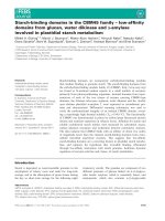
The lipocalins share a structurally conserved b-barrel
as their central folding motif, which is composed of
eight antiparallel b-strands that wind around a central
axis (Fig. 1). At its open end the cup-like structure
supports four loops, which form the entrance to the
ligand pocket. The opposite end of the b-barrel is
closed by short loops, and densely packed amino acid
side chains form the hydrophobic core in this region.
As another typical feature, a C-terminal a-helix packs
against the b-barrel from one side. Despite extremely
low mutual sequence homology, the b-barrel is struc-
turally highly conserved among the lipocalins. In con-
trast, the loop region around the ligand-binding site
exhibits large mutual differences, both in amino acid
sequence and length, and in the conformation of the
four polypeptide segments [5].
This structural property reflects the many ligand
specificities observed for this protein family and resem-
bles the hypervariable region that forms the antigen-
binding site of antibodies [6]. In the immunoglobulins,
six hypervariable loops, also called complementarity-
determining regions (CDRs), are supported by the
structurally rigid b-sandwich framework of the paired
variable domains of the light and heavy chains. These
CDRs come together at the tips of the Y-shaped mole-
cule to form a contiguous interface for antigen bind-
ing. On the basis of this structural resemblance,
lipocalins should offer the same potential for molecu-
lar recognition as do antibodies. In contrast, natural li-
pocalins cannot benefit from the mechanisms of
somatic gene recombination and hypermutation, which
lead to the vast number of different antibodies gener-
ated by the immune system. However, the methods of
combinatorial biochemistry can be employed in order
to engineer artificial lipocalins with novel specificities
for prescribed targets, which were hence dubbed
‘anticalins’ [7,8].
Properties and potential of anticalins
Engineered lipocalins offer several advantages over
immunoglobulins. Their size, of < 20 kDa, is much
smaller than that of antibodies, whose extended molec-
ular dimensions hamper efficient tissue penetration.
RBP ApoD
Tlc NGAL
Superposition of Lipocalins
Fig. 1. Molecular architecture of human lipocalins and structural variability of their binding sites. Ribbon representation of the crystal struc-
tures of four human lipocalins: retinol-binding protein (RBP; PDB entry 1RBP), apolipoprotein D (ApoD; PDB entry 2HZQ), tear lipocalin (Tlc;
PDB entry 1XKI) and neutrophil gelatinase-associated lipocalin (NGAL; PDB entry 1L6M). Lipocalins share a conserved b -barrel of eight
antiparallel b-strands (cyan). The four exposed loops at its open end (red), which form the natural ligand-binding site, exhibit high structural
variability, which is illustrated by the superposition shown to the right.
Anticalins A. Skerra
2678 FEBS Journal 275 (2008) 2677–2683 ª 2008 The Author Journal compilation ª 2008 FEBS
Furthermore, lipocalins have a rather simple composi-
tion, which is based on a single polypeptide chain. In
contrast, antibodies comprise two different polypep-
tides (i.e. the light and heavy chains), which leads to
unstable domain association when dealing with small
Fv fragments and which also requires complicated
cloning steps for recombinant expression. With four
structurally variable loops, the binding site of lipoca-
lins is less complex and easier to manipulate [5] than
the CDR of antibodies, which is composed of alto-
gether six non-sequential loop segments from both
immunoglobulin chains [6].
Naturally, lipocalins lack the constant Fc region,
which mediates immunological effector functions but
often causes undesired interactions of antibodies while
being crucial only for a few biopharmaceutical applica-
tions. Finally, many lipocalins lack glycosylation and
can thus be produced as authentic proteins in micro-
bial expression systems, whereas the manufacture of
glycosylated full-size antibodies requires expensive
eukaryotic cell culture, whose optimization and fer-
mentation is time-consuming and prone to limited
capacities [9]. While some of these benefits have also
been claimed for engineered single-chain variable frag-
ments of antibodies or isolated VHH domains of cam-
eloid immunoglobulins, for example their practical
applicability compared with intact antibodies, espe-
cially for medical purposes, is still unclear [10].
Similarly to the immunoglobulins, human lipocalins
occur as soluble proteins in the plasma and other
tissue fluids, with concentrations up to approximately
1.0 mgÆmL
)1
. Most of the lipocalins are freely distrib-
uted in the body, where they exert a ligand buffer or
transport function. This predestines this family of
proteins not only as carrier vehicles or scavengers for
pharmaceutically active compounds but also, especially
when engineered for novel binding functions, as thera-
peutic drugs on their own [11].
Both natural and engineered lipocalins are often
surprisingly stable, with melting temperatures above
70 °C [12], and they are easily produced in Escherichia
coli in a functional state [4]. The recombinant lipoca-
lins can be recovered as soluble monomeric proteins,
even when lacking natural glycosylation (eg: ApoD
and NGAL). Lipocalins are typical secretory proteins,
both in vertebrates and in lower organisms such as
insects, and thus they often carry one or two disul-
phide bonds. Consequently, bacterial production via a
secretory route is the method of choice [4,13], albeit
several recombinant lipocalins were also successfully
isolated from the soluble cytoplasmic extract of E. coli
[14,15]. As the disulphide bridges are not buried in the
hydrophobic interior of lipocalins – but rather serve
for cross-linking the N- and C-terminus to the b-barrel
[5] – they are not as crucial for folding as is the case
for immunoglobulins. Indeed, several natural lipocalins
devoid of disulphide bonds exist (eg: the human epi-
didymal and the bacterial lipocalins), and in other li-
pocalins (e.g. Tlc) the single disulphide bond can be
eliminated without much loss of protein stability.
Interestingly, especially among the human lipocalins,
many members carry an additional free Cys residue.
Its reactive thiol side chain sometimes serves for
cross-linking to other plasma proteins, although the
physiological function is often not known. Thus, for
application as research reagents, or in medical diagnos-
tics as well as therapy, it is usually advisable to substi-
tute the unpaired Cys residue with an inert amino
acid, such as Ser [4]. On the other hand, a free Cys res-
idue can be used for the site-specific covalent attach-
ment of functional groups via maleimide chemistry,
including fluorescent labels or poly(ethylene glycol),
which can serve for plasma half-life extension [16].
Lipocalins are also well suited for the construction of
functional fusion proteins. The fusion of anticalins
with alkaline phosphatase, for example, leads to useful
reporter reagents [17]. Anticalins may even be fused
with each other, yielding either bivalent or bispecific-
binding proteins, so-called ‘duocalins’ [18].
Anticalins recognizing small molecules
Initially, the structurally and biochemically well char-
acterized bilin-binding protein (BBP) of Pieris brassi-
cae [19] was employed to engineer an artificial binding
site for ligands such as fluorescein and digoxigenin, as
well as other small molecules and peptides. This lipoc-
alin comprises 174 residues and exhibits a rather wide
and shallow ligand pocket, where biliverdin IX
c
is
complexed as natural ligand. Sixteen residues distrib-
uted across all four loop segments, whose side chains
form the centre of the binding site, were identified by
molecular modelling and subjected to concerted
random mutagenesis, followed by phagemid display
selection for variants with novel binding activities [7].
In the case of fluorescein, which was chosen as a
well-known immunological hapten, several variants
with high specificity and dissociation constants as low
as 35.2 nm were identified. Following X-ray structural
analysis of the complex between the engineered lipoca-
lin and this ligand [20], improved variants with K
D
values for fluorescein of approximately 1 nm were
rationally engineered just by optimizing two side
chains in the binding pocket [21]. Thus, it was demon-
strated that engineered lipocalins with novel specifici-
ties (i.e. anticalins) can provide hapten-binding
A. Skerra Anticalins
FEBS Journal 275 (2008) 2677–2683 ª 2008 The Author Journal compilation ª 2008 FEBS 2679
proteins with affinities in a range that was so far con-
sidered typical for antibodies. Notably, the BBP vari-
ants appeared to recognize fluorescein – or other small
molecule targets – as true haptens, without measurable
context-dependence concerning the carrier protein that
served for ligand immobilization during the phage-dis-
play panning process. With their ability to provide
deep and highly complementary ligand pockets, antica-
lins distinguish themselves from most other protein
scaffolds that are currently under investigation [22].
From the same BBP mutant library an anticalin with
specificity for the cardiac steroid digoxigenin was
selected [17]. Its initially moderate affinity was subse-
quently raised by selective random mutagenesis of the
first hypervariable loop, followed by phagemid display
and colony screening under more stringent conditions,
thus resulting in a 10-fold improved K
D
value of
30.2 nm. Attempts to raise the affinity for digoxigenin
even further were made in a combinatorial approach
using a ‘loop-walking’ randomization strategy [12] and
also by rational protein design based on the crystal
structure of this engineered lipocalin [23]. These
approaches allowed the identification of several point
mutations, leading to K
D
values as low as 800 pm for
digoxin (i.e. the natural glycosylated derivative of
digoxigenin) [11].
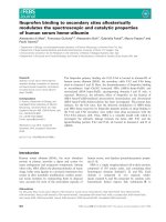
The crystal structures of these first anticalins in com-
plex with their ligands – and in one instance also as
apo-protein – which were solved at resolutions of
2.0 A
˚
or better [20,23], provided interesting insight into
the mechanism and specificity of molecular recognition
by engineered lipocalins (Fig. 2). Most importantly,
the extensive replacement of side chains, affecting 10%
of all residues in the BBP, did not impair the b-barrel
fold. The randomized loops, on the other hand,
adopted dramatically altered conformations compared
with the wild-type lipocalin. Both fluorescein and
digoxigenin are bound at the bottom of the cleft that
harbours biliverdin IX
c
in the BBP [24]. Thus, while
the overall topology of the lipocalin, comprising the
b-barrel with the a-helix attached to it, remained con-
served for both anticalins, the set of four loops at the
entrance to the ligand pocket exhibited pronounced
conformational differences in comparison with each
other and with the BBP. These structural changes seem
to be triggered by the amino acid substitutions that
were introduced during the combinatorial engineering
of the anticalins rather than by complex formation
with the ligand, thus illustrating the inherent structural
plasticity of the lipocalin loop region.
Indeed, the mechanism of complex formation, at
least with low-molecular-weight ligands, appears to be
similar to the interaction between antibodies and hap-
tens, except that the ligand can be buried more deeply
in the engineered lipocalin pocket. Shape complemen-
tarity is mainly generated by means of aromatic side
chains, and specific interactions arise from suitably
placed hydrogen-bond donors or acceptors, sometimes
mediated by buried water molecules. Notably, in the
case of the digoxigenin-binding anticalin, DigA16, the
bound steroid ligand is sandwiched between one Trp
and two Tyr side chains, very similar to a monoclonal
antibody directed against digoxin, which provides a
nice example of ‘convergent’ in vitro evolution [23]. In
addition, a His side chain at the bottom of the ligand
pocket displays an induced fit upon complex formation
with digoxigenin, an effect so far regarded as common
in antibodies. Further to the pronounced backbone
plasticity in the loop region, comparison of the pri-
mary sequences of many engineered lipocalins revealed
that all randomized amino acid positions essentially
tolerate the entire set of natural side chains.
Apart from these fundamental insights into the struc-
ture–function relationships of lipocalins and their simi-
larity to immunoglobulins, the resulting anticalin,
which was designated Digical, may be applicable as a
therapeutic agent for the treatment of digitalis intoxica-
tions. Although digitalis is widely applied in conjunc-
tion with heart insufficiency and arrhythmias [25], it
has a very narrow therapeutic window, and precise
adjustment of digoxin plasma levels is mandatory to
prevent poisoning with fatal outcome. Indeed, when
Digical was employed for studies in a guinea-pig animal
model of digitalis intoxication, the anticalin appeared
N
Loop #4
Loop #1
C
Loop #2
Loop #3
Fig. 2. 3D structure of an anticalin in complex with its cognate
ligand. Ribbon representation of the crystal structure of the digoxi-
genin-binding anticalin DigA16 (PDB entry 1LKE). The bound ligand
is shown in a space-filling representation in yellow, whereas the 16
amino acid side chains in the four hypervariable loops – as well as
the adjoining regions of the b-barrel – which were randomized in
the naive combinatorial library derived from the BBP used for the
anticalin selection, are depicted in orange. The N-terminus (N) and
the C-terminus (C) of the polypeptide chain are labelled.
Anticalins A. Skerra
2680 FEBS Journal 275 (2008) 2677–2683 ª 2008 The Author Journal compilation ª 2008 FEBS
to be effective in reversing the digoxin-induced toxicity
after administering just a moderate stoichiometric
excess [11], thus demonstrating the acute protective
effect of this anticalin on the cardiovascular system and
its suitability as an antidote against digoxin.
Furthermore, the anticalin FluA, which possesses
high affinity for fluorescein, has the interesting prop-
erty of almost completely quenching the fluorescence
emission of this widely applied reagent [7]. The reason
for the disappearance of the stationary ligand fluores-
cence seems to be an ultrafast electron transfer
between the excited fluorescein dianion and a Trp side
chain in the binding site of the engineered lipocalin,
which closely packs against the xanthenolone moiety
[26]. This phenomenon opens interesting applications
in biophysics. Such an ‘anti-fluorescent’ protein could
also be useful as a reagent for the specific quenching
of background signals that arise from fluorescein
groups surrounding a cell, for example when deter-
mining the topology of a site-specifically labelled
membrane protein.
Anticalins directed at proteins
Considering medical applications, extracellular proteins
or cell-surface receptors are the predominant class of
biomolecules that currently provide relevant targets for
biopharmaceuticals such as antibodies. Consequently,
in recent years anticalin libraries were specifically
developed for the recognition of such protein ‘anti-
gens’. In addition, to reduce immunogenic side effects
upon prolonged treatment, these libraries were con-
structed on the basis of natural human lipocalins, in
particular ApoD [27,28], NGAL [29] and Tlc [30]
(Fig. 1).
To this end, 16–24 amino acid residues located at
exposed positions, close to the tips of the four hyper-
variable loops, were subjected to random mutagenesis
in order to allow tight contact formation with a mac-
romolecular target, which cannot penetrate as deeply
into the ligand-binding site as a small molecule. Using
these libraries, anticalins with high specificity and
affinities in the subnanomolar range were successfully
selected against a variety of disease-related protein
antigens, including immunological receptors such as
cytotoxic T-lymphocyte antigen-4 (CTLA-4) [11] and
soluble growth factors such as vascular endothelial
growth factor (VEGF) [31].
Recently, the crystal structure of the complex
between a cognate anticalin and the extracellular
domain of CTLA-4 was solved, demonstrating that a
macromolecular ‘protein antigen’ can be effectively
bound at the cup-shaped binding site of an engineered
lipocalin, even though its natural counterparts almost
exclusively recognize low-molecular-weight substances.
All four randomized loops of NGAL – which had
served as a lipocalin scaffold in this case – contribute
to the formation of the molecular complex, thus vali-
dating the design of the anticalin library.
CTLA-4 (CD152) is an activation-induced, trans-
membrane T-cell coreceptor with an inhibitory effect
on T-cell-mediated immune responses [32]. CTLA-4
antagonizes the CD28-dependent costimulation of T
cells, whereby CTLA-4 and CD28 share the same
counter-receptors on antigen-presenting cells (i.e. B7.1
and B7.2). Notably, the bound anticalin shields the
CTLA-4 epitope that is involved in the interaction
both with B7.1 and B7.2. Indeed, an antagonistic
activity of the anticalin towards CTLA-4 was con-
firmed in several in vitro cell culture tests, where T-cell
proliferation was stimulated in a manner comparable
to that of commercially available antibodies directed
against the same target. Thus, the CTLA-4-specific
anticalin is a promising drug candidate for the immu-
notherapy of cancer, similarly to corresponding anti-
bodies that are already in clinical trials [33]. Apart
from its much smaller size and probably better tissue
penetration, the lack of immunological effector func-
tions – which reside in the antibody Fc region – for
the anticalin should limit off-target toxicity because
only the antagonistic activity is needed. In fact, this is
the case for many relevant targets involved in the regu-
lation of the immune response and inflammation as
well as neoangiogenesis.
Another promising drug candidate is an anticalin
with strong antagonistic activity towards VEGF.
VEGF is a well-characterized mediator of tumor
angiogenesis and other neovascular diseases [34], for
example age-related macular degeneration (AMD).
The selected anticalin exhibits a favorable binding and
activity profile in direct comparison with currently
approved VEGF antagonists [31]. A half-life extended
version of the anticalin has demonstrated excellent effi-
cacy in three animal models assessing VEGF-induced
enhanced vascular permeability, angiogenesis and anti-
xenograft tumor activity. As immunological effector
functions again appear to be irrelevant for biomedical
activity, an anticalin with proven VEGF-antagonistic
function should offer an interesting alternative to full-
size antibodies, especially in the light of its presumably
better distribution.
Conclusions and prospects
Engineered lipocalins offer binding sites with surpris-
ingly high structural plasticity and an extended
A. Skerra Anticalins
FEBS Journal 275 (2008) 2677–2683 ª 2008 The Author Journal compilation ª 2008 FEBS 2681
molecular interface for target recognition which is
comparable in size to that of antibodies. Anticalins
with high specificity and affinity, down to picomolar
dissociation constants, can be readily generated against
haptens, peptides and proteins. Thus, regarding their
range of addressable targets they surpass other protein
scaffolds that are presently pursued [22]. Available
structural and functional data suggest that anticalins
are able to recognize a diverse set of epitopes on differ-
ent target proteins and therefore have considerable
potential as specific antagonistic reagents in general.
Consequently, anticalins constitute promising
reagents for therapeutic applications. As anticalins
can be derived from human lipocalin scaffolds, the
risk of immunogenicity is minimized and further
reformatting – such as by CDR grafting for the
‘humanization’ of antibodies – is not required. The
absence of immunological effector functions prevents
many potential side effects known for antibodies.
Furthermore, the monovalent binding activity of
anticalins decreases the risk of intermolecular cross-
linking of cellular receptor targets that could lead to
unwanted signal triggering.
Natural lipocalins, as well as engineered lipocalins,
are quickly cleared by renal filtration, as a result of
their small size of approximately 20 kDa, if they circu-
late as monomeric proteins. When conjugated with
radioactive isotopes for in vivo diagnostics, for exam-
ple, such properties should lead to images of high
contrast soon after administration. Nevertheless, for
medical indications that require prolonged treatment,
the simple architecture and robustness of the lipocalin
scaffold facilitates the preparation of fusion proteins
or of site-directed conjugates to decelerate clearance.
In principle, several established techniques are avail-
able to extend the plasma half life of anticalins, for
example by the production of fusion proteins with
serum albumin, with an albumin-binding domain or
peptide or via pegylation.
Anticalins display both their N-terminus and C-ter-
minus in an accessible manner and remote from the
binding site, which differs from the situation with sin-
gle-chain variable fragments of antibodies, where the
N-terminus often forms part of the paratope. Thus,
anticalins are well suited for fusion with other func-
tional domains without compromising their engineered
binding activities. Fusion proteins of anticalins that
address a specific receptor on solid tumors with
enzymes which generate a cytotoxic compound from
an inactive precursor (prodrug) might be of special
interest as an alternative to antibody-directed enzyme
prodrug therapy (ADEPT). Furthermore, a dimeric
binding mode utilizing either a duocalin that has twice
the same target specificity or a fusion protein between
an anticalin and a dimerization domain may be
employed to enhance binding avidity.
Hence, owing to their adaptable binding site and
their simple and robust molecular architecture, specifi-
cally engineered anticalins promise a future as versatile
reagents for research, biotechnology and medicine.
References
1A
˚
kerstro
¨
m B, Borregaard N, Flower DA & Salier J-S
(2006) Lipocalins. Landes Bioscience, Georgetown, TX.
2 Finn RD, Mistry J, Schuster-Bockler B, Griffiths-Jones
S, Hollich V, Lassmann T, Moxon S, Marshall M,
Khanna A, Durbin R et al. (2006) Pfam: clans, web
tools and services. Nucleic Acids Res 34, D247–D251.
3 Berman HM, Westbrook J, Feng Z, Gilliland G, Bhat
TN, Weissig H, Shindyalov IN & Bourne PE (2000)
The Protein Data Bank. Nucleic Acids Res 28, 235–242.
4 Breustedt DA, Scho
¨
nfeld DL & Skerra A (2006) Com-
parative ligand-binding analysis of ten human lipoca-
lins. Biochim Biophys Acta 1764, 161–173.
5 Skerra A (2000) Lipocalins as a scaffold. Biochim Bio-
phys Acta 1482, 337–350.
6 Skerra A (2003) Imitating the humoral immune
response. Curr Opin Chem Biol 7, 683–693.
7 Beste G, Schmidt FS, Stibora T & Skerra A (1999)
Small antibody-like proteins with prescribed ligand
specificities derived from the lipocalin fold. Proc Natl
Acad Sci U S A 96, 1898–1903.
8 Skerra A (2001) ‘Anticalins’: a new class of engineered
ligand-binding proteins with antibody-like properties.
J Biotechnol 74, 257–275.
9 Werner RG (2004) Economic aspects of commercial
manufacture of biopharmaceuticals. J Biotechnol 113,
171–182.
10 Holliger P & Hudson PJ (2005) Engineered antibody
fragments and the rise of single domains. Nat Biotech-
nol 23, 1126–1136.
11 Schlehuber S & Skerra A (2005) Lipocalins in drug dis-
covery: from natural ligand-binding proteins to ‘antica-
lins’. Drug Discov Today 10, 23–33.
12 Schlehuber S & Skerra A (2002) Tuning ligand affinity,
specificity, and folding stability of an engineered lipoca-
lin variant – a so-called ‘anticalin’ – using a molecular
random approach. Biophys Chem 96, 213–228.
13 Mu
¨
ller HN & Skerra A (1993) Functional expression of
the uncomplexed serum retinol-binding protein in
Escherichia coli. Ligand binding and reversible unfold-
ing characteristics. J Mol Biol 230, 725–732.
14 Coles M, Diercks T, Muehlenweg B, Bartsch S, Zolzer
V, Tschesche H & Kessler H (1999) The solution struc-
ture and dynamics of human neutrophil gelatinase-
associated lipocalin. J Mol Biol 289, 139–157.
Anticalins A. Skerra
2682 FEBS Journal 275 (2008) 2677–2683 ª 2008 The Author Journal compilation ª 2008 FEBS
15 Holzfeind P, Merschak P, Rogatsch H, Culig Z, Feicht-
inger H, Klocker H & Redl B (1996) Expression of the
gene for tear lipocalin ⁄ von Ebner’s gland protein in
human prostate. FEBS Lett 395, 95–98.
16 Harris JM & Chess RB (2003) Effect of pegylation on
pharmaceuticals. Nat Rev Drug Discov 2, 214–221.
17 Schlehuber S, Beste G & Skerra A (2000) A novel type
of receptor protein, based on the lipocalin scaffold, with
specificity for digoxigenin. J Mol Biol 297, 1105–1120.
18 Schlehuber S & Skerra A (2001) Duocalins: engineered
ligand-binding proteins with dual specificity derived
from the lipocalin fold. Biol Chem 382, 1335–1342.
19 Schmidt FS & Skerra A (1994) The bilin-binding pro-
tein of Pieris brassicae. cDNA sequence and regulation
of expression reveal distinct features of this insect
pigment protein. Eur J Biochem 219, 855–863.
20 Korndo
¨
rfer IP, Beste G & Skerra A (2003) Crystallo-
graphic analysis of an ‘‘anticalin’’ with tailored specific-
ity for fluorescein reveals high structural plasticity of
the lipocalin loop region. Proteins Struct Funct Genet
53, 121–129.
21 Vopel S, Mu
¨
hlbach H & Skerra A (2005) Rational engi-
neering of a fluorescein-binding anticalin for improved
ligand affinity. Biol Chem 386, 1097–1104.
22 Skerra A (2007) Alternative non-antibody scaffolds for
molecular recognition. Curr Opin Biotechnol 18, 295–
304.
23 Korndo
¨
rfer IP, Schlehuber S & Skerra A (2003) Struc-
tural mechanism of specific ligand recognition by a
lipocalin tailored for the complexation of digoxigenin.
J Mol Biol 330, 385–396.
24 Huber R, Schneider M, Mayr I, Mu
¨
ller R, Deutzmann
R, Suter F, Zuber H, Falk H & Kayser H (1987)
Molecular structure of the bilin binding protein (BBP)
from Pieris brassicae after refinement at 2.0 A
˚
resolu-
tion. J Mol Biol 198, 499–513.
25 Hauptman PJ & Kelly RA (1999) Digitalis. Circulation
99, 1265–1270.
26 Go
¨
tz M, Hess S, Beste G, Skerra A & Michel-Beyerle
ME (2002) Ultrafast electron transfer in the complex
between fluorescein and a cognate engineered lipocalin
protein, a so-called anticalin. Biochemistry 41, 4156–
4164.
27 Eichinger A, Nasreen A, Kim HJ & Skerra A (2007)
Structural insight into the dual ligand specificity and
mode of high density lipoprotein association of apolipo-
protein D. J Biol Chem 282, 31068–31075.
28 Vogt M & Skerra A (2004) Construction of an artificial
receptor protein (‘‘anticalin’’) based on the human
apolipoprotein D. ChemBioChem 5, 191–199.
29 Goetz DH, Holmes MA, Borregaard N, Bluhm ME,
Raymond KN & Strong RK (2002) The neutrophil
lipocalin NGAL is a bacteriostatic agent that interferes
with siderophore-mediated iron acquisition. Mol Cell
10, 1033–1043.
30 Breustedt DA, Korndo
¨
rfer IP, Redl B & Skerra A
(2005) The 1.8-A
˚
crystal structure of human tear lipoca-
lin reveals an extended branched cavity with capacity
for multiple ligands. J Biol Chem 280, 484–493.
31 Hohlbaum AM & Skerra A (2007) Anticalins: the lipoc-
alin family as a novel protein scaffold for the develop-
ment of next-generation immunotherapies. Expert Rev
Clin Immunol 3, 491–501.
32 Leach DR, Krummel MF & Allison JP (1996) Enhance-
ment of antitumor immunity by CTLA-4 blockade.
Science 271, 1734–1736.
33 Tarhini AA & Kirkwood JM (2007) Tremelimumab, a
fully human monoclonal IgG2 antibody against CTLA4
for the potential treatment of cancer. Curr Opin Mol
Ther 9, 505–514.
34 Ferrara N, Gerber HP & LeCouter J (2003) The biol-
ogy of VEGF and its receptors. Nat Med 9, 669–676.
A. Skerra Anticalins
FEBS Journal 275 (2008) 2677–2683 ª 2008 The Author Journal compilation ª 2008 FEBS 2683