Báo cáo khoa học: Incorporation of ZP1 into perivitelline membrane after in vivo treatment with exogenous ZP1 in Japanese quail (Coturnix japonica) doc
Bạn đang xem bản rút gọn của tài liệu. Xem và tải ngay bản đầy đủ của tài liệu tại đây (433.21 KB, 10 trang )
Incorporation of ZP1 into perivitelline membrane after
in vivo treatment with exogenous ZP1 in Japanese quail
(Coturnix japonica)
Mihoko Kinoshita
1
, Kaori Mizui
1
, Tsukasa Ishiguro
1
, Mamoru Ohtsuki
1
, Norio Kansaku
2
, Hiroshi
Ogawa
3
, Akira Tsukada
4
, Tsukasa Sato
1
and Tomohiro Sasanami
1
1 Department of Applied Biological Chemistry, Faculty of Agriculture, Shizuoka University, Japan
2 Laboratory of Animal Genetics and Breeding, Azabu University, Fuchinobe, Sagamihara, Japan
3 Laboratory of Wild Animals, Tokyo University of Agriculture, Atsugi-shi, Japan
4 Graduate School of Bioagricultural Sciences, Nagoya University, Japan
The avian vitelline membrane in laid eggs consists of
three layers: the outer layer; the continuous membrane;
and the inner layer [1]. The outer layer, which is com-
posed of a varying number of sublayers of latticed fine
fibrils, is formed in the infundibulum part of the ovi-
duct [2]. The continuous membrane, which is a very
thin granular membrane, is also formed in the infun-
dibulum [2]. The inner layer, a 3D network of coarse
fibers, is found between the granulosa cells and the
oocyte in follicles before ovulation and is called the
perivitelline membrane (PL) [3]. The PL is a homo-
logue of the egg envelope in other vertebrates, the
zona pellucida in mammals, the vitelline membrane in
amphibians and the chorion in teleosts. These egg
Keywords
Japanese quail; matrix assembly;
perivitelline membrane; zona pellucida; ZP1
Correspondence
T. Sasanami, Department of Applied
Biological Chemistry, Faculty of Agriculture,
Shizuoka University, 836 Ohya, Shizuoka
422-8529, Japan
Fax ⁄ Tel: +81 54 238 4526
E-mail:
(Received 20 March 2008, revised 7 May
2008, accepted 13 May 2008)
doi:10.1111/j.1742-4658.2008.06503.x
In birds, the egg envelope surrounding the oocyte prior to ovulation is
called the perivitelline membrane and it plays important roles in fertiliza-
tion. In a previous study we demonstrated that one of the components of
the perivitelline membrane, ZP3, which is secreted from the ovarian granu-
losa cells, specifically interacts with ZP1, another constituent that is synthe-
sized in the liver of Japanese quail. In the present study, we investigated
whether ZP1 injected exogenously into the blood possesses the ability to
reconstruct the perivitelline membrane of Japanese quail. When ZP1 puri-
fied from the serum of laying quail was injected into other female birds,
the signal of this exogenous ZP1 was detected in the perivitelline mem-
brane. In addition, we revealed, by means of ligand blot analysis, that
serum ZP1 interacts with both ZP1 and ZP3 of the perivitelline membrane.
By contrast, when ZP1 derived from the perivitelline membrane was
administered, it failed to become incorporated into the perivitelline mem-
brane. Interestingly, serum ZP1 recovered from other Galliformes, includ-
ing chicken and guinea fowl, could be incorporated into the quail
perivitelline membrane, but the degree of interaction between quail ZP3
and ZP1 of the vitelline membrane of laid eggs from chicken and guinea
fowl appeared to be weak. These results demonstrate that exogenous ZP1
purified from the serum, but not ZP1 from the perivitelline membrane, can
become incorporated into the perivitelline membrane upon injection into
other types of female birds. To our knowledge, this is the first demon-
stration that the egg envelope component, when exogenously administered
to animals, can reconstruct the egg envelope in vivo.
Abbreviations
CBB, Coomassie Brilliant Blue; DIG, digoxigenin; PL, perivitelline membrane; PVDF, poly(vinylidene difluoride); ZP, zona pellucida.
3580 FEBS Journal 275 (2008) 3580–3589 ª 2008 The Authors Journal compilation ª 2008 FEBS
envelopes are mainly constructed of glycoproteins
belonging to different subclasses of the zona pellucida
(ZP) gene family [4–7]. The components of this matrix
include three glycoproteins (ZP1, ZP2 and ZP3) in
mouse [4,8] and four glycoproteins (ZP1, ZP2, ZP3
and ZP4) in several other organisms, including
humans, bonnet monkeys and rats [9–11]. The ZP gene
family proteins are characterized by a highly conserved
amino acid sequence called the ZP domain, consisting
of about 260 amino acid residues with 8 or 10 con-
served Cys residues [12].
Two glycoproteins, ZP3 and ZP1, have been iden-
tified as major components of the PL in quail
oocytes [13], and each of these glycoproteins is an
essential structural component of the extracellular
coat. In addition to these major glycoproteins, we
recently cloned the cDNA encoding ZPD (GenBank
accession number: AB301422) and ZP2 (GenBank
accession number: AB295393), which are also
expressed in the ovary of laying quail (M. Kinoshita
& T. Sasanami, unpublished results). We previously
reported that the ZP3 protein was produced in the
granulosa cells of developing follicles by the stimu-
lation of follicle-stimulating hormone [14,15] and
testosterone [16]. On the other hand, the other con-
stituent, ZP1, is synthesized in the liver of the laying
female, and the expression of ZP1 mRNA was stim-
ulated by in vivo treatment with diethylstilbestrol
[17].
In a previous study, we demonstrated that the ZP3
secreted from the granulosa cells specifically binds
with ZP1, and that this phenomenon might be
involved in the formation of insoluble PL fibers in
the quail ovary [18]. We also provided evidence dem-
onstrating that the C-terminal half of the ZP domain
of ZP1 contains a binding site for ZP3 [19]. Similarly,
Okumura et al. also demonstrated that ZP1 in the
serum of laying hens specifically interacts with ZP3
[20] and that this interaction induces the formation of
fibrous aggregates, which are visible under optical
microscopy [21].
In the present study, we aimed to clarify whether
ZP1, when injected exogenously into the blood, pos-
sesses the ability to reconstruct the PL of Japanese
quail. To achieve this, we purified ZP1 proteinfrom
the serum and from the PL lysate and then injected
the proteins intravenously into birds, which were
then analyzed to establish the level of ZP1 incorpo-
ration in the PL. Moreover, we compared the differ-
ences in the incorporation of the ZP1 purified from
Japanese quail with the incorporation of ZP1 recov-
ered from other Galliformes (chicken and guinea
fowl). To our knowledge, this is the first report to
demonstrate that exogenous ZP1 derived from the
serum, but not from the PL, can be successfully
incorporate into the PL.
Results
Incorporation of exogenously injected ZP1 into
quail PL
To determine whether ZP1 injected intravenously into
a bird was incorporated into the PL of the ovarian fol-
licles, we first purified the 97-kDa ZP1 from either the
serum or the PL lysate of laying quail and labeled the
samples with digoxigenin (DIG). As shown in Fig. 1,
presence of the purified serum ZP1 (Fig. 1A, lane 1), as
well as of the purified PL ZP1 (Fig. 1B, lane 1), pro-
duced a dominant band migrating at a molecular mass
of 97 kDa. In addition, both 97-kDa bands reacted
strongly with anti-ZP1 serum (Fig. 1A,B, lane 2) as
well as with anti-DIG immunoglobulin (Fig. 1A,B, lane
3). These results suggest that each glycoprotein purified
by means of the methods in this study was practically
pure and was successfully labeled with DIG.
The DIG-labeled ZP1 was injected into laying
females, and the PL lysates isolated from the largest
follicles 6 h after the injection were probed with anti-
DIG immunoglobulin. The results are shown in Fig. 2.
As shown in Fig. 2A, the PL lysate from the oocytes
of birds injected with DIG-labeled PL ZP1 (Fig. 2A,
lane 1) or DIG-labeled serum ZP1 (Fig. 2A, lane 2)
showed similar banding patterns after staining the gel
with Coomassie Brilliant Blue R250 (CBB). When the
sample was analysed by western blotting and probed
with anti-DIG immunoglobulin (Fig. 2B), immuno-
reactive bands migrating at around 175 and 97 kDa,
which corresponds to the apparent molecular mass
values of dimeric and monomeric ZP1, respectively,
were detected in the PL lysate from the animals
injected with DIG-labeled serum ZP1 (lane 2). The
immunoreactive bands were observed to migrate at a
level consistent with a molecular mass of approxi-
mately 120 kDa (Fig. 2B, lane 2). This immunoreactive
protein might be the 97-kDa ZP1 complex, which was
formed through an intermolecular disulfide bond with
the ZP1 fragment or with other partner(s), because this
band shifted to 97 kDa when the proteins were sepa-
rated under reducing conditions [19]. Interestingly,
the PL lysate obtained from the DIG-labeled PL
ZP1-treated birds contained no immunoreactive mate-
rials (Fig. 2B, lane 1). The samples obtained from the
birds treated with NaCl ⁄ P
i
alone also had no immuno-
reactive bands (lane 3). These results suggest that the
intravenously injected serum ZP1, but not PL ZP1, is
M. Kinoshita et al. Incorporation of ZP1 into perivitelline membrane
FEBS Journal 275 (2008) 3580–3589 ª 2008 The Authors Journal compilation ª 2008 FEBS 3581
incorporated into the PL of the quail ovary and the
intravenously injected DIG-labeled serum ZP1 could
be detected in the endogenous PL 6 h after injection.
In subsequent experiments, we investigated the accu-
mulation of the intravenously injected ZP1 protein in
the PL during follicular development by western blot
analysis. As shown in Fig. 3, the F3 lysate contains a
small amount of immunoreactive protein (Fig. 3B, lane
1), but the intensity of the band increases in mature
follicles (F2: Fig. 3, lane 2; F1: Fig. 3, lane 3). The PL
lysate isolated from the control animals, which were
injected with NaCl ⁄ P
i
alone, contained no immuno-
reactive bands (Fig. 3A). These results suggest that the
exogenously injected serum ZP1 is accumulated in the
final stage of follicular growth.
To investigate the localization of the injected ZP1
protein in the follicles, we prepared paraffin sections
of pre-ovulatory follicles and analyzed them by
immunofluorescence microscopy. As shown in Fig. 4,
the immunoreactive material recognized by anti-DIG
immunoglobulin accumulated in the PL apposed to
the apical surface of the granulosa cells of the largest
follicle (Fig. 4A). No such intense immunostaining
was seen when the specimens prepared from the
control animals were stained with anti-DIG immuno-
globulin (Fig. 4B). Taken together, these results
clearly demonstrate that the serum ZP1 intravenously
injected into the animals is transported into the
ovary via the blood circulation, and that the
protein accumulates in the PL of mature follicles
in vivo.
200 kDa
117 kDa
97 kDa
66 kDa
45 kDa
31 kDa
200 kDa
117 kDa
97 kDa
66 kDa
45 kDa
31 kDa
M1 2 3
M1 2
3
A
B
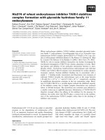
Fig. 1. Purification of ZP1 from the serum or the PL of laying quail.
The ZP1 proteins purified from the serum of laying quail (A) or
recovered from the PL lysate of the largest follicles (B) were
labeled with DIG and separated by SDS-PAGE (1 lg per lane). The
proteins were then detected using silver staining (lane 1). The
same sample was also western blotted then probed using anti-ZP1
serum (lane 2) or anti-DIG immunoglobulin (lane 3). The molecular
mass markers were also separated by SDS-PAGE and detected
using silver staining (lane M). Molecular mass values of the mark-
ers are given on the left of the respective lanes. Representative
results of repeated experiments are shown.
200 kDa
117 kDa
97 kDa
66 kDa
45 kDa
31 kDa
21.5 kDa
200 kDa
117 kDa
97 kDa
66 kDa
45 kDa
31 kDa
M12
12
3
AB
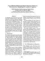
Fig. 2. Detection of exogenously injected ZP1 in the PL lysates.
(A) The PL lysates isolated from the largest follicles of the quail
administered DIG-labeled PL (lane 1) or serum ZP1 (lane 2) were
separated on SDS-PAGE (10 lg per lane) and were stained
with CBB. The molecular mass marker was also separated by
SDS-PAGE and detected using CBB staining (lane M). (B) The
same samples in (A) were probed with anti-DIG immunoglobulin
(lanes 1 and 2). The PL lysate of the quail (10 lg per lane)
injected with vehicle alone was used as a negative control
(lane 3). Representative results of repeated experiments are
shown.
Incorporation of ZP1 into perivitelline membrane M. Kinoshita et al.
3582 FEBS Journal 275 (2008) 3580–3589 ª 2008 The Authors Journal compilation ª 2008 FEBS
Both ZP1 and ZP3 in the PL interact with
serum ZP1
To identify the binding partner of serum ZP1 in the
PL, we identified the PL lysate of the largest follicles
by means of ligand blotting (Fig. 5). When the sample
was probed using DIG-labeled serum ZP1, bands
migrating at 175, 97 and 35 kDa, which are the
expected molecular mass values of dimeric ZP1, mono-
meric ZP1 and ZP3, respectively, were visualized (lane
1). When the same sample was probed using DIG-
labeled bovine serum albumin (lane 2) or without
ligand (lane 3, blocking buffer alone), no such staining
was seen. These results suggest that the serum ZP1
interacts with both ZP1 and ZP3, and that this interac-
tion might be responsible for the incorporation of
serum ZP1 into the PL.
Species-specific interaction of quail ZP3 with ZP1
from various species
In our previous study we demonstrated that tritium-
labeled ZP3, which is present in the conditioned med-
ium of the granulosa cells, binds specifically to ZP1
of the PL in Japanese quail [18,19]. To investigate
whether the interaction of ZP3 and ZP1 occurs in
other bird species, we analyzed the lysates of vitelline
membrane of laid eggs from various species using
ligand blotting and probing with radiolabelled ZP3.
To achieve this, we first confirmed the presence of
ZP3 and ZP1 proteins in the lysate of the vitelline
membrane by western blot analysis. As shown in
Fig. 6A, our anti-ZP3 serum reacted strongly with the
bands of approximately 33 kDa molecular mass in
the lysate of the vitelline membrane of Japanese quail
(lane 1), blue-breasted quail (lane 2), chicken (lane 3),
turkey (lane 4), and guinea fowl (lane 5). Similarly,
anti-ZP1 serum also recognized dimeric (around
175 kDa molecular mass) as well as monomeric
(around 97 kDa molecular mass) ZP1s in the lysates
of all vitelline membranes tested (Fig. 6B). These
results suggest that the vitelline membrane of laid
200 kDa
117 kDa
97 kDa
66 kDa
45 kDa
31 kDa
200 kDa
117 kDa
97 kDa
66 kDa
45 kDa
31 kDa
12 3
31 kDa
12 3
A
B
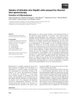
Fig. 3. Western blot analysis of exogenous ZP1 during follicular
development. (A) The PL lysate (10 lg per lane) isolated from the
third-largest (lane 1), the second-largest (lane 2), or the largest (lane
3) follicles of the control bird (vehicle alone), was transblotted onto
a PVDF membrane after separation on SDS-PAGE and then probed
with anti-DIG immunoglobulin (A). The samples (10 lg per lane)
prepared from the third-largest (lane 1), the second-largest (lane 2),
or the largest (lane 3) follicles of the bird receiving intravenous
injection of DIG-labeled serum ZP1 were probed with anti-DIG
immunoglobulin (B). The blots shown are representative of three
independent experiments.
A
B
Fig. 4. Immunohistochemical analysis of exogenous ZP1 in the fol-
licular wall. Sections of follicular wall obtained from the largest folli-
cles of the animals treated with DIG-labeled serum ZP1 (A) or with
vehicle alone (B) were processed for immunohistochemical obser-
vation using anti-DIG immunoglobulin (1 : 300 dilution). Shown are
the representative results of three experiments. Bar = 50 lm.
M. Kinoshita et al. Incorporation of ZP1 into perivitelline membrane
FEBS Journal 275 (2008) 3580–3589 ª 2008 The Authors Journal compilation ª 2008 FEBS 3583
eggs from these birds contains ZP3 and ZP1, like that
of Japanese quail.
Upon examination of the poly(vinylidene difluoride)
(PVDF) membrane that had been used to immobilize
the aliquots of SDS-solubilized vitelline membrane
proteins of Japanese quail, dimeric and monomeric
ZP1s were detected by autoradiography (Fig. 6D,
lane 1). This result is consistent with previous reports
showing that the dimeric as well as the monomeric
ZP1s were visualized by ligand blotting with radio-
labelled ZP3 [18,19]. Comparable staining intensity of
the ZP1 bands was observed when the lysates from
blue-breasted quail (lane 2) and turkey (lane 4) were
incubated with quail ZP3. In comparison with that of
Japanese quail, the ZP1 bands in the vitelline mem-
brane of chicken (lane 3) and guinea fowl (lane 5) were
stained weakly. The weaker staining intensity observed
in the case of the chicken and guinea fowl was not the
result of a lower abundance of ZP1 in the vitelline
membrane because comparable amounts of ZP1 were
detected in the lysates when the samples were stained
with CBB (Fig. 6C). These results indicate that the
interaction of ZP3 and ZP1 might occur in a species-
specific manner.
Incorporation of heterologous ZP1 into quail PL
In the next set of experiments, we investigated whether
there is species specificity in the incorporation of serum
ZP1 into the PL. For this purpose, we purified serum
ZP1 from chicken and guinea fowl in addition to Japa-
nese quail, and the purified ZP1 of these species was
injected intravenously into several laying quail, sever-
ally. As shown in Fig. 7A, reruns of the purified quail
(lanes 1 and 4), chicken (lanes 2 and 5) and guinea
fowl (lanes 3 and 6) ZP1 proteins showed a dominant
band after western blotting and probing using anti-
ZP1 serum (lanes 1-3) as well as after silver staining
(lanes 4-6).
As shown in Fig. 7B, the PL lysate of the birds that
had been injected with quail (lane 1), chicken (lane 2),
or guinea fowl (lane 3) ZP1 contained materials that
were immunoreactive with anti-DIG immunoglobulin
and migrated close to dimeric and monomeric ZP1. It
should be noted that the immunoreactive monomeric
ZP1 in the bird injected with serum ZP1 from a differ-
ent species showed distinct disparity in mobility on
SDS-PAGE. The mobilities of the monomeric ZP1 pro-
teins corresponded with those of the purified protein in
the silver-stained gel (Fig. 7A, lanes 4-6). Although the
reasons are unclear, the composition of the materials
immunoreactive with anti-DIG immunoglobulin in the
PL lysate isolated from the oocyte of the animals
injected with quail ZP1 was different from that shown
in Fig. 2B, lane 2, in spite of being the same prepara-
tion. On the other hand, no distinct difference in band-
ing pattern was observed when the total protein lysate
of each sample was visualized using CBB (Fig. 7C).
Taken together with the results shown in Fig. 5, these
results suggest that the incorporation of serum ZP1 into
the PL can occur between different species, and that the
specificity of the incorporation might be relatively low
in comparison with that of ZP3 incorporation.
Discussion
As reported previously in our studies using Japanese
quail, ZP3 secreted from cultured granulosa cells spe-
cifically interacts with ZP1, which is synthesized in the
liver, and this interaction appears to be involved in the
formation of the insoluble fibers of the PL [18,19]. In
the present study, we showed, for the first time, that
exogenous ZP1 purified from the serum of laying quail
was incorporated into the PL when it was injected into
other female birds. To our knowledge, this is the first
demonstration that the egg envelope component, when
exogenously administered to birds, actually recon-
structs the egg envelope in vivo. In accordance with
200 kDa
117 kDa
97 kDa
66 kDa
45 kDa
31 kDa
123
Fig. 5. Ligand blot analysis of PL lysate. (A) Aliquots (10 lg per
lane) of the SDS-solubilized PL isolated from the largest pre-ovula-
tory follicles of Japanese quail were subjected to ligand blot analy-
sis using DIG-labeled serum ZP1 (lane 1), DIG-labeled bovine serum
albumin (lane 2), or no ligand (lane 3). Representative blots of three
independent experiments are shown.
Incorporation of ZP1 into perivitelline membrane M. Kinoshita et al.
3584 FEBS Journal 275 (2008) 3580–3589 ª 2008 The Authors Journal compilation ª 2008 FEBS
our data, Okumura et al. recently demonstrated that
chicken ZP3 and ZP1 can specifically associate to form
a heterocomplex of ZP3 and ZP1, and this interaction
induces ZP3–ZP1 fibrous aggregates in vitro ; however,
they did not confirm this phenomenon by an in vivo
study [20,21].
In the present study we found that when ZP1 puri-
fied from the PL of the largest follicles was injected
intravenously into birds, it did not become incorpo-
rated into the PL. This was not a result of denatur-
ation of the protein during the purification process
using SDS-PAGE, because the serum ZP1 was also
123
AB
4
200 kDa
117 kDa
97 kDa
66 kDa
45 kDa
31 kDa
21.5 kDa
200 kDa
117 kDa
97 kDa
66 kDa
45 kDa
31 kDa
21.5 kDa
1234
C
12 3
200 kDa
117 kDa
97 kDa
66 kDa
45 kDa
31 kDa
21.5 kDa
45 6
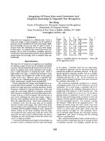
Fig. 7. Cross-species incorporation of serum ZP1 into quail PL. (A) ZP1 was purified from the sera of Japanese quail (lanes 1 and 4), chicken
(lanes 2 and 5), or guinea fowl (lanes 3 and 6). They were separated on SDS-PAGE (1 lg per lane) and detected with western blotting using
anti-ZP1 immunoglobulin (lanes 1–3) or silver staining (lanes 4–6). (B, C) The PL lysate isolated from the largest follicles of the animals, which
were injected with 10 lg of DIG-labeled quail ZP1 (lane 1), chicken ZP1 (lane 2), guinea fowl ZP1 (lane 3), or vehicle alone (lane 4), were
separated on SDS-PAGE (1 lgÆlane
)1
) and detected with anti-DIG immunoglobulin (B). The same lysates (20 lg per lane) were separated
on SDS-PAGE and stained with CBB (panel C). The results representative of three experiments are shown.
123
A
200 kDa
117 kDa
97 kDa
66 kDa
45 kDa
31 kDa
ZP3
dimeric ZP1
monomeric ZP1
ZP3
dimeric ZP1
monomeric ZP1
dimeric ZP1
monomeric ZP1
200 kDa
117 kDa
97 kDa
66 kDa
45 kDa
31 kDa
21.5 kDa
200 kDa
117 kDa
97 kDa
66 kDa
45 kDa
31 kDa
21.5 kDa
200 kDa
117 kDa
97 kDa
66 kDa
45 kDa
31 kDa
21.5 kDa
B
C
D
45
123 45
12 3 45
12 345
Fig. 6. (A, B) Western blot analysis of the
lysate of vitelline membrane of a laid egg.
The SDS-solubilized vitelline membrane
(1 lg per lane) isolated from the laid egg of
a Japanese quail (lane 1), blue-breasted quail
(lane 2), chicken (lane 3), turkey (lane 4) and
guinea fowl (lane 5) were subjected to wes-
tern blot analysis with anti-ZP3 immunoglob-
ulin (A) or anti-ZP1 immunoglobulin (B).
Representative blots of three experiments
are shown. (C, D) Ten micrograms of pro-
tein from the vitelline membrane of laid
eggs from a Japanese quail (lane 1), blue-
breasted quail (lane 2), chicken (lane 3), tur-
key (lane 4) and guinea fowl (lane 5) were
subjected to SDS-PAGE and detected with
CBB staining (C), or by ligand blotting and
probing with radiolabeled ZP3 (D). Results
representative of three experiments are
shown.
M. Kinoshita et al. Incorporation of ZP1 into perivitelline membrane
FEBS Journal 275 (2008) 3580–3589 ª 2008 The Authors Journal compilation ª 2008 FEBS 3585
recovered from SDS-PAGE gels. Although the final
fate of the injected PL ZP1 was not elucidated, this
unexpected result indicates the possibility that the
properties of ZP1 necessary for incorporation into the
PL might change after the serum ZP1 arrives at
the PL. The authors of a recent study demonstrated
that C-terminal proteolytic processing of the egg
envelope protein of rainbow trout (Oncorhynchus
mykiss), VEa,VEb and VEc, which are synthesized
by the liver and transported in the bloodstream to
the ovary, occurs after the arrival of precursor pro-
teins at the egg [22]. These authors did not determine
the source of the enzyme responsible for the cleavage
of VE protein in the ovary; however, they speculated
that the enzyme is associated with the oocyte plasma
membrane close to the innermost layer of the egg
envelope into which nascent VE proteins are incorpo-
rated. In an analogous situation, we also showed that
N-terminal proteolytic processing of quail ZP1 might
take place after the arrival of ZP1 at the ovary, and
the resulting products – the 46-kDa protein as well as
the cleaved N-terminal fragments of ZP1 – are incor-
porated into the PL [23]. Although the question
remains unanswered of whether such structural
changes have relevance regarding the capability of
serum ZP1 to be incorporated into the PL, efforts are
currently underway to reveal the differences between
the serum ZP1 and the PL ZP1. In addition to these
observations, we found that the accumulation of
DIG-labeled ZP1 tended to be high in mature follicles
(e.g. the largest and the second-largest follicles) com-
pared with that in the third-largest follicles, and the
signal intensity was belowthe detection level when we
analysed the PL lysates isolated from the small yellow
follicles (data not shown). This pattern is similar to
that of the accumulation of ZP1 in the PL during fol-
licular development, in that the ZP1 was first detected
as a 97-kDa protein by means of western blot analy-
sis when the granulosa layer was isolated from the
fourth-largest follicle, and the intensity of the band
was dramatically increased after the follicle matured
to being the third-largest one [24]. Taken together,
these observations strongly suggest that the data
obtained in the current study reflect the events that
occur in vivo for the formation of the insoluble fibers
of the PL.
Our results also provide evidence, for the first time,
that serum ZP1 can interact with both ZP3 and ZP1 in
the PL of Japanese quail. Our previous study demon-
strated that the accumulation of ZP1 was not synchro-
nized with that of ZP3 in the PL during follicular
development [24]. Morever, the accumulation of ZP3
in the PL precedes that of ZP1 during follicular devel-
opment in the quail ovary. Based on these observa-
tions, we suggest that the accretion of ZP3 protein on
the surface of the oocyte by an unknown mechanism
might trigger the binding of serum ZP1 to the PL, and
that the accumulation of ZP1 initiates the formation
of a ZP1–ZP1 complex in addition to the ZP3–ZP1
heterocomplex in vivo. Although direct evidence is not
available at present, the presence of two forms of ZP1
in the SDS polyacrylamide gels under non-reducing
conditions, that is, monomeric (i.e. ZP1, which might
interact with ZP3 in the PL) and dimeric (i.e. ZP1,
which binds with ZP1 itself) ZP1s, could support this
possibility. In accordance with this expectation, the
monomeric ZP1 first appeared as a dominant band in
the lysate of the third-largest follicles when the exoge-
nous ZP1 in the lysates was detected using anti-DIG
immunoglobulin (Fig. 3B).
In the present study, all the vitelline membrane
isolated from the eggs of various birds contained ZP3
and ZP1 homologue (Fig. 6A,B). However, when the
lysates were analyzed using ligand blotting and probed
with radiolabeled quail ZP3, a distinct difference in the
intensity of the ZP1 band was observed between the
Japanese quail eggs and the chicken and guinea fowl
eggs, whereas comparable intensity was obtained in the
case of turkey ZP1 (Fig. 6D). These results indicate that
the affinity of quail ZP3 for chicken and guinea fowl
ZP1 might be weak. Although the direct relationship is
not known, the amino acid sequence similarity of ZP1
between quail and turkey is higher than that of quail
and chicken (quail versus turkey, 91.4%; and quail ver-
sus chicken, 87.6%). By contrast, when we injected
intravenously the chicken or guinea fowl ZP1 purified
from the serum of a laying female, a signal of ZP1, simi-
lar in intensity to that of Japanese quail, was seen in the
lysate of the PL of the largest follicles of the birds
(Fig. 7B). These results indicate that the serum ZP1
purified from different species of birds could incorpo-
rate into the PL in a manner similar to that of Japanese
quail. Although we did not perform a quantitative anal-
ysis, this process appears to be different from that of
the ZP3–ZP1 interaction. Although we did not compare
the differences in binding affinity of ZP1s for ZP3
between quail and other birds, in light of these facts, we
consider that the interaction between ZP3 and ZP1 is
relatively species-specific, whereas the specificity for
ZP1–ZP1 binding is comparatively low. In a previous
study we demonstrated that N-linked glycans on ZP1
play an important role in triggering the acrosome reac-
tion in Japanese quail, whereas ZP3 failed to induce the
acrosome reaction at any concentration tested [25]. On
the other hand, Stewart et al. [26] observed the inter-
action of chicken spermatozoa with the PL from
Incorporation of ZP1 into perivitelline membrane M. Kinoshita et al.
3586 FEBS Journal 275 (2008) 3580–3589 ª 2008 The Authors Journal compilation ª 2008 FEBS
different avian species in vitro by counting the number
of holes on the PL produced by the hydrolase from the
spermatozoa. They found that the number of holes on
the PL of Galliformes, including turkey, quail and
guinea fowl, produced by chicken spermatozoa, was
equal to or greater than 100% of that observed on
chicken PL. The induction of the acrosome reaction of
the spermatozoa, probably caused by the effect of ZP1
in birds, also did not display precise species specificity
in avian species. We would not rule out a role of the
ZP3–ZP1 complex during fertilization as well as in the
formation of the PL in avian species; this heterocomplex
in the PL could exert a specific function in these events.
Recent transgenic experiments with null mice suggested
that the binding of sperm depends on the supramolecu-
lar structure on the zona pellucida, not on an individual
glycoprotein [27], although the machinery underlying
the sperm–egg binding in mice is under debate [28]. The
question of whether the ZP3–ZP1 complex, as well as
the supramolecular structure of the PL, is responsible
for these events in birds remains to be resolved.
Materials and methods
Preparation of birds and of tissue
Female Japanese quail, Coturnix japonica, 15–30 weeks of
age (Tokai-Yuki, Toyohashi, Japan), were maintained indi-
vidually under a photoperiod of 14 h light : 10 h dark (with
the light on at 05:00 h) and were provided with water and
a commercial diet (Tokai-Hokuriku Nosan, Chita, Japan)
ad libitum. Female chickens (Gallus gallus), turkeys (Melea-
gris gallopavo) and guinea fowl (Numida meleagris) were
housed in a room under a lighting schedule of 14 h light
and 10 h darkness and were provided with tap water and a
commercial diet (Tokai-Kigyo, Toyohashi, Japan) ad libi-
tum. The birds were killed by bleeding from the carotid
artery, and the serum and pre-ovulatory follicles were
collected. The laid eggs of blue-breasted quail (Cotur-
nix chinensis ) were generous gifts from T. Ono (Shinshu
University). The granulosa layer of Japanese quail was iso-
lated as a sheet of granulosa cells sandwiched between the
PL and the basal laminae, as previously described [29]. The
PL was isolated using a procedure described by Sasanami
et al. [30]. All experimental procedures for the use and the
care of birds in the present study were approved by the
Animal Care Committee of Shizuoka University (approval
number 19-13).
Purification of ZP1
The isolated PL was dissolved overnight at room tempera-
ture in 1% SDS, which was buffered at pH 6.8 with 70 mm
Tris–HCl. After centrifugation at 10 000 g for 10 min, the
supernatants were used as a PL lysate, and the protein con-
centrations of the samples were measured using a BCA pro-
tein assay kit (Pierce, Rockford, IL, USA). The PL lysate
was separated on 1D SDS-PAGE, performed as described
by Laemmli [31], under non-reducing conditions with 12%
polyacrylamide as the separating gel. The samples (750 lg
of protein per gel) were applied to a 5% stacking gel, with-
out a comb, for lane casting. After electrophoresis, the gel
was stained with Copper Stain (Bio-Rad Laboratories,
Hercules, CA, USA), and the 97-kDa (monomeric ZP1)
band was excised. The proteins were eluted by incubating
the gel slices in 0.1% SDS buffered at pH 8.0 with 100 mm
Tris–HCl, overnight at 4 °C with constant shaking. The
eluent was then extensively dialyzed against water, lyophi-
lized and dissolved in NaCl⁄ P
i
. The protein concentrations
of the samples were measured as described above, using a
BCA protein assay kit (Pierce).
To prepare the affinity gel for the separation of serum
ZP1, the IgG fractionated from anti-ZP1 serum [32], using a
HiTrap Protein A FF affinity column (Amersham Pharma-
cia Biotech, Piscataway, NJ, USA), was covalently coupled
to 3-o-succinyl-s-aminocaproic acid-N-hydroxy-succinimide
ester (NHS)-activated sepharose (Amersham Pharmacia
Biotech) according to the manufacturer’s instructions. The
serum of laying birds was incubated with the affinity gel for
16 h at 4 °C. After extensive washing with NaCl ⁄ P
i
, the gel
was eluted with elution buffer (1 m CH
3
COOH, 0.1 m m
glycine, pH 2.5) and the eluent containing serum ZP1 was
applied to a 5% stacking gel, without a comb, for lane
casting. The 97-kDa serum ZP1 was purified using the same
procedure as that used to purify PL ZP1. The purity of the
ZP1 proteins was confirmed using silver staining after
separation of the protein by SDS-PAGE.
Labeling and administration of ZP1
The purified serum ZP1 was labeled with DIG using a
DIG protein-labeling kit (Roche, Penzberg, Germany)
according to the manufacturer’s instructions. Briefly, the
purified serum ZP1 (100 lg), dissolved in 1 mL of
NaCl ⁄ P
i
, was mixed with 100 lL of labeling solution
containing 70 lg of digoxigenin (DIG)-NHS, and the mix-
ture was incubated at room temperature with constant
agitation. Non-reacted DIG-NHS in the mixture was sepa-
rated by gel filtration on a prepared Sephadex G-25 col-
umn. The labeled proteins were pooled and stored at
)80 °C until required for use. The purified PL ZP1 was
also labeled with DIG using the same procedure for the
labeling of the serum ZP1. The protein concentration of
the DIG-labeled ZP1 was measured, as described above,
using a BCA protein assay kit (Pierce).
The birds were injected intravenously with 10 lg of the
DIG-labeled serum or with PL ZP1 dissolved in NaCl ⁄ P
i
.
The volume of NaCl ⁄ P
i
injected was kept at 0.1 mL per
100 g of body weight. Six hours after injection the birds
M. Kinoshita et al. Incorporation of ZP1 into perivitelline membrane
FEBS Journal 275 (2008) 3580–3589 ª 2008 The Authors Journal compilation ª 2008 FEBS 3587
were decapitated and the pre-ovulatory follicles were dis-
sected. The PL was isolated as described above.
Gel electrophoresis and western blot analysis
SDS-PAGE was carried out under non-reducing conditions
as described previously [31], using 12 and 5% polyacryl-
amide for resolving and stacking gels, respectively. For wes-
tern blotting, proteins separated on SDS-PAGE were
transferred to a PVDF membrane (Immobilon-P; Millipore
Bedford, MA, USA) [33]. The membrane was reacted with
anti-ZP1 serum (1 : 10 000 dilution) or anti-ZP3 serum
(1 : 10 000 dilution) [34] and visualized by means of a
chemiluminescent technique (Amersham Pharmacia Bio-
tech) using horseradish peroxidase-conjugated anti-rabbit
IgG (1 : 16 000 dilution; Cappel, Durham, NC, USA) as a
secondary antibody. To detect the exogenously injected
ZP1, the membrane was incubated with horseradish peroxi-
dase-conjugated anti-DIG immunoglobulin (1 : 1000 dilu-
tion; Roche), and the bands were visualized as described
above.
Ligand blot analysis
To identify the binding partner for serum ZP1 in the PL,
the PL lysate (6 lg of protein) was separated on SDS-
PAGE and transferred onto PVDF membrane, as described
above. Non-specific binding was inhibited by incubating the
membrane with 5% non-fat skim milk in saline buffered at
pH 7.4 with 10 mm Tris–HCl containing 0.1% Tween 20
(blocking buffer), and the membrane was incubated with
0.006 lg of DIG-labeled serum ZP1, DIG-labeled bovine
serum albumin in blocking buffer, or blocking buffer alone,
overnight at room temperature. After washing several
times, the DIG-labeled proteins were detected using horse-
radish peroxidase-conjugated anti-DIG immunoglobulin
(1 : 1000 dilution; Roche), and the bands were visualized as
described above, using a chemiluminescent technique
(Amersham Pharmacia Biotech).
To detect the binding of quail ZP3 to the ZP1 in the
laid eggs of various species, we performed a ligand blot
analysis using radiolabeled ZP3, as described previously
[18,19].
Immunofluorescence microscopy
For localization of exogenously injected ZP1 in the follicle,
the largest pre-ovulatory follicles were dissected, fixed in
Bouin’s fixative and embedded in Paraplast (Oxford Lab-
ware, St Louis, MO, USA). Immunohistochemical tech-
niques using fluorescein isothiocyanate (FITC)-conjugated
anti-DIG immunoglobulin (1 : 300 dilution; Roche) were as
described previously [24]. The immunolabeled sections were
examined under a fluorescence microscope (BX50; Olym-
pus, Tokyo, Japan), equipped with a fluorescence mirror
unit (U-MNIB2; Olympus).
Acknowledgements
The authors are grateful to Miss Maki Joho for techni-
cal assistance. This work was supported, in part, by a
Grant-in-Aid for Scientific Research (18780210 to
T. S.) from the Ministry of Education, Culture, Sports,
Science and Technology, Japan.
References
1 Bellairs R, Harkness M & Harkness RD (1963) The vitel-
line membrane of the hen’s egg: a chemical and electron
microscopical study. J Ultrastruct Res 8, 339–359.
2 Bain JM & Hall JM (1969) Observations on the devel-
opment and structure of the vitelline membrane of the
hen’s egg: an electron microscope study. Aust J Biol Sci
22, 653–665.
3 Wyburn GM, Aitken RNC & Johnston HS (1965) The
ultrastructure of the zona radiata of the ovarian follicle
of the domestic fowl. J Anat 99, 469–484.
4 Spargo SC & Hope RM (2003) Evolution and nomen-
clature of the zona pellucida gene family. Biol Reprod
68, 358–362.
5 Conner SJ, Lefievre LL, Hughes DC & Barratt CLR
(2005) Cracking the egg: increased complexity in the
zona pellucida. Hum Reprod 20, 1148–1152.
6 Smith J, Paton IR, Hughes DC & Burt DW (2005) Iso-
lation and mapping the chicken zona pellucida genes:
an insight into the evolution of orthologous genes in
different species. Mol Reprod Dev 70, 133–145.
7 Hughes DC (2007) ZP genes in avian species illustrate
the dynamic evolution of the vertebrate egg envelope.
Cytogenet Genome Res 117, 86–91.
8 Monne M, Han L & Jovine L (2006) Tracking down
the ZP domain: from the mammalian zona pellucida to
the molluscan vitelline envelope. Semin Reprod Med 24,
204–216.
9 Lefievre L, Conner SJ, Salpekar A, Olufowobi O, Ash-
ton P, Pavlovic B, Lenton W, Afnan M, IBrewis IA,
Monk M et al. (2004) Four zona pellucida glycopro-
teins are expressed in the human. Hum Reprod 19,
1580–1586.
10 Ganguly A, Sharma RK & Gupta SK (2008) Bonnet
monkey (Macaca radiata) ovaries, like human oocytes,
express four zona pellucida glycoproteins. Mol Reprod
Dev 75, 156–166.
11 Hoodbhoy T, Joshi S, Boja ES, Williams SA, Stanley
P & Dean J (2005) Human sperm do not bind to
rat zonae pellucidae despite the presence of four
homologous glycoproteins J Biol Chem 280, 12721–
12731.
Incorporation of ZP1 into perivitelline membrane M. Kinoshita et al.
3588 FEBS Journal 275 (2008) 3580–3589 ª 2008 The Authors Journal compilation ª 2008 FEBS
12 Bork P & Sander C (1992) A large domain common to
sperm receptors (ZP2 and ZP3) and TGF-b type III
receptor. FEBS Lett 300, 237–240.
13 Mori M & Masuda N (1993) Proteins of the vitelline
membrane of quail (Coturnix coturnix japonica) eggs.
Poult Sci 72, 1566–1572.
14 Sasanami T, Pan J & Mori M (1999) FSH enhanced
production of ZPC homologue of inner perivitelline
membrane by quail granulosa cells. J Poult Sci 36, 343–
353.
15 Pan J, Sasanami T & Mori M (2003) Stimulation of
ZPC production by follicle-stimulating hormone in the
granulosa cells of Japanese quail (Coturnix japonica).
J Poult Sci 40, 202–211.
16 Pan J, Sasanami T, Kono Y, Matsuda T & Mori M
(2001) Effects of testosterone on production of perivitel-
line membrane glycoprotein ZPC by granulosa cells of
Japanese quail (Coturnix japonica). Biol Reprod 64,
310–316.
17 Sasanami T, Pan J & Mori M (2003) Expression of
perivitelline membrane glycoprotein ZP1 in the liver
of Japanese quail (Coturnix japonica) after in vivo treat-
ment with diethylstilbestrol. J Steroid Biochem Mol Biol
84, 109–116.
18 Ohtsuki M, Hanafy AM, Mori M & Sasanami T (2004)
Involvement of interaction of ZP1 and ZPC in the for-
mation of quail perivitelline membrane. Cell Tissue Res
318, 565–570.
19 Sasanami T, Ohtsuki M, Ishiguro T, Matsushima K,
Hiyama G, Kansaku N, Doi Y & Mori M (2006) Zona
pellucida domain of ZPB1 controls specific binding of
ZPB1 and ZPC in Japanese quail (Coturnix japonica).
Cell Tissue Organ 183, 41–52.
20 Okumura H, Aoki N, Sato C, Nadano D & Matsuda T
(2007) Heterocomplex formation and cell-surface accu-
mulation of Hen’s serum zona pellucida B1 (ZPB1) with
ZPC expressed by a mammalian cell line (Cos-7): a pos-
sible initiating step of egg-envelope matrix construction.
Biol Reprod 76, 9–18.
21 Okumura H, Okajima T, Nadano D & Matsuda T
(2007) Association of chicken zona pellucida glycopro-
tein (ZP) B1 with ZPC induces formation of ZPB1-ZPC
fibrous aggregates containing disulfide-bridged ZPB1
dimer. Biochem Biophys Res Commun 364, 682–688.
22 Darie CC, Biniossek ML, Gawinowicz MA, Milgrom
Y, Thumfart JO, Jovine L, Litscher ES & Wassarman
PM (2005) Mass spectrometric evidence that proteolytic
processing of rainbow trout egg vitelline envelope pro-
teins takes place on the egg. J Biol Chem 280, 37585–
37598.
23 Sasanami T, Ohtsuki M, Doi Y, Ryota T, Fujishima A
& Mori M (2008) Analysis of 46-kDa protein in the
perivitelline membrane of Japanese quail (Coturnix
japonica). Anim Sci J 79, 104–110.
24 Sasanami T, Ohtsuki M, Hanafy AM & Mori M (2004)
Accumulation of ZP1 and ZPC in quail perivitelline
membrane during follicular development. J Poult Sci
41, 289–293.
25 Sasanami T, Murata T, Ohtsuki M, Matsushima K,
Hiyama G, Kansaku N & Mori M (2007) Induction of
sperm acrosome reaction by perivitelline glycoprotein
ZP1 in Japanese quail (Coturnix japonica). Reproduction
133, 41–49.
26 Stewart SG, Bausek N, Wohlrab F, Schneider WJ, Hor-
rocks A & Wishart G (2004) Species specificity in avian
sperm: perivitelline interaction. Comp Biochem Physiol
A 137, 657–663.
27 Hoodbhoy T & Dean J (2004) Insights into the mole-
cular basis of sperm-egg recognition in mammals.
Reproduction 127, 417–422.
28 Wassarman PM (2005) Contribution of mouse egg zona
pellucida glycoproteins to gamete recognition during
fertilization. J Cell Physiol 204, 388–391.
29 Gilbert AB, Evans AJ, Perry MM & Davidson MH
(1977) A method for separating the granulosa cells, the
basal lamina and the theca of the preovulatory ovarian
follicle of the domestic fowl (Gallus domesticus).
J Reprod Fertil 50, 179–181.
30 Sasanami T, Pan J, Doi Y, Hisada M, Kohsaka T &
Toriyama M (2002) Secretion of egg envelope protein
ZPC after C-terminal proteolytic processing in quail
granulosa cells. Eur J Biochem 269, 2223–2231.
31 Laemmli UK (1970) Cleavage of structural proteins
during the assembly of the head of bacteriophage T4.
Nature 227, 680–685.
32 Ohtsuki M, Hiyama G, Kansaku N, Ogawa H, Mori M
& Sasanami T (2008) Cloning of perivitelline membrane
protein; ZP1 in turkey (Meleagris gallopavo). J Poult
Sci 45, 67–74.
33 Matsudaira P (1987) Sequence from picomole quantities
of proteins electroblotted onto polyvinylidene difluoride
membranes. J Biol Chem 262, 10035–10038.
34 Kuroki M & Mori M (1995) Origin of 33 kDa protein
of vitelline membrane of quail egg: immunological
studies. Develop Growth Differ 37, 545–550.
M. Kinoshita et al. Incorporation of ZP1 into perivitelline membrane
FEBS Journal 275 (2008) 3580–3589 ª 2008 The Authors Journal compilation ª 2008 FEBS 3589