Báo cáo khoa học: Membrane trafficking of CD98 and its ligand galectin 3 in BeWo cells ) implication for placental cell fusion pot
Bạn đang xem bản rút gọn của tài liệu. Xem và tải ngay bản đầy đủ của tài liệu tại đây (636.9 KB, 13 trang )
Membrane trafficking of CD98 and its ligand galectin 3
in BeWo cells ) implication for placental cell fusion
Paola Dalton
1
, Helen C. Christian
1
, Christopher W. G. Redman
2
, Ian L. Sargent
2
and C. A. R. Boyd
1
1 Department of Physiology, Anatomy and Genetics, University of Oxford, UK
2 Nuffield Department of Obstetrics and Gynaecology, John Radcliffe Hospital, Oxford, UK
CD98, a multifunctional membrane protein originally
discovered on the surface of activated T cells [1], is
now known to be present in many cell types and all
malignant cell lines [2]. The CD98 antigen (also known
as FRP-1 and 4F2) is a dimeric structure consisting of
a type 2 heavily glycosylated integral membrane pro-
tein of around 80 kDa (heavy chain) covalently
attached to a nonglycosylated integral membrane pro-
tein of 40 kDa (light chain); there are six possible light
chains, which are expressed differentially according to
the tissue of origin [3,4]. The heavy and light chains
are linked by a single extracellular disulfide bond. In
this heterodimeric form, the CD98 protein is an amino
acid transporter transferring specific groups of amino
acids across the plasma membrane, the group and the
mechanism depending on the properties of the specific
light chain. Transfection studies in mammalian cells
have indicated that whereas CD98hc can be expressed
on the plasma membrane on its own, trafficking of the
light chain to the cell surface is possible only in the
heterodimeric form and apparently independently of
disulfide linkage [5].
Although roles for CD98 in cellular differentiation,
adhesion, growth, apoptosis and amino acid transport
have been reported, plausible mechanisms underlying
most of these functions are only starting to emerge,
Keywords
brefeldin A; CD98; cell fusion; galectin 3;
trafficking
Correspondence
P. Dalton, Department of Physiology,
Anatomy and Genetics, University of
Oxford, Oxford OX1 3QX, UK
Fax: +44 186 527 2420
Tel: +44 795 286 8502
E-mail:
(Received 28 December 2006, revised 6
March 2007, accepted 23 March 2007)
doi:10.1111/j.1742-4658.2007.05806.x
CD98 heavy chain (CD98hc), expressed at high levels in developing human
trophoblasts, is an integral membrane protein with multiple N-linked gly-
cosylation sites and known to be important for cell fusion, adhesion, and
amino acid transport. Western blotting and flow cytometry were used to
study the effect of brefeldin A, an inhibitor of protein translocation
through the Golgi, on CD98hc in the human placental trophoblast cell line
BeWo. Although brefeldin A treatment caused increased cell surface
expression of CD98hc, a novel partially glycosylated form of the protein
was found and, concomitantly, cell fusion was reduced. Western blotting
showed that CD98 and galectin 3, a proposed ligand for the glycosylated
extracellular domain of CD98hc, co-immunoprecipitated, and double-label
immuno-electron microscopy confirmed that CD98hc associated with galec-
tin 3. Furthermore, cell fusion was reduced (specifically) by the disacchar-
ide lactose, a known ligand for the carbohydrate recognition domain of
galectin 3, suggesting that the association was functional. Taken together,
the data suggest that N-glycosylation of CD98 and subsequent interaction
with galectin 3 is critical for aspects of placental cell biology, and provides
a rationale for the observation that, in the mouse, truncation of the
CD98hc extracellular domain leads to early embryonic lethality [Tsum-
ura H, Suzuki N, Saito H, Kawano M, Otake S, Kozuka Y, Komada H,
Tsurudome M & Ito Y (2003) Biochem Biophys Res Commun 308,
847–851].
Abbreviations
BFA, brefeldin A; CRD, carbohydrate recognition domain; EM, electron microscopy; ER, endoplasmic reticulum; FACS, fluorescence
activated cell sorting; Lac, lactose; PFA, paraformaldehyde.
FEBS Journal 274 (2007) 2715–2727 ª 2007 The Authors Journal compilation ª 2007 FEBS 2715
and formation of activated complexes with other pro-
teins, in particular b
1
-integrin, galectin 3 and CD147,
has been proposed by various investigators [6–9].
CD98 expression is also necessary for virus-induced
cell fusion and for osteoclast formation [10–12] and,
importantly, it is found in cytotrophoblasts and on the
plasma membrane of the syncytiotrophoblast of the
human placenta [13,14]. Furthermore, manipulation of
CD98 expression by antisense oligoneucleotide and
small interfering RNA affects both amino acid trans-
port and cell fusion in BeWo cells [15–17]. More
recently, we have shown that CD98 involvement in the
process of cell fusion that is necessary for syncytiotro-
phoblast formation is a distinct function from its role
in amino acid transport. Indeed, by crosslinking
CD98hc with monoclonal antibodies to CD98, we have
shown increased surface expression of this molecule
and increased fusion of BeWo cells (a well-established
choriocarcinoma cell line that can undergo fusion and
morphologic differentiation similar to the formation of
syncytiotrophoblast by the cytotrophoblasts in the pla-
centa). In contrast, LAT1 (one of the six known light
chains) surface expression and amino acid transport
were disrupted [18].
The macrocyclic lactone brefeldin A (BFA) is a
metabolite of the fungus Eupenicillium brefeldianum
and has antiviral, antibacterial and antifungal activit-
ies. Most importantly, though, it specifically and
reversibly blocks protein transport from the endoplas-
mic reticulum (ER) to the Golgi apparatus in many
cell types and species. Distinct morphologic changes
accompany several specific and reversible effects on
cellular protein traffic; however, the precise effects of
BFA vary among cell types. Because of its numerous
and reversible effects on protein transport and process-
ing, BFA has become an important tool for cell biolo-
gists [19,20]. We decided to employ this drug to
perturb the protein trafficking and function of CD98
and galectin 3, which has been proposed as an endog-
enous crosslinker for CD98 [21–23]. Galectin 3 was
originally found by Ho and Springer as a surface mar-
ker called Mac2, which is present on the cell surface of
inflammatory macrophages [24]. Galectins belong to a
b-galactoside-binding family of proteins defined by
their conserved peptide sequence elements, which are
crucial for the carbohydrate-binding activity of those
lectins. Fourteen galectins (galectin 1–14) have been
found in mammals so far, and are also known in birds,
amphibians, fish, nematodes, Drosophila, sponges, and
fungi. A common feature of all galectins is the strong
modulation of their expression during development.
Galectin 3 is expressed widely in epithelial and immune
cells, and its expression is correlated with cancer
aggressiveness and metastasis. It is reported to be
involved in various biological phenomena, including
cell growth, adhesion, differentiation, angiogenesis,
and apoptosis (indeed, it is the only antiapoptotic
galectin family member). Galectin 3 is composed of
one carbohydrate recognition domain (CRD), consist-
ing of 130 amino acids, and of an additional non-
CRD domain, which is involved in the oligomerization
of galectin 3. The oligomerization results in the forma-
tion of a galectin 3 molecule that possesses multivalent
CRDs. Oligomerization enables galectin 3 to mediate
crosslinking of its ligands. In order to crosslink surface
ligands to exert its activities, galectin 3, which is
mainly intracellular, has to be released extracellularly;
however, this protein contains no hydrophobic
sequences that may function as signal sequences or
transmembrane domains, and is secreted by unknown
mechanisms [21,25] (although alternative spliced forms
of galectin 3 that contain transmembrane domains
have been detected in chicken osteoblast-like cells and
in intestine [26]). Finally, there is evidence for galec-
tin 3 as a factor in RNA splicing, based on the local-
ization of the protein in the nucleus [27].
In this article, we further examine to what extent the
functions of CD98, other than amino acid transport,
are independent of dimerization with the light chain
LAT1, and whether interaction with galectin 3 is
necessary to facilitate fusion. The properties of and
putative relationship between these two molecules are
discussed in the context of cellular distribution and cel-
lular fusion.
Results
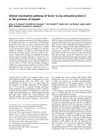
Expression of CD98 increases over time almost
linearly after forskolin treatment
Fusion of BeWo cells is enhanced by forskolin treat-
ment, which, by activating adenylyl cyclase, results in
an increase in intracellular cAMP concentration. We
have previously shown, using single-color flow cytome-
try [fluorescence activated cell sorting (FACS)], a signi-
ficant increase of CD98 expression on intact BeWo
cells after forskolin treatment for 24 h [18]. Here, we
determined the levels of expression of CD98 by west-
ern blotting in cell extracts from BeWo cells cultured
with or without forskolin for 12 h, 24 h, 36 h, or 48 h
(Fig. 1A,B): CD98 expression in cells cultured in the
presence of the vehicle (dimethylsulfoxide, control
cells) was not substantially different from that in cells
treated with 100 lm forskolin after 12 h of culture.
After 24 h, whereas there was no increase in control
cells, the addition of forskolin produced a 20%
CD98 and galectin 3 membrane trafficking P. Dalton et al.
2716 FEBS Journal 274 (2007) 2715–2727 ª 2007 The Authors Journal compilation ª 2007 FEBS
increase in CD98 expression. This stimulation
increased almost linearly up to 48 h, at which time
there was approximately 35% more CD98 in BeWo
cells cultured in forskolin-containing medium than in
control cells. The mean CD98 expression of the two
types of culture was significantly different, with a
P-value of 0.032 (two-tailed paired t-test).
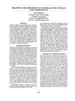
CD98 surface expression increases after cell
treatment with BFA
In this series of experiments, BeWo cells were cultured
in six-well plates in the presence of the vehicle or
100 lm forskolin for 24 h. At 20 h, 22 h, 23 h, and
23.5 h (for a total of 4 h, 2 h, 1 h, and 30 min, respect-
ively), BFA was added to half of the wells to a final
concentration of 5 lgÆmL
)1
, and the cells were
returned to culture for the remaining period. Single-
color FACS, while confirming that forskolin stimula-
tion significantly enhanced CD98 surface expression as
compared to control cells (dimethylsulfoxide), clearly
showed, contrary to expectation, a time-dependent
increase of CD98 surface expression on intact BeWo
cells after BFA treatment for both control and forsko-
lin-incubated cells (Fig. 2A). However, this was not
due to an increase in the amount of CD98, as total
(surface plus cytoplasm) CD98 expression did not sig-
nificantly change (Fig. 2B), suggesting that BFA treat-
ment had increased CD98 trafficking to the cell
surface.
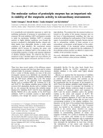
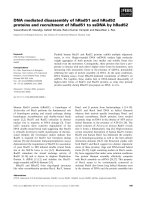
Detection of partially glycosylated ⁄
unglycosylated CD98 after cell treatment
with BFA and tunicamycin
We then used SDS ⁄ PAGE and western blotting to
look at CD98 expression in cell lysates from BeWo
cells cultured for 24 h as above but with or without
BFA (5 lgÆmL
)1
) only for the last 4 h. We speculated
whether BFA, known to produce distinct morpho-
logic ⁄ structural effects at the ER–Golgi level, could
cause alteration not only of CD98 trafficking to the
plasma membrane but also of its structure.
Interestingly, after incubation of the blots with
rabbit anti-(human CD98), we observed an extra
band that ran lower than the normal CD98 band of
80 kDa (reduced blots) or 110–120 kDa (nonre-
duced blots) and had an approximate molecular mass
of 64 kDa or 80 kDa, depending on whether the
gels had been run under reducing or nonreducing
conditions. The extra band was present only in the
samples treated with BFA in either control or forsk-
olin-stimulated cells (Fig. 3A,B), and presumably cor-
responds to partially glycosylated CD98 proteins that
failed to complete the complex process of N-glycosy-
lation in the ER–Golgi apparatus. This is consistent
with the results obtained when we treated BeWo cells
for 24 h with tunicamycin, an antibiotic that inhibits
the first steps of N-linked glycosylation and blocks
the formation of new N-glycosidic protein–carbo-
hydrate linkages. Under reducing conditions, in the
absence of forskolin, an extra band of lower mole-
cular mass ( 53 kDa) was detected in these lysates
(Fig. 3A2). After forskolin treatment, known to sti-
mulate CD98 expression, in addition to the 53 kDa
band, a tight band running at approximately 49 kDa
was clearly identifiable. This, we suggest, corresponds
to the fully unglycosylated CD98 molecule, and is
compatible with the theoretical 30 kDa mobility shift
that is predicted based on the four potential extracel-
lular N-glycosylation sites. The number of bands seen
in Fig. 3A
2
must reflect the total population of
immunoreactive CD98 molecules after 24 h of tunica-
mycin treatment; these molecules normally will only
be present transiently, and thus the duration of
A
B
12.5
Forskolin
dimethylsulfoxide
-F +F -F -F
- 24 hrs 12 hrs - - 48 hrs 36 hrs -
+F
97
64
97
64
CD98hc
+F -F +F
10.0
7.5
Absorbance
5.0
2.5
0.0
12 hours 24 hours 36 hours 48 hours
Fig. 1. The expression of CD98 increases over time almost linearly
after forskolin treatment. Western blotting on a 4–12% Bis ⁄ Tris
NuPage gel run under reducing conditions with Mops running buffer:
BeWo cells at 50–60% confluence were treated with dimethyl-
sulfoxide (vehicle control, – F) or 100 l
M forskolin (+ F) for the indi-
cated times at 37 °C. (A) Immunoblot after incubation with rabbit
anti-(human CD98) (Santa Cruz, 1 : 200), horseradish peroxidase-
conjucated goat anti-rabbit IgG and 3,3¢-diaminobenzidine (DAB);
the data shown are representative of two independent experiments
performed in triplicate. (B) Absorbance of the 80 kDa band quanti-
fied by densitometry. The data are the means of two independent
experiments performed in triplicate ± SEM. The mean CD98
expression of the two types of culture was significantly different,
with a P-value of 0.032 (two-tailed paired t-test).
P. Dalton et al. CD98 and galectin 3 membrane trafficking
FEBS Journal 274 (2007) 2715–2727 ª 2007 The Authors Journal compilation ª 2007 FEBS 2717
Fig. 2. CD98 surface expression increases after BFA treatment. BeWo cells at 50–60% confluence were incubated in medium containing di-
methylsulfoxide (vehicle control) or 100 l
M forskolin (Forsk) for 24 h at 37 °C. BFA was added for the indicated times before harvesting, and
CD98 was detected by single-color flow cytometry. Cells were labeled with goat anti-(human CD98) and rabbit anti-(goat IgG) conjugated
with fluorescein isothiocyanate. (A) Labeling of surface antigens on intact BeWo cells. (B) Labeling of surface and intracellular antigens after
cell permeabilization. n ¼ number of cell samples.
A1 B
A
2
Fig. 3. Detection of partially glycosylated and unglycosylated CD98 after cell treatment with BFA and tunicamycin. Western blotting under redu-
cing (A) or non reducing (B) conditions on a 10% Bis ⁄ Tris NuPage gel: BeWo cells at 50–60% confluence were treated with dimethylsulfoxide
(vehicle control, – Forskolin) or with 100 l
M forskolin (+ Forskolin) for 24 h at 37 °C. Immunoblots were incubated with rabbit anti-(human CD98)
(1 : 200), horseradish peroxidase-conjucated goat anti-rabbit IgG and DAB. A novel band (arrow) of 64 kDa (A
1
)or 80 kDa (B) was present in
whole cell lysates from BFA-treated cells in both control and forskolin-stimulated cells, presumably a partially glycosylated form of CD98. A band
of lower molecular mass ( 53 kDa) was present in both control and forskolin-stimulated cell lysates after tunicamycin treatment (A
2
), with an
additional band of 49 kDa after forskolin treatment. Single representative blots from two experiments run in duplicate.
CD98 and galectin 3 membrane trafficking P. Dalton et al.
2718 FEBS Journal 274 (2007) 2715–2727 ª 2007 The Authors Journal compilation ª 2007 FEBS
glycosidase inhibition will determine the precise pat-
tern observed.
Cell fusion decreases after pulse treatment
with BFA for 4 h
To investigate the relationship between the changes
observed in CD98 expression and structure after BFA
treatment of BeWo cells with functional alterations, we
used two-color FACS to quantify cellular fusion.
We used 3,3¢-dioctadecyloxacarbocyanine perchlo-
rate (DiO) cell-labeling solution, a lipophilic tracer that
is weakly fluorescent in water but highly fluorescent
and quite photostable when incorporated into mem-
branes, and Mitotracker deep red 633, a cell-permeant
mitochondrion-selective dye, to uniformly label suspen-
ded BeWo cells as previously described [18]. Briefly,
flow cytometry analysis of a 50 : 50 mixed cell popula-
tion from cells stained either with DiO or Mitotracker
red and then cultured together allows us to quantify
cellular fusion ⁄ stable aggregation, which is represented
by double-positive cells. To better evaluate the effects
of BFA treatment on cell fusion in this group of
experiments, BFA was added for 4 h in the middle of
a 24–26 h culture to cells incubated with or without
100 lm forskolin. The medium was then changed back
to dimethylsulfoxide or forskolin alone for the remain-
ing culture time. We note that, inevitably, the magni-
tude of the effect will be reduced by the diverse
cellular stages (proliferation, aggregation, fusion, etc.)
of the BeWo cell population in the window of the
pulse of BFA. Two-color FACS analysis showed a
decrease in cellular fusion after pulse BFA treatment
as compared to both groups of BFA-untreated cells
(P ¼ 0.048, one-way ANOVA) (Fig. 4). However, when
we measured CD98 surface expression in control or
forskolin-stimulated cells, we found that this was still
increased in the presence of BFA (data not shown).
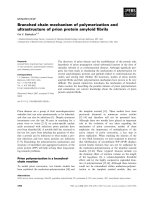
Galectin 3 and CD98 co-immunoprecipitate
We have previously postulated a role for galectin 3,
an S-type lectin containing a carbohydrate-binding
domain, as a physiological ligand of CD98 in vivo [18].
Figure 5A shows the primary intracellular distribu-
tion of galectin 3 in the cytoplasm and nucleus of
BeWo cells, determined by indirect immunofluores-
cence. To investigate the possible ligand-binding role of
galectin 3 in relation to CD98, BeWo cell lysates were
incubated with a goat polyclonal antibody against
CD98 or a goat polyclonal or mouse monoclonal anti-
body against galectin 3. Original whole cell lysates and
immunoprecipitates were then subjected to SDS ⁄ PAGE
and western blotting (Fig. 5B), and cut into single
strips as described in Experimental procedures. Whole
lysates of BeWo cells (lanes 2 and 7) were probed with
rabbit anti-(human CD98) (lane 2) or with goat anti-
(human galectin 3) (lane 7). Goat anti-(human CD98)
immunoprecipitates (lanes 3–6) were probed with rab-
bit anti-(human CD98) (lane 3), goat anti-(human
galectin 3) (lane 4), mouse anti-(phosphatidylinositol
3-kinase) (lane 5, as irrelevant control antibody), and
rabbit IgG (lane 6, as negative control). Goat anti-
(human galectin 3) immunoprecipitates (lanes 8, 9 and
11) were probed with mouse anti-(human galectin 3)
(lane 8), rabbit anti-(human CD98) (lane 9), and rabbit
IgG (lane 11). Mouse anti-(human galectin 3) immuno-
precipitate (lane 10) was probed with rabbit
anti-(human CD98). The results clearly demonstrate
galectin 3 and CD98 co-immunoprecipitation in BeWo
cell extracts. A band equivalent to the molecular mass
of galectin 3 monomers ( 28–30 kDa) was present in
Fig. 4. Cell fusion decreases after pulse treatment with BFA for 4 h.
Double-color flow cytometry assays for detection of fused (double-
positive) cells. BeWo cells were prestained with DiO (maximum
emission 501 nm) or Mitotracker deep red 633 (maximum emission
665 nm) dye. Single-color cells or a 50 : 50 mixture of both cells
were then cultured for 12–14 h in medium containing
dimethylsulfoxide or 100 l
M forskolin (Forsk) at 37 °C, and BFA (final
concentration 5 lgÆmL
)1
) was then added to the medium for 4 h.
After that, the medium was replaced with fresh medium containing
dimethylsulfoxide or 100 l
M forskolin, and cells were cultured for a
further 8–10 h. The graph shows data normalized to dimethylsulfox-
ide control (dimethylsulfoxide ¼ 10%); n ¼ number of cell samples.
Statistical analysis: one-way
ANOVA, P-value 0.048.
P. Dalton et al. CD98 and galectin 3 membrane trafficking
FEBS Journal 274 (2007) 2715–2727 ª 2007 The Authors Journal compilation ª 2007 FEBS 2719
CD98 immunoprecipitates (with an additional band of
higher molecular mass, probably corresponding to
galectin 3 dimers). A band equivalent to the molecular
mass of CD98 ( 80 kDa) was present in the reverse
immunoprecipitation experiment, whether or not the
immunoprecipitates were prepared using the goat or
the mouse anti-(human galectin 3).
CD98 and galectin 3 co-localize in the plasma
membrane, cytoplasm and nucleus
We have previously shown CD98 expression and distri-
bution by immuno-electron microscopy (immuno-EM)
[18]. In the current study, we performed immuno-EM
of galectin 3. We found that galectin 3 was uniformly
distributed in the cytoplasm and nucleus, even if it was
scarce on the cellular membrane, in both the dimethyl-
sulfoxide-treated and the forskolin-treated groups,
although in the latter, sporadic clustering of immuno-
reactivity was observed (Fig. 6A). To further confirm
the close localization of the galectin 3 and CD98 mole-
cules, we used immuno-EM and a standard double gold
technique: double immunoreactivity was determined
using an appropriate secondary antibody)10 nm gold
complex to detect anti-CD98 (smaller-diameter parti-
cles) and an appropriate secondary antibody)15 nm
gold complex to detect anti-galectin 3 (larger-diameter
particles). The electronmicrographs in Fig. 6B,C clearly
show co-localization of these two molecules in the
plasma membrane, in the cytoplasm and in the nucleus
of forskolin-treated BeWo cells.
Inhibition of galectin 3 binding to membrane
glycoproteins affects cellular fusion
We then investigated whether the close proximity of
CD98 and galectin 3 in several cellular locations was
indicative of a functional association.
Galectin 3, like most members of the galectin family,
acts as a receptor for ligands containing poly(N-acetyl-
lactosamine) sequences through the C-terminus CRD.
We used the high affinity of galectin 3 for lactose
(Lac) to inhibit binding between the glycosylated sites
of CD98 and galectin 3 CDR domains, and measured
its effect on cellular fusion. The cells were labeled
either with DiO and Mitotracker Deep Red 633, or
A
B
Fig. 5. Galectin 3 is detected in BeWo cells
and co-immunoprecipitates with CD98. (A)
Immunofluorescence: galectin 3 (fluorescein
isothiocyanate) primary distribution in the
cytoplasm and nucleus of BeWo cells; nuc-
lei are stained with DAPI. (B) Co-immuno-
precipitation: western blotting of a 10%
Bis ⁄ Tris NuPage gel run under reducing con-
ditions with Mes running buffer. BeWo cell
original total lysate (lanes 2 and 7) was
probed with rabbit anti-(human CD98)
(lane 2) or with goat anti-(human galectin 3)
(lane 7). Goat anti-(human CD98) immuno-
precipitates (lanes 3–6) were probed with
rabbit anti-(human CD98) (lane 3), goat anti-
(human galectin 3) (lane 4), mouse anti-
(human PI3Kinase) (lane 5, irrelevant control
antibody) and rabbit IgG (lane 6). Goat anti-
(human galectin 3) immunoprecipitates
(lanes 8, 9 and 11) were probed with mouse
anti-(human galectin 3) (lane 8), rabbit anti-
(human CD98) (lane 9) and rabbit IgG
(lane 11). Mouse anti-(human galectin 3) im-
munoprecipitate (lane 10) was probed with
rabbit anti-(human CD98). Arrows indicate
CD98 immunoreactivity (upper arrows) or
galectin 3 immunoreactivity (lower arrows).
CD98 and galectin 3 membrane trafficking P. Dalton et al.
2720 FEBS Journal 274 (2007) 2715–2727 ª 2007 The Authors Journal compilation ª 2007 FEBS
with DiO and 1,1¢-dioctadecyl-3,3,3¢,3¢-tetramethyl-
indodicarbocyanine perchlorate (DiD), another lipo-
philic tracer with markedly red-shifted fluorescence
excitation and emission spectra in the same range as
Mitotracker Deep Red. We followed the protocol
employed for two-color FACS after BFA treatment;
here, however, pulsed incubation with BFA for 4 h
was substituted by an equal incubation time with
50 mm Lac or, in some experiments, with 50 mm malt-
ose, which has a much lower affinity for galectin 3.
Interestingly, we observed a small but significant
reduction in cell fusion in the presence of Lac (Fig. 7).
Discussion
Syncytial fusion is a rare event in cell biology.
In humans, we typically find three syncytial tissues:
syncytiotrophoblast, striated muscle fibers and
Fig. 7. Inhibition of galectin 3 crosslinking membrane glycoproteins
affects cellular fusion. Double-color flow cytometry assays for
detection of fused (double-positive) cells. BeWo cells were pre-
stained with DiO (em. 501 nm) or DiD (em. 665 nm) dye. Single-
color cells or a 50 : 50 mixture of both cells were then cultured for
12–14 h in medium containing dimethylsulfoxide or 100 l
M forsko-
lin (Forsk) at 37 °C, when Lac (final concentration 50 m
M) or malt-
ose (Malt, final concentration 50 m
M) was added to the medium for
4 h. The medium was then replaced with fresh medium containing
dimethylsulfoxide or 100 l
M forskolin, and cells were cultured for a
further 8–10 h. The graph shows data normalized to dimethylsulfox-
ide control (dimethylsulfoxide ¼ 10%); n ¼ number of cell samples.
Statistical analysis: one-way
ANOVA, P-value 0.0148.
A
B
C
Fig. 6. Galectin 3 co-localizes with CD98. (A). Immuno-EM: electron
micrograph of forskolin-treated cell (· 25 000) showing galectin 3
(arrows); note occasional clustering of gold particles. (B, C) Double
labeling immuno-EM: electron micrographs of forskolin-treated cells
showing co-localization of galectin 3 and CD98 at the plasma mem-
brane (B,C), in the nucleus (B), and in the cytoplasm (C) (arrows).
Sections were sequentially stained using as secondary antibodies
anti-(goat IgG))10 nm gold complex to detect anti-CD98 (smaller-
diameter particles) and anti-(rabbit IgG))15 nm gold complex to
detect anti-galectin 3 (larger-diameter particles). Three representa-
tive fields; scale bars 200 nm.
P. Dalton et al. CD98 and galectin 3 membrane trafficking
FEBS Journal 274 (2007) 2715–2727 ª 2007 The Authors Journal compilation ª 2007 FEBS 2721
chondro-osteoclast. Syncytiotrophoblast forms during
implantation, and is then maintained at the villous
maternal–fetal interface throughout pregnancy. A useful
model of trophoblast syncytialization is the choriocarci-
noma cell line BeWo; these cells are able to fuse, and
fusion can be also enhanced by forskolin treatment.
Recently, the use of the fungal metabolite BFA to
cause Golgi breakdown showed that part of Golgi gly-
cosylation enzymes recycle to the ER, whereas Golgi
matrix proteins are retained in a set of cytoplasmic
membranes; this has led to the suggestion that BFA
disrupts a dynamic membrane-recycling pathway
between the ER and cis ⁄ medial Golgi, effectively
blocking membrane transport out of but not back to
the ER [28]. However, both the dynamic interaction
between ER and Golgi and the mechanism of action
of BFA are still subjects of intense discussion.
N-linked glycosylation of membrane proteins is
acquired as a post-translational modification in the
ER, and further processing takes place in the Golgi
before the proteins reach the cell surface. CD98 has
been previously shown to be involved, among its many
other functions, in trophoblast fusion [17,18]. As gly-
cosylation seems to be important for correct protein
folding and for ligand–receptor interactions, and
because CD98 is an N-glycosylated protein [29,30], in
this study we investigated the effect of BFA on the
expression and the function of CD98 and its direct
and indirect effect on galectin 3, which binds glycosyl-
ated proteins through its CDR site [25].
By analysing BeWo cells at different time points with
SDS ⁄ PAGE, we showed that, as a consequence of fors-
kolin treatment, there is a time-dependent increase in
CD98 protein expression comparable to that of the
CD98 mRNA previously observed [31]. Unexpectedly,
we found that CD98 surface expression was also incre-
ased in a time-dependent manner in BeWo cells treated
with BFA. This result implied the existence, for CD98,
of an alternative route to the plasma membrane that is
independent of the classic secretory pathway through
the trans-Golgi apparatus, which is used by most secre-
tory and transmembrane proteins and can be inhibited
by BFA. Furthermore, analysis of western blots probed
with CD98 antibody, under reducing and non reducing
conditions, showed the presence of an additional band
in the BFA-treated cell lysates; this band presumably
corresponded to a partially glycosylated form of CD98,
after breakdown of the Golgi apparatus, in the last 4 h
of culture. Taken together, these two findings would
suggest that a partially glycosylated form of CD98 is
capable of reaching and inserting into the cell membrane
via an unknown mechanism of transport that is inde-
pendent of the ER–trans-Golgi pathway.
We next investigated whether CD98 glycosylation
was necessary for its role in cellular fusion. For this
purpose, it was important to add BFA to the culture
when the cells were just starting to fuse; previous
experiments had indicated that this occurs after 12–
14 h. Moreover, we had found that BeWo cells
undergo morphologic changes followed by detachment
and death if cultured with BFA for over 6–8 h (data
not shown). However, BFA effects are reversible if the
drug is removed. We decided, therefore, to add BFA
for 4 h in the middle of the culture time, to remove it,
and then to observe the number of cells that under-
went fusion as compared to untreated cells. This could
be quantified by two-color FACS of BeWo cells previ-
ously labeled with one of two well-separated fluores-
cent dyes and then calculation of the number of
double-fluorescent cells [18]. The results showed that
glycosylation of CD98 is important for the fusion of
BeWo cells as, although the molecule was still over-
expressed on the surface of BFA-treated cells (data not
shown), cellular fusion was decreased as compared to
untreated cells. However, as anticipated, the magnitude
of the observed effect was moderate.
It has been suggested that galectin 3 is an endo-
genous crosslinker for CD98 and may promote CD98
dimerization (and consequent integrin activation) [21].
We have shown in BeWo cells, both by immunofluo-
rescence and by immuno-EM, that galectin 3 is
expressed in all three cellular compartments. Immuno-
precipitation of BeWo cell total lysates with either goat
anti-(human CD98) or goat or mouse anti-(human
galectin 3) has also shown that CD98 and galectin 3
co-immunoprecipitate. Consequently, we used imm-
uno-EM to confirm the relative positions of galectin 3
and CD98 in the cells. We showed unambiguous
co-localization of the two molecules, both intracellular-
ly, in the nucleus and cytoplasm, and at the plasma
membrane. The relative abundance of CD98 molecules
as compared to that of galectin 3 molecules at the
same location supports the hypothesis of dimerization
of CD98 molecules through linking with either mono-
meric or oligomeric forms of galectin 3. In the latter
case, galectin 3 would have several CDR sites and be
able to interact with many CD98 molecules.
If there is an association between CD98 and galec-
tin 3, then disturbing it should disrupt cellular fusion.
Indeed, by blocking galectin 3 CDR sites with 50 mm
Lac, we showed that we could reduce the fusion of
BeWo cells.
Getting proteins to the correct place at the right
time is a logistical challenge for any cell. Proteins des-
tined for the classic secretory pathway, such as immu-
noglobulins, typically contain N-terminal signal
CD98 and galectin 3 membrane trafficking P. Dalton et al.
2722 FEBS Journal 274 (2007) 2715–2727 ª 2007 The Authors Journal compilation ª 2007 FEBS
peptides that mediate membrane translocation into the
lumen of the ER followed by ER–Golgi-dependent
transport to the cell surface. On the other hand, a
growing number of proteins (angiogenic growth fac-
tors, galectins, inflammatory cytokines, viral proteins)
lack a signal peptide but are still secreted from the cell.
These proteins do not contain modifications such as
glycosylation (which happen at the ER–Golgi level),
and their secretion is not inhibited by BFA or similar
inhibitors of the classic secretory pathway. In recent
years, several distinct ‘nonclassic’ secretory pathways
have been demonstrated [32].
As any morphologic and functional modification of
the ER–Golgi–trans-Golgi complex would affect the
proteins using this pathway, both structurally (incom-
plete or null secondary modifications) and functionally
(as a result of the failure to reach correct cell loca-
tions), in this study we used BFA to investigate CD98
function and protein interactions.
Our data suggest that CD98 can traffic to the
plasma membrane via at least two distinct transport
mechanisms in BeWo cells, one dependent upon the
classic secretory pathway (glycosylated protein), and
the other on an alternative route (nonglycosylated pro-
tein). Furthermore, we demonstrate that CD98 glyco-
sylation is necessary for cell fusion and that this in
turn requires interaction between CD98 and galectin 3.
This lectin, like CD98, is present both in the cytotro-
phoblasts and in the syncytiotrophoblast [33,34].
Hence, crosslinking of these two molecules in vivo
could be an essential molecular mechanism to enable
syncytiotrophoblast formation. Our findings now need
to be investigated in the intact placenta, e.g. by look-
ing for co-localization of these two molecules in nor-
mal placental tissue and in primary cell lines.
The results reported in this article fit unexpectedly
with a recent study on CD98hc knockout mice that
suggested an essential role for CD98 in early mouse
development. Embryonic lethality was found when the
transgene encoding the molecule was truncated at the
extracellular domain, leaving intact both the intracellu-
lar and the transmembrane parts of the molecule [35].
Our work emphasizes the way in which the external
domain of CD98 may play a critical role in tropho-
blast cell biology.
Experimental procedures
Primary antibodies
Rabbit anti-(human galectin 3) was obtained from Chem-
icon Europe Ltd (Chandlers Ford, UK). Goat anti-(human
CD98) (C-20), rabbit anti-(human CD98) (H-300), normal
goat IgG and normal rabbit IgG (isotype-matched con-
trols), goat anti-(human galectin 3) (D-20) and mouse
anti-(human galectin 3) (B-2) were obtained from Santa
Cruz Biotechnology Inc. (Santa Cruz, CA, USA). Mouse
anti-(human PI3Kinase p85a) was obtained from Serotec
(Kidlington, UK).
Secondary antibodies
Horseradish peroxidase-conjugated goat anti-(rabbit IgG),
horseradish peroxidase-conjugated rabbit anti-(mouse IgG)
and fluorescein isothiocyanate-conjugated swine anti-(rabbit
IgG) were obtained from Dako (Glostrup, Denmark).
Horseradish peroxidase-conjugated donkey anti-(goat IgG)
and protein A ⁄ G plus agarose were obtained from Santa
Cruz Biotechnology Inc. Fluorescein isothiocyanate-conju-
gated rabbit anti-(goat IgG) was obtained from Sigma
(Gillingham, UK). Rabbit anti-(goat IgG))10 nm gold
complex to detect anti-CD98 and goat anti-(rabbit
IgG))15 nm gold complex to detect anti-galectin 3 were
obtained from British Biocell (Cardiff, UK).
Cell culture
BeWo cells were cultured at 37 °C as monolayers in
F-12K Nutrient Mixture (Kaighn’s modification) supple-
mented with 10% fetal bovine serum, 2 mml-glutamine
(all Gibco, Paisley, UK), 100 UÆmL
)1
penicillin and
100 UÆmL
)1
streptomycin (Sigma) in a humidified atmo-
sphere of 5% CO
2
and 95% air. Confluent cells were har-
vested by trypsinization with trypsin ⁄ EDTA in HBSS
without Ca
2+
and Mg
2+
(Gibco), resuspended in fresh
medium, and plated in six-well culture plates (BD Falcon,
Oxford, UK). When the cells reached 65–70% confluence,
forskolin (Sigma) or vehicle (dimethylsulfoxide) was added
in fresh medium at a final concentration of 100 lm for
24 h, unless otherwise indicated. In some wells, BFA
(Sigma) (final concentration 5 lgÆmL
)1
), tunicamycin
(Sigma) (final concentration 10 lgÆmL
)1
), 50 mm Lac or
50 mm maltose were added as indicated in the different
experiments. For the two-color FACS experiments, before
plating, viable cells were counted by the trypan blue
(Sigma) method, resuspended in serum-free medium, and
stained with either 10 lL of vybrant DiO or 5 lLof
vybrant DiD cell labeling solutions (1 mm) (Molecular
Probes, Invitrogen, Paisley, UK) per 10
6
cellsÆmL
)1
cells
for 30 min, or with MitoTracker Deep Red633 (Molecular
Probes) at a concentration of 25 nm per 10
6
cellsÆmL
)1
cells for 15 min; labeling was carried out at 37 °C in the
dark with gentle shaking. After extensive washing with
warm serum-free medium, each group of stained cells was
resupended in complete growth medium and plated either
on its own or in a 50 : 50 mixture (DiO-labeled and Mito-
tracker Red-labeled or DiO-labeled and DiD-labeled cells)
in six-well culture plates.
P. Dalton et al. CD98 and galectin 3 membrane trafficking
FEBS Journal 274 (2007) 2715–2727 ª 2007 The Authors Journal compilation ª 2007 FEBS 2723
SDS ⁄ PAGE and western blotting
Confluent cultures from six-well plates were washed with ice-
cold Ca
2+
-free and Mg
2+
-free Dulbecco’s phosphate-buf-
fered saline (D-NaCl ⁄ P
i
) (Gibco) and then lysed at 4 °Cin
ice-cold modified RIPA buffer containing 50 mm Tris ⁄ HCl
(pH 7.4), 1% NP-40, 0.25% sodium deoxycholate, 150 mm
NaCl, 1 mm EDTA, and 10 lL of protease inhibitor mixture
(Sigma), for 15 min on a rocker. Samples were sonicated
three times for 30 s, and clarified by centrifugation at
10 000 g for 15 min at 4 °C (Beckman GS15-R, rotor F402,
Beckman Coulter Ltd., High Wycombe, UK). Supernatants
(10 lg of protein) were retained, solubilized in NuPAGE
sample buffer (Invitrogen), with or without reducing agent,
warmed for 10 min at 75 °C, and then run on 10% Novex
Bis ⁄ Tris NuPAGE gels (Invitrogen). The proteins were trans-
ferred to nitrocellulose membranes, blocked using 5% (w ⁄ v)
nonfat dry milk in 0.01 m NaCl ⁄ P
i
(Sigma) with 0.05% (v ⁄ v)
Tween 20 for 1 h at room temperature, and then incubated
with rabbit anti-(human CD98) (H-300, 1 : 200) overnight at
4 °C. Horseradish peroxidase-conjugated goat anti-(rabbit
IgG) was used for secondary labeling. Immunoreactive bands
were identified by SIGMAFAST 3,3¢-diaminobenzidine tab-
lets (Sigma) according to the manufacturer’s instructions.
Co-immunoprecipitation
Whole cell lysates were prepared as described above. Aliqu-
ots (10 lg of protein) were retained and solubilized in Nu-
PAGE sample buffer (Invitrogen) for analysis by western
blotting. The remaining lysates were precleared with 1 lgof
goat IgG and 10 lL of protein A ⁄ G plus agarose [for goat
anti-(human CD98) and goat anti-(human galectin 3) immu-
noprecipitates] or with 10 lL of protein A ⁄ G plus agarose
[for mouse anti-(human galectin 3)] for 10 min at 4 °C; the
agarose beads were removed by centrifugation at 4000 g
(Beckman GS15-R, rotor F4202), and the cleared lysates
were incubated overnight with 2 lg of goat anti-(human
CD98) or goat anti-(human galectin 3) or mouse anti-
(human galectin 3) at 4 °C. Immune complexes were cap-
tured using 20 lL of protein A ⁄ G agarose beads for 2 h at
4 °C, and then washed three times with lysis buffer. Follow-
ing elution with NuPAGE buffer, samples were boiled for
5 min to dissociate beads from the immunocomplexes, and
centrifuged at 100 000 g (Eppendorf 5415C, Eppendorf UK
Ltd., Cambridge, UK); associated proteins in the superna-
tants were resolved on a 10% Novex Bis ⁄ Tris NuPAGE gel
(Invitrogen) under reducing condition with Mes running buf-
fer (Invitrogen). The proteins were transferred to a nitrocellu-
lose membrane, and this was cut into strips corresponding to
single protein lanes (revealed after reversibly staining with
Ponceau red). Single strips were blocked using either 5%
(w ⁄ v) nonfat dry milk in 0.01 m NaCl ⁄ P
i
(Sigma) with 0.05%
(v ⁄ v) Tween 20 (for strips to be probed with anti-CD98), or
0.01 m NaCl ⁄ P
i
(Sigma) with 0.05% (v ⁄ v) Tween 20 plus
10% normal serum of the host species of the secondary anti-
body for 1 h at room temperature, and then incubated with
either rabbit anti-(human CD98) (H-300, 1 : 200), goat anti-
(human galectin 3) (1 : 100), mouse anti-(human galectin 3)
(1 : 100), mouse anti-(human PI3 kinase) (1 : 100) as irre-
levant control antibody, normal rabbit IgG or normal
goat IgG (1 : 100) overnight at 4 °C. Horseradish peroxi-
dase-conjugated goat anti-(rabbit IgG), horseradish peroxi-
dase-conjugated donkey anti-(goat IgG) or horseradish
peroxidase-conjugated rabbit anti-(mouse IgG) were used for
secondary labeling. Immunoreactive bands were identified by
SIGMAFAST 3,3¢-diaminobenzidine tablets (Sigma) accord-
ing to the manufacturer’s instructions.
Flow cytometry ) surface staining on intact cells
Cells from six-well plates were detached with trypsin ⁄ EDTA
(Gibco). Aliquots of 1 · 10
6
cells were washed in NaCl ⁄ P
i
and resuspended in 250 lL of FACS buffer (NaCl ⁄ P
i
,1%
fetal bovine serum, 0.1% NaN
3
) with goat anti-(human
CD98) (C-20, 1 : 20) or isotype control IgG or no primary
antibody. Cells were incubated for 45 min on ice, and then
washed three times with FACS buffer. Samples were then
incubated with fluorescein isothiocyanate-conjugated rabbit
anti-(goat IgG) (1 : 50) for 45 min on ice and washed three
times. Samples were finally resuspended in FACS buffer and
2% paraformaldehyde (PFA), and the number of events
was analyzed by flow cytometry using a FACSCalibur (BD
Biosciences, Oxford, UK) flow cytometer and cell quest
software and ⁄ or an EPICS Altra (Beckman Coulter Ltd.,
High Wycombe, UK) flow cytometer and expo32 software.
Flow cytometry ) surface and intracellular
staining
Cell suspensions were fixed in 2% PFA for 20 min at room
temperature, washed once in NaCl ⁄ P
i
, permeabilized with
1% saponin in FACS buffer for 15 min at room tempera-
ture, and then stained following the surface staining proto-
col. After the final wash, samples were fixed again in 2%
PFA before analysis.
Immunofluorescence
Cells (1 · 10
3
) were plated onto chamber wells (Lab-Tek,
Fisher Scientific UK Ltd., Loughborough, UK), grown for
24 h, washed with NaCl ⁄ P
i
, and fixed with 2% PFA and rin-
sed. Nonspecific binding sites were blocked with blocking
buffer (NaCl ⁄ P
i
, 0.05% Tween 20, 10% fetal bovine serum,
10% goat serum) for 20 min at room temperature. Cells were
then incubated with rabbit anti-(human galectin 3), 1 : 1000
in diluting buffer (NaCl ⁄ P
i
, 0.05% Tween 20, 1% fetal
bovine serum, 1% goat serum) for 1 h at room temperature,
washed three times for 5 min, and then incubated with
CD98 and galectin 3 membrane trafficking P. Dalton et al.
2724 FEBS Journal 274 (2007) 2715–2727 ª 2007 The Authors Journal compilation ª 2007 FEBS
fluorescein isothiocyanate-conjugated rabbit anti-(goat IgG).
After three more washes, chambers were removed, and slides
mounted with ProLong Gold antifade reagent with 4¢,6-dia-
midino-2-phenylindole (DAPI) (Molecular Probes). Images
were captured with a Leica DC 500 digital camera on a Leica
DMR microscope (Leica Microsystem Digital Imaging,
Cambridge, UK).
Immunogold EM
Cells were prepared for EM by standard methods [36].
Briefly, cell pellets were postfixed in osmium tetroxide (1%
w ⁄ v in 0.1 m sodium phosphate buffer), contrasted with
uranyl acetate (2% w ⁄ v in distilled water), dehydrated
through increasing concentrations of ethanol (70–100%),
and embedded in LR Gold resin (Agar Scientific, Reading,
UK). Ultrathin sections (50–80 nm) were prepared by use
of a Reichert Ultracut S microtome (Reichert, Vienna, Aus-
tria), and mounted on 200-mesh nickel grids. For immuno-
gold detection of CD98 and galectin 3, sections were
incubated with either goat anti-(human CD98) (1 : 100) or
rabbit anti-(human galectin 3) (1 : 100) for 2 h and for 1 h
with protein A)15 nm gold complex. For control sections,
the primary antibody was omitted and replaced with a
matching dilution of the respective nonimmune serum. Sec-
tions were then lightly counterstained with uranyl acetate
and lead citrate. All antibodies were diluted in 0.1 m phos-
phate buffer containing 1% w ⁄ v egg albumin. The sections
were viewed with a JEOL 1010 transmission electron micro-
scope (JEOL, Peabody, MA, USA), and representative
micrographs were prepared. The area of each cellular com-
partment of interest was determined by point counting
morphometry, and the number of gold particles over each
compartment was counted. The density of immunogold
(particles per lm
2
) was then calculated. For double labeling
of CD98 and galectin 3, sections were sequentially stained
as above using rabbit anti-(goat IgG))10 nm gold complex
to detect anti-CD98 and goat anti-(rabbit IgG))15 nm gold
complex to detect anti-galectin 3.
Statistical analysis
Results are presented as means ± SE. The significance of
the differences between means was assessed using the two-
tailed Student’s t-test or one-way anova. P values < 0.05
were considered to be significant.
Acknowledgements
We are grateful to Dr Paul Klenerman (Peter Medawar
Building for Pathogen Research, Oxford University)
for assistance with the flow cytometry, and we thank
Lynne Scott for expert technical help with the immuno-
EM. This work was funded by the Wellcome Trust.
References
1 Haynes BF, Hemler ME, Man DL, Eisenbarth GS,
Shelhamer J, Mostowski HS, Thomas CA, Strominger
JL & Fauci AS (1981) Characterization of a monoclonal
antibody (4F2) that binds to human monocytes and to
a subset of activated lymphocytes. J Immunol 126,
1409–1414.
2 Deves R & Boyd CAR (2000) Surface antigen CD98
(4F2): not a single membrane protein, but a family of
proteins with multiple functions. J Membr Biol 173,
165–177.
3 Mastroberardino L, Spindler B, Pfeiffer R, Skelly PJ,
Loffing J, Shoemaker CB & Verrey F (1998) Amino
acid transport by heterodimers of 4F2hc ⁄ CD98 and
members of a permease family. Nature 395, 288–291.
4 Hemler ME & Strominger JL (1982) Characterization of
antigen recognized by the monoclonal antibody (4F2):
different molecular forms on human T and B lympho-
blastoid cell lines. J Immunol 129, 623–628.
5 Nakamura E, Sato M, Yang H, Miyagawa F, Harasaki
M, Tomita K, Matsuoka S, Noma A, Iwai K & Minato
N (1999) 4F2 (CD98) heavy chain is associated cova-
lently with an amino acid transporter and controls
intracellular trafficking and membrane topology of 4F2
heterodimer. J Biol Chem 274, 3009–3016.
6 Cho JY, Fox DA, Horejsi V, Sagawa K, Skubitz KM,
Katz DR & Chain B (2001) The functional interactions
between CD98, 1-integrins, and CD147 in the induction
of U937 homotypic aggregation. Blood 98, 374–382.
7 Rintoul RC, Buttery RC, Mackinnon AC, Wong WS,
Mosher D, Haslett C & Sethi T (2002) Cross-linking
CD98 promotes integrin-like signaling and anchorage-
independent growth. Mol Biol Cell 8, 2841–2852.
8 Suga K, Katagiri K, Kinashi T, Harazaki M, Iizuka T,
Hattori M & Minato N (2001) CD98 induces LFA-1-
mediated cell adhesion in lymphoid cells via activation
of Rap1. FEBS Lett 489, 249–253.
9 Warren AP, Patel K, Miyamoto Y, Wygant JN,
Woodside DG & McIntyre BW (2000) Convergence
between CD98 and integrin-mediated T-lymphocyte
co-stimulation. Immunology 99, 62–68.
10 Tajima M, Higuchi S, Higuchi Y, Miyamoto N, Uchida
A, Ito M, Nishio M, Komada H, Kawano M,
Kusagawa S et al. (1999) Suppression of FRP-1 ⁄ CD98-
mediated multinucleated giant cell and osteoclast forma-
tion by an anti-FRP-1 ⁄ CD98 mAb, HBJ 127, that
inhibits c-src expression. Cell Immunol 193, 162–169.
11 Mori K, Nishimura M, Tsurudome M, Ito M, Nishio
M, Kawano M, Kozuka Y, Yamashita Y, Komada H,
Uchida A et al. (2004) The functional interaction
between CD98 and CD147 in regulation of virus-
induced cell fusion and osteoclast formation. Med
Microbiol Immunol (Berl) 193, 155–162.
P. Dalton et al. CD98 and galectin 3 membrane trafficking
FEBS Journal 274 (2007) 2715–2727 ª 2007 The Authors Journal compilation ª 2007 FEBS 2725
12 Tsurudome M & Ito Y (2000) Function of fusion regu-
latory proteins (FRPs) in immune cells and virus-
infected cells. Crit Rev Immunol 20, 167–196.
13 Ayuk PT, Sibley CP, Donnai P, D’Souza SW & Glazier
JD (2000) Development and polarization of cationic
amino acid transporters and regulators in the human
placenta. Am J Physiol Cell Physiol 278, C1162–C1171.
14 Okamoto Y, Sakata M, Ogura K, Yamamoto T,
Yamaguchi M, Tasaka K, Kurachi H, Tsurudome M &
Murata Y (2002) Expression and regulation of 4F2hc
and hLAT1 in human trophoblasts. Am J Physiol Cell
Physiol 282, C196–C204.
15 Kudo Y, Boyd CAR, Millo J, Sargent IL & Redman
CW (2003) Manipulation of CD98 expression affects
both trophoblast cell fusion and amino acid transport
activity during syncytialization of human placental
BeWo cells. J Physiol 550, 3–9.
16 Kudo Y & Boyd CAR (2004) RNA interference-induced
reduction in CD98 expression suppresses cell fusion dur-
ing syncytialization of human placental BeWo cells.
FEBS Lett 577, 473–477.
17 Kudo Y, Boyd CAR, Kimura H, Cook PR, Redman
CW & Sargent IL (2003) Quantifying the syncytialisa-
tion of human placental trophoblast BeWo cells grown
in vitro. Biochim Biophys Acta 1640, 25–31.
18 Dalton P, Christian HC, Redman CWG, Sargent IL &
Boyd CAR (2006) Differential effect of cross-linking the
CD98 heavy chain on fusion and amino acid transport
in the human placental trophoblast (BeWo) cell line.
Biochim Biophys Acta 1768, 401–410.
19 Klausner RD, Donaldson JG & Lippincott-Schwartz J
(1992) Brefeldin A: insights into the control of membrane
traffic and organelle structure. J Cell Biol 116, 71–80.
20 Zeghouf M, Guibert B, Zeeh JC & Cherfils J (2005)
Arf, Sec7 and brefeldin A: a model towards the thera-
peutic inhibition of guanine nucleotide-exchange factors.
Biochem Soc Trans 33, 1265–1268.
21 Hughes RC (2001) Galectins as modulators of cell adhe-
sion. Biochimie 83, 667–676.
22 Dong S & Hughes RC (1996) Galectin 3 stimulates
uptake of extracellular Ca2+ in human Jurkat T-cells.
FEBS Lett 395, 165–169.
23 Dong S & Hughes RC (1997) Macrophage surface
glycoproteins binding to galectin 3 (Mac-2-antigen).
Glycoconj J 14, 267–274.
24 Ho M & Springer T (1982) Mac-2, a novel 32,000 Mr
mouse macrophage subpopulation-specific antigen
defined by monoclonal antibodies. J Immunol 128,
1221–1228.
25 Sato S (2002) Galectins as molecules of danger signal,
which could evoke an immune response to infection.
Trends Glycosci Glycotechnol 14, 285–301.
26 Gorski J, Liu F, Artigues A, Castagna L & Osdoby P
(2002) New alternatively spliced form of galectin 3, a
member of the beta-galactoside-binding animal lectin
family, contains a predicted transmembrane-spanning
domain and a leucine zipper motif. J Biol Chem 277,
18840–18848.
27 Patterson RJ, Wang W & Wang JL (2004) Understand-
ing the biochemical activities of galectin-1 and galectin
3 in the nucleus. Glycoconjugate J 19, 499–506.
28 Lippincott-Schwartz J, Yuan LC, Bonifacino JS &
Klausner RD (1989) Rapid redistribution of Golgi
proteins into the ER in cells treated with brefeldin A:
evidence for membrane cycling from Golgi to ER. Cell
56, 801–813.
29 Barclay AN, Brown MH, Law SKA, McKnight AJ,
Tomlinson MG & van der Merwe PA (1997) CD98. In
The Leucocyte Antigen Facts Book (Barclay AN, Brown
MH, Law SKA, McKnight AJ, Tomlinson MG & van
der Merwe PA eds), pp. 369–370. Academic Press, San
Diego.
30 Teixeira S, Di Grandi S & Kuhn LC (1987) Primary
structure of the human 4F2 antigen heavy chain predicts
a transmembrane protein with a cytoplasmic NH2
terminus. J Biol Chem 262 , 9574–9580.
31 Kudo Y, Boyd CAR, Sargent IL, Redman CW, Lee JM
& Freeman TC (2004) An analysis using DNA microar-
ray of the time course of gene expression during syncy-
tialization of a human placental cell line (BeWo).
Placenta 25, 479–488.
32 Nickel W (2005) Unconventional secretory routes: direct
protein export across the plasma membrane of mamma-
lian cells. Traffic 6, 607–614.
33 Maquoi E, van den Brule FA, Castronovo V & Foidart
JM (1997) Changes in the distribution pattern of galec-
tin-1 and galectin 3 in human placenta correlates with
the differentiation pathways of trophoblasts. Placenta
18, 433–439.
34 Vicovac L, Jankovic M & Cuperlovic M (1998) Galec-
tin-1 and -3 in cells of the first trimester placental bed.
Hum Reprod 13, 730–735.
35 Tsumura H, Suzuki N, Saito H, Kawano M, Otake S,
Kozuka Y, Komada H, Tsurudome M & Ito Y (2003)
The targeted disruption of the CD98 gene results in
embryonic lethality. Biochem Biophys Res Commun 308,
847–851.
36 Christian HC, Taylor AD, Flower RJ, Morris JF &
Buckingham JC (1997) Characterization and localiza-
tion of lipocortin 1-binding sites on rat anterior pitui-
tary cells by fluorescence-activated cell analysis ⁄ sorting
and electron microscopy. Endocrinology 138, 5341–5351.
Supplementary material
The following supplementary material is available
online:
Fig. S1. Whole immunoblot of CD98 time-course
experiment in Fig. 1.
CD98 and galectin 3 membrane trafficking P. Dalton et al.
2726 FEBS Journal 274 (2007) 2715–2727 ª 2007 The Authors Journal compilation ª 2007 FEBS
Fig. S2. SDS ⁄ PAGE: whole membrane after transfer
and staining with Ponceau Red, showing equal protein
loading for CD98 time-course experiment in Fig. 1.
This material is available as part of the online article
from
Please note: Blackwell Publishing is not responsible
for the content or functionality of any supplementary
materials supplied by the authors. Any queries (other
than missing material) should be directed to the corres-
ponding author for the article.
P. Dalton et al. CD98 and galectin 3 membrane trafficking
FEBS Journal 274 (2007) 2715–2727 ª 2007 The Authors Journal compilation ª 2007 FEBS 2727