Báo cáo khoa học: Peroxin Pex21p interacts with the C-terminal noncatalytic domain of yeast seryl-tRNA synthetase and forms a specific ternary complex with tRNASer potx
Bạn đang xem bản rút gọn của tài liệu. Xem và tải ngay bản đầy đủ của tài liệu tại đây (527.72 KB, 12 trang )
Peroxin Pex21p interacts with the C-terminal noncatalytic
domain of yeast seryl-tRNA synthetase and forms a
specific ternary complex with tRNA
Ser
Vlatka Godinic
1
, Marko Mocibob
1
, Sanda Rocak
1
, Michael Ibba
2
and Ivana Weygand-Durasevic
1
1 Department of Chemistry, Faculty of Science, University of Zagreb, Croatia
2 Department of Microbiology, The Ohio State University, Columbus, OH, USA
Accurate aminoacylation of tRNA by aminoacyl-tRNA
synthetases (aaRSs) is a crucial step in the faithful
translation of mRNA. In addition to their fundamental
role in translation, aaRSs are now known to participate
in a wide variety of other cellular processes [1]. These
noncanonical activities vary with the type of aaRS and
its cellular location [2]. The expansion of synthetase
function well beyond protein synthesis, and in many
cases the participation of synthetases in the control of
the efficiency and fidelity of the catalytic reaction,
depend on terminally appended or inserted noncatalytic
domains [3–5]. For example, specialized tRNA-binding
domains attached to the N-terminal or C-terminal ends
guide the productive docking of cognate tRNAs [6–8].
Editing domains, appended to the ends or inserted into
the core domains, catalyze the tRNA-dependent hydro-
lysis of incorrectly attached amino acids [9]. The role of
catalytically dispensable peptides is also to promote
association between the synthetases [10]. Such multi-
synthetase complexes are a common feature of all
higher eukaryotic cells or tissues [3,11,12]. These assem-
blies often involve different nonsynthetase proteins
[13,14], which may affect the efficacy of the aminoacy-
lation reaction in trans. In addition, many novel func-
tions unrelated to protein synthesis have been ascribed
to aaRS-interacting multifunctional proteins and aaRSs
in higher eukaryotes [2]. For example, fragments of
TyrRS stimulate angiogenesis, whereas those of TrpRS
Keywords
peroxin; protein biosynthesis; protein–
protein interaction; seryl-tRNA synthetase;
yeast two-hybrid
Correspondence
I. Weygand-Durasevic, Department of
Chemistry, Faculty of Science, University of
Zagreb, Horvatovac 102a, 10000 Zagreb,
Croatia
Fax: +385 1 460 6401
Tel: +385 1 460 6230
E-mail:
(Received 22 December 2006, revised 24
February 2007, accepted 27 March 2007)
doi:10.1111/j.1742-4658.2007.05812.x
The seryl-tRNA synthetase from Saccharomyces cerevisiae interacts with
the peroxisome biogenesis-related factor Pex21p. Several deletion mutants
of seryl-tRNA synthetase were constructed and inspected for their ability
to interact with Pex21p in a yeast two-hybrid assay, allowing mapping of
the synthetase domain required for complex assembly. Deletion of the 13
C-terminal amino acids abolished Pex21p binding to seryl-tRNA synthe-
tase. The catalytic parameters of purified truncated seryl-tRNA synthetase,
determined in the serylation reaction, were found to be almost identical to
those of the native enzyme. In vivo loss of interaction with Pex21p was con-
firmed in vitro by coaffinity purification. These data indicate that the C-ter-
minally appended domain of yeast seryl-tRNA synthetase does not
participate in substrate binding, but instead is required for association with
Pex21p. We further determined that Pex21p does not directly bind tRNA,
and nor does it possess a tRNA-binding motif, but it instead participates
in the formation of a specific ternary complex with seryl-tRNA synthetase
and tRNA
Ser
, strengthening the interaction of seryl-tRNA synthetase with
its cognate tRNA
Ser
.
Abbreviations
aaRS, aminocyl-tRNA synthetase; EMSA, electrophoretic mobility shift assay; Gal-X, 5-bromo-4-chloroindol-3-yl b-
D-galactopyranoside;
Gal-ONp, 2-nitrophenyl b-
D-galactopyranoside; RS, tRNA synthetase; Sc, Saccharomyces cerevisiae; SerRS, seryl-tRNA synthetase; Zmc,
Zea mays cytosolic.
2788 FEBS Journal 274 (2007) 2788–2799 ª 2007 The Authors Journal compilation ª 2007 FEBS
inhibit angiogenesis [15]. Thus, the unusual versatility
of aaRSs in higher eukaryotes is further increased by
their interactions with nonsynthetase proteins.
The association of the synthetases into multicompo-
nent complexes has also been described in lower eukary-
otes [16,17], archaea [18,19], and bacteria [20,21]. As in
higher eukaryotes, some of these complexes also include
nonsynthetase accessory proteins. In yeast, the tRNA-
binding protein Arc1p is associated with MetRS and
GluRS [17,22,23], and enhances the binding affinity for
cognate tRNAs. The association of an aaRS with a puta-
tive metabolic protein Mj1338 has recently been identi-
fied in archaea [19], whereas Trbp111 and CsaA are the
best known bacterial tRNA chaperones [24–26]. On the
other hand, the recent discovery of autonomous editing
proteins that are efficient at hydrolyzing misacylated
products provides a direct link between ancestral aaRSs
consisting solely of the catalytic core and extant enzymes
to which functionally independent modules are appen-
ded [27,28]. Factors unrelated to the translation machin-
ery have also been found to associate with aaRSs. Yeast
TyrRS interacts with Knr4, a protein involved in cell
wall synthesis [29], and heat shock protein 90 interacts
with human glutamyl-prolyl-tRNA synthetase and medi-
ates protein–protein interactions during the association
of several human synthetases [30,31].
We previously identified the yeast peroxin Pex21p
as a protein that interacts with seryl-tRNA synthetase
(SerRS) (EC 6.1.1.11), and this was confirmed by an
in vitro binding assays using truncated Pex21p fused to
glutathione-S-transferase [32]. Pex21p is part of a two-
member peroxin family (Pex18p and Pex21p) specific-
ally required for peroxisomal targeting of the Pex7p
peroxisomal signal recognition factor and import of
peroxisomal targeting signal 2-type peroxisomal matrix
proteins [33,34]. The observation that yeast (Saccharo-
myces cerevisiae, Sc) SerRS, like all eukaryotic
cytosolic SerRSs, contains a dispensable C-terminal
extension that influences the enzyme’s stability and
possibly its substrate recognition properties [35,36]
prompted us to investigate possible interactions
between this domain and SerRS. Our results revealed
that the interaction with the peroxin is mediated by a short
C-terminal peptide (consisting of 13–20 amino acids) that
facilitates formation of a ternary complex with tRNA
Ser
.
Results
Pex21p participates in ternary complex formation
with SerRS and tRNA
Ser
As shown by our previous experiments, the C-terminal
extension was dispensable for cell viability [35], but
truncated SerRS was unstable and displayed somewhat
altered kinetic parameters towards its substrates [36].
In contrast, the interaction with Pex21p slightly increa-
ses serylation by the full-length synthetase [32]. There-
fore, we investigated the potential involvement of
Pex21p in SerRS substrate binding. We were partic-
ularly interested in whether the peroxin mediates cap-
ture or positioning of tRNA by the synthetase, and
whether it interacts directly with tRNA
Ser
. As sequence
analysis of Pex21p did not reveal a tRNA-binding
motif, it seemed unlikely that Pex21p was a tRNA-
binding protein. On the other hand, an RNA-binding
motif could be hidden due to its bipartite nature, as in
other cis-acting or trans-acting synthetase cofactors,
where two or more relatively weak tRNA-binding
regions work in synergy [7,37–39]. It seemed plausible
that both the synthetase and protein cofactor could
bind tRNA, as interactions with two proteins might
enable more efficient tRNA turnover. We coincubated
tRNA
Ser
and the proteins at 25 °C, and then per-
formed gel-shift analysis, monitoring by western blot
whether the rate of protein migration was increased or
shifted upon tRNA binding. Whereas SerRS (Fig. 1,
lane 1) forms stable binary complexes both with cog-
nate tRNA
Ser
(Fig. 1, lane 2) and with Pex21p (Fig. 1,
lane 4), we did not observe Pex21p–tRNA
Ser
interac-
tions by the gel-shift analysis (Fig. 2A, lane 3). In this
experimental setup, it is not possible to better separate
SerRS and SerRS–Pex21p complex (Fig. 1, compare
lanes 1 and 4), but complex formation was confirmed
by several other approaches [32]. The addition of
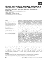
Fig. 1. Gel-shift analysis of SerRS and Pex21p protein with tRNA
Ser
or total yeast tRNA. Full-size His
6
-Pex21p was purified from Escheri-
chia coli and SerRS was purified from yeast and incubated with
tRNA
Ser
transcript or total yeast tRNA in the absence or presence
of Pex21p. Complexes were subjected to nondenaturing polyacryla-
mide gel electrophoresis, transferred to nitrocellulose membrane,
incubated with antibodies to SerRS, and visualized by chemilumi-
nescence. The dotted arrow indicates a faster-migrating SerRS–
tRNA
Ser
complex. The full arrow indicates a Pex21p–SerRS–tRNA
Ser
supershifted complex.
V. Godinic et al. Interaction between SerRS and Pex21p
FEBS Journal 274 (2007) 2788–2799 ª 2007 The Authors Journal compilation ª 2007 FEBS 2789
Pex21p further retarded the complex between SerRS
and its cognate tRNA (Fig. 1, lane 2), indicative of
ternary complex formation (Fig. 1, lanes 5 and 6). In
support of this finding, a retarded complex was
observed with antibodies against the His tag in His
6
-
Pex21p (not shown). A ternary complex was also
detected after coincubation of SerRS and Pex21p with
total yeast tRNA, as indicated by the occurrence of a
supershifted band of the same intensity and mobility
(Fig. 1, lane 6).
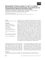
A supershifted band denoting a ternary complex
was also obtained in the experiment conducted with
3¢-radiolabeled tRNA
Ser
transcript (Fig. 2A, lane 4).
Equal amounts of Pex21p and SerRS were preincuba-
ted for 10 min, prior to addition of
32
P-labeled tRNAs.
Reaction mixtures were subjected to electrophoresis on
a nondenaturing polyacrylamide gel. Free and protein-
bound tRNA was visualized by radiography, and the
ternary complex was only detected with cognate
tRNA
Ser
. Faint bands in the upper part of the gel are
artefacts of macromolecular aggregation occurring
upon entry into the gel. Yeast tRNA
Phe
, which was
used as a control, did not affect the migration of the
protein binary complex (Fig. 2A, lane 5), suggesting
that ternary complex formation was specific for
tRNA
Ser
.
The formation of binary and ternary complexes was
assayed under various ionic conditions. Magnesium
ions are of great importance in stabilizing the tRNA
tertiary structure, and influence aaRS–tRNA complex
formation; as a result, the complex between SerRS and
tRNA
Ser
cannot be detected at low Mg
2+
concentra-
tions [40]. In agreement, the complex obtained at
150 mm sodium chloride and 2.5 mm MgCl
2
was
rather weak (Fig. 2A, lane 1), and it seems to be sta-
bilized by addition of Pex21p. When the ionic strength
was reduced to 30 mm NaCl and the concentration of
Mg
2+
was increased (8 mm MgCl
2
), the stability of the
binary complex was increased (Fig. 2B).
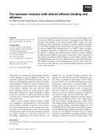
Pex21p increases tRNA binding by SerRS
To further investigate the influence of Pex21p on tRNA
binding by SerRS, we compared the extent of tRNA
binding by isolated enzyme and by the SerRS–Pex21p
binary complex. Labeled tRNA
Ser
was mixed with
increasing amounts of preformed SerRS–Pex21p com-
plex, and the resulting tRNA–protein complexes were
resolved and quantified (the values for bound tRNA
were estimated by deducting uncomplexed tRNA from
total tRNA). The percentage of bound tRNA
Ser
was
plotted as a function of enzyme concentration (Fig. 3),
and indicated that tRNA binding by SerRS was eleva-
ted in the presence of Pex21p.
Mapping the Pex21p interaction domain in SerRS
Eukaryotic cytosolic SeRSs comprise positively charged
noncatalytic peptides appended to conserved catalytic
cores. As shown in Fig. 4, extensions are characterized
by high lysine content, but their primary sequences are
not conserved.
In order to map the interaction domain of SerRS
that is involved in complex assembly with Pex21p,
we prepared a number of truncated yeast SerRS vari-
ants (Fig. 5A). By aligning the primary structures of
A
B
Fig. 2. (A) Formation of a ternary complex, Pex21p–SerRS–tRNA
Ser
.
Equal amounts of Pex21p and SerRS were preincubated for 10 min
prior to addition of 3¢-radiolabeled tRNA
Ser
in binding buffer contain-
ing 150 m
M sodium chloride and 2.5 mM MgCl
2
. The complexes
were analyzed by 5% native PAGE, and submitted to phosphorimag-
ing. Lane 1, bottom bracket: free tRNA
Ser.
Lane 2, middle bracket:
tRNA
Ser
shifted in SerRS–tRNA
Ser
complex (the stability of this
complex is discussed further in the text). Lane 3: labeled tRNA
Ser
with Pex21p added in a binding reaction. Lane 4, upper bracket:
larger ternary complex Pex21p–SerRS–tRNA
Ser
trapped at the top of
the gel. Lane 5: SerRS and Pex21p in a binding reaction with radio-
labeled tRNA
Phe
used as a control. Lane 6: tRNA
Phe
only. (B) tRNA
Ser
shifted in the SerRS–tRNA
Ser
complex in the presence of 30 mM
NaCl and 8 mM MgCl
2
. Lane 1, bottom bracket: radiolabeled tRNA
Ser.
Lanes 2–5, upper bracket: SerRS–tRNA
Ser
complex. The concentra-
tion of
32
P-tRNA
Ser
was 0.15 lM, and the concentration of SerRS in
lanes 2–5 was 0.4, 0.7, 1.0 and 2.5 l
M, respectively. In lane 5, almost
all of the tRNA
Ser
was sequestered in SerRS–tRNA
Ser
complex.
Interaction between SerRS and Pex21p V. Godinic et al.
2790 FEBS Journal 274 (2007) 2788–2799 ª 2007 The Authors Journal compilation ª 2007 FEBS
available SerRS proteins, and on the basis of the struc-
tural data available for Escherichia coli and Thermus
thermophilus SerRS [41,42], we attempted to delete
the C-terminal extension and subsequently the struc-
tural motifs III, II, and I. Finally, the shortest trun-
cated protein (ScSerRSD356) comprised only the
presumed N-terminal coiled-coil domain (denoted
COIL in Fig. 5A).
To investigate the role of the C-terminal extension,
two truncated mutants were designed. ScSerRSDC20
lacked the 20 C-terminal amino acids, whereas in
ScSerRSDC13, only the fragment of 13 amino acids
(containing seven lysines) was cut off. Truncated yeast
SerRS constructs were fused to the C-terminal end of
the LexA DNA-binding domain (LexAbd), yielding a
series of LexA-fusion proteins, which were then used
as baits in the two-hybrid assay. Upon transformation
of baits into the yeast strain L40 coexpressing full-
length Pex21p fused to the transcription activator
GAL4-activation domain (GAL4ad), which yields
GAL4–Pex21p, they were tested in a yeast two-hybrid
system to assay for Pex21p binding in vivo. The
fusion proteins denoted LexA–ScSerRS, LexA–
ScSerRSDC13, LexA–ScSerRSDC20, LexA–ScSerRSDC82,
Fig. 3. Binding of tRNA
Ser
to free SerRS and to the preformed
Pex21p–SerRS complex. Labeled in vitro-transcribed tRNA
Ser
(50 nM)
was titrated with increasing concentrations of SerRS (0.5–2.5 l
M)in
the presence of BSA (10 l
M; j) or Pex21p (2 lM; m) in binding
buffer, and electrophoresed in 5% native polyacrylamide gel (as des-
cribed in Experimental procedures). The curve corresponds to the
percentage of bound tRNA
Ser
as a function of enzyme concentration.
The results were scanned using a phosphorimager and analyzed by
IMAGEQUANT software. The data were fitted to a single-site binding
equation.
Fig. 4. Alignment of C-terminal regions of SerRS proteins from different domains of life: AA, Aquifex aeolicus; BS, Bacillus subtilis; LP, Leg-
ionella pneumophila; EC, E. coli; TD, Thiobacillus denitrificans; TT, Thermus thermophilus; TP, Thermofilum pendens; AP, Aeropyrum pernix;
PF, Pyrococcus furiosus; HS, Halobacterium salinarum; MT, Methanothermobacter thermautotrophicus; HS, Homo sapiens; BS, Bos taurus;
MM, Mus musculus; GG, Gallus gallus; AT, Arabidopsis thaliana; ZM, Z. mays; CA, Candida albicans; SC, S. cerevisiae; SP, Schizosaccha-
romyces pombe. Only eukaryotic cytosolic enzymes contain positively charged C-terminal extensions. The C-terminal sequence of S. cerevis-
iae and Z. mays cytosolic SerRSs is shown in bold letters. The sequence truncated in the yeast SerRSDC13 mutant is underlined.
V. Godinic et al. Interaction between SerRS and Pex21p
FEBS Journal 274 (2007) 2788–2799 ª 2007 The Authors Journal compilation ª 2007 FEBS 2791
LexA–ScSerRSDC202 and LexA–ScSerRSDC356 were
stably expressed in yeast, as confirmed by western blot
using antibodies to LexA (Fig. 5B). In order to test the spe-
cificity of t he S erRS–Pex21p i nteraction, the f ull-length
maize (Zea mays cytosoli c, Zmc) Se rRS (LexA–Zmc SerRS)
and its truncated varian t (LexA–Zm cSerRSDC26), lacking
26 C-terminal amino acids, were also analyzed. In a western
blot, equal amounts of protein extract were loaded per
well for each construct, indicating that the level of
expression was the same for all d eletion mutants and full-
length SerRSs, except for LexA–ScSerRSD35 6 and LexA–
ZmcSerRS, whose expression was h igher (Fig. 5B).
The basic C-terminal extension of yeast SerRS
is required for interaction with Pex21p in vivo
To test activation of the reporter gene HIS3, transfor-
mants were inspected for growth after 4 days of incu-
bation on minimal plates (–Trp-Leu-His) containing
30 mm 3-amino-1,2,4-triazole (Fig. 6A,B, left panels).
To verify the interaction and subsequently quantify it,
we tested the activation of the second reporter gene
lacZ by colony-lift filter assays (Fig. 6A,B, middle pan-
els). The expression of the lacZ reporter gene, indica-
ting the strength of protein–protein interactions, was
quantified using 2-nitrophenyl b-d-galactopyranoside
(Gal-ONp) as a substrate for b-galactosidase (Fig. 6C).
Screening variants for 3-amino-1,2,4-triazole resistance
and a lacZ-positive phenotype revealed that C-terminal
truncation of SerRS abolished Pex21p binding. The
interaction is lost upon deletion of the positively
charged C-terminal fragment (mutant C13) within the
extension. The deletion of the whole C-terminal exten-
sion of yeast SerRS also resulted in noninteracting trun-
cated SerRS (mutant C20). Accordingly, all other more
truncated variants (see deletion scheme in Fig. 5A)
failed to interact with Pex21p (Fig. 6A,C). Interestingly,
maize cytosolic SerRS, which also possesses the C-ter-
minal extension, did not interact with Pex21p in the
two-hybrid assays (Fig. 6B). It is pertinent to note that
positively charged C-terminally appended fragments of
yeast and maize SerRSs do not share sequence homo-
logy. Thus, the C-terminal domain of yeast SerRS func-
tions as the specific Pex21p-binding site.
Truncation of a short C-terminal fragment
of SerRS abolishes Pex21p binding without
affecting catalytic activity
A pull-down assay was used to verify that Pex21p spe-
cifically recognizes the C-terminal appendix of yeast
SerRS in vitro.Ni
2+
–nitrilotriacetic acid agarose was
A
B
Fig. 5. Schematic representation and west-
ern blot analysis of full-length and deletion
constructs of yeast (S. cerevisiae, Sc) and
maize cytosolic (Z. mays, Zmc) SerRS. (A)
The names of the constructs used as baits
in the two-hybrid system are shown on the
left. The number of truncated amino acids
from the C-terminus of full-length ScSerRS
or ZmSerRS is indicated. (B) Western blot of
protein extracts comprising fusion proteins
in the L40 yeast strain. Lane 1: LexAbd
(25.5 kDa). Lane 2: LexA–ScSerRSDC356
(38 kDa). Lane 3: LexA–ScSerRSDC202
(55.3 kDa). Lane 4: LexA–ScSerRSDC82
(69.4 kDa). Lane 5: LexA–ScSerRSDC20
(76.5 kDa). Lane 6: LexA–ZmcSerRS (a full-
length Z. mays cytosolic SerRS; 77.2 kDa).
Lane 7: LexA–ZmcSerRSDC26 (74.9 kDa).
Lane 8: S. cerevisiae strain L40, nontrans-
formed. Lane 9: LexA–ScSerRS (a full-length
S. cerevisiae SerRS; 78.8 kDa). Lane 10:
LexA–ScSerRSDC13 (77.3 kDa). Calculated
molecular masses of the LexA-fusion
proteins are in parentheses.
Interaction between SerRS and Pex21p V. Godinic et al.
2792 FEBS Journal 274 (2007) 2788–2799 ª 2007 The Authors Journal compilation ª 2007 FEBS
saturated with crude E. coli extract containing recom-
binant Pex21p, which was immobilized on the resin by
its N-terminal His-tag. Ni
2+
–nitrilotriacetic acid
agarose precharged with Pex21p was incubated with
yeast or maize SerRS variants. Proteins bound to
resin were eluted with buffer containing 300 mm
imidazole and analyzed by SDS ⁄ PAGE followed by
Coomassie Brilliant Blue staining (Fig. 7). As expected,
only full-length yeast SerRS binds to resin previously
saturated with Pex21p (Fig. 7, lane 6). Truncated yeast
SerRSDC13 or maize SerRS were not pulled down on
the resin precharged with Pex21p (Fig. 7, lanes 7 and
8). In the absence of Pex21p, purified SerRS deriva-
tives were also not retained on the resin (Fig. 7, lanes
3 and 4).
To ensure that SerRSDC13 was stable and correctly
folded in pull-down experiments, we determined kinetic
parameters for full-length Saccharomyces cerevisiae
(Sc)SerRS and truncated enzyme in standard amino-
acylation reactions. K
m
and k
cat
values with respect to
both tRNA and serine (K
m
¼ 51±6lm and k
cat
¼
0.47 ± 0.02 s
)1
for serine; K
m
¼ 0.65 ± 0.07 lm and
k
cat
¼ 0.54 ± 0.02 s
)1
for tRNA
Ser
) were unchanged
as compared to those obtained for intact ScSerRS
(K
m
¼ 47 ± 5 lm and k
cat
¼ 0.46 ± 0.02 s
)1
for serine;
K
m
¼ 0.71 ± 0.11 lm and k
cat
¼ 0.58 ± 0.03 s
)1
for
A
B
C
Fig. 6. In vivo interaction of Pex21p with homologous and heterologous SerRS variants. Yeast strain L40 coexpressing GAL4–Pex21p
and full-length or C-terminally truncated yeast (A) or maize (B) LexA–SerRS variants were plated onto medium that lacks histidine to test for
histidine prototrophy (far left plates). The same transformants were also subjected to b-galactosidase colony-lift filter assay using Gal-X as a
substrate (see middle plates). The appearance of a blue color indicates protein–protein interactions. Yeast cells were transformed with
various constructs as indicated on the right part of the panel, which shows the orientation on the plates that were tested. b-Galactosidase
activity was quantified using the Gal-ONp assay (C). The bars indicate Miller units showing the strength of interaction of bait proteins with
GAL4–Pex21p. LexA ⁄ GAL4–Pex21p and ScSerRS–GAL4 were used as controls.
V. Godinic et al. Interaction between SerRS and Pex21p
FEBS Journal 274 (2007) 2788–2799 ª 2007 The Authors Journal compilation ª 2007 FEBS 2793
tRNA
Ser
). As ScSerRSDC13 is catalytically fully active,
we assume that the overall structure of SerRS is unaf-
fected by deletion of its C-terminal appendix. The
similarity of kinetic parameters for the full-length
SerRS and the ScSerRSDC13 truncation mutant indi-
cates that the C-terminal peptide, composed of 13
amino acids, was not important in substrate recogni-
tion, but was primarily involved in protein cofactor
binding. Thus, 13 C-terminal amino acids of yeast
SerRS function as the binding domain for Pex21p, as
revealed by yeast two-hybrid and pull-down assays.
Discussion
Role of the C-terminal extensions of eukaryotic
SerRSs
We previously identified the peroxin Pex21p as an inter-
action partner of SerRS [32]. Here we examined whether
the positively charged C-terminal region of the synthe-
tase, a characteristic of all eukaryotic SerRSs, is
involved in protein binding. Our studies revealed that
the removal of 13 amino acids from the C-terminus gen-
erates a stable ScSerRSDC13 variant that exhibits essen-
tially the same kinetic parameters for serylation as the
wild-type, but has lost the ability to interact with
Pex21p. Thus, in contrast to the idiosyncratic extensions
of several eukaryotic aaRSs, which serve as additional
tRNA-binding domains, due to their high lysine content
and overall positive charge [6,7,43], the yeast SerRS C-
terminal extension does not participate directly in sub-
strate binding, but instead mediates protein binding.
Whereas SerRS binds both tRNA and Pex21p, the
stoichiometry of the ternary complex is not yet known.
In agreement with our previously reported findings
that dimeric yeast SerRS binds cognate tRNA
Ser
anti-
cooperatively [44,45], and that the serylation efficiency
is moderately enhanced when approximately two mole-
cules of Pex21p are bound per dimeric SerRS [32], our
current model suggests cross-subunit binding of one
tRNA per two SerRS subunits, whereas each subunit
interacts with one molecule of Pex21p. In this complex,
Pex21p may possibly contribute to tRNA binding by
enhancing the affinity of the enzyme for the second
tRNA molecule. However, whether the peroxin affects
the affinity, stability or stoichiometry of the heterotri-
meric Pex21p–SerRS–tRNA
Ser
complex remains an
open question.
Role of Pex21p in enhancing cognate tRNA
binding by SerRS
Pex21p does not bind tRNA
Ser
(Fig. 2A, lane 3) or any
other component of total yeast tRNA (not shown), and
thus cannot be considered a tRNA-binding protein. On
the other hand, SerRS binds both cognate tRNA
Ser
and Pex21p, resulting in the formation of a ternary
complex. The Haemophilus influenzae YbaK protein,
which hydrolyzes misacylated Cys-tRNA
Pro
in trans,
also appears to lack specific tRNA recognition capabil-
ity [45], but, like Pex21p, forms a binary complex with
the corresponding synthetase (ProRS) and a ternary
complex with the synthetase–tRNA pair. It is possible
that Pex21p induces a conformational change in the
synthetase, facilitating additional contacts between the
tRNA and SerRS. Moreover, higher levels of tRNA
(15–20%) were found in the heterotrimeric than in the
binary complex, in agreement with our previous obser-
vation that Pex21p moderately stimulates the amino-
acylation efficiency of SerRS [32]. This suggests that
Pex21p-induced contacts between tRNA and the syn-
thetase may contribute to tRNA binding, in synergy
with the major interaction between the N-terminal
a-helical coiled coil of SeRS and the long extra arm of
tRNA
Ser
. The failure to obtain discrete upshifted bands
in the gel mobility shift assay, which is probably a
consequence of the rapid dissociation kinetics of the
components in the complex, precluded K
d
determin-
ation, and the precise mechanism by which ternary
Fig. 7. Pull-down assay of SerRSs on Ni
2+
–nitrilotriacetic acid
agarose precharged with crude E. coli extract containing recombin-
ant His-tagged Pex21p. Lanes 1 and 2: purified yeast SerRS (lane 1)
and ZmcSerRS (lane 2). Lanes 3 and 4: proteins eluted from Ni
2+
–
nitrilotriacetic acid agarose saturated with crude E. coli extract con-
taining no Pex21p and incubated with purified yeast SerRS (lane 3) or
maize SerRS (lane 4). Lanes 5–8: proteins eluted from Ni
2+
–
nitrilotriacetic acid agarose precharged with crude E. coli extract con-
taining recombinant His-tagged Pex21p (lane 5) and incubated with
full-length yeast SerRS (lane 6), truncated SerRSDC13 (lane 7) or
maize SerRS (lane 8). Only full-length yeast SerRS binds to Pex21p
immobilized on Ni
2+
–nitrilotriacetic acid agarose (lane 6). Nonspecific
adsorption of yeast SerRS or ZmcSerRS to Ni
2+
–nitrilotriacetic acid
agarose was not observed (lanes 3 and 4, respectively). Positions of
molecular mass markers are indicated on the left.
Interaction between SerRS and Pex21p V. Godinic et al.
2794 FEBS Journal 274 (2007) 2788–2799 ª 2007 The Authors Journal compilation ª 2007 FEBS
complex formation stimulates serylation remains
unclear. In vivo, the lack of Pex21p does not signifi-
cantly perturb peroxisomal biogenesis, as it is function-
ally redundant with Pex18p [34]. The level of SerRS in
a Dpex21 strain is only slightly decreased compared to
the wild-type, as quantified by immunoblot analysis
(data not shown). It seems, therefore, that the two
genes are not coregulated, and that Pex21p does not
act as a transactivator of SES1 gene transcription.
Available RNA microarray data [47–49] indicate that
Pex21p is overexpressed under oxidative stress condi-
tions [50,51], whereas the level of SerRS is decreased.
On the other hand, upon stress-related overexpression
of Pex21p, the peroxin may bind SerRS (or SerRS–
tRNA
Ser
) and target this component of the transla-
tional machinery for degradation by an as yet unknown
mechanism. Interestingly, in addition to the catalysis of
the aminoacylation reaction, SerRS has also been
found to participate in the synthesis and turnover of
diadenosine oligophosphates (Ap
n
A) [52], which plays
an important role in the response of bacterial and
eukaryotic cells to a variety of stress conditions. These
adenylylated nucleotides may be alarmones, i.e. regula-
tory molecules, alerting cells to the onset of oxidation
stress [53]. Therefore, aaRSs could be important coor-
dinators in stress signaling networks.
Experimental procedures
Plasmid constructions
The full-length gene encoding S. cerevisiae SeRS was inserted
in-frame to the 3¢-end of the coding sequence of the tran-
scription factor LexA-binding domain (LexAbd) in the yeast
expression vector pAB151 [32]. Furthermore, all truncated
constructs for bait proteins were created using PCR, and
checked by sequencing. Inserted genes for C-terminal-dele-
tion mutants were expressed in-frame with the LexA DNA-
binding domain. The gene for Pex21p was cloned in pET15b
(Novagen, Madison, WI, USA) for protein purification, and
in pACT2 (Clontech, Mountain View, CA, USA) for two-
hybrid analysis. Full-length and truncated SerRS genes were
inserted in pCJ11 for protein overexpression and purifica-
tion. The Zea mays SerRS gene was inserted in pET28b
(Novagen).
Yeast two-hybrid analysis
Recombinant plasmids were introduced by the lithium
acetate transformation procedure as previously described
[32] into the L40 S. cerevisiae strain (MATa his3-D200
trp1-D901 leu2–3, 112 ade2 LYS2::(lexAop)
4
-HIS3 URA3::
(lexAop)
8
-lacZ), kindly provided by I. Stagljar (Department
of Biochemistry, University of Toronto, Canada). Trans-
formants were allowed to grow at 30 °C for 2–3 days
on – Trp-Leu (absence of the amino acids Trp and Leu)
plates, and then transferred to selective – Trp-Leu-His
(absence of the amino acids Trp, Leu and His) plates, with
30 mm 3-amino-1,2,4-triazole for testing activation of the
reporter gene HIS3. Then, transformants were tested for
b-galactosidase activity by colony-lift filter assay using
5-bromo-4-chloro-3-indolyl-b-d-galactopyranoside (Gal-X).
The filters were incubated at room temperature, and
checked periodically for the appearance of a blue color that
developed between 30 min and 8 h. b-Galactosidase activity
was quantified using Gal-ONp as a substrate in assays car-
ried out according to the manufacturer’s instructions (Clon-
tech), and expressed in Miller units under the trial
conditions pH 7.0 and temperature 30 °C. Each determin-
ation was performed in triplicate.
Western blotting
To probe the expression levels of fusion proteins, yeast
whole cell protein lysates were separated on a 9% SDS ⁄
PAGE gel, transferred to nitrocellulose membrane, and
immunoblotted with rabbit anti-LexA (Invitrogen, Carls-
bad, CA, USA) and rabbit anti-SerRS raised against yeast
SerRS. Anti-LexA was diluted 1 : 5000 (v ⁄ v) and anti-
SerRS 1 : 500 in Tris-buffered saline (NaCl ⁄ Tris) contain-
ing 0.2% (v ⁄ v) Tween-20. Secondary anti-rabbit IgG
(Novagen) conjugated with horseradish peroxidase was
diluted 1 : 10 000 (v ⁄ v), and anti-mouse IgG (Novagen)
conjugated with horseradish peroxidase was diluted
1 : 5000 (v ⁄ v). Immunoreactive bands were subsequently
visualized using chemiluminescence (KPL, Gaithersburg,
MD, USA). Nondenaturing gels were also subjected to
western blot analysis using antibodies to His-tag (Novagen)
for detection of His-tagged Pex21p, or antibodies to SerRS
for detection of yeast SerRS.
Purification of proteins
pET15bPEX21 plasmid was introduced into the bacterial
strain BL21(DE3)pLysS (Novagen). Cells were harvested,
resuspended in ice-cold lysis buffer [50 mm NaCl, 50 mm
Tris ⁄ HCl, pH 7.5, 5% (v ⁄ v) glycerol, 5 mm dithiothreitol
and 0.2 mm phenylmethanesulfonyl fluoride], lysed on ice
by mild sonication, and centrifuged (10 000 g, 15 min, 4 °C,
6K1s centrifuge, Sigma, Osterode am Hartz, Germany).
The lysate was subjected to centrifugation (20 000
g
, 30 min
at 4 °C, 6K1s centrifuge) to remove cell debris, and protein
was purified on Ni
2+
–nitrilotriacetate agarose (Qiagen Inc.
Valencia, CA, USA) according to the manufacturer’s proto-
col. For analysis of purified Pex21p, aliquots were boiled in
sample buffer and loaded onto SDS–polyacrylamide gel.
The eluant was dialyzed against 1 L of 20 mm Tris ⁄ HCl
V. Godinic et al. Interaction between SerRS and Pex21p
FEBS Journal 274 (2007) 2788–2799 ª 2007 The Authors Journal compilation ª 2007 FEBS 2795
(pH 7.5), 100 mm NaCl, 1 mm dithiothreitol and 20%
(v ⁄ v) glycerol, and used for electrophoretic mobility shift
assay (EMSA). The overproduction of ScSerRSDC13 and
full-length ScSerRS enzymes was achieved as described
[34]. The enzymes were purified by a two-step chromato-
graphic procedure on FPLC MonoQ and MonoS columns
(Pharmacia Biotech Inc., Uppsala, Swedan). Z. mays
SerRS was overexpressed in E. coli strain BL21(DE3)
after 2.5 h of induction with 1 mm isopropyl thio-b-d-gal-
actoside at 30 °C, and purified by ion exchange chroma-
tography on a MonoQ HR 10 ⁄ 10 column (Pharmacia
Biotech).
Aminoacylation assays
Reaction mixtures contained 50 m m Tris ⁄ HCl (pH 7.5),
4mm dithiothreitol, 15 mm MgCl
2
,5mm ATP, 0.4 mgÆmL
)1
BSA, variable concentrations of [
14
C]l-serine (32–40
CiÆmol
)1
), and unfractionated yeast tRNA. Kinetic parame-
ters (k
cat
and K
m
) for serine were determined in the presence
of 4.3 mgÆmL
)1
unfractioned yeast tRNA (containing 5 lm
tRNA
Ser
) and 10–400 lm [
14
C]l-serine. For tRNA kinetic
parameter determination, the concentration of [
14
C]l-serine
was kept constant (100 lm) and the unfractioned yeast
tRNA concentration was varied (0.17–3.4 mgÆmL
)1
, corres-
ponding to 0.2–4.0 lm tRNA
Ser
). The amount of tRNA
Ser
isoacceptors in unfractioned yeast tRNA was determined
from the maximal acceptor activity of unfractioned yeast
tRNA towards purified ScSerRS and [
14
C]l-serine. The
enzyme concentration was 7.5–10 nm for serine and 4 nm for
tRNA kinetic parameter determination. Reactions were per-
formed at 30 °C. Kinetic parameters were calculated from
initial velocities for different substrate concentrations using
nonlinear regression.
EMSA
For radioactive EMSA, yeast tRNA
Ser
transcript was gener-
ated and purified as before [54]. The tRNA transcript was
charged with [
14
C]l-serine, and the aminoacylation plateau
was measured with homologous SerRS, giving a serine-accept-
ing activity of 700 pmol of serylated tRNA per A
260
.The
CCA 3¢-end from tRNA
Ser
was removed prior to the labeling
reaction by incubation for 2 h at room temperature with
73 lgÆmL
)1
snake venom exonuclease (phosphodiesterase I)
from Crotalus atrox (Sigma-Aldrich, St Louis, MO, USA) in
40 mm sodium glycinate (Na-Gly) buffer (pH 9.0) and 10 mm
magnesium acetate. The reaction product was extracted with
phenol ⁄ chloroform, desalted by gel filtration through a Se-
phadex G25 column (Amersham Biosciences, Piscataway, NJ,
USA), and precipitated with ethanol. The CCA 3¢-end of the
tRNA
Ser
transcript was reconstituted and labeled with
[
32
P]ATP[aP] by incubation for 10 min at 37 °Cwith0.5lm
snake venom-treated tRNA in 50 mm Na-Gly (pH 9.0),
10 mm MgCl
2
,10lm CTP, 9 lm ATP, 1 lm [
32
P]ATP[aP]
and 3 lgÆmL
)1
E. coli tRNA-terminal nucleotidyltransferase
in a final volume of 20 lL. The reaction was stopped by the
addition of one volume of phenol, and the resulting mixture
was gel filtered twice through a G25 column. Prior to complex
formation, tRNA
Ser
was freshly renatured by heating to 80 °C
for 2 min; MgCl
2
was then added to 10 mm, and the reaction
mixture was further placed on ice.
Proteins Pex21p, SerRS (250 nm) and ⁄ or BSA were incu-
bated for 10 min at 25 °C in a binding buffer containing
20 mm Tris ⁄ HCl (pH 8.0), 8 mm (or 2.5 mm) MgCl
2
,30mm
(or 150 mm) NaCl, and 5% (v ⁄ v) glycerol. After addition of
32
P-labeled tRNA
Ser
or
32
P-labeled tRNA
Phe
(150 nm), incu-
bation was continued for an additional 15 min at room
temperature in a final binding reaction volume of 25 lL.
Finally, tRNA
Ser
–protein complexes were resolved by elec-
trophoresis in a 5% native polyacrylamide gel (37.5 :1 acryl-
amide ⁄ bisacrylamide) in 0.5 · Tris-borate buffer (90 mm
Tris, pH 8.3, and 65 mm boric acid) with 5 mm MgCl
2
. The
gel was vacuum-dried for 30 min in a gel-dryer, and tRNA
was detected by Phosphorimager (Amersham Biosciences).
For nonradioactive EMSA, the protein concentration was
2 lm, the tRNA concentration was 3 lm, and complexes
were formed by coincubating tRNA
Ser
and proteins for
10 min at 25 °C in the presence of 30 mm KCl, 5 mm
MgCl
2
, and 20 mm Tris ⁄ HCl (pH 8.0). After electrophor-
esis, proteins were transferred to nitrocellulose membrane
and subjected to western blotting using specific antibodies.
In vitro binding assay
Ni
2+
–nitrilotriacetic acid agarose (Qiagen) was equilibrated
with lysis buffer containing 10 mm imidazole, and saturated
with E. coli BL21(DE3) crude extract containing expressed
His-tagged Pex21p. The resin was washed extensively with
buffer containing 40 mm imidazole to remove unbound pro-
teins, and this was followed by equilibration with buffer for
SerRS binding (40 mm imidazole, 5 mm MgCl
2
, and 5 mm
2-mercaptoethanol). Resin saturated with Pex21p was dis-
pensed in small batches (15 lL) and incubated with 30 lgof
purified ScSerRS, ScSerRSDC13 or ZmcSerRS for 10 min at
room temperature. The resin was thoroughly washed with
binding buffer (40 mm imidazole, 5 m m MgCl
2
, and 5 mm
2-mercaptoethanol), and bound proteins were eluted with
300 mm imidazole. The eluate was analyzed by SDS ⁄ PAGE.
Nonspecific binding of SerRS to Ni
2+
–nitrilotriacetic acid
agarose and the presence of impurities bound to resin were
tested on resin saturated with E. coli BL21(DE3) crude
extract without any recombinant protein expressed.
Acknowledgements
This work was supported by grants from the Ministry of
Science, Education and Sports of the Republic of Cro-
atia (I. Weygand-Durasevic), and the National Institute
Interaction between SerRS and Pex21p V. Godinic et al.
2796 FEBS Journal 274 (2007) 2788–2799 ª 2007 The Authors Journal compilation ª 2007 FEBS
of General Medical Sciences (M. Ibba). We thank Pro-
fessor I. Stagljar (Department of Biochemistry, Univer-
sity of Toronto, Canada) for the yeast two-hybrid strain
and plasmid. We are grateful to J. Jaric and J. Ling for
generous gifts of tRNA transcripts.
References
1 Francklyn C, Perona JJ, Puetz J & Hou JM (2002)
Aminoacyl-tRNA synthetases: versatile players in the
changing theater of translation. RNA 8, 1363–1372.
2 Park SG, Ewalt KL & Kim S (2005) Functional expan-
sion of aminoacyl-tRNA synthetases and their interact-
ing factors: new perspectives on housekeepers. Trends
Biochem Sci 30, 569–574.
3 Mirande M (1991) Aminoacyl-tRNA synthetase family
from prokaryotes and eukaryotes: structural domains
and their implications. Prog Nucleic Acid Res Mol Biol
40, 95–142.
4 Cusack S (1995) Eleven down and nine to go. Nat
Struct Biol 2, 824–831.
5 Mirande M (2005) Multi-aminoacyl-tRNA complexes.
In The Aminoacyl-tRNA Synthetases (Ibba M,
Francklyn C & Cusack S, eds), pp. 298–308. Landes
Bioscience, Georgetown, TX.
6 Frugier M, Moulinier L & Giege R (2000) A domain in
the N-terminal extension of class IIb eukaryotic ami-
noacyl-tRNA synthetases is important for tRNA bind-
ing. EMBO J 19, 2371–2380.
7 Francin M, Kaminska Kerjan P & Mirande M (2002)
The N-terminal domain of mammalian lysyl-tRNA
synthetase is a functional tRNA-binding domain. J Biol
Chem 277, 1762–1769.
8 Kim S, Landro JA, Gale AJ & Schimmel P (1993)
C-terminal peptide appendix in a class I tRNA synthe-
tase needed for acceptor–helix contacts and microhelix
aminoacylation. Biochemistry 32, 13026–13031.
9 Geslain R & Ribas de Pouplana L (2004) Regulation of
RNA function by aminoacylation and editing? Trends
Genet 20, 604–610.
10 Ling C, Yao YN, Zheng YG, Wei H, Wang L, Wu XF
& Wang ED (2005) The C-terminal appended domain
of human cytosolic leucyl-tRNA synthetase is indispen-
sable in its interaction with arginyl-tRNA synthetase in
the multi-tRNA synthetase complex. J Biol Chem 280,
34755–34763.
11 Cerini C, Kerjan P, Astier M, Gratecos D, Mirande M
& Semeriva M (1991) A component of the multisynthe-
tase complex is a multifunctional aminoacyl-tRNA
synthetase. EMBO J 10, 4267–4277.
12 Filonenko VV & Deutscher MP (1994) Evidence for
similar structural organization of the multienzyme ami-
noacyl-tRNA synthetase complex in vivo and in vitro.
J Biol Chem 269, 17375–17378.
13 Quevillon S, Agou F, Robinson JC & Mirande M
(1997) The p43 component of the mammalian multi-
synthetase complex is likely to be the precursor of the
endothelial monocyte-activating polypeptide II cytokine.
J Biol Chem 272, 32573–32579.
14 Rho SB, Kim MJ, Lee JS, Seol W, Motegi H, Kim S &
Shiba K (1999) Genetic dissection of protein–protein
interactions in multi-tRNA synthetase complex. Proc
Natl Acad Sci USA 96, 4488–4493.
15 Tzima E & Schimmel P (2006) Inhibition of tumor
angiogenesis by a natural fragment of a tRNA synthe-
tase. Trends Biochem Sci 31, 7–10.
16 Harris CL & Kolanko CJ (1995) Aminoacyl-tRNA
synthetase complex in Saccharomyces cerevisiae. Bio-
chem J 309, 321–324.
17 Deinert K, Fasiolo F, Hurt EC & Simos G (2001)
Arc1p organizes the yeast aminoacyl-tRNA synthetase
complex and stabilizes its interaction with the cognate
tRNAs. J Biol Chem 276, 6000–6008.
18 Praetorius-Ibba M, Rogers TE, Samson R, Kelman Z &
Ibba M (2005) Association between Archaeal prolyl-
and leucyl-tRNA synthetases enhances tRNA(Pro)
aminoacylation. J Biol Chem 280, 26099–26104.
19 Lipman RS, Chen J, Evilia C, Vitseva O & Hou YM
(2003) Association of an aminoacyl-tRNA synthetase
with a putative metabolic protein in archaea. Biochemis-
try 42, 7487–7496.
20 Harris CL (1987) An aminoacyl-tRNA synthetase com-
plex in Escherichia coli. J Bacteriol 169, 2718–2723.
21 Buddha MR, Keery KM & Crane BR (2004) An unu-
sual tryptophanyl tRNA synthetase interacts with nitric
oxide synthase in Deinococcus radiodurans. Proc Natl
Acad Sci USA 101, 15881–15886.
22 Simos G, Segref A, Fasiolo F, Hellmuth K, Shevchenko
A, Mann M & Hurt EC (1996) The yeast protein Arc1p
binds to tRNA and functions as a cofactor for the
methionyl- and glutamyl-tRNA synthetases. EMBO J
15, 5437–5448.
23 Simos G, Sauer A, Fasiolo F & Hurt EC (1998) A con-
served domain within Arc1p delivers tRNA to aminoa-
cyl-tRNA synthetases. Mol Cell 1, 235–242.
24 Morales AJ, Swairjo MA & Schimmel P (1999) Struc-
ture-specific tRNA-binding protein from the
extreme thermophile Aquifex aeolicus. EMBO J 18,
3475–3483.
25 Nomanbhoy T, Morales AJ, Abraham AT, Vortler CS,
Giege R & Schimmel P (2001) Simultaneous binding of
two proteins to opposite sides of a single transfer RNA.
Nat Struct Biol 8, 344–348.
26 Kawaguchi S, Muller J, Linde D, Kuramitsu S, Shibata
T, Inoue Y, Vassylyev DG & Yokoyama S (2001) The
crystal structure of the ttCsaA protein: an export-related
chaperone from Thermus thermophilus. EMBO J 20,
562–569.
V. Godinic et al. Interaction between SerRS and Pex21p
FEBS Journal 274 (2007) 2788–2799 ª 2007 The Authors Journal compilation ª 2007 FEBS 2797
27 Ahel I, Korencic D, Ibba M & Soll D (2003) Trans-edit-
ing of mischarged tRNAs. Proc Natl Acad Sci USA
100, 15422–15427.
28 Korencic D, Ahel I, Schelert J, Sacher M, Ruan B,
Stathopoulos C, Blum P, Ibba M & So
¨
ll D (2004) A
freestanding proofreading domain is required for pro-
tein synthesis quality control in Archaea. Proc Natl
Acad Sci USA 101, 10260–10265.
29 Dagkessamanskaia A, Martin-Yken H, Basmaji F, Briza
P & Francois J (2001) Interaction of Knr4 protein, a
protein involved in cell wall synthesis, with tyrosine
tRNA synthetase encoded by TYS1 in Saccharomyces
cerevisiae. FEMS Microbiol Lett 200, 53–58.
30 Kang J, Kim T, Ko YG, Rho SB, Park SG, Kim MJ,
Kwon HJ & Kim S (2000) Heat shock protein 90
mediates protein–protein interactions between human
aminoacyl-tRNA synthetases. J Biol Chem 275,
31682–31688.
31 Han JM, Lee MJ, Park SG, Lee SH, Razin E, Choi EC
& Kim S (2006) Hierarchical network between the
components of the multi-tRNA synthetase complex:
implications for complex formation. J Biol Chem 281,
38663–38667.
32 Rocak S, Landeka I & Weygand-Durasevic I (2002)
Identifying Pex21p as a protein that specifically interacts
with yeast seryl-tRNA synthetase. FEMS Microbiol Lett
214, 101–106.
33 Purdue PE, Yang X & Lazarow PB (1998) Pex18p and
Pex21p, a novel pair of related peroxins essential for
peroxisomal targeting by the PTS2 pathway. J Cell Biol
143, 1859–1869.
34 Einwachter H, Sowinski S, Kunau WH & Schliebs W
(2001) Yarrowia lipolytica Pex20p, Saccharomyces
cerevisiae Pex18p ⁄ Pex21p and mammalian Pex5pL
fulfil a common function in the early steps of the
peroxisomal PTS2 import pathway. EMBO Rep 2,
1035–1039.
35 Weygand-Durasevic I, Lenhard B, Filipic S & Soll D
(1996) The C-terminal extension of yeast seryl-tRNA
synthetase affects stability of the enzyme and its sub-
strate affinity. J Biol Chem 271, 2455–2461.
36 Lenhard B, Praetorius-Ibba M, Filipic S, Soll D &
Weygand-Durasevic I (1998) C-terminal truncation of
yeast SerRS is toxic for Saccharomyces cerevisiae due to
altered mechanism of substrate recognition. FEBS Lett
439, 235–240.
37 Kaminska M, Deniziak M, Kerjan P, Barciszewski J &
Mirande M (2000) A recurrent general RNA binding
domain appended to plant methionyl-tRNA synthetase
acts as a cis-acting cofactor for aminoacylation. EMBO
J 19, 6908–6917.
38 Kaminska M, Shalak V & Mirande M (2001) The
appended C-domain of human methionyl-tRNA synthe-
tase has a tRNA-sequestering function. Biochemistry 40,
14309–14316.
39 Shalak V, Kaminska M, Mitnacht-Kraus R,
Vandenabeele P, Clauss M & Mirande M (2001) The
EMAPII cytokine is released from the mammalian
multisynthetase complex after cleavage of its p43 ⁄
proEMAPII component. J Biol Chem 276, 23769–23776.
40 Gruic-Sovulj I, Rokov-Plavec J, Mocibob M, Kamenski
T & Weygand-Durasevic I (2004) Stability of the com-
plex between yeast seryl-tRNA synthetase and tRNASer
under different electrophoretic conditions. Croat Chem
Acta 77, 599–604.
41 Cusack S, Berthet-Colominas C, Ha
¨
rtlein M, Nassar N
& Leberman R (1990) A second class of synthetase
structure revealed by X-ray analysis of Escherichia
coli seryl-tRNA synthetase at 2.5 A
˚
. Nature 347
,
249–255.
42 Fujinaga M, Berthet-Colominas C, Yaremchuk AD,
Tukalo MA & Cusack S (1993) Refined crystal
structure of the seryl-tRNA synthetase from Thermus
thermophilus at 2.5 A
˚
resolution. J Mol Biol 234,
222–233.
43 Hammamieh R & Yang DC (2001) Magnesium
ion-mediated binding to tRNA by an amino-terminal
peptide of a class II tRNA synthetase. J Biol Chem 276,
428–433.
44 Gruic-Sovulj I, Ludemann HC, Hillenkamp F,
Weygand-Durasevic I, Kucan Z & Peter-Katalinic J
(1997) Detection of noncovalent tRNA.aminoacyl-
tRNA synthetase complexes by matrix-assisted laser
desorption ⁄ ionization mass spectrometry. J Biol Chem
272, 32084–32091.
45 Gruic-Sovulj I, Landeka I, Soll D & Weygand-Durase-
vic I (2002) tRNA-dependent amino acid discrimination
by yeast seryl-tRNA synthetase. Eur J Biochem 269,
5271–5279.
46 An S & Musier-Forsyth K (2004) Trans-editing of
Cys-tRNAPro by Haemophilus influenzae YbaK pro-
tein. J Biol Chem 279, 42359–42362.
47 Gasch AP, Spellman PT, Kao CM, Carmel-Harel O,
Eisen MB, Storz G, Botstein D & Brown PO (2000)
Genomic expression programs in the response of yeast
cells to environmental changes. Mol Biol Cell 11,
4241–4257.
48 Smith JJ, Marelli M, Christmas RH, Vizeacoumar FJ,
Dilworth DJ, Ideker T, Galitski T, Dimitrov K,
Rachubinski RA & Aitchison JD (2002) Transcriptome
profiling to identify genes involved in peroxisome
assembly and function. J Cell Biol 158, 259–271.
49 Travers KJ, Patil CK, Wodicka L, Lockhart DJ,
Weissman JS & Walter P (2000) Functional and
genomic analyses reveal an essential coordination
between the unfolded protein response and ER-asso-
ciated degradation. Cell 101, 249–258.
50 Brown LA & Baker A (2003) Peroxisome biogenesis
and the role of protein import. J Cell Mol Med 7,
388–400.
Interaction between SerRS and Pex21p V. Godinic et al.
2798 FEBS Journal 274 (2007) 2788–2799 ª 2007 The Authors Journal compilation ª 2007 FEBS
51 Lopez-Huertas E, Charlton WL, Johnson B, Graham
IA & Baker A (2000) Stress induces peroxisome biogen-
esis genes. EMBO J 19, 6770–6777.
52 Belrhali H, Yaremchuk A, Tukalo M, Berthet-Colomi-
nas C, Rasmussen B, Bo
¨
secke P, Diat O & Cusack S
(1995) The structural bases for seryl-adenylate and
Ap
4
A synthesis by seryl-tRNA synthetase. Structure 3,
341–352.
53 Lee PC, Bochner BR & Ames BN (1983) AppppA,
heat-shock stress, and cell oxidation. Proc Natl Acad
Sci USA 80, 7496–7500.
54 Gruic-Sovulj I, Jaric J, Dulic M, Cindric M & Wey-
gand-Durasevic I (2006) Shuffling of discrete tRNASer
regions reveals differently utilized identity elements in
yeast and methanogenic archaea. J Mol Biol 361,
128–139.
V. Godinic et al. Interaction between SerRS and Pex21p
FEBS Journal 274 (2007) 2788–2799 ª 2007 The Authors Journal compilation ª 2007 FEBS 2799