Báo cáo khoa học: Characterization and structural modeling of a new type of thermostable esterase from Thermotoga maritima ppt
Bạn đang xem bản rút gọn của tài liệu. Xem và tải ngay bản đầy đủ của tài liệu tại đây (664.68 KB, 11 trang )
Characterization and structural modeling of a new type
of thermostable esterase from Thermotoga maritima
Mark Levisson, John van der Oost and Serve
´
W. M. Kengen
Laboratory of Microbiology, Wageningen University, the Netherlands
Enzymes play an important role in modern biotechno-
logy because of their specificity, selectivity, efficiency
and sustainability. One of the industrially most fre-
quently used groups of biocatalysts are the esterases
and lipases, which are exploited in various processes,
such as the stereospecific hydrolysis of drugs and ester
synthesis for food ingredients (flavors) [1–4]. Esterases
and lipases catalyse the hydrolysis of an ester bond
resulting in the formation of an alcohol and a carboxy-
lic acid. Both types of enzymes belong to the family of
serine hydrolases and share structural and functional
characteristics, including a catalytic triad, an a ⁄ b-
hydrolase fold and a cofactor independent activity.
The catalytic triad usually consists of a nucleophilic
serine in a GXSXG pentapeptide motif and an acidic
residue (aspartic acid or glutamic acid) that is hydro-
gen bonded to a histidine residue [1,2].
In the presence of water, esterases and lipases may
be used for specific ester hydrolysis but, in anhydrous
solvents, the reverse reaction or a transesterification
reaction becomes possible. The use of organic cosol-
vents, however, puts high constraints on the enzymes’
stability, resulting in a growing demand for esterases
with improved stability for industrial application.
Enzymes from extremophiles and thermophiles in par-
ticular are promising in this respect because these
enzymes have a high intrinsic thermal and chemical sta-
bility [5]. The hyperthermophilic archaea Archaeoglobus
fulgidus, Pyrococcus furiosus and Pyrobaculum calidi-
fontis have been shown to contain such thermostable
esterases [6–8]. From the hyperthermophilic bacteria,
only few esterases have been described to date, viz. two
acetyl xylan esterases from a Thermoanaerobacterium
species [9], an esterase from Thermoanaerobacter
Keywords
esterase; hyperthermophile; lipase;
thermostable; Thermotoga maritima
Correspondence
M. Levisson, Laboratory of Microbiology,
Wageningen University, Hesselink van
Suchtelenweg 4, 6703 CT, Wageningen,
the Netherlands
Fax: +31 0317 483829
Tel: +31 0317 483748
E-mail:
Website:
(Received 16 January 2007, revised 30
March 2007, accepted 2 April 2007)
doi:10.1111/j.1742-4658.2007.05817.x
A bioinformatic screening of the genome of the hyperthermophilic bacter-
ium Thermotoga maritima for ester-hydrolyzing enzymes revealed a protein
with typical esterase motifs, though annotated as a hypothetical protein.
To confirm its putative esterase function the gene (estD) was cloned, func-
tionally expressed in Escherichia coli and purified to homogeneity. Recom-
binant EstD was found to exhibit significant esterase activity with a
preference for short acyl chain esters (C4–C8). The monomeric enzyme has
a molecular mass of 44.5 kDa and optimal activity around 95 °C and at
pH 7. Its thermostability is relatively high with a half-life of 1 h at 100 °C,
but less stable compared to some other hyperthermophilic esterases. A
structural model was constructed with the carboxylesterase Est30 from
Geobacillus stearothermophilus as a template. The model covered most of
the C-terminal part of EstD. The structure showed an a ⁄ b-hydrolase fold
and indicated the presence of a typical catalytic triad consisting of a serine,
aspartate and histidine, which was verified by site-directed mutagenesis and
inhibition studies. Phylogenetic analysis showed that EstD is only distantly
related to other esterases. A comparison of the active site pentapeptide
motifs revealed that EstD should be grouped into a new family of esterases
(Family 10). EstD is the first characterized member of this family.
Abbreviation
COGs, clusters of orthologous groups.
2832 FEBS Journal 274 (2007) 2832–2842 ª 2007 The Authors Journal compilation ª 2007 FEBS
tengcongensis [10] and, recently, a carboxylesterase
from Thermotoga maritima [11].
Traditionally, active biocatalysts have been discov-
ered by screening for the desired activity but, because
of the availability of an ever increasing number of
complete genome sequences, bioinformatics has
become an important tool in the discovery and identifi-
cation of novel industrial biocatalysts [12,13]. In order
to extract a maximal amount of information from the
available genome sequences, conserved genes have
been classified according to their homologous relation-
ships, which resulted in the delineation of clusters
of orthologous groups (COGs) [14,15]. The purpose of
the COG system is to facilitate the annotation of
newly sequenced genomes and to functionally charac-
terize individual proteins.
Here, a bioinformatic analysis of the genome of the
hyperthermophilic bacterium T. maritima was per-
formed to find new thermostable esterases. Several
ORFs that potentially encode esterases or lipases were
identified, including one (estD, TM0336) that has
been annotated as a conserved hypothetical protein,
although it does possess characteristics of an ester
hydrolyzing enzyme. Interestingly, EstD belongs to a
COG (1073) that comprises proteins only predicted to
have an a ⁄ b-hydrolase fold, whose function has not
yet been experimentally determined. To confirm the
anticipated function of EstD and to support COG1073
with experimental evidence, estD was cloned and
expressed in Escherichia coli. The recombinant enzyme
was characterized, including structural modeling and
experimental analysis of the catalytic triad.
Results
Identification and in silico analysis
Thermotoga maritima is a bacterium growing optimally
at a temperature of 80 °C. Its genome has been
sequenced [16] and revealed 1877 predicted coding
regions, of which approximately 40% are still of
unknown function. While performing BLAST searches
with sequences of known esterases from other hyper-
thermophilic microorganisms against the T. maritima
genome, an amino acid sequence (locus tag: TM0336)
has been identified that had a pentapeptide consensus
sequence, Gly-Xaa-Ser-Xaa-Gly, typical for serine
hydrolases. The ORF was annotated as a conserved
hypothetical protein [16]. The gene encodes a protein
of 412 amino acids and has a calculated molecular
mass of 46.5 kDa. BLAST-P analysis revealed the
highest similarity to other hypothetical proteins and
putative hydrolases. The most significant hits of a
BLAST search analysis include a hypothetical protein
of Solibacter usitatus (36% identity), a hypothetical
protein of Bacteroides fragilis (33% identity) and puta-
tive hydrolases of several Bacillus species (up to 34%
identity).
Analysis using prosite interproscan (http://www.
ebi.ac.uk/interpro) revealed a possible esterase domain
(IPR000379) and lipase active site (IPR008262). A
kegg ssdb Motif Search showed that EstD is com-
posed of two possible domains: an N-terminal domain
(AA 17–121) which has homology to a MecA_N
domain and a C-terminal domain (AA 150–400) which
showed predicted domains for esterase or general
hydrolase. The MecA gene is involved in bacterial
resistance to antibiotics; however, the N-terminal
domain of MecA seems unlikely to have enzymatic
activity and its role remains unknown [17]. The con-
served domains present in the encoded protein were
analyzed using the NCBI Conserved Domain Search.
EstD belongs to the COG1073, comprising hydrolases
of the a ⁄ b superfamily. Furthermore, the C-terminal
part of this protein is also related to COG1506
(dipeptidyl aminopeptidases ⁄ acylaminoacyl-peptidases),
COG1647 (esterase ⁄ lipase) and COG2267 (lysophos-
pholipases), which are all subfamilies of the serine hy-
drolase family [18]. The characteristics of serine
hydrolases include a tertiary structure called the
a ⁄ b-hydrolase fold and a catalytic triad consisting of a
serine, aspartate and histidine residue. A comparison
of TM0336 with the amino acid sequences of the most
significant hits in the blast search, as well as with the
carboxylesterase Est2 of Alicyclobacillus acidocaldarius
and the carboxylesterase Est30 of Geobacillus stearo-
thermophilus, identified the three amino acids that
potentially constitute the catalytic triad (Ser243,
Asp347 and His378) (supplementary Table S1).
Cloning and purification of recombinant EstD
N-terminal sequence analysis using the SignalP 3.0 Ser-
ver ( ⁄ ) revealed
that the first 18 amino acids form a signal peptide.
The predicted mature gene was cloned into the
expression vector pET-26b. The enzyme EstD was
purified to homogeneity from heat-treated cell extracts
of E. coli BL21(DE3) ⁄ pSJS1244 ⁄ pWUR353 by immo-
bilized metal affinity chromatography. The recombin-
ant protein was purified 115-fold with a yield of 66%.
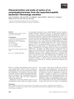
Homogeneity of the protein was checked by SDS ⁄
PAGE and confirmed a molecular subunit mass of
44.5 kDa (mature enzyme) (Fig. 1A). Activity stain-
ing of the SDS ⁄ PAGE gels using a-naphtyl acetate
confirmed the identity of the EstD band (Fig. 1B).
M. Levisson et al. New thermostable esterase from Thermotoga maritima
FEBS Journal 274 (2007) 2832–2842 ª 2007 The Authors Journal compilation ª 2007 FEBS 2833
Native-PAGE showed a single band that was con-
firmed to possess esterase activity by means of an
activity stain. Size exclusion chromatography showed
that the enzyme existed mainly as a monomer and, to
some extent as dimer, with estimated masses of
48 kDa and 93 kDa, respectively.
Substrate specificity and kinetics
The substrate specificity of purified EstD was analyzed
using p-nitrophenyl esters. The highest specific activity
with EstD was found towards short chain p-nitro-
phenyl esters of butyrate (C4) and valerate (C5). Little
activity was found towards long chain p-nitrophenyl
esters of decanoate (C10) to myristate (C14). In gen-
eral, activity of the enzyme on shorter (£ C10) and
longer fatty acids (‡ C10) is referred to as esterase
activity and lipase activity, respectively [1]. The kinetic
properties of EstD were determined for p-nitrophenyl
esters of acetate (C2), butyrate (C4), valerate (C5),
octanoate (C8) and decanoate (C10) (Table 1). The
catalytic efficiency represented by the value of k
cat
⁄ K
m
indicated that p-nitrophenyl valerate and p-nitrophenyl
octanoate were the best substrates for EstD among
the p-nitrophenyl esters tested. Hence, on the basis of
its substrate profile, EstD should be classified as an
esterase.
Neither proteolytic activity using casein as substrate,
nor peptidase activity when assayed with l-leucine
p-nitroanilide and l-proline p-nitroanilide was detected.
Effect of temperature and pH on enzyme activity
and thermal stability
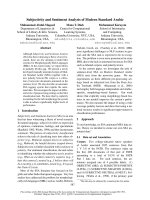
The effect of temperature on EstD activity was studied
using p -nitrophenyl valerate as a substrate. The est-
erase activity increased from 45 °C upwards until
95 °C (Fig. 2). An Arrhenius analysis resulted in a lin-
ear plot in the temperature range of 45–85 °C (Fig. 2,
inset), with a calculated activation energy for the
formation of the enzyme ⁄ substrate complex of 15
kJÆmol
)1
. EstD has a high resistance to thermal inacti-
vation, with a half-life value of approximately 1 h at
100 °C. To determine the optimal pH for the esterase,
the activity of EstD was measured in a pH range of 5–
9. EstD displayed > 70% of its maximal activity in
the pH range of 5–9, with an optimal pH at approxi-
mately 7.0 (not shown).
Effect of metals, detergents, solvents and
inhibitors
The effect of metal ions on EstD activity was tested
using various metal ions: Ca
2+
,Ni
2+
,Co
2+
,Cu
2+
,
kDa
200
116
91
66
45
33
M
AB
1234 1 234M
Fig. 1. SDS ⁄ PAGE of EstD fractions. Samples were separated by
SDS ⁄ PAGE in duplicate. One gel was stained with Coomassie Brilli-
ant Blue (A) and the other was stained for activity using a-naphtyl
acetate after renaturation (B). Lane M relative molecular mass
standards; lane 1, cell free extract; lane 2, heat-stable cell free
extract; lane 3, EstD after immobilized metal affinity chromatogra-
phy; lane 4, purified EstD. A second band at approximately 90 kDa
is corresponding to the EstD dimer. The dimer is believed to be
catalytically active as well.
Table 1. Kinetic parameters for hydrolysis of various p-nitrophenyl
esters. Kinetic assays were performed in 50 m
M citrate-phosphate
buffer pH 7 at 70 °C.
p-nitrophenyl
esters K
m
(mM) k
cat
(s
)1
)
k
cat
⁄ K
m
(s
)1
ÆmM
)1
)
Acetate (C2) 0.148 ± 0.025 1.0 ± 0.05 6.8 ± 1.2
Butyrate (C4) 0.227 ± 0.017 14.9 ± 0.40 65.6 ± 5.2
Valerate (C5) 0.066 ± 0.006 10.2 ± 0.20 154.5 ± 14.4
Octanoate (C8) 0.011 ± 0.003 1.6 ± 0.15 145.5 ± 12.1
Decanoate (C10) 0.072 ± 0.012 1.3 ± 0.06 18.1 ± 0.5
40
25
15
5
20
10
0
50 60 70
Temperature (°C)
Specific activity (U/mg)
LOG (U/mg)
80 90
1000 / T (K)
2.75
0.5
0.9
1.3
3.152.95
100
Fig. 2. Effect of temperature on esterase activity. The effect of
temperature on esterase activity was studied using pNP-valerate as
a substrate at temperatures ranging from 45 °Cto95°C. The inset
shows the temperature dependence as an Arrhenius plot.
New thermostable esterase from Thermotoga maritima M. Levisson et al.
2834 FEBS Journal 274 (2007) 2832–2842 ª 2007 The Authors Journal compilation ª 2007 FEBS
Fe
2+
,Zn
2+
,Mn
2+
and Mg
2+
at concentrations of
1mm. No significant stimulation or reduction of activ-
ity of EstD was observed. The effect of inhibitors on
EstD activity is shown in Table 2. Phenylmethylsulfo-
nyl fluoride, a serine protease inhibitor, strongly inhib-
ited enzyme activity. Diethyl pyrocarbonate, a histidine
modifier, also inhibited the reaction, albeit less pro-
nounced than phenylmethylsulfonyl fluoride. This indi-
cates that serine as well as histidine residues are
important for EstD activity. Activity was also strongly
inhibited by mercury chloride and to some extent by N-
ethylmaleimide. In contrast, dithiothreitol did not
affect enzyme activity and neither did EDTA, which
agrees with the metal tests.
The effect of detergents and solvents on EstD activ-
ity was tested in the standard assay with final concen-
trations of either 1% or 10% (v ⁄ v) (Table 3). In the
detergents test, activity was decreased by more than
50% when 1% Tween 20 was present and was com-
pletely inhibited by 1% SDS. Addition of the organic
solvents methanol, ethanol and isopropanol resulted in
a decrease in activity ranging from more than 70% to
less than 20% residual activity, respectively. On the
other hand, addition of glycerol in the assay did not
seem to have an effect on activity.
Structural modeling
In the absence of a 3D structure of EstD, it was deci-
ded to build a 3D-model of EstD. Since there are no
close structural homologs of EstD, modeling was
based on threading. A model of EstD was made using
the 3D-structural threading program phyre [19]. A
threading algorithm seeks a template protein in a data-
base that structurally fits well to a query sequence.
Unlike homology modeling, a certain sequence similar-
ity between the query sequence and a template protein
is not necessary. Several structural fits were found. The
thermophilic carboxylesterase Est30 of G. stearother-
mophilus (PDB code 1TQH) [20] was used to build the
model of EstD. Est30 consists of 247 amino acid resi-
dues and the crystal structure showed a large domain
with a modified a ⁄ b-hydrolase core including a seven-,
rather than an eight-stranded b-sheet, and a smaller
domain comprising three a-helices. Like EstD, Est30
has a preference for short acyl chain substrates, with
an optimum for C4–C8. The main difference between
Est30 and EstD is their amino acid sequence length.
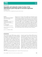
The final model for EstD covered the C-terminal
domain of EstD (amino acid residues 150–412). The
schematic structural model consists of six a-helixes and
has one central b-sheet made up of six b-strand-strands
(Fig. 3A). The first and second b-strand of the a⁄ b-
hydrolase fold have not been modeled.
The quality of the model towards stereochemistry
and geometry was analyzed by procheck analysis [21].
The Ramachandran plot (not shown) indicated that
most (92%) of the residues are in the core and allowed
regions. Bond lengths, bond angles and torsion angles
were evaluated with the what if program [22] and
were considered good (a RMS z-score for a normally
restrained data set is expected to be around 1.0). Bond
lengths were found to deviate slightly less than normal
from the mean standard bond length (a RMS z-score
of 0.7). Bond angles and torsion angles were found to
deviate normally (RMS z-scores around 1.0).
A first secondary structural alignment indicated the
residues Ser243, Asp347 and His378 as the probable
catalytic triad. In the obtained model, Ser243, Asp347
and His378 were indeed located in close proximity,
most likely representing the actual active site. Ser243 is
located within a nucleophile elbow connecting strand
b5 and helix a3, while Asp347 and His378 are located
on loops between b7–a7 and b8–a8, respectively
(Fig. 3A).
In the crystal structure of Est30, a covalently bound
ligand is present. This ligand, propylacetate, was
modeled into the active site of the EstD model. The
ligand is covalently bound to the side-chain of Ser243,
Table 2. Effect of inhibitors on EstD activity.
Inhibitors Relative activity (%)
None 100
Phenylmethylsulfonyl fluoride 4
Diethyl pyrocarbonate 53
N-ethylmaleimide 83
HgCl
2
3
EDTA 97
Dithiothreitol 99
b-Mercaptoethanol 97
Table 3. Effect of detergents and solvents on EstD activity.
Detergents and solvents
Concentration
(v ⁄ v%)
Relative activity
(%)
Control – 100
Methanol 10 73
Ethanol 10 37
2-Propanol 10 18
Glycerol 1 87
10 96
Dimethylsulfoxide 10 84
Tween 20 1 43
SDS 1 0
M. Levisson et al. New thermostable esterase from Thermotoga maritima
FEBS Journal 274 (2007) 2832–2842 ª 2007 The Authors Journal compilation ª 2007 FEBS 2835
His378 acts as proton carrier and Asp347 is the charge
relay network. The ligand is stabilized by hydrogen
bond interactions with the amides of Leu244 and
Gly164, which likely form the oxyanion hole (Fig. 3B).
The putative substrate binding pocket extends in a
cleft on both sides of Ser243. The alcohol side of the
substrate is in a groove pointed towards the entrance
of the pocket and extends approximately 10 A
˚
from
Ser243. The acyl-side of the ligand fits in a less
exposed pocket of approximately 6 A
˚
wide and 9 A
˚
long, consistent with the observed activity on sub-
strates with acyl chain length C2–C12. The hydro-
phobic side-chains in this pocket are Met247, Ala265,
Pro267, Ala268, Pro270, Leu271, Leu279, Phe320 and
Val350. One polar residue Gln349 is located at the
edges of the pocket and might have a role in substrate
recognition. Gln349 and adjacent residues are well
conserved in the closest homologues (supplementary
Table S1), suggesting an important structural role. The
EstD substrate binding pocket is very similar to that
of Est30 and structurally related esterases. This com-
prises an open accessible binding cleft and a relatively
large cap domain, consisting of one small and two
large helices on the N-terminal side of the central
b-sheet. This structural similarity between EstD and
Est30 corresponds with their very similar substrate
preference.
To confirm the predictions of the catalytic triad,
these residues were substituted by site-directed muta-
genesis. The mutants Ser243Ala, Asp347Asn and
His378Asn were expressed and purified using heat
treatment. The enzymes remained stable during heat
treatment. However, no activity was observed with the
mutants, confirming the importance of these three resi-
dues for the activity of EstD.
Discussion
In this contribution the cloning, expression, and char-
acterization of a new type of esterase from the hyper-
thermophilic bacterium T. maritima is described. The
encoding gene (estD) was originally annotated as a
hypothetical protein, but a more detailed sequence
analysis revealed the presence of an a ⁄ b-hydrolase fold
and a nucleophilic serine in a pentapeptide motif, sug-
gesting a possible role in ester hydrolysis. After func-
tional expression in E. coli, the esterase activity could
indeed be confirmed. When EstD was assayed with
p-nitrophenyl esters, it showed a preference for sub-
strates with shorter chain lengths, indicating that it
should be classified as an esterase and not as a lipase.
Highest activity was seen on esters of butyrate and val-
erate, which is comparable to esterases from other
hyperthermophiles, viz. T. tengcongensis [10], Sulfolo-
bus solfatoricus [32], Sulfolobus shibatae [24] and Sul-
folobus tokodaii [25]. The determined k
cat
values of
EstD, however, were found to be 100–1000-fold lower
compared to the hyperthermophilic esterases. The K
m
,
Leu244
Gly164
Gly166
PA
His378
His378
Asp347
Asp347
Ser243
Ser243
Ser165
Oxyanion
hole
A
B
Fig. 3. 3D model of EstD. (A) The overall structure of the C-ter-
minal domain of EstD. The central b-sheet and surrounding
a-helixes are shown in black and grey, respectively. Residues of
the catalytic triad are indicated. (B) The active site region of the
EstD model with bound ligand. Interatomic interactions are shown
in dashed lines. The ligand, propylacetate (PA), is covalently bound
to Ser243. The NH groups of Leu244 and Gly164 most likely form
the oxyanion hole.
New thermostable esterase from Thermotoga maritima M. Levisson et al.
2836 FEBS Journal 274 (2007) 2832–2842 ª 2007 The Authors Journal compilation ª 2007 FEBS
on the other hand, was relatively low. The low k
cat
may indicate that the artificial p-nitrophenyl sub-
strates differ substantially from the enzyme’s natural
substrate. However, the physiological function of EstD
is not known, as is the case for most described
esterases.
As to be expected for a hyperthermophilic enzyme,
EstD showed a temperature optimum around 95 °C,
which is comparable to that of the P. furiosus esterase
[7] and the Pyrobaculum calidifontis esterase [8]. The
Arrhenius plot for EstD was linear at temperatures in
the range 45–85 °C, indicating that the conformation
of EstD does not change throughout this temperature
range. The enzyme was very stable at high tempera-
tures, with a half-life of approximately 1 h at 100 °C.
EstD is less stable than the esterase from P. furiosus
(half-life value of 34 h at 100 °C) [7], but substantially
more stable than the esterase from T. tengcongensis
(half-life value of 15 min at 80 °C) [10] or the esterase
from T. maritima (half-life value of 30 min at 80 °C)
[11], which makes EstD the most stable bacterial est-
erase to date. EstD exhibited activity in the presence
of 10% organic solvents, which is comparable to the
activity of the Pyrobaculum calidifontis carboxyl-
esterase [8]. The high thermal stability and activity in
the presence of organic solvents makes EstD an
attractive catalyst for future applications in industry.
To gain more knowledge on the presence of essential
catalytic or structural amino acids, EstD activity was
tested upon incubation with various chemicals. The
inhibition by phenylmethylsulfonyl fluoride and diethyl
pyrocarbonate indicated that serine and histidine resi-
dues might be involved at the catalytic site of the
enzyme, in agreement with the anticipated catalytic
triad. Different metals and EDTA did not inhibit
activity indicating that there is no requirement for
divalent cations. The inhibition by HgCl
2
and N-ethyl-
maleimide suggests that the only free thiol group
which is present (Cys42), is important for the correct
functioning of the enzyme. The presence of a single
thiol makes oxidation to a disulfide not possible, which
is confirmed by the observation that neither dithio-
threitol nor b-mercaptoethanol enhanced the activity
of the enzyme. The single cysteine is not included in
the EstD model; however, it may be close to active site
residues and, as such, can influence activity when
modified with chemicals. Altogether, the inhibition
pattern is similar to that described for the esterases
from Pyrobaculum calidifontis [8], S. solfataricus [23]
and T. maritima [11].
Based on the alignment and the site-directed muta-
genesis experiments, EstD was shown to contain the
typical catalytic triad, consisting of a serine in a
GXSXG pentapeptide, an acidic aspartate, and a histi-
dine residue. The structural modeling was expected to
be difficult due to the lack of 3D structures of homol-
ogous esterases. Despite the very low sequence identity
(16% identity over the C-terminal part); EstD could
be modeled using Est30 from G. stearothermophilis as
a template. However, modeling was only possible with
the C-terminal domain of EstD, which also contains
all the active site residues. The N-terminal domain of
EstD has similarity to the MecA N-terminus but could
not be modeled. The function of the N-terminus
remains unclear. It might be involved in selection of
the substrates, either by binding of the substrate or by
narrowing the entrance to the active site.
The low sequence homology of EstD to character-
ized proteins was the reason that it was initially anno-
tated as a hypothetical protein. Nevertheless, the
results described here show that EstD has esterase
activity and also exhibits the typical structural features
of this type of enzyme. Bacterial esterases and lipases
have been classified into eight families based on a com-
parison of their amino acid sequences and some funda-
mental biological properties [26]. Enzymes in Family 1
are called true lipases and are further classified into six
subfamilies. Enzymes belonging to Family 2–8 are est-
erases. However, a homology search with the EstD
sequence against public databases revealed the highest
similarity to hypothetical proteins and putative hydro-
lases that are not grouped in any of the eight families.
Moreover, EstD showed no sequence identity to any
of the members of the previously classified families of
microbial lipases and esterases. A phylogenetic analysis
showed that EstD is indeed grouped into a new separ-
ate family (data not shown), which also includes
enzymes from several Bacillus species, B. fragilis and
S. usitatus. This divergence from the current families
can be viewed best by aligning the pentapeptide con-
sensus sequences (Fig. 4). EstD and related sequences
show a high pentapeptide homology (GHSLG), which
is different from the consensus of the esterase families.
These data suggest that EstD is a member of a new
family of esterases, designated as Family 10. EstD is
the third esterase that cannot be grouped into one of
the eight families. Because of absence of significant
amino acid homology, Handrick et al. [28], suggested
that PhaZ7 of Paucimonas lemoignei should be classi-
fied into a new family of esterases (Family 9: extracel-
lular PHA depolymerases) and also Liu et al. [20],
suggested that Est30 of G. stearothermophilus repre-
sents a new family of carboxylesterases (Fig. 4). EstD
is the first characterized member of the proposed
new family and, as such, also the first characterized
enzyme of COG1073, which will contribute to a better
M. Levisson et al. New thermostable esterase from Thermotoga maritima
FEBS Journal 274 (2007) 2832–2842 ª 2007 The Authors Journal compilation ª 2007 FEBS 2837
understanding of the function of the other enzymes in
this COG.
Experimental procedures
Chemicals
All chemicals were purchased from Sigma-Aldrich
(St Louis, MO, USA) or Acros Organics (Geel, Belgium).
The restriction enzymes were obtained from Invitrogen
(Carlsbad, CA, USA). Pfu Turbo and T4 DNA ligase were
purchased from Invitrogen and Stratagene (La Jolla, CA,
USA), respectively.
Strains and plasmids
The vector pGEM-t-easy (Promega, Madison, WI, USA)
was used for the cloning of PCR products. For hetero-
logous expression, the vector pET-26b (Kanamycin-resist-
ant; Novagen, San Diego, CA, USA) and the tRNA helper
plasmid pSJS1244 (Spectinomycin-resistant) [29,30] were
used. Escherichia coli strain XL1-Blue (Stratagene) was
used as a host for cloning. Escherichia coli strain
BL21(DE3) (Novagen) was used as an expression host.
Both strains were grown under standard conditions [31] fol-
lowing the instructions of the manufacturers.
Data mining
The genome of T. maritima MSB8 [16] was screened for
possible esterases and lipases. Sequences coding for esterases
and lipases were identified by performing BLAST searches
with sequences from characterized esterases ⁄ lipases (http://
www.ncbi.nlm.nih.gov/blast/) [32] and Motif (http://www.
expasy.org/prosite/) searches. The conserved domains were
analyzed with cd-search ( />Structure/cdd/wrpsb.cgi) [33] and kegg ssdb Motif Search
( [34]. The N-terminal
sequence analysis of the translational product of TM0336
was performed using the SignalP 3.0 Server (http://
www.cbs.dtu.dk/services/SignalP/) [35]. Phylogenetic analysis
was performed by aligning EstD, close homologues and
sequences of the esterase and lipase families using the Tcoffee
server ( />cgi) [36]. The alignment was further corrected by hand. A
bootstrapped phylogenetic tree was constructed and dis-
played using the neighbor-joining method with treeview,
version 1.6.5 [37]. A three dimensional structure of EstD was
modeled using the phyre protein fold recognition Server
( [19]. The model was
evaluated for stereochemical quality using the programs
procheck ( />procheck.html) [21] and what if ( />WIWWWI/) [22]. pymol was used to analyze and visualize
the structure [38].
Cloning and expression
The gene TM0336 (GenBank accession number NP_228147)
was PCR-amplified, without the sequence encoding its sig-
nal peptide (the first 18 amino acids) and its stop codon
using chromosomal DNA of T. maritima as a template and
the following two primers: 5¢-GCGGCGC
CATATGGAT
CAGGAAGCGTTTCTC-3¢ (sense, underlined NdeI restric-
tion site) and 5¢-GCGCG
CTCGAGTTTTACCATCCACC
TGGC-3¢ (antisense, underlined XhoI restriction site). The
PCR product generated was modified using the A-tailing
procedure [39] and ligated into the pGEM-t-easy vector.
E. coli XL1-blue was transformed with this construct
(pWUR349). The recombinant plasmid was digested by
NdeI and XhoI and the product was purified and inserted
into pET-26b digested with the same restriction enzymes.
The construct was designed with a hexahistidine-tag engin-
eered at the C-terminus of the enzyme to facilitate purifica-
tion. Subsequently, E. coli BL21(DE3), harboring the
tRNA helper plasmid pSJS1244, was transformed with the
resulting plasmid (pWUR353). The sequence of the expres-
sion clone was confirmed by sequence analysis of both
DNA strands.
Mutagenesis
Mutants of EstD were created to confirm the identity of the
active site residues. Mutants Ser243Ala, Asp347Asn and
His378Asn were generated using Quickchange (Stratagene)
site-directed mutagenesis with the following primers 5¢-GT
GCTGGGACAC
GCCCTCGGTGCGATGC-3¢ and 5¢-GC
ATCGCACCGAG
GGCGTGTCCCAGCAC-3¢,5¢-GATCT
TCGGCGGCAGA
AACTACCAGGTGACTG-3¢ and 5¢-CA
Fig. 4. Alignment of the esterase lipase ⁄ pentapeptide motif of
EstD with related enzymes and consensus sequences. Consensus
sequences of the different lipase and esterase families [35, 36] and
the two enzymes discussed in the text, PhaZ7 [28] and Est30 [20]
are indicated.
New thermostable esterase from Thermotoga maritima M. Levisson et al.
2838 FEBS Journal 274 (2007) 2832–2842 ª 2007 The Authors Journal compilation ª 2007 FEBS
GTCACCTGGTAGTTACTGCCGCCGAAG-3¢,5¢-CGAC
GATCTCAAT
AACTTGATGATTTCAGG-3¢ and 5¢-CTC
CTGAAATCATCAA
GTTATTGAGATCGTCG-3¢, respec-
tively (the underlining indicates the modified codon). Muta-
tions were confirmed by sequence analysis of both DNA
strands.
Production and purification
Escherichia coli BL21(DE3) ⁄ pSJS1244 was transformed
with pWUR353. A single colony was used to inoculate
4 mL of Luria–Bertani medium containing kanamycin and
spectinomycin (both 50 lgÆmL
)1
) and incubated overnight
at 37 °C while shaking. Next, the preculture was used to
inoculate (1 : 1000) two times 1500 mL of Luria–Bertani
medium containing kanamycin and spectinomycin (both
50 lgÆmL
)1
) in 2 L conical flasks and incubated in a rotary
shaker at 37 °C for 8 h. The culture was then induced by
adding isopropyl thio- b-d-galactoside to a final concentra-
tion of 0.1 mm. The culture was further incubated at 37 °C
for another 16 h. Cells were harvested by centrifugation at
10 000 g (Sorvall RC-6 centrifuge with SLA3000 rotor) and
4 °C for 15 min. The cell pellet was resuspended in 25 mL
lysis buffer (50 mm Tris ⁄ HCl buffer (pH 7.8), 300 mm
NaCl, 10 mm imidazole), and passed twice through a
French press at 110 MPa. The crude cell extract was DNase
treated for 30 min at room temperature to become less vis-
cous. The extract was centrifuged at 43 000 g (Sorvall RC-6
centrifuge with SS34 rotor) and 4 °C for 25 min 20 mL
lysis buffer was added to the resulting supernatant (cell free
extract) and heated for 25 min at 70 °C and subsequently
centrifuged at 43 000 g (Sorvall RC-6 centrifuge with SS34
rotor) and 4 °C for 25 min. The supernatant (heat-stable
cell free extract) was filtered (0.45 lm) and applied at a
flow rate of 2 mLÆmin
)1
to a Ni-chelating column (20 mL)
equilibrated in 50 mm Tris ⁄ HCl buffer (pH 7.8) containing
300 mm NaCl. The column was washed with 20 mm imi-
dazole in the same buffer and subsequently proteins were
eluted with a linear gradient of 20–500 mm imidazole and
fractions (2 mL) were collected. The most active fractions
were pooled and applied at a flow rate of 10 mLÆmin
)1
to a
HiPrep desalting column (53 mL) (Amersham Biosciences,
Piscataway, NJ, USA), equilibrated in 50 mm Tris ⁄ HCl
buffer (pH 7.8) containing 150 mm NaCl in order to
remove imidazole. Fractions of 5 mL were collected.
Size exclusion chromatography
The molecular mass of the purified enzyme was determined
by size exclusion chromatography on a Superdex 200 high-
resolution 10 ⁄ 30 column (24 mL) (Amersham Biosciences)
equilibrated in 50 mm Tris ⁄ HCl (pH 7.8) containing 100 mm
NaCl. Two hundred microliters of enzyme solution in 50 mm
Tris ⁄ HCl and 150 mm NaCl (pH 7.8) buffer was loaded
at a flow rate of 0.7 mLÆmin
)1
onto the column and frac-
tions (0.5 mL) were collected. Proteins used for calibration
were blue dextran 2000 (> 2000 kDa), ferritin (440 kDa),
catalase (232 kDa), aldolase (158 kDa), bovine serum albu-
min (67 kDa), ovalbumin (43 kDa), chymotrypsinogen A
(25 kDa), and ribonuclease A (13.7 kDa).
SDS ⁄ PAGE, native PAGE and activity staining
SDS ⁄ PAGE was performed with gels containing 10% acryl-
amide using a MiniProtean III system (Bio-Rad, Hercules,
CA, USA). Samples containing loading buffer (0.1 m
sodium phosphate buffer, 4% SDS, 10% 2-mercaptoetha-
nol, 20% glycerol, pH 6.8), were prepared by heating for
10 min at 100 °C. Gels were stained with Coomassie
Brilliant Blue. The molecular mass was estimated using the
Bio-Rad broad range protein marker. Native PAGE was
performed with gels containing 6% acrylamide. Native
PAGE and SDS ⁄ PAGE gels were stained for esterase activ-
ity by a modified version of the staining technique of Sobek
[40]. A renaturation procedure was carried out after
SDS ⁄ PAGE by incubating the gel two times for 15 min in
50 mm Tris ⁄ HCl (pH 7.8) ⁄ isopropanol (4 : 1, v ⁄ v%), sub-
sequently rinsed three times for 15 min in 50 mm Tris ⁄ HCl
(pH 7.8) and then rinsed again with water. The gel was
stained at 37 °C in the dark by incubating it in a 100 mL
solution of 50 mm Tris ⁄ HCl (pH 7.8) buffer containing
50 mg of Fast Blue BB plus and 1 mL of acetone solution
containing 10 mg of a-naphtyl acetate. When esterase active
bands began to color deep brown, the reactions were
stopped by rinsing the gel with tap water, followed by fix-
ation in 3% (v ⁄ v) acetic acid.
Enzyme assays
Esterase activity was determined by measuring the amount
of p-nitrophenol released during enzymatic hydrolysis of
different p-nitrophenyl esters. The release of p-nitrophenol
was continuously monitored at 405 nm using a Hitachi
UV2001 spectrophotometer (Hitachi Ltd, Tokyo, Japan)
with a temperature controlled cuvette holder. Unless other-
wise indicated, in a standard assay, esterase activity was
measured with 0.2 mm p-nitrophenyl valerate (pNP-C5)
as a substrate in 50 mm citrate-phosphate buffer (pH 7)
containing 1% isopropanol at 70 °C. Stock solutions of
p-nitrophenyl esters were prepared by dissolving substrates
in isopropanol. After preincubation, the reaction was star-
ted by adding enzyme to the reaction mix. One unit of
esterase activity was defined as the amount of protein
releasing 1 lmolÆ min
)1
of p-nitrophenol from pNP-C5.
Measurements were corrected for background hydrolysis in
the absence of enzyme. Measurements were carried out at
least three times and the molar extinction coefficient of
p-nitrophenol was determined for every condition prior to
each measurement. Activity was determined from the initial
rate of the hydrolysis reaction. The protein concentration
M. Levisson et al. New thermostable esterase from Thermotoga maritima
FEBS Journal 274 (2007) 2832–2842 ª 2007 The Authors Journal compilation ª 2007 FEBS 2839
was measured at 280 nm using a NanoDrop ND-1000
Spectrophotometer (NanoDrop, Wilmington, DE, USA).
Peptidase activity was assayed with 0.2 mml-leucine
p-nitroanilide and l-proline p-nitroanilide as substrates in a
standard assay as described above.
The proteolytic activity of EstD was assayed using 1%
(w ⁄ v) casein in 50 mm Tris ⁄ HCl (pH 8). Casein hydrolysis
assays were performed for up to 1 h at 70 °C. The reaction
was terminated with 10% (v ⁄ v) trichloroacetic acid and
incubated on ice for 30 min. The absorbance of the centri-
fuged supernatant was measured at 280 nm. A blank with-
out esterase was incubated under the same conditions.
Acyl chain length preference
Substrate specificity of the enzyme towards the acyl chain
length of different p-nitrophenyl esters was investigated by
using p-nitrophenyl acetate, p-nitrophenyl butyrate, p-nitro-
phenyl valerate, p-nitrophenyl octanoate, p-nitrophenyl
decanoate, p-nitrophenyl dodecanoate, and p-nitrophenyl
myristate in the standard assay.
pH and temperature optimum
The effect of pH on esterase activity was studied by meas-
uring activities on p-nitrophenyl valerate for a pH range of
4.0–9.5. The buffers used were 50 mm citrate-phosphate
(pH 4.0–8.0) and 50 mm Caps buffer (pH 9.5). The effect
of temperature on esterase activity was studied in the range
45–95 °C using 1 mm p-nitrophenyl valerate in the standard
assay. The pH of the buffers was set at 25 °C, and tempera-
ture corrections were made using their temperature coeffi-
cients ()0.0028 pHÆ°C
)1
for citrate-phosphate buffer and
)0.018 pHÆ°C
)1
for CAPS buffer) [41].
Thermostability
Enzyme thermostability was determined by incubating the
enzyme in a 50 mm Tris ⁄ HCl, 150 mm NaCl (pH 7.8) buf-
fer at 100 °C for various time intervals. Residual activity
was assayed under the standard condition.
Inhibition studies
The effect of metal ions on esterases activity was deter-
mined using different metal salts (CaCl
2
, NiCl
2
, CuCl
2
,
MnCl
2
, MgCl
2
, FeSO
4
and ZnSO
4
) at final concentrations
of 1 mm using the standard activity assay. The activity of
EstD without addition of metal ions was defined as 100%.
The effect of inhibitors on esterase activity was determined
using EDTA, dithiothreitol, b-mercaptoethanol and merc-
uric chloride. The effect of modifying agents for serine and
histidine was determined using phenylmethylsulfonyl fluor-
ide and diethyl pyrocarbonate, respectively. The enzyme
was preincubated in 50 mm citrate phosphate buffer (pH 7)
in the presence of the inhibitor (1 mm)at37°C for 60 min.
Subsequently, samples were cooled on ice and the residual
activities were measured using the standard method. Stabil-
ity against organic solvents and detergents was measured in
the presence of 1% solvents and detergents within the
standard activity assay, viz. glycerol, SDS, Tween 20 and
10% solvents and detergents, viz. methanol, ethanol, 2-pro-
panol, glycerol and dimethylsulfoxide.
Kinetic measurements
The EstD kinetic parameters K
m
and V
max
were calculated
from multiple measurements (substrate concentrations used
were 0.001, 0.005, 0.01, 0.02, 0.05, 0.1, 0.15, 0.2, 0.4, 0.6,
0.8 and 1.0 mm) by a computer-aided direct fit to the
Michaelis–Menten curve (tablecurve 2d, version 5.0;
Systat Software Inc., San Jose, CA, USA).
Acknowledgements
This work was supported by a grant from the graduate
school Voeding, Levensmiddelentechnologie, Agrobio-
technologie en Gezondheid (VLAG).
References
1 Jaeger KE, Dijkstra BW & Reetz MT (1999) Bacterial
biocatalysts: molecular biology, three-dimensional struc-
tures, and biotechnological applications of lipases. Annu
Rev Microbiol 53, 315–351.
2 Krishna SWH & Karanth NG (2002) Lipases and
lipase-catalyzed esterification reactions in non-aqueous
media. Catal Rev 44, 499–591.
3 Bornscheuer UT (2002) Microbial carboxyl esterases:
classification, properties and application in biocatalysis.
FEMS Microbiol Rev 26, 73–81.
4 Jaeger KE & Eggert T (2002) Lipases for biotechnology.
Curr Opin Biotechnol 13, 390–397.
5 Vieille C & Zeikus GJ (2001) Hyperthermophilic
enzymes: sources, uses, and molecular mechanisms for
thermostability. Microbiol Mol Biol Rev 65, 1–43.
6 Manco G, Giosue E, D’Auria S, Herman P, Carrea G &
Rossi M (2000) Cloning, overexpression, and properties
of a new thermophilic and thermostable esterase with
sequence similarity to hormone-sensitive lipase subfamily
from the archaeon Archaeoglobus fulgidus. Arch Biochem
Biophys 373, 182–192.
7 Ikeda M & Clark DS (1998) Molecular cloning of extre-
mely thermostable esterase gene from hyperthermophilic
archaeon Pyrococcus furiosus in Escherichia coli. Bio-
technol Bioeng 57, 624–629.
8 Hotta Y, Ezaki S, Atomi H & Imanaka T (2002) Extre-
mely stable and versatile carboxylesterase from a
New thermostable esterase from Thermotoga maritima M. Levisson et al.
2840 FEBS Journal 274 (2007) 2832–2842 ª 2007 The Authors Journal compilation ª 2007 FEBS
hyperthermophilic archaeon. Appl Environ Microbiol 68,
3925–3931.
9 Shao W & Wiegel J (1995) Purification and characteri-
zation of two thermostable acetyl xylan esterases from
Thermoanaerobacterium sp. strain JW ⁄ SL-YS485. Appl
Environ Microbiol 61, 729–733.
10 Zhang J, Liu J, Zhou J, Ren Y, Dai X & Xiang H
(2003) Thermostable esterase from Thermoanaerobacter
tengcongensis: high-level expression, purification and
characterization. Biotechnol Lett 25, 1463–1467.
11 Kakugawa S, Fushinobu S, Wakagi T & Shoun H
(2007) Characterization of a thermostable carboxylester-
ase from the hyperthermophilic bacterium Thermotoga
maritima. Appl Microbiol Biotechnol 74, 585–591.
12 Kwoun Kim H, Jung YJ, Choi WC, Ryu HS, Oh TK &
Lee JK (2004) Sequence-based approach to finding func-
tional lipases from microbial genome databases. FEMS
Microbiol Lett 235, 349–355.
13 Atomi H & Imanaka T (2004) Thermostable carboxyles-
terases from hyperthermophiles. Tetrahedron: Asymme-
try 15, 2729–2735.
14 Tatusov RL, Koonin EV & Lipman DJ (1997) A genomic
perspective on protein families. Science 278, 631–637.
15 Tatusov RL, Fedorova ND, Jackson JD, Jacobs AR,
Kiryutin B, Koonin EV, Krylov DM, Mazumder R,
Mekhedov SL, Nikolskaya AN,et al. (2003) The COG
database: an updated version includes eukaryotes. BMC
Bioinformatics 4, 41.
16 Nelson KE, Clayton RA, Gill SR, Gwinn ML, Dod-
son RJ, Haft DH, Hickey EK, Peterson JD, Nelson
WC, Ketchum KA, et al. (1999) Evidence for lateral
gene transfer between archaea and bacteria from gen-
ome sequence of Thermotoga maritima. Nature 399,
323–329.
17 Lim D & Strynadka A (2002) Structural basis for the
b-lactam resistance of PBP2a from methicillin-resistant
Staphylococcus aureus. Nat Struct Biol 9, 870–876.
18 Holmquist M (2000) Alpha ⁄ beta-hydrolase fold
enzymes: structures, functions and mechanisms. Curr
Protein Pept Sci 1, 209–235.
19 Bates PA, Kelley LA, MacCallum RM & Sternberg MJ
(2001) Enhancement of protein modeling by human
intervention in applying the automatic programs 3D-
JIGSAW and 3D-PSSM. Proteins 45, 39–46.
20 Liu P, Wang YF, Ewis HE, Abdelal AT, Lu CD,
Harrison RW & Weber IT (2004) Covalent reaction
intermediate revealed in crystal structure of the Geoba-
cillus stearothermophilus carboxylesterase Est30. J Mol
Biol 342, 551–561.
21 Laskowski RA, MacArthur MW, Moss DS & Thornton
JM (1993)
procheck: a program to check the stereo-
chemical quality of protein structures. J Appl Cryst 26,
283–291.
22 Vriend G (1990) what if: a molecular modeling and
drug design program. J Mol Graph 8, 52–56.
23 Kim S & Lee SB (2004) Thermostable esterase from
a thermoacidophilic archaeon: purification and
characterization for enzymatic resolution of a
chiral compound. Biosci Biotechnol Biochem 68,
2289–2298.
24 Ejima K, Liu J, Oshima Y, Hirooka K, Shimanuki S,
Yokota Y, Hemmi H, Nakayama T & Nishino T (2004)
Molecular cloning and characterization of a thermo-
stable carboxylesterase from an archaeon, Sulfolobus
shibatae DSM5389: non-linear kinetic behaviour of a
hormone-sensitive lipase family enzyme. J Biosci Bioeng
98, 445–451.
25 Suzuki Y, Miyamoto K & Ohta H (2004) A novel ther-
mostable esterase from the thermoacidophilic archaeon
Sulfolobus tokodaii strain 7. FEMS Microbiol Lett 236,
97–102.
26 Arpigny JL & Jaeger KE (1999) Bacterial lipolytic
enzymes: classification and properties. Biochem J 343,
177–183.
27 Cruz H, Pe
´
rez C, Wellington E, Castro C & Servı
´
n-
Gonza
´
lez L (1994) Sequence of the Streptomyces albus
G lipase-encoding gene reveals the presence of a prokar-
yotic lipase family. Gene 144, 141–142.
28 Handrick R, Reinhardt S, Focarete ML, Scandola M,
Adamus G, Kowalczuki M & Jendrossek D (2001) A
new type of thermoalkalophilic hydrolase of Paucimonas
lemoignei with high specificity for amorphous polyesters
of short chain-length hydroxyalkanoic acids. J Biol
Chem 276, 36215–36224.
29 Sorensen HP, Sperling-Petersen HU & Mortensen KK
(2003) Production of recombinant thermostable proteins
expressed in Escherichia coli: completion of protein
synthesis is the bottleneck. J Chromatogr B Anal Tech-
nol Biomed Life Sci 786, 207–214.
30 Kim R, Sandler SJ, Goldman S, Yokota H, Clark AJ
& Kim SH (1998) Overexpression of archaeal
proteins in Escherichia coli. Biotechnol Lett 20,
207–210.
31 Sambrook J, Fritsch EF & Maniatis T (1989) Molecu-
lar Cloning: A Laboratory Manual, 2nd edn. Cold
Spring Harbor Laboratory Press, Cold Spring Harbor,
NY.
32 Altschul SF, Madden TL, Scha
¨
ffer AA, Zhang J,
Zhang Z, Miller W & Lipman DJ (1997) Gapped
BLAST and PSI-BLAST: a new generation of protein
database search programs. Nucl Acids Res 25,
3389–3402.
33 Marchler-Bauer A, Anderson JB, Cherukuri PF,
DeWeese-Scott C, Geer LY, Gwadz M, He S, Hurwitz
DI, Jackson JD, Ke Z et al. (2005) CDD: a Conserved
Domain Database for protein classification. Nucl Acids
Res 33, 192–196.
34 Kanehisa M, Goto S, Kawashima S & Nakaya A
(2002) The KEGG databases at GenomeNet. Nucl Acids
Res 30, 42–46.
M. Levisson et al. New thermostable esterase from Thermotoga maritima
FEBS Journal 274 (2007) 2832–2842 ª 2007 The Authors Journal compilation ª 2007 FEBS 2841
35 Bendtsen JD, Nielsen H, von Heijne G & Brunak S
(2004) Improved prediction of signal peptides: SignalP
3.0. J Mol Biol 340, 783–795.
36 Notredame C, Higgins D & Heringa J (2000) T-Coffee:
a novel method for multiple sequence alignments. J Mol
Biol 302, 205–217.
37 Page RDM (1996) TREEVIEW: an application to view
phylogenetic trees on personal computers. Comput Appl
Biosci 12, 357–358.
38 DeLano WL (2002) The PyMOL molecular graphics sys-
tem. DeLano Scientific, Palo Alto, CA.
39 Kobs G (1997) Cloning blunt-end DNA fragments into
the pGEMÒ-T vector systems. Promega Notes 62, 15.
40 Sobek H & Gorisch H (1988) Purification and charac-
terization of a heat-stable esterase from the thermoaci-
dophilic archaebacterium Sulfolobus acidocaldarius.
Biochem J 250, 453–458.
41 Beynon R & Easterby J (2003) Buffer Solutions. Taylor
& Francis Group, London.
Supplementary material
The following supplementary material is available
online:
Fig. S1. Amino acid sequence alignment of EstD.
This material is available as part of the online article
from
Please note: Blackwell Publishing is not responsible
for the content or functionality of any supplementary
materials supplied by the authors. Any queries (other
than missing material) should be directed to the corres-
ponding author for the article.
New thermostable esterase from Thermotoga maritima M. Levisson et al.
2842 FEBS Journal 274 (2007) 2832–2842 ª 2007 The Authors Journal compilation ª 2007 FEBS