Báo cáo khoa học: Modulation of Ca2+ entry and plasma membrane potential by human TRPM4b pptx
Bạn đang xem bản rút gọn của tài liệu. Xem và tải ngay bản đầy đủ của tài liệu tại đây (747.67 KB, 10 trang )
Modulation of Ca
2+
entry and plasma membrane potential
by human TRPM4b
Ralf Fliegert
1
,Gu
¨
nter Glassmeier
2
, Frederike Schmid
1
, Kerstin Cornils
1
, Selda Genisyuerek
1
,
Angelika Harneit
1
,Ju
¨
rgen R. Schwarz
2
and Andreas H. Guse
1
1 Calcium Signalling Group, Institute of Biochemistry and Molecular Biology I: Cellular Signal Transduction, Center of Experimental Medicine,
University Medical Center Hamburg-Eppendorf, Hamburg, Germany
2 Institute of Applied Physiology, Center of Experimental Medicine, University Medical Center Hamburg-Eppendorf, Hamburg, Germany
Changes in the free cytoplasmic Ca
2+
concentration
modulate a plethora of cellular processes, ranging from
exocytosis to proliferation [1]. In nonexcitable cells, the
activation of several second messenger systems leads
to the release of Ca
2+
from intracellular stores, most
likely the endoplasmic reticulum, and depletion of these
stores has in turn been shown to activate capacitative
Ca
2+
entry via as yet poorly identified channels [2].
The amount of Ca
2+
entering the cell is not only deter-
mined by the number of open channels but also
depends on the electrochemical driving force for Ca
2+
.
Therefore, changes in the membrane potential are
important in the regulation of Ca
2+
entry in nonexcita-
ble cells. The essential role of voltage-gated and Ca
2+
-
activated K
+
channels in regulating Ca
2+
entry has
been firmly established, especially for lymphocytes [3].
TRPM4b is a member of the family of melastatin-
related transmembrane receptor potential channels. The
eight members of this family differ largely in their ion
selectivity and mode of activation [4]. TRPM4b (tran-
sient receptor potential channel, melastatin subfamily)
is a Ca
2+
-activated and voltage-dependent nonselective
cation channel that has been shown to be broadly
expressed in excitable as well as nonexcitable cells
[5,6]. The Ca
2+
sensitivity of TRPM4b is regulated
in a complex manner by intracellular nucleotides [7],
Keywords
Ca
2+
; calcium; membrane potential; TRPM4;
TRP channel
Correspondence
A. H. Guse, Institute of Biochemistry and
Molecular Biology I: Cellular Signal
Transduction, Center of Experimental
Medicine, University Medical Center
Hamburg-Eppendorf, Martinistr. 52,
D-20246 Hamburg, Germany
Fax: +49 40 42803-9880
Tel: +49 40 42803 2828
E-mail:
(Received 31 August 2006, revised 20
November 2006, accepted 22 November
2006)
doi:10.1111/j.1742-4658.2006.05614.x
TRPM4b is a Ca
2+
-activated, voltage-dependent monovalent cation chan-
nel that has been shown to act as a negative regulator of Ca
2+
entry and
to be involved in the generation of oscillations of Ca
2+
influx in Jurkat
T-lymphocytes. Transient overexpression of TRPM4b as an enhanced
green fluorescence fusion protein in human embryonic kidney (HEK) cells
resulted in its localization in the plasma membrane, as demonstrated by
confocal fluorescence microscopy. The functionality and plasma membrane
localization of overexpressed TRPM4b was confirmed by induction of
Ca
2+
-dependent inward and outward currents in whole cell patch clamp
recordings. HEK-293 cells stably overexpressing TRPM4b showed higher
ionomycin-activated Ca
2+
influx than wild-type cells. In addition, analysis
of the membrane potential using the potentiometric dye bis-(1,3-dibutylbar-
bituric acid)-trimethine oxonol and by current clamp experiments in the
perforated patch configuration revealed a faster initial depolarization after
activation of Ca
2+
entry with ionomycin. Furthermore, TRPM4b expres-
sion facilitated repolarization and thereby enhanced sustained Ca
2+
influx.
In conclusion, in cells with a small negative membrane potential, such as
HEK-293 cells, TRPM4b acts as a positive regulator of Ca
2+
entry.
Abbreviations
CHO, Chinese hamster ovary; DAPI, 4,6-diamidino-2-phenylindole; DiBAC
4
(3), bis-(1,3-dibutylbarbituric acid)-trimethine oxonol; ECL,
enhanced chemoluminescence; EGFP, enhanced green fluorescence protein; HEK, human embryonic kidney; TBS-T, Tris-buffered saline
with 0.1% Tween-20; TRP, transient receptor potential.
704 FEBS Journal 274 (2007) 704–713 ª 2006 The Authors Journal compilation ª 2006 FEBS
phosphorylation by protein kinase C, and binding of
Ca
2+
–calmodulin and phosphatidylinositol 4,5-bisphos-
phate [8,9]. Nilius et al. observed Ca
2+
-dependent in-
activation of TRPM4b in whole cell patch clamp
experiments [8]. This has been shown to be due to the
loss of phosphatidylinositol 4,5-bisphosphate and a resul-
tant reduction in Ca
2+
sensitivity of TRPM4b [9,10].
Launay et al. proposed that depolarization of cells after
activation of TRPM4b by Ca
2+
might be involved in
the negative regulation of Ca
2+
entry [5]. For Jurkat
T-lymphocytes, it has been shown that TRPM4b is
involved in generating oscillations of Ca
2+
influx neces-
sary for cytokine secretion and proliferation [11,12].
Here we show for the first time that expression of
TRPM4b in human embryonic kidney (HEK)-293 cells
facilitates Ca
2+
entry by counteracting the depolariza-
tion resulting from sustained Ca
2+
entry. As TRPM4b
is a nonselective cation channel with a slightly out-
ward-rectifying characteristic, the impact of TRPM4b
activation on Ca
2+
entry is dependent on the resting
membrane potential of the cell.
Results
Localization and electrophysiologic
characterization of TRPM4b–enhanced green
fluorescence protein (EGFP) fusion proteins
Confocal microscopy of Chinese hamster ovary (CHO)
(Fig. 1A), COS-7 (Fig. 1B) and HEK-293 (Fig. 1C)
cells transiently expressing a TRPM4b–EGFP fusion
protein (EGFP fused to the C-terminus of TRPM4b)
showed intense staining of intracellular membranes,
especially close to the nucleus, but also of small vesicu-
lar structures throughout the cell body and in the
plasma membrane. These data indicate de novo expres-
sion of the TRPM4–EGFP fusion protein and subse-
quent transport via the endoplasmic reticulum–Golgi
network to the plasma membrane.
The TRPM4b–EGFP fusion protein was fully active
as a cation channel in HEK-293 cells. In patch clamp
recordings in the whole cell mode, 10 lm Ca
2+
activa-
ted the current (Fig. 1B). The Ca
2+
dependence, the
rapid inactivation and the slight outward-rectifying
characteristic with a reversal potential close to 0 mV
suggest almost no influence of fused EGFP on channel
function (compare Fig. 1D with Fig. 2A,B).
Generation of HEK-293 clones with stable
expression of TRPM4b
To investigate the effect of TRPM4b expression on
Ca
2+
entry and changes of the transmembrane poten-
tial, HEK-293 clones stably expressing TRPM4b were
generated. Clones derived by selection for neomycin
resistance and limiting dilution cloning were subse-
quently analyzed for the expression of TRPM4b by
RT-PCR and western blotting. Six out of nine clones
expressed the transcript from the vector pTRPM4b
(supplementary Fig. S1). The membrane fractions of
these clones were probed for TRPM4b expression by
SDS ⁄ PAGE and western blot analysis, using an affin-
ity-purified antibody raised against a peptide present
in the published splice variants TRPM4b [5,6],
TRPM4a [13] and TRPM4c (GenBank accession no.
AY297046). Three of six clones expressing the tran-
script were also shown to express the protein in western
blot analysis (supplementary Fig. S1). TRPM4b-
expressing cells also responded to Ca
2+
infusion with
typical transient currents (Fig. 2B), indicating that
TRPM4b was functioning as expected. TRPM4b has
been shown to be permeable to Cs
+
[6], so we used a
pipette solution with CsCl to block K
+
currents.
Under these experimental conditions, the reversal
potential was near 0 mV. Immunostaining showed
localization of TRPM4b in the plasma membrane
(Fig. 2C). Expression of endogenous TRPM4b was not
detected at the protein level in western blot experi-
ments (supplementary Fig. S1). This biochemical result
was confirmed by the lack of Ca
2+
-activated
TRPM4b-like currents in patch clamp experiments in
wild-type cells (Fig. 2A).
Influence of TRPM4b on Ca
2+
entry induced by
ionomycin
The influence of TRPM4b expression on the Ca
2+
sig-
nal induced by ionomycin was investigated using the
ratiometric Ca
2+
indicator Fura2. Ionomycin at the
concentration used here mainly induces Ca
2+
release
from intracellular stores. This pool depletion in turn
activates the Ca
2+
-induced Ca
2+
entry current (I
crac
[14]). A Ca
2+
-free ⁄ Ca
2+
-readdition protocol was used
to distinguish between the release of Ca
2+
from intra-
cellular stores and Ca
2+
influx across the plasma mem-
brane.
Whereas Ca
2+
release did not significantly differ
between wild-type and TRPM4b-expressing HEK-293
cells, Ca
2+
entry after Ca
2+
readdition was signifi-
cantly higher in TRPM4b-expressing HEK-293 cells
(Fig. 3). Readdition of Ca
2+
in the absence of iono-
mycin only slightly increased [Ca
2+
]
i
(Fig. 3A). The
stimulatory effect of TRPM4b expression was true for
both the initial overshoot (Fig. 3B; Influx peak) and
the sustained Ca
2+
entry phase (Fig. 3B; Influx plat-
eau). These results were observed in three different
R. Fliegert et al. Modulation of Ca
2+
entry by TRPM4b
FEBS Journal 274 (2007) 704–713 ª 2006 The Authors Journal compilation ª 2006 FEBS 705
TRPM4b-expressing HEK-293 clones (clones 5, 4 and
9), ruling out the possibility that the cell-cloning proce-
dure itself caused this alteration in Ca
2+
entry follow-
ing store depletion.
Influence of TRPM4b on ionomycin-induced
membrane depolarization
Ca
2+
entry induced by depletion of intracellular Ca
2+
stores by ionomycin also induced marked depolariza-
tion of the plasma membrane (Fig. 4A,B). In current
clamp experiments in the perforated-patch configur-
ation, the resting potential of the HEK-293 cells was
determined to be ) 50 ± 1.6 mV (n ¼ 4). TRPM4b
expression had a slight stimulatory effect on the initial
kinetics of depolarization (Fig. 4A,B). In addition,
TRPM4b allowed for rapid and sustained repolari-
zation of the plasma membrane (Fig. 4A,C). The
membrane potential after repolarization in TRPM4b-
expressing clone 9 was ) 30 ± 6.5 mV (n ¼ 6), as
compared to ) 18 ± 3.3 mV (n ¼ 4) in wild-type cells
(Fig. 4B). The effect of TRPM4b expression on
plasma membrane potential was dependent on the
expression level (Fig. 4C, inset western blot), strongly
indicating that TRPM4b is indeed involved in this
process.
A
C
D
B
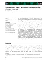
Fig. 1. Subcellular localization and character-
ization of transiently expressed TRPM4b-
EGFP. CHO, COS-7 and HEK-293 cells were
grown on coverslips and transfected with
pEGFPN1-TRPM4b as detailed in Experi-
mental procedures. (A–C) Confocal immuno-
fluorescence images were taken 24–72 h
post-transfection. Scale bars ¼ 10 lm. (D)
Activation of TRPM4b–EGFP fusion proteins
by Ca
2+
was analyzed in patch clamp experi-
ments in HEK-293 cells. Voltage ramps from
) 100 mV to + 100 mV were applied every
2 s (during cell-attached mode, the time
variance between consecutive data points
was due to automatic compensation).
Arrows indicate breakthrough from the cell-
attached into the whole cell configuration.
The panels on the left show current devel-
opment of inward currents at ) 100 mV and
outward currents at + 80 mV against time.
The panels on the right show I– V relation-
ships obtained from voltage ramps after full
development of the current. Data are pre-
sented as mean ± SEM (n ¼ 3–12).
Modulation of Ca
2+
entry by TRPM4b R. Fliegert et al.
706 FEBS Journal 274 (2007) 704–713 ª 2006 The Authors Journal compilation ª 2006 FEBS
Discussion
In this study, we confirmed that, (a) overexpressed
TRPM4b is localized in the plasma membrane of
HEK-293, CHO and COS-7 cells; and (b) infusion of
Ca
2+
into TRPM4b- or TRPM4b–EGFP-overexpress-
ing HEK-293 cells induces a typical transient current
with an outward-rectifying characteristic. We also
found that in a model cell with a more depolar-
ized ⁄ less negative resting membrane potential, the
HEK293 cell, TRPM4b, (a) displays a complex, dose-
dependent influence on changes in membrane potential
A
B
CD
Fig. 2. Ca
2+
activation and localization of
TRPM4b. Activation of TRPM4b by Ca
2+
was analyzed in patch clamp experiments.
Voltage ramps from ) 120 mV to + 100 mV
were applied every 2 s (during cell-attached
mode, the time variance between consecu-
tive data points was due to automatic com-
pensation). Arrows indicate breakthrough
from the cell-attached into the whole cell
configuration. The panels on the left show
current development of inward currents at
) 100 mV and outward currents at + 80 mV
against time. The panels on the right show
I–V relationships obtained from voltage
ramps after full development of the current.
(A) Wild-type HEK-293 cells. (B) HEK-293
clone 9 stably overexpressing TRPM4b.
Data are presented as mean ± SEM
(n ¼ 3–6). (C, D) For localization of TRPM4b,
cells from clone 9 (C) or wild-type HEK-293
cells (D) were stained using affinity-purified
anti-peptide serum against TRPM4 and a
rhodamine-conjugated secondary antibody
(red). Nuclei were visualized using DAPI
(blue). Fluorescence images were taken and
deconvolved as detailed in Experimental
procedures. DAPI and rhodamine images
were merged after deconvolution. Scale
bar ¼ 15 lm.
R. Fliegert et al. Modulation of Ca
2+
entry by TRPM4b
FEBS Journal 274 (2007) 704–713 ª 2006 The Authors Journal compilation ª 2006 FEBS 707
induced by ionomycin; and (b) facilitates Ca
2+
entry
induced via store depletion by ionomycin.
TRPM4b-expressing HEK-293 cells show signifi-
cantly higher Ca
2+
entry after ionomycin stimulation
than do wild-type cells. Ionomycin releases Ca
2+
from
intracellular Ca
2+
stores and thus induces electrogenic
Ca
2+
entry across the plasma membrane via the capa-
citative Ca
2+
entry mechanism [14]. This Ca
2+
entry,
either alone or in combination with additional, less
specific store-operated cation entry pathways, depolar-
izes the cell, as can be seen in Fig. 4A,C. Both the
onset of depolarization and the rise in [Ca
2+
]
i
activate
TRPM4b. Usually, in cells with a negative membrane
potential () 70 to ) 90 mV), TRPM4b activation
enhances depolarization by inflow of Na
+
, and thus
reduces the driving force for Ca
2+
entry, as has been
demonstrated for Jurkat T-cells [11]. In contrast, our
data indicate that in cells with a more depolarized ⁄ less
negative resting membrane potential, such as HEK-293
cells (about ) 40 to ) 50 mV; Fig. 4B), Na
+
influx
through TRPM4b first facilitates depolarization of the
cell, but as soon as the potential becomes more posi-
tive, more K
+
than Na
+
passes the outward-rectify-
ing, nonselective channel, and the net efflux of cations
then repolarizes the cell, as shown in Fig. 4A,B. This
partial repolarization results in a higher driving force
for Ca
2+
, and thus enhances Ca
2+
entry into cells
expressing TRPM4b as compared to wild-type cells.
This was a surprising result, as Launay et al. [5] des-
cribed TRPM4b as a negative regulator of Ca
2+
entry.
According to their model, activation of TRPM4b by
the starting depolarization and the rise in [Ca
2+
]
i
results in entry of Na
+
, further depolarization, and
thus diminished Ca
2+
entry. In cells expressing volt-
age-gated K
+
channels, such as lymphocytes, this
would ultimately result in oscillating Ca
2+
influx [11].
The results of Launay et al. were obtained either in
Jurkat T-lymphocytes, which normally have a very
negative membrane potential [11], or in HEK-293 cells
that were clamped to a negative membrane potential
[5]. It therefore seems that the impact of TRPM4b
expresssion on Ca
2+
signaling depends on the resting
membrane potential of the respective cell.
Does our finding have any general relevance to cell
physiology? The resting membrane potentials of euk-
aryotic cells vary widely. Most quiescent cells expres-
sing K
+
inward-rectifier channels have a membrane
potential near the equilibrium potential for K
+
,
whereas most cells lacking K
+
inward-rectifier channel
expression are characterized by membrane potentials
between ) 30 and ) 40 mV [15]. Dividing cells, especi-
ally cells derived from tumors, often show even
less negative ⁄ more depolarized membrane potentials
[16,17]. This holds especially true when cells are kept
in serum-containing medium instead of Ringer solution
[16]. Interestingly, the gene encoding TRPM4 has been
shown to be one of the genes upregulated during the
transition from prostatic intraepithelial neoplasia to
invasive prostate cancer [18]. It is conceivable that the
change in resting membrane potential that occurs
during the transition from a quiescent to a rapidly
dividing cell switches TRPM4b from a negative to a
positive regulator of Ca
2+
entry. The upregulation
of TRPM4b might then be involved in the further
progression of the tumor. Such a possible role of
A
B
Fig. 3. Ca
2+
release and Ca
2+
entry induced by ionomycin in
TRPM4b-expressing HEK-293 cells. The cells were loaded with
Fura2 ⁄ AM, and [Ca
2+
]
i
was determined using the Ca
2+
-free ⁄ Ca
2+
-
readdition protocol as detailed in Experimental procedures. (A)
Averaged tracings of cell suspensions of wild-type HEK-293 (gray
lines) or a HEK-293 clone stably overexpressing TRPM4b (black
lines) either activated by ionomycin (addition of 1 l
M final concen-
tration of ionomycin indicated by arrow; solid lines) or untreated
(dashed lines). (B) Quantitative analysis of the amplitude of Ca
2+
released by ionomycin (left) and the amplitude of Ca
2+
entry after
readdition of Ca
2+
(right). Values for the Ca
2+
influx plateau were
determined after 800 s. Data are presented as mean ± SEM
(n ¼ 5–8). Significant differences from the wild type are
marked with asterisks (Student’s t-test, P ¼ 0.05). #Student’s
t-test, P ¼ 0.07.
Modulation of Ca
2+
entry by TRPM4b R. Fliegert et al.
708 FEBS Journal 274 (2007) 704–713 ª 2006 The Authors Journal compilation ª 2006 FEBS
TRPM4b in tumor development will be an interesting
subject for future investigations.
Experimental procedures
Cell culture
Wild-type HEK-293 and COS-7 cells were cultured in
DMEM supplemented with Glutamax I, 10% (v ⁄ v) fetal
bovine serum (Biochrom, Berlin, Germany), 100 unitsÆmL
)1
penicillin, and 50 lgÆmL
)1
streptomycin (termed ‘complete’
DMEM). For the culture of stably transfected HEK-293
cells, 400 lgÆmL
)1
G418-Sulfat was added to the complete
DMEM. CHO cells were cultured in a-MEM with
Glutamax I, 10% (v ⁄ v) fetal bovine serum (Biochrom),
100 unitsÆmL
)1
penicillin, and 50 lgÆmL
)1
streptomycin. All
cells were kept at 37 °C in a humidified atmosphere con-
taining 5% CO
2
in air.
Preparation of plasmids and transfection
Total RNA was isolated from HEK-293 cells (0.5–2 · 10
7
)
using the RNeasy kit with on-column DNase digest
(Qiagen, Hilden, Germany) according to the manufacturer’s
instructions. Three overlapping cDNA segments containing
the total ORF of TRPM4c (accession no. AY297046) were
separately amplified by one-step RT-PCR using the Titan
one-Tube RT-PCR system (Roche, Mannheim, Germany),
A
C
B
Fig. 4. Ionomycin-induced depolarization of the plasma membrane in TRPM4b-expressing HEK-293 cells. Changes in membrane potential of
cells preincubated with DiBAC
4
(3) were determined as detailed in Experimental procedures. (A) Averaged tracings of cell suspensions of
HEK-293 (black line) or HEK-293 clones stably overexpressing TRPM4b (gray lines) activated by ionomycin (iono, 1 l
M final concentration).
Cells were treated with gramicidin (gram, 1 l
M final concentration) at the end of each experiment. Fluorescence tracings were normalized to
minimal depolarization (before addition of ionomycin) and maximal depolarization (after addition of gramicidin). Current clamp experiments in
the perforated patch configuration were performed as detailed in Experimental procedures. (B) Development of membrane potential after
addition of ionomycin (iono, 1 l
M final concentration) in individual representative cells from the wild type or the TRPM4b-expressing clone 9
(n ¼ 4–6). (C) Quantitative analysis of the depolarization of the plasma membrane 200 s after addition of ionomycin. The results of a western
blot experiment showing the different levels of expression of TRPM4b in the individual clones are shown for comparison. Data are presen-
ted as mean ± SD (n ¼ 5). Significant differences from the wild type are marked with an asterisk (Student’s t-test, P ¼ 0.05).
R. Fliegert et al. Modulation of Ca
2+
entry by TRPM4b
FEBS Journal 274 (2007) 704–713 ª 2006 The Authors Journal compilation ª 2006 FEBS 709
cloned, and recombined using internal BspEI (position 633)
and HindIII (position 1329) sites (primers used: segment 1,
5¢-GTCGACCTGGGCTGCAGGAGGTTG-3¢ and 5¢-GT
ACCTCGCAGGGAACGAG-3¢; segment 2, 5¢-AGCCT
GGATTGTCACTGG-3¢ and 5¢-GGGCTCCTCTTCTGA
TTTCC-3¢; and segment 3, 5¢-GACCCTGGAAGACAC
TCTGG-3¢ and 5¢-AGGAATCTGTGAGTGGTGAGG-3¢).
A segment of human chromosome 19 containing exon 17
(missing from TRPM4c) was amplified from genomic DNA
(primers used: 5¢-AGTGGGCAGGAAGGATGAG-3¢ and
5¢-TCAGGCAGGGTGAGATGTG-3¢), cloned into pGEM
T-easy (Promega, Munich, Germany), and sequenced.
Integration of exon 17 was achieved by fusion PCR [19]
(mega-primers: 5¢-TCATTAATGGGGAAGGGCCTGTCG
GGACGGCGGACCCAGCCGAGAAGA-3¢ and 5¢-AGG
TGGTACAAACCCGGGGTCAGCCGGCAGCCCACGCC
CAGGAGGAAGC-3¢). For the expression of TRPM4b
N-terminally fused to EGFP, the coding sequence was sub-
cloned into the SalI and BamHI sites of the multiple clo-
ning site of pEGFP-N1 (Clontech, Heidelberg, Germany).
The resulting vector was termed pEGFP-N1-TRPM4b).
For the expression of untagged TRPM4b, the sequence
between AscI and XbaI encoding EGFP was replaced by a
short fragment containing the original stop codon of
TRPM4; the resulting vector was termed pTRPM4b.
Both expression vectors were sequenced using the Big-
DYE terminator kit (PerkinElmer Life Sciences, Weiter-
stadt, Germany) and appropriate primers. CHO, COS-7
and HEK-293 cells were transfected by calcium phosphate
precipitation as described by Ausubel et al. [20]. Briefly, for
fluorescence microscopy, CHO, COS-7 or HEK-293 cells
were plated on 35 mm glass-bottomed culture dishes
(MatTek, Ashland, MA, USA), transfected with pEGFP-
N1-TRPM4b by calcium phosphate precipitation, and cul-
tured for an additional 24–72 h. For cloning, HEK-293
cells were plated on 35 mm culture dishes (Nunc, Wiesba-
den, Germany) and transfected with pTRPM4b. For the
electrophysiologic analysis of EGFP–TRPM4b, cells were
lipofected either with Metafectene Pro (Biontex, Martins-
ried, Germany) or Lipofectamine (Invitrogen, Karlsruhe,
Germany). Cells were subjected to patch clamp approxi-
mately 24 h post-transfection.
Immunostaining
For immunostaining of TRPM4, cells were fixed with 4%
p-formaldehyde for 15 min and permeabilized with meth-
anol for 5 min at room temperature. Unspecific binding
was blocked by incubation in 1% BSA for 30 min. The
primary antibody against TRPM4 (custom-made affinity-
purified anti-peptide serum from Sigma, Deisenhofen, Ger-
many) was applied at 7.3 lgÆmL
)1
for 1 h. For detection,
rhodamine-conjugated goat anti-rabbit serum (Invitrogen,
R6394) was used at 40 lgÆmL
)1
(1 h of incubation). To
stain the nuclei 10 lgÆmL
)1
4,6-diamidino-2-phenylindole
(DAPI) was added during the incubation with the primary
antibody. For microscopy, slides were mounted using Per-
mafluor (Beckman Coulter, Krefeld, Germany).
Confocal fluorescence microscopy
Confocal microscopy of cells either expressing TRPM4b–
EGFP fusion proteins or expressing untagged TRPM4b and
immunostained as described above was performed using a
monochromator-based imaging system (Improvision, Tu
¨
bin-
gen, Germany) built around a Leica (Solms, Germany) DM
IRBE microscope at either 40-fold or 100-fold magnification.
Images were taken with 12-bit gray-scale CCD cameras
(either C4742-95-12NRB or C4742-95-12ER; Hamamatsu,
Enfield, UK) on at least 10 consecutive horizontal (z) planes
with a distance of 0.2 lm. The excitation wavelength was
488 nm (EGFP), 570 nm (rhodamine) or 358 nm (DAPI),
and emission light was filtered at 525 nm (EGFP), 590 nm
(rhodamine) or 461 nm (DAPI). Raw data images were
stored on hard disk. To obtain digital confocal images,
mathematical deconvolution based on the point-spread algo-
rithm was carried out using the openlab confocal imaging
software module [21]; Improvision). Usually, deconvolution
was carried out using five neighbors on each side of the
central horizontal plane of the cell. Fifty to seventy per cent
of stray light was removed. In the case of the DAPI images, a
no-neighbor-deconvolution algorithm was applied.
Cloning procedure
Twenty-four hours post-transfection, G418-resistant cells
were selected first in bulk culture. The G418 concentration
was 400 l gÆmL
)1
. The surviving cells were then subcloned
using the limiting dilution procedure, as follows. Cells were
seeded at 0.3 cells per well in 96-well plates in complete
DMEM supplemented with 400 lgÆmL
)1
G418-Sulfat. Sur-
viving clones were then expanded in the same medium and
analyzed for the expression of TRPM4b by RT-PCR and
western blotting.
RT-PCR
Total RNA was isolated, and RT-PCR was performed as
described above. The primer pair used recognized only the
transcript of pTRPM4b and not endogenous transcripts
(forward primer, 5¢-AGGCAATTGTGCAGGCGACC-3¢,
and reverse primer, 5¢-TTATGTTTCAGGTTCAGGGG-3¢).
As a negative control, reverse transcriptase was inactivated
by heat. Products were separated on 1% agarose gel.
Preparation of membrane fraction
HEK-293 cells (4 · 10
8
) were detached from the flask with
2mm EDTA in physiological phosphate buffered NaCl
Modulation of Ca
2+
entry by TRPM4b R. Fliegert et al.
710 FEBS Journal 274 (2007) 704–713 ª 2006 The Authors Journal compilation ª 2006 FEBS
solution, washed, and resuspended in 5 mL of 20 mm
Hepes (pH 7.5) and 110 mm NaCl. The cells were homo-
genized on ice in the presence of protease inhibitor mixture
(Roche complete) using a tight Potter-Elvehjem homogeniz-
er (1500 unitsÆmin
)1
, 30 strokes; IKA-Labortechnik, Stau-
fen, Germany). All further steps were carried out at 4 °C.
After removal of cell debris, unbroken cells and nuclei by
low-speed centrifugation using a Sorvall Superspeed RC2-B
with SM-24 rotor (Sorvall/Thermo Electron, Langensel-
bold, Germany; 2000 g, 10 min), the supernatant was ultra-
centrifuged using a Beckman Coulter UC L7-80 with 80 TI
rotor (Beckman Coulter; 90 min, 100 000 g,4°C) to obtain
the membrane fraction. This fraction was stored in aliquots
at ) 70 °C.
Protein assay
For the determination of the protein concentration, the
Bio-Rad assay (Bio-Rad, Munich, Germany) was used as
microassay with BSA (fraction V; Sigma) as standard.
SDS
⁄
PAGE and western blot
Membrane fractions (75 lg of protein per lane) were sub-
jected to reducing SDS ⁄ PAGE on a 7.5% separation gel
(3.9% stacking gel). Protein transfer to nitrocellulose mem-
brane was carried out by tank blotting with 100 V for 1 h
at 10 °C. Unspecific binding to the membrane was then
blocked by 5% (w ⁄ v) dry milk powder (Merck, Darmstadt,
Germany) in Tris-buffered saline with 0.1% Tween-20
(TBS-T) at room temperature for 1 h. Immunostaining was
performed with affinity-purified rabbit anti-TRPM4 serum
incubated for 1 h in 5% (w ⁄ v) dry milk powder in TBS-T
at room temperature with repeated rinsing and a further
1 h of incubation using horseradish peroxidase-conjugated
goat anti-rabbit serum (Dianova, Hamburg, Germany). The
membranes were washed and developed using the enhanced
chemoluminescence (ECL) system (Amersham Biosciences,
Freiburg, Germany) according to the manufacturer’s
instructions.
Electrophysiology
Membrane currents and membrane potentials were recor-
ded in the whole cell and in the perforated-patch configur-
ation of the patch clamp technique [22,23]. An EPC9 patch
clamp amplifier was used in conjunction with the pulse sti-
mulation and data acquisition software (HEKA Elektronik,
Lamprecht, Germany). The patch electrodes were made
from 1.5-mm-diameter borosilicate glass capillaries and
filled with intracellular solution. All experiments were per-
formed at room temperature. For the whole cell experi-
ments, the Ca
2+
-free pipette solution contained 156 mm
CsCl, 1 mm MgCl
2
,10mm Hepes and 10 mm EGTA,
adjusted to pH 7.2 with CsOH. For activation of TRPM4b,
the free Ca
2+
concentration in the pipette solution was
adjusted to 10 lm (calculated using maxchelator [24])
with CaCl
2
. The external solution contained 156 mm NaCl,
5mm CaCl
2
,10mm Hepes and 10 mm glucose, adjusted to
pH 7.2 with NaOH. The cells were held at ) 60 mV, and
current–voltage (I–V) relationships were obtained using
250 ms voltage ramps from ) 120 to + 100 mV. For the
recording of membrane potentials in the perforated patch
configuration, the pipette solution contained 140 mm KCl,
2mm MgCl
2
,1mm CaCl
2
, 2.5 mm EGTA and 10 mm
Hepes. The pH was adjusted to 7.3 with KOH. Nystatin
was dissolved in dimethylsulfoxide. Its final concentration
in the pipette solution was 0.2 mgÆmL
)1
. The external solu-
tions contained 140 mm NaCl, 2 mm MgCl
2
,2mm CaCl
2
,
5mm KCl, 10 mm Hepes and 10 mm glucose, buffered to
pH 7.3 with NaOH.
Determination of intracellular Ca
2+
concentration
[Ca
2+
]
i
was determined as described by Zhu et al. [25].
Briefly, after detachment, 2.4 · 10
7
HEK-293 cells were pel-
leted for 5 min at 500 g and the supernatant was removed.
Cells were washed in an extracellular solution composed of
140 mm NaCl, 5 mm KCl, 1 mm MgCl
2
, 1.8 mm CaCl
2
,
10 mm glucose, 0.1% BSA and 15 mm Hepes (pH 7.4) and
resuspended in 1 mL of extracellular solution supplemented
with 4 lm Fura2 ⁄ AM. Cells were incubated for 30 min at
37 °C, washed once, and resuspended in extracellular solu-
tion at 2 · 10
6
cellsÆmL
)1
. Aliquots of 2 mL were kept in
the dark at room temperature until use. Before each meas-
urement, cells were washed twice in extracellular solution
without CaCl
2
. Changes in Fura2 fluorescence were meas-
ured using a Hitachi F-2000 spectrofluorometer (Hitachi-
Colora, Lorch, Germany). In a quartz cuvette (Hellma,
Mu
¨
llheim, Germany), 2 mL of cell suspension was continu-
ously stirred at room temperature. Emission at 510 nm was
detected with alternating excitation at 340 nm and 380 nm
at 5 s intervals. [Ca
2+
]
i
was calculated according to
Grynkiewicz et al. [26] after calibration, using 0.1% Triton
X-100 to obtain the maximal ratio and EGTA ⁄ Tris
(8 mm ⁄ 60 mm) to obtain the minimal ratio.
Analysis of membrane potential
Changes in the transmembrane potential were analyzed
using the slow response dye bis-(1,3-dibutylbarbituric
acid)-trimethine oxonol [DiBAC
4
(3)] (Molecular Probes).
HEK-293 cells were detached, washed, and resuspended in
extracellular solution at 1 · 10
6
cellsÆmL
)1
. Cells were kept
at room temperature until use. Before each measurement,
200 nm DiBAC
4
(3) was added. Changes in DiBAC
4
(3)
fluorescence were followed using a Hitachi F-2000 spectro-
fluorimeter (excitation wavelength 490 nm, emission
R. Fliegert et al. Modulation of Ca
2+
entry by TRPM4b
FEBS Journal 274 (2007) 704–713 ª 2006 The Authors Journal compilation ª 2006 FEBS 711
wavelength 520 nm). At the end of each experiment, cells
were treated with gramicidin (1 lm final concentration),
and fluorescence readings were normalized to minimal
depolarization (before addition of ionomycin) and maximal
depolarization (after addition of gramicidin).
Acknowledgements
We are grateful to Professor Pongs (Hamburg) for
support with the patch clamp set-up.
References
1 Berridge MJ, Lipp P & Bootman MD (2000) The versa-
tility and universality of calcium signalling. Nat Rev
Mol Cell Biol 1, 11–21.
2 Parekh AB & Penner R (1997) Store depletion and cal-
cium influx. Physiol Rev 77, 901–930.
3 Fanger CM, Rauer H, Neben AL, Miller MJ, Rauer
H, Wulff H, Rosa JC, Ganellin CR, Chandy KG &
Cahalan MD (2001) Calcium-activated potassium
channels sustain calcium signaling in T
lymphocytes. Selective blockers and manipulated
channel expression levels. J Biol Chem 276, 12249–
12256.
4 Kraft R & Harteneck C (2005) The mammalian mela-
statin-related transient receptor potential cation chan-
nels: an overview. Pflugers Arch 451, 204–211.
5 Launay P, Fleig A, Perraud AL, Scharenberg AM,
Penner R & Kinet JP (2002) TRPM4 is a Ca
2+
-activa-
ted nonselective cation channel mediating cell membrane
depolarization. Cell 109, 397–407.
6 Nilius B, Prenen J, Droogmans G, Voets T, Vennekens
R, Freichel M, Wissenbach U & Flockerzi V (2003)
Voltage dependence of the Ca
2+
-activated cation chan-
nel TRPM4. J Biol Chem 278, 30813–30820.
7 Nilius B, Prenen J, Voets T & Droogmans G (2004)
Intracellular nucleotides and polyamines inhibit the
Ca
2+
-activated cation channel TRPM4b. Pflugers Arch
448, 70–75.
8 Nilius B, Prenen J, Janssens A, Owsianik G, Wang C,
Zhu MX & Voets T (2005) The selectivity filter of the
cation channel TRPM4. J Biol Chem 280, 22899–22906.
9 Nilius B, Mahieu F, Prenen J, Janssens A, Owsianik G,
Vennekens R & Voets T (2006) The Ca
2+
-activated cat-
ion channel TRPM4 is regulated by phosphatidylinosi-
tol 4,5-biphosphate. EMBO J 25, 467–478.
10 Zhang Z, Okawa H, Wang Y & Liman ER (2005)
Phosphatidylinositol 4,5-bisphosphate rescues TRPM4
channels from desensitization. J Biol Chem 280, 39185–
39192.
11 Launay P, Cheng H, Srivatsan S, Penner R, Fleig A &
Kinet J (2004) TRPM4 regulates calcium oscillations
after T cell activation. Science 306, 1374–1377.
12 Takezawa R, Cheng H, Beck A, Ishikawa J, Launay P,
Kubota H, Kinet J, Fleig A, Yamada T & Penner R
(2006) A pyrazole derivative potently inhibits lympho-
cyte Ca
2+
influx and cytokine production by facilitating
transient receptor potential melastatin 4 channel activ-
ity. Mol Pharmacol 69, 1413–1420.
13 Xu XZ, Moebius F, Gill DL & Montell C (2001) Regu-
lation of melastatin, a TRP-related protein, through
interaction with a cytoplasmic isoform. Proc Natl Acad
Sci USA 98, 10692–10697.
14 Morgan AJ & Jacob R (1994) Ionomycin enhances
Ca
2+
influx by stimulating store-regulated cation entry
and not by a direct action at the plasma membrane.
Biochem J 300(3), 665–672.
15 Chilton L, Ohya S, Freed D, George E, Drobic V,
Shibukawa Y, Maccannell KA, Imaizumi Y, Clark
RB, Dixon IMC et al. (2005) K
+
currents regulate
the resting membrane potential, proliferation, and con-
tractile responses in ventricular fibroblasts and myofi-
broblasts. Am J Physiol Heart Circ Physiol 288,
H2931–H2939.
16 Kunzelmann K (2005) Ion channels and cancer. J Mem-
br Biol 205, 159–173.
17 Binggeli R & Weinstein RC (1986) Membrane potentials
and sodium channels: hypotheses for growth regulation
and cancer formation based on changes in sodium chan-
nels and gap junctions. J Theor Biol 123, 377–401.
18 Ashida S, Nakagawa H, Katagiri T, Furihata M,
Iiizumi M, Anazawa Y, Tsunoda T, Takata R,
Kasahara K, Miki T et al. (2004) Molecular features
of the transition from prostatic intraepithelial
neoplasia (PIN) to prostate cancer: genome-wide
gene-expression profiles of prostate cancers and PINs.
Cancer Res 64, 5963–5972.
19 Karreman C (1998) Fusion PCR, a one-step variant of
the ‘megaprimer’ method of mutagenesis. Biotechniques
24, 736–742.
20 Ausubel F, Brent R, Kingston R, Moore D, Seidman J,
Smith J & Struhl K (1995) Short Protocols in Molecular
Biology, 3rd edn. John Wiley & Sons, New York.
21 Atherton T, Wilson S, Morrey R & Waterfall A (1997)
Digital Confocal Imaging. Research Report RR333.
Department of Computer Science, University of War-
wick, Coventry, UK.
22 Hamill OP, Marty A, Neher E, Sakmann B & Sigworth
FJ (1981) Improved patch-clamp techniques for
high-resolution current recording from cells and cell-free
membrane patches. Pflugers Arch 391, 85–100.
23 Horn R & Marty A (1988) Muscarinic activation of
ionic currents measured by a new whole-cell recording
method. J Gen Physiol 92, 145–159.
24 Patton C, Thompson S & Epel D (2004) Some precau-
tions in using chelators to buffer metals in biological
solutions. Cell Calcium 35, 427–431.
Modulation of Ca
2+
entry by TRPM4b R. Fliegert et al.
712 FEBS Journal 274 (2007) 704–713 ª 2006 The Authors Journal compilation ª 2006 FEBS
25 Zhu X, Jiang M & Birnbaumer L (1998) Receptor-acti-
vated Ca
2+
influx via human Trp3 stably expressed
in human embryonic kidney (HEK) 293 cells. Evidence
for a non-capacitative Ca
2+
entry. J Biol Chem 273,
133–142.
26 Grynkiewicz G, Poenie M & Tsien RY (1985) A
new generation of Ca
2+
indicators with greatly
improved fluorescence properties. J Biol Chem 260,
3440–3450.
Supplementary material
The following supplementary material is available
online:
Fig. S1. Analysis of TRPM4b overexpression in clonal
HEK-293 lines transfected with pTRPM4b. (A) RT-
PCR was performed using 200 ng of total RNA as
a template. The forward primer was specific for
TRPM4b, whereas the reverse primer was directed
against a part of the 3¢-UTR derived from the vector.
Aliquots of the RT-PCR reaction were analyzed on a
1% agarose gel. The expected amplicon was 763 bp in
length. In lanes marked ‘–’, the reverse transcriptase
was inactivated, whereas in lanes marked ‘+’, reverse
transcriptase was active. (B) Wild-type HEK-293 cells
and cells of the indicated HEK-293 clones transfected
with pTRPM4b were homogenized, and P100 mem-
brane fractions were prepared as described in Experi-
mental procedures. Protein (75 lg per lane) was
separated by reducing SDS ⁄ PAGE (3.9% stacking gel,
7.5% seperating gel) and tank-blotted onto a nitrocel-
lulose membrane (see Experimental procedures).
TRPM4 was detected with affinity-purified polyclonal
rabbit anti-TRPM4 serum and a secondary, horserad-
ish peroxidase-conjugated goat anti-rabbit serum using
the ECL System (Amersham Pharmacia, Freiburg,
Germany). To estimate the molecular mass, a pre-
stained marker (Bio-Rad) was used (indicated on the
right). The lanes for clone 9 and wild-type HEK-293
were part of the same western blot (an irrelevant lane
has been deleted for clarity).
This material is available as part of the online article
from
Please note: Blackwell Publishing is not responsible
for the content or functionality of any supplementary
materials supplied by the authors. Any queries (other
than missing material) should be directed to the corres-
ponding author for the article.
R. Fliegert et al. Modulation of Ca
2+
entry by TRPM4b
FEBS Journal 274 (2007) 704–713 ª 2006 The Authors Journal compilation ª 2006 FEBS 713