Báo cáo khoa học: Substrate specificity of the human UDP-glucuronosyltransferase UGT2B4 and UGT2B7 Identification of a critical aromatic amino acid residue at position 33 doc
Bạn đang xem bản rút gọn của tài liệu. Xem và tải ngay bản đầy đủ của tài liệu tại đây (372.39 KB, 9 trang )
Substrate specificity of the human
UDP-glucuronosyltransferase UGT2B4 and UGT2B7
Identification of a critical aromatic amino acid residue at
position 33
Lydia Barre
1
, Sylvie Fournel-Gigleux
1
, Moshe Finel
2
, Patrick Netter
1
, Jacques Magdalou
1
and
Mohamed Ouzzine
1
1 UMR 7561 CNRS, Universite
´
Henri Poincare
´
– Nancy I, Faculte
´
de Me
´
decine, Vandoeuvre-le
`
s-Nancy, France
2 Drug Discovery and Development Technology Center (DDTC), Faculty of Pharmacy, University of Helsinki, Finland
UDP-glucuronosyltransferases (UGT) constitute a super-
family of enzymes that are involved in the phase II
detoxification pathway of many drugs, pollutants pre-
sent in our environment and numerous exogenous
compounds [1]. They catalyze the formation of glu-
curonides by the transfer of glucuronic acid, from the
high energy donor UDP-glucuronic acid, to hydroxyl,
carboxyl or amine groups of structurally diverse mole-
cules. The hydrophilic glucuronides are readily
excreted from the body via urine and bile. Endogenous
compounds, such as bilirubin, fatty acids, steroids and
retinoic acid are also substrates of UGTs. Thus, these
enzymes that are expressed in several tissues, such as
liver, lung, brain, kidney and gastro-intestinal tract,
play a major role in both physiological and toxicologi-
cal processes [2].
UGTs have been classified into two main sub-
families, UGT1A and UGT2B, based on similarities
between their amino acid sequences and gene organiza-
tion. Molecular cloning of cDNAs has identified to
date up to 16 human UGT isoforms, most of which
have been extensively characterized in terms of sub-
strate specificity upon heterologous expression [3].
Determination of their activity towards series of sub-
stances led to the conclusion that most of them present
distinct, but frequently overlapping substrate specifici-
ties [4]. Interestingly, this redundancy provides an effi-
cient protection against toxicity of drugs, pollutants
Keywords
site-directed mutagenesis; substrate
specificity; UDP-glucuronosyltransferase;
UGT2B4; UGT2B7
Correspondence
M. Ouzzine, UMR 7561 CNRS-UHP-Nancy I,
Faculte
´
de Me
´
decine, BP 184, F-54505
Vandoeuvre-le
`
s-Nancy cedex, France
Fax: +33 3 83683959
Tel: +33 3 83683972
E-mail:
(Received 10 November 2006, revised 21
December 2006, accepted 22 December
2006)
doi:10.1111/j.1742-4658.2007.05670.x
The human UDP-glucuronosyltransferase (UGT) isoforms UGT2B4 and
UGT2B7 play a major role in the detoxification of bile acids, steroids and
phenols. These two isoforms present distinct but overlapping substrate spe-
cificity, sharing common substrates such as the bile acid hyodeoxycholic
acid (HDCA) and catechol-estrogens. Here, we show that in UGT2B4, sub-
stitution of phenylalanine 33 by leucine suppressed the activity towards
HDCA, and impaired the glucuronidation of several substrates, including
4-hydroxyestrone and 17-epiestriol. On the other hand, the substrate speci-
ficity of the mutant UGT2B4F33Y, in which phenylalanine was replaced
by tyrosine, as found at position 33 of UGT2B7, was similar to wild-type
UGT2B4. In the case of UGT2B7, replacement of tyrosine 33 by leucine
strongly reduced the activity towards all the tested substrates, with the
exception of 17-epiestriol. In contrast, mutation of tyrosine 33 by phenyl-
alanine exhibited similar or even somewhat higher activities than wild-
type UGT2B7. Hence, the results strongly indicated that the presence of an
aromatic residue at position 33 is important for the activity and substrate
specificity of both UGT2B4 and UGT2B7.
Abbreviations
HDCA, hyodeoxycholic acid; UGT, UDP-glucuronosyltransferase.
1256 FEBS Journal 274 (2007) 1256–1264 ª 2007 The Authors Journal compilation ª 2007 FEBS
and harmful endogenous compounds. When the activ-
ity of one isoform is impaired by mutations or upon
inhibition, other UGTs can often act as a relay to
overcome the deficiency. Such redundancy in substrate
specificity is clearly observed for the human UGT2B4
and UGT2B7.
UGT2B4 is mainly involved in the glucuronidation
of the bile acid, hyodeoxycholic acid (HDCA) [5] and
catechol-estrogens, such as 17-epiestriol and 4-hydroxy-
estrone [6]. In addition to the substrates accepted by
UGT2B4, UGT2B7 is able to glucuronidate various
steroid hormones (androsterone, epitestosterone) and
fatty acids [7]. UGT2B4 and UGT2B7 therefore play a
key role in the detoxification of cholestatic bile acids
and may prevent the formation of proximal carcino-
gens such as quinone estrogens. In addition, UGT2B7
is also able to conjugate major classes of drugs such as
analgesics (morphine), carboxylic nonsteroidal anti-
inflammatory drugs (ketoprofen) and anticarcinogens
(all-trans retinoic acid). However, the molecular basis
of the overlapping substrate specificity of these enzymes
remains to be elucidated.
Several studies have highlighted the role of the N-ter-
minal domain of UGTs in substrate specificity, and
many lines of evidence indicated that it may contain the
major structural determinants for substrate recognition.
The organization of the UGT1A complex locus suggests
that the N-terminal part encoded by separate exons 1
governs the individual substrate specificity of each iso-
form, whereas the identical C-terminal halves, encoded
by exons 2–5, would interact with the common co-sub-
strate, UDP-glucuronic acid [8]. In addition, Mackenzie
[9] showed that exchanging the N-terminal half between
two rat UGT2B isoforms, UGT2B2 and UGT2B3,
resulted in a switch-over of their respective substrate
selectivity. In agreement, Li et al. [10] showed that
replacement of the C-terminal part of rabbit UGT2B16
with its counterpart in UGT2B13 did not change the
specificity of this isoform.
The aim of this study was to identify amino acid res-
idues that are involved in substrate specificity of
UGTs 2B4 and 2B7 in order to better understand the
molecular basis of substrate recognition and catalysis
by these enzymes. Attention was paid to amino acids
at the N-terminal end of these UGTs, as this region is
believed to interact with the substrates, although the
contribution of the C-terminal part cannot be totally
excluded. Mutation of phenylalanine at position 33 at
the N-terminus of UGT2B4 was specifically carried
out, as we have discovered that this residue was substi-
tuted by leucine, in a UGT2B4 variant cDNA that
was previously described by Jin et al. [11] to encode a
UGT2B4 deficient in HDCA glucuronidation activity.
As the phenylalanine residue at position 33 in
the UGT2B4 isoform was replaced by tyrosine in
UGT2B7, the mutation of this residue into leucine
in UGT2B7 was also performed. We also mutated the
phenylalanine 33 residue of UGT2B4 into the tyrosine
residue found at the same position in UGT2B7 and
carried out the corresponding mutations in UGT2B7,
namely UGT2B7Y33L and UGT2B7Y33F. The results
demonstrated the critical importance of an aromatic
amino acid at position 33 for the activity and substrate
specificity of both UGT2B4 and UGT2B7.
Results
The phenylalanine residue at position 33 of UGT2B4 is
important for substrate specificity of the enzyme
towards HDCA. Investigation of the deficiency in
HDCA glucuronidation by the UGT2B4 variant
described by Jin et al. [11] led to the discovery of the
previously unreported mutation of phenylalanine resi-
due 33 to leucine. Sequence alignment showed that all
UGT2B members contained either a phenylalanine or
tyrosine residue at this position (Fig. 1). In order to
determine the effect of phenylalanine at position 33 on
HDCA glucuronidation, this residue was replaced by
leucine, creating the UGT2B4F33L mutant and
expressed in baculovirus-infected insect cells. As illustra-
ted in Fig. 2, immunoblot analysis of the membrane
fraction of these cells showed that the full-length protein
was produced. The expression level of each UGT in the
current set of recombinant enzymes, mutants as well as
wild-types, was determined by dot-blot analyses using
monoclonal antibodies, as previously described [19].
The substrate specificity of UGT2B4F33L mutant was
evaluated towards HDCA and a range of steroids and
phenolic compounds in addition to carboxylic acids and
was compared to that of the wild-type UGT2B4
(Fig. 3). The results confirmed that both 17-epiestriol
Fig. 1. Sequence alignment of the region encompassing residue 33
of several UGTs of subfamily 2B. The alignment was performed
using the program resident in
GCG DNA and Protein Analysis Package
(Promega, Madison, WI, USA). The fully conserved amino acids in
this alignment are indicated by bold font.
L. Barre et al. Substrate specificity in UGT2B4 and UGT2B7
FEBS Journal 274 (2007) 1256–1264 ª 2007 The Authors Journal compilation ª 2007 FEBS 1257
and HDCA were efficiently glucuronidated by this iso-
form (Fig. 3A). In addition, we show here that
UGT2B4 could also glucuronidate bulky and planar
phenols (eugenol, 4-hydroxybiphenyl and 1-naphthol).
In contrast, other steroids such as testosterone and
17a-ethynylestradiol were not accepted. The carboxylic
nonsteroidal anti-inflammatory drug ketoprofen or the
anti-HIV drug 3¢-azido-3¢-deoxythymidine were conju-
gated at a very low rate (Fig. 3A). Altogether, the
results of this substrate screening indicated that
UGT2B4 is able to transfer glucuronic acid onto struc-
turally diverse substrates, with a marked preference for
17-epiestriol, HDCA and phenolic substrates.
The activity profile of the UGT2B4F33L mutant
showed a selective change in substrate preference
(Fig. 3B). Indeed, the mutant was unable to glucuroni-
date HDCA, and its activity towards phenolic sub-
strates, as well as the steroids 4-hydroxyestrone and
17-epiestriol was strongly affected (Fig. 3B). Apparent
kinetic constants of the wild-type UGT2B4 and of the
mutant were evaluated and V
max
values were normal-
ized according to the level of protein expression
(Table 1). In the case of 4-hydroxyestrone and 17-epi-
estriol, the K
m
values of the mutant enzymes were
increased by six- and two-fold, respectively, compared
Fig. 2. Western blot analyses of the enzymes included in this
study. The gels were loaded with 2, 10 or 100 lg of membrane
proteins for UGT2B4, UGT2B4F33L and UGT2B4F33Y, respectively
(A), or 12, 15 or 15 lg of membrane proteins for UGT2B7, UGT2-
B7Y33L and UGT2B7Y33F, respectively (B). The UGTs were probed
with primary antibody directed against the His-tag, and the second
antibodies were horseradish-peroxidase-conjugated anti-mouse Ig.
The blot was then developed using LumiGLO
TM
.
A
Enzyme activity
(pmol/min/mg protein)
0
25
50
75
100
1 2 3 4 5 6 7 8 9 10 11 12 13 14 15 16
UGT2B4
Substrates
1 2 3 4 5 6 7 8 9 1011121314 1516
B
Normalized enzyme activity
(pmol/min/mg protein)
UGT2B4F33L
Substrates
0
0.5
1.0
1.5
2.0
123456789
10
11 12 13 14 15 16
C
Substrates
0
25
50
75
100
Normalized enzyme activity
(pmol/min/mg protein)
UGT2B4F33Y
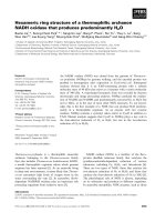
Fig. 3. Glucuronidation activity of UGT2B4 (A) and UGT2B4 mutants
(B, C) for the probe substrates. 1, 4-Methylumbelliferone; 2, euge-
nol; 3, hyodeoxycholic acid (HDCA); 4, androsterone; 5, testoster-
one; 6, epitestosterone; 7, b-estradiol; 8, 17a-ethynylestradiol; 9, 4-
hydroxyestrone; 10, 17-epiestriol; 11, 4-hydroxybiphenyl; 12, 4-iso-
propylphenol; 13, 4-nitrophenol; 14, 1-naphthol; 15, RS-ketoprofen;
16, 3¢-azido-3¢deoxytymidine. The enzyme reaction was carried out
with 50 lg protein and was incubated with 0.02 m
M UDP-glucuron-
ic acid containing 0.1 lCi UDP-[
14
C]glucuronic acid and 0.5 mM sub-
strate as indicated in Experimental procedures. The glucuronides
were separated by thin layer chromatography, visualized by auto-
radiography (shown in insert) and quantitated by liquid scintillation
counting. The rate values are the mean of three experiments. The
film was exposed for four days in the case of UGT2B4 and
UGT2B4F33Y and for one week in the case of UGT2B4Y33L.
Substrate specificity in UGT2B4 and UGT2B7 L. Barre et al.
1258 FEBS Journal 274 (2007) 1256–1264 ª 2007 The Authors Journal compilation ª 2007 FEBS
with wild-type UGT2B4. The V
max
values showed a
decrease of 19- and 34-fold for 4-hydroxyestrone and
17a-epiestriol, respectively (Table 1).
The primary structure of UGT2B7 is 87% identical
to that of UGT2B4 with 76 differences out of 528
amino acids, including 55 differences in the first 300
amino acids of the N-terminus. Both enzymes share
common substrates, including HDCA [6]. This led us
to compare the N-terminal amino acid sequence of
UGT2B4 and UGT2B7, predicted from its cDNA, in
the region encompassing residue 33 (Fig. 1). The ana-
lysis revealed that residue F33 of UGT2B4 was
replaced by Y33 in UGT2B7. Therefore, we have also
constructed and expressed in Sf9 cells a UGT2B4
mutant in which F33 was replaced by tyrosine, gener-
ating the mutant UGT2B4F33Y (Fig. 2). Analysis of
the glucuronidation activity of this mutant showed an
activity profile similar to the wild-type UGT2B4.
Moreover, HDCA and 4-hydroxyestrone were even
more efficiently glucuronidated by the mutant
(Fig. 3C, Table 1). Kinetic analysis indicated that the
K
m
and V
max
values of UGT2B4F33Y towards HDCA
and 4-hydroxyestrone were increased by 3.5- and two-
fold, and by 4.6- and three-fold, respectively, com-
pared with UGT2B4 (Table 1). These results led us to
hypothesize that the aromatic tyrosine residue at posi-
tion 33 in UGT2B4 may play an important role in the
substrate specificity of the isoform.
Importance of amino acid residue tyrosine 33
in the substrate specificity of UGT2B7
The wild-type UGT2B7 efficiently glucuronidates
17-epiestriol and eugenol and, in comparison with
UGT2B4, it exhibited a marked preference for
4-hydroxyestrone and HDCA (Table 1). In addition,
UGT2B7 efficiently glucuronidated androsterone and
epitestosterone (Fig. 4A). To investigate whether the
tyrosine residue at position 33 in UGT2B7 plays a
role in HDCA glucuronidation and substrate specifi-
city, we substituted this residue by leucine, as found in
the HDCA-deficient UGT2B4 variant, and expressed
the mutant in insect cells (Fig. 2).
Analysis of the activity of the UGT2B7Y33L mutant
towards various substrates showed that replacement of
Y33 by leucine resulted in a dramatic change in activity
and substrate specificity of UGT2B7 (Fig. 4, compare
parts A and B). Indeed, the mutation abolished glucu-
ronidation of several substrates including phenols such
as 1-naphthol and steroids such as androsterone and
b-estradiol (Fig. 4B), and greatly reduced the activity
towards HDCA and 4-hydroxyestrone (Fig. 4B,
Table 1). In addition, glucuronidation of bulky phen-
ols, 4-hydroxybiphenyl and 4-isopropylphenol, and the
steroid epitestosterone was dramatically decreased. On
the other hand, the activity towards 17-epiestriol was
increased by the Y33L mutation in UGT2B7 (Table 1).
These data showed that the presence of a leucine resi-
due at position 33, instead of tyrosine, led to an
enzyme with restricted and somewhat modified specific-
ity. Further kinetic characterization of this mutant
indicated that the K
m
values towards HDCA and
17-epiestriol were in the same range as that of the wild-
type. However, the K
m
value towards 4-hydroxyestrone
was decreased by six-fold (Table 1). Furthermore, the
V
max
values underwent major changes, with 20- and
25-fold decrease for HDCA and 4-hydroxyestrone,
respectively, and 1.2-fold increase for 17-epiestriol.
In contrast to leucine residue, replacement of tyro-
sine by phenylalanine at position 33 of UGT2B7 had
Table 1. Apparent K
m
and normalized V
max
values for glucuronidation of selected substrates by wild-type UGT2B4 and UGT2B7 and
mutants. Kinetic parameters were evaluated from initial velocity values of the reaction performed in triplicates using varying concentrations
of substrates (0–1 m
M) at a constant concentration of UDP-glucuronic acid (0.5 mM). Expression of wild-type and mutants was evaluated as
described in the Experimental procedures and expressed relative to UGT2B4 or UGT2B7. ND, not determined, due to lack of detectable
activity.
UGT
HDCA
4-Hydroxy-
estrone 17-Epiestriol
Relative protein
expression (%)
V
max
(pmolÆmin
)1
Æ
mg
)1
Æprotein)
K
m
lM
V
max
(pmolÆmin
)1
Æ
mg
)1
Æprotein)
K
m
lM
V
max
(pmolÆmin
)1
Æ
mg
)1
Æprotein)
K
m
lM
2B4 26 ± 1 25 ± 3 19 ± 1 28 ± 5 276 ± 7 42 ± 5 100
2B4F33 L ND ND 1 173 ± 29 8 93 ± 12 44
2B4F33Y 48 ± 1 91 ± 10 57 ± 4 131 ± 26 182 ± 5 71 ± 7 4
2B7 1164 ± 36 21 ± 4 2365 ± 55 81 ± 8 570 ± 10 52 ± 4 100
2B7Y33 L 56 ± 2 29 ± 6 94 ± 3 12 ± 2 670 ± 17 45 ± 5 63
2B7Y33F 2156 ± 114 39 ± 9 1260 ± 33 117 ± 11 3283 ± 56 159 ± 8 60
L. Barre et al. Substrate specificity in UGT2B4 and UGT2B7
FEBS Journal 274 (2007) 1256–1264 ª 2007 The Authors Journal compilation ª 2007 FEBS 1259
only a minor effect on activity and substrate specifi-
city. The mutant UGT2B7Y33F exhibited similar sub-
strate specificity as wild-type UGT2B7 (Fig. 4C) and
kinetic analysis indicated that the K
m
values towards
HDCA and 17-epiestriol were increased by about two-
and three-fold, respectively. The V
max
value towards
4-hydroxyestrone was decreased by two-fold and it
was increased by two- and six-fold for HDCA and
17-epiestriol, respectively (Table 1). These experiments
highlighted the importance of an aromatic residue at
position 33 in the capacity of UGT2B7 to glucuroni-
date a broad range of aglycone substrates.
Discussion
A major property of the UGTs is their large and over-
lapping substrate specificity, which confers to glucuroni-
dation a significant role in the detoxification processes.
This characteristic feature is typically illustrated from
comparison of the activity of UGT2B4 and UGT2B7,
which are both able to glucuronidate HDCA and cate-
chol-estrogens as well as xenobiotics, as shown in this
and other studies [5]. However, UGT2B7 has a broader
specificity than UGT2B4 and it is able to accommodate
various steroids such as androsterone and epitestoster-
one. The molecular basis of the substrate selectivity of
these enzymes is difficult to understand because no com-
mon structural features between the substrates glucuro-
nidated by each isoform were thus far found [12].
This general assessment prompted us to identify
amino acids that may account for the substrate specific-
ity of these UGTs. The high sequence homology
between UGT2B4 and UGT2B7, in combination with a
marked difference in substrate specificity, especially
towards steroid substrates, was favorable for attempt-
ing to pinpoint the amino acid residues that are critical
for the substrate specificity. In the current study, we
have shown that the presence of an aromatic residue at
position 33 of UGT2B4 and UGT2B7 is important in
that respect. This conclusion is based on the following
lines of evidence: (a) the UGT2B4F33L mutant exhib-
ited a strong decrease in HDCA glucuronidation; (b)
the UGT2B4F33Y mutant was able to sustain the
glucuronidation of both HDCA and 4-hydroxyestrone;
(c) mutation of residue Y33 of UGT2B7 to leucine led
to an enzyme with a restricted substrate specificity; and
(d) the mutant UGT2B7Y33F exhibited similar activity
and substrate specificity to those of UGT2B7. Interest-
ingly, Villeneuve et al. [13] recently reported a novel
polymorphism of the UGT1A9 isoform, whose muta-
tion M33T (corresponding to position 31 in UGT2B4)
was responsible for a large decrease in the activity (by
96%) of the glucuronidation of the anticancer drug,
SN-38. In contrast, the activity measured with flavo-
piridol was unaffected, indicating that, similar to our
findings, a single mutation can affect enzyme activity
for a subset of aglycones substrates. The above study
by Villeneuve et al. [13] and our work emphasize the
crucial role of the region encompassing residue at posi-
tion 33 in the substrate specificity of UGT isoforms.
A
Substrates
2 3 4 5 6 7 8 9 10 11 1213 1415 161
0
150
300
600
750
450
UGT2B7
Enzyme activity
(pmol/min/mg protein)
B
Substrates
UGT2B7Y33L
162 3 4 5 6 7 8 9 10 11 121314151
0
50
75
100
25
Normalized enzyme activity
(pmol/min/mg protein)
C
Normalized enzyme activity
(pmol/min/mg protein)
UGT2B7Y33F
2 3 4 5 6 7 8 9 10 11 12 13 14 15 161
Substrates
0
600
1500
300
750
450
150
Fig. 4. Glucuronidation activity of UGT2B7 (A) and UGT2B7Y33L
mutant (B) for probe substrates. Numbers refer to substrates as in
Fig. 3. The insert shows the glucuronides separated by thin layer
chromatography and visualized by autoradiography. (All the films
were exposed for 4 days.) Activities were measured as indicated in
the legend to Fig. 3 and are the mean of three experiments.
Substrate specificity in UGT2B4 and UGT2B7 L. Barre et al.
1260 FEBS Journal 274 (2007) 1256–1264 ª 2007 The Authors Journal compilation ª 2007 FEBS
The changes in specificity observed for the different
mutants were characterized further by kinetic analyses.
The results with UGT2B4F33L revealed that the
impairment in 4-hydroxyestrone and 17-epiestriol glucu-
ronidation efficacy resulted from a large increase in K
m
values, along with a decrease in the V
max
values. These
data suggest that the mutations primarily affect binding
of the substrates, but they do not rule out the possibility
of a reduced access of the substrate to the catalytic site
upon mutation. On the other hand, replacement of F33
by tyrosine led to mutant UGT2B4F33Y with similar
substrate specificity as the wild-type enzyme support-
ing the idea that a tyrosine can substitute to the
wild-type phenylalanine residue. Moreover, mutant
UGT2B4F33Y exhibited enhanced glucuronidation
towards HDCA and 4-hydroxyestrone compared with
wild-type. The kinetic parameters of the mutant indica-
ted an increase in both V
max
and K
m
values (Table 1).
In the case of UGT2B7, substitution of Y33 to leu-
cine led to a severe restriction in aglycones accepted by
the enzyme. In fact, the effects of replacing the aroma-
tic residue at position 33 by leucine on the substrate
specificity of UGT2B7 were even more dramatic than
in UGT2B4. Only three out of the 12 compounds pre-
viously glucuronidated by UGT2B7 remained effi-
ciently glucuronidated by the UGT2B7Y33L mutant.
Nonetheless, the K
m
value for HDCA was not signifi-
cantly different from that obtained for the wild-type
enzyme, suggesting that the affinity of the enzyme for
HDCA was not largely altered by the mutation. In the
case of 4-hydroxyestrone glucuronidation, the K
m
indi-
cated an enhanced apparent affinity of the mutant.
For both substrates, the mutation decreased the V
max
values. On the other hand, the V
max
of the mutant
towards 17-epiestriol was slightly increased and the K
m
was not significantly modified.
Replacement of the Y33 residue of UGT2B7 by phe-
nylalanine led to a mutant, UGT2B7Y33F, with even
more enhanced glucuronidation activity towards
HDCA and 17-epiestriol compared with the wild-type.
Analyses of the kinetic parameters of the UGT2-
B7Y33F mutant indicated enhanced V
max
and K
m
values, except for 4-hydroxyestrone, which showed a
two-fold decrease in the V
m
value (Table 1).
Taken together, the results of this study are consis-
tent with the notion that residue 33 is involved in the
interactions of the enzyme with the substrate in both
UGT2B4 and UGT2B7.
In contrast to the F33L mutation, which reduces the
activity of UGT2B4 and UGT2B7, exchanging F33 for
tyrosine sustained the enzyme activity and specificity.
Although a leucine residue can establish hydrophobic
interactions, it will produce more steric hindrance than
an aromatic residue such as phenylalanine or tyrosine.
In agreement with this proposal, a tyrosine residue at
position 33 in UGT2B4 was able to support glucuroni-
dation of HDCA, thus suggesting that p-stacking
interactions and ⁄ or steric hindrance conferred by an
aromatic residue are critical for access or recognition
of this substrate. Steric hindrance by a critical residue
has been proposed as an underlying principle that can
regulate substrate and ⁄ or product specificities of
enzymes catalyzing the metabolism of hydrophobic
substrates. For example, the phenylalanine residue at
position 87 of cytochrome P450 BM-3 was suggested
to act through steric hindrance to determine the regio-
and stereospecificity of the arachidonic acid epoxy-
genase activity [14]. Such a situation is also exemplified
in the case of estrogen sulfotransferase, which posses-
ses two critical aromatic residues forming a gate-like
structure that was suggested to confer estrogen specifi-
city to this enzyme [15].
The involvement of several residues in determining
the substrate specificity probably also stands true for
the UGTs. Coffman et al. [16] reported the important
role of the aspartic residue at position 99 of UGT2B7
in the binding of morphine. When this charged amino
acid was substituted with alanine, a dramatic decrease
in activity was observed. In agreement, the structure–
function analysis of UGT2B15 and UGT2B17 sugges-
ted that a set of residues (including residue 121) is
implicated in the steroid specificity of these isoenzymes
[17]. These studies, along with our work, indicate that
substitution of a single amino acid can substantially
affect substrate recognition, but multiple differences
between two related isoforms probably contribute to
their individual specificity.
In conclusion, this study shows, for the first time, that
an aromatic residue at position 33 is critical for the sub-
strate specificity of UGT2B4 and UGT2B7. The data
provide the basis with which to modulate the substrate
specificity of human UGT isoforms by protein engineering.
Experimental procedures
Chemicals and reagents
4-Methylumbelliferone (free acid), 1-naphthol, 4-nitro-
phenol, 4-hydroxybiphenyl, 4-hydroxyestrone, 17a-ethinyl-
estradiol, testosterone, eugenol, HDCA, androsterone,
epitestosterone, b-estradiol, 17-epiestriol, isopropylphenol,
ketoprofen, 3¢-azido-3¢-deoxythymidine and UDP-glucuro-
nic acid (sodium salt) were purchased from Sigma (L’Isle
d’Abeau, St. Quentin Fallavier, France). UDP-[U-
14
C]-
glucuronic acid (418 mCiÆmmol
)1
) was obtained from NEN
(Perkin Elmer, Courtaboeuf, France). Restriction enzymes
L. Barre et al. Substrate specificity in UGT2B4 and UGT2B7
FEBS Journal 274 (2007) 1256–1264 ª 2007 The Authors Journal compilation ª 2007 FEBS 1261
were provided by New England Biolabs (Hitchin, UK). The
QuikChange site-directed mutagenesis kit was from Strata-
gene (La Jolla, CA, USA), LumiGLO
TM
was from Cell
Signaling (Beverly, MA, USA), and AdvantageÒ 2 poly-
merase mix was from Clontech (Palo Alto, CA, USA). All
other reagents were of the best quality and commercially
available.
Expression vectors constructions
Expression vectors used to express human UGT2B4 and
UGT2B7 with an apparent molecular mass of about
53 kDa in baculovirus-infected insect cells were previously
described [18]. The short C-terminal extension, including a
His-tag, was added by subcloning the respective cDNAs
into the modified shuttle vector pFBXHA to generate 2B4-
XHA and 2B7-XHA expression vectors [18].
Site-directed mutagenesis
Construction of amino acid substituted mutants of
UGT2B4 and UGT2B7 were performed using the Quik-
Change site-directed mutagenesis kit according to the
recommendations of the manufacturer. 2B4-XHA and 2B7-
XHA expression vectors were used as a template. The
sequence of the sense and antisense mutation primers is
indicated in Table 2. Full-length mutated cDNAs were sys-
tematically checked by DNA sequencing.
Heterologous expression in insect Sf9 cells
Wild-type UGT2B4 and UGT2B7 and mutants expression
vectors were transfected in the Escherichia coli strain
DH10Bac for the generation of recombinant ‘bacmids’
that, in turn, were employed for the production of recom-
binant baculovirus stocks according to the Bac-to-Bac
procedure (Invitrogen, Cergy Pontoise, France). The pro-
duction of recombinant proteins was carried out following
optimization trials in which the suitable amount of virus
from the new stocks for the infection of insect Sf9
cells was estimated. The relative expression level of
each UGT in microsomal membranes was evaluated by
immunodetection using the monoclonal anti-His-tag anti-
body Tetra-His (Qiagen, Hilden, Germany) as described
previously [19].
Western blot analysis was performed by loading onto the
gel 2, 10 and 100 lg of membrane proteins for UGT2B4,
UGT2B4F33L and UGT2B4F33Y, respectively, and 12, 15
and 15 lg of membrane proteins for UGT2B7, UGT2-
B7Y33L and UGT2B7Y33F, respectively. The proteins
were separated in 10% SDS ⁄ PAGE gels, transferred to a
polyvinylidene difluoride membrane (Millipore, Eschborn,
Germany), and subsequently blocked in Tris-buffer saline-
Tween 20 containing 5% nonfat milk. Membranes were
incubated overnight with monoclonal anti-His-tag antibody
Tetra-His directed against His-tag followed by incubation
with horseradish-peroxidase-conjugated secondary antibod-
ies. The blot was then developed using LumiGLO
TM
according to the instructions of the manufacturer (Cell
Signaling, Danvers, MA, USA).
Analysis of glucuronidation activity
Protein concentration was measured as previously described
[20] with the Bio-Radä reagent (Bio-Rad, Hercules, CA,
USA). The activity of the recombinant wild-type and
mutant UGT2B4 and UGT2B7 towards several substrates
was determined as described [21]. Briefly, incubation in
Eppendorf tubes (total volume 40 lL) consisted of 50 lgof
microsomal proteins for UGT2B7, UGT2B7Y33L, UGT2-
B7Y33F and UGT2B4 and 200 l g for UGT2B4F33Y and
UGT2B4F33L in 100 mm Tris ⁄ HCl buffer (pH 7.4), 10 mm
MgCl
2
containing 0.02 mm UDP-glucuronic acid and
0.1 lCi UDP-[U-
14
C]glucuronic acid. The reaction was star-
ted by addition of substrate (0.5 mm final concentration)
dissolved in 2 lL dimethylsulfoxide. A control was per-
formed in which the substrate was omitted and dimethyl-
sulfoxide added. After incubation for 1 h at 37 ° C, the
proteins were precipitated by 40 lL ethanol in ice, and
removed by centrifugation at 4000 g for 10 min at 4 °C.
The supernatant was loaded onto thin layer chromato-
graphy plates (LK6DF silica gel, 250 lm; Whatman, Clif-
ton, NJ, USA). The plates were developed with n-butanol,
acetone, acetic acid, aqueous ammoniac (28%), water
Table 2. Sequence of the primers used for site-directed mutagenesis. Mutant amino acid codons are underlined.
Mutant Primer Sequence (5’- to 3’)
2B4F33 L Sense CTGGTGTGGCCCACAGAA
CTCAGCCACTGGATGAATATAAAG
Antisense CTTTATATTCATCCAGTGGCT
GAGTTCTGTGGGCCACACCAG
2B4F33Y Sense CTGGTGTGGCCCACAGAA
TACAGCCACTGGATGAATATAAAG
Antisense CTTTATATTCATCCAGTGGCT
GTATTCTGTGGGCCACACCAG
2B7Y33 L Sense CTGGTGTGGGCAGCAGAA
CTCAGCCATTGGATGAATATAAAG
Antisense CTTTATATTCATCCAATGGCT
GAGTTCTGCTGCCCACACCAG
2B7Y33F Sense CTGGTGTGGGCAGCAGAA
TACAGCCATTGGATGAATATAAAG
Antisense CTTTATATTCATCCAATGGCT
GTATTCTGCTGCCCACACCAG
Substrate specificity in UGT2B4 and UGT2B7 L. Barre et al.
1262 FEBS Journal 274 (2007) 1256–1264 ª 2007 The Authors Journal compilation ª 2007 FEBS
(70 : 50 : 18 : 1.5 : 60 v ⁄ v). They were dried and sprayed
with 1% (v ⁄ v) 2-(4-t-butylphenyl)-5() 4-biphenyl)-1,3,
4-oxadiazole in toluene. The radioactivity associated with
the glucuronide was visualized by autoradiography with
X-Omat Kodak films (Sigma) for 3 days at )20 °C. The sil-
ica gel areas of the glucuronides were scraped off and the
associated radioactivity was quantified on a LKB spectro-
meter using Fluoran Safe Ultima Gold scintillant cocktail
(Packard, Rungis, France). The decomposition per min
value in a given sample was considered significant when it
was at least two-fold of that of the blank sample.
Kinetic analysis of the data
Kinetic parameters were evaluated from initial velocity val-
ues of the reaction performed as described above. Varying
concentrations of the substrates (0–1 mm) at a constant
concentration of UDP-GlcA (0.5 mm) were used. K
m
and
V
max
values for HDCA, 4-hydroxyestrone and 17a-epiestriol
were determined using nonlinear least square analysis of
the data fitted to Michaelis-Menten rate equation (v ¼
V
max
· [S] ⁄ K
m
+ [S]), where S is the substrate and v is
the velocity, using the curve-fitter program sigmaplot 9.0
[22].
Acknowledgements
This work was supported by grants from Ligue Contre
le Cancer Re
´
gion Lorraine, Agence Nationale de la
Recherche (ANR number NT05-3_42251) and Re
´
gion
Lorraine, as well as the Academy of Finland (Project
210933). We thank J, Mosorin for excellent technical
assistance and PI. Mackenzie (Flinders University,
Adelaide, Australia) for kindly providing the UGT2B4
variant cDNA.
References
1 Clarke DJ & Burchell B (1994) The Uridine Diphosphate
Glucuronosyltransferase Multigene Family: Function
and Regulation Springer-Verlag, Berlin, Heidelberg,
New York.
2 Tukey RH & Strassburg CP (2000) Human UDP-
glucuronosyltransferases: metabolism, expression, and
disease. Annu Rev Pharmacol Toxicol 40 , 581–616.
3 Guengerich FP, Parikh A, Johnson EF, Richardson
TH, von Wachenfeldt C, Cosme J, Jung F, Strassburg
CP, Manns MP, Tukey RH et al. (1997) Heterologous
expression of human drug-metabolizing enzymes. Drug
Metab Dispos 25, 1234–1241.
4 Radominska-Pandya A, Czernik PJ, Little JM, Battaglia
E & Mackenzie PI (1999) Structural and functional stu-
dies of UDP-glucuronosyltransferases. Drug Metab Rev
31, 817–899.
5 Fournel-Gigleux S, Jackson MR, Wooster R & Burchell
B (1989) Expression of a human liver cDNA encoding a
UDP-glucuronosyltransferase catalysing the glucuroni-
dation of hyodeoxycholic acid in cell culture. FEBS Lett
243, 119–122.
6 Ritter JK, Chen F, Sheen YY, Lubert RA & Owens IS
(1992) Two human liver cDNAs encode UDP-glucurono-
syltransferases with 2 log differences in activity toward
parallel substrates including hyodeoxycholic acid and
certain estrogen derivatives. Biochemistry 31, 3409–3414.
7 Radominska-Pandya A, Little JM & Czernik PJ (2001)
Human UDP-glucuronosyltransferase 2B7. Curr Drug
Metab 2, 283–298.
8 Ritter JK, Chen F, Sheen YY, Tran HM, Kimura S,
Yeatman MT & Owen IS (1992) A novel complex locus
UGT1 encodes human bilirubin, phenol, and other
UDP-glucuronosyltransferase isozymes with identical
carboxyl termini. J Biol Chem 267, 3257–3261.
9 Mackenzie PI (1990) Expression of chimeric cDNAs in
cell culture defines a region of UDP-glucuronosyltrans-
ferase involved in substrate selection. J Biol Chem 265,
3432–3435.
10 Li Q, Lou X, Peyronneau MA, Obermayer-Straub P &
Tukey RH (1997) Expression and functional domains of
rabbit liver UDP-glucuronosyltransferase 2B16 and
2B13. J Biol Chem 272, 3272–3279.
11 Jin C, Miners JO, Lillywhite KJ & Mackenzie PI (1993)
Complementary deoxyribonucleic acid cloning and
expression of human liver uridine diphosphate-glucuro-
nosyltransferase glucuronidating carboxylic acid-con-
taining drugs. J Pharm Exp Ther 264, 475–479.
12 Jin CJ, Mackenzie PI & Miners JO (1997) The regio-
and stereo-selectivity of C19 and C21 hydroxysteroid
glucuronidation by UGT2B7 and UGT2B11. Arch Bio-
chem Biophys 341, 207–211.
13 Villeneuve L, Girard H, Fortier LC, Gagne JF & Guil-
lemette C (2003) Novel functional polymorphisms in the
UGT1A7 and UGT1A9 glucuronidating enzymes in
Caucasian and African-American subjects and their
impact on the metabolism of 7-ethyl-10-hydroxycamp-
tothecin and flavopiridol anticancer drugs. J Pharmacol
Exp Ther 307, 117–128.
14 Graham-Lorence S, Truan G, Peterson JA, Falck JR,
Wei S, Helvig C & Capdevila JH (1997) An active site
substitution, F87V, converts cytochrome P450 BM-3
into a regio- and stereoselective (14S,15R)-arachidonic
acid epoxygenase. J Biol Chem 272, 1127–1135.
15 Petrotchenko EV, Doerflein ME, Kakuta Y, Pedersen
LC & Negishi M (1999) Substrate gating confers steroid
specificity to estrogen sulfotransferase. J Biol Chem 274,
30019–30022.
16 Coffman BL, Kearney WR, Goldsmith S, Knosp BM &
Tephly TR (2003) Opioids bind to the amino acids
84–118 of UDP-glucuronosyltransferase UGT2B7. Mol
Pharmacol
63, 283–288.
L. Barre et al. Substrate specificity in UGT2B4 and UGT2B7
FEBS Journal 274 (2007) 1256–1264 ª 2007 The Authors Journal compilation ª 2007 FEBS 1263
17 Dubois SG, Beaulieu M, Levesque E, Hum DW &
Belanger A (1999) Alteration of human UDP-glucuro-
nosyltransferase UGT2B17 regio-specificity by a single
amino acid substitution. J Mol Biol 289, 29–39.
18 Kurkela M, Garcia-Horsman JA, Luukkanen L,
Morsky S, Taskinen J, Baumann M, Kostiainen R,
Hirvonen J & Finel M (2003) Expression and character-
ization of recombinant human UDP-glucuronosyltrans-
ferases (UGTs). UGT1A9 is more resistant to detergent
inhibition than other UGTs and was purified as an
active dimeric enzyme. J Biol Chem 278, 3536–3544.
19 Kurkela M, Hirvonen J, Kostiainen R & Finel M
(2004) The interactions between the N-terminal and
C-terminal domains of the human UDP-glucuronosyl-
transferases are partly isoform-specific, and may involve
both monomers. Biochem Pharmacol 68, 2443–2450.
20 Bradford MM (1976) A rapid and sensitive method for
the quantitation of microgram quantities of protein util-
izing the principle of protein-dye binding. Anal Biochem
72, 248–254.
21 Bansal SK & Gessner T (1980) A unified method for
the assay of uridine diphospho-glucuronyltransferase
activity toward various aglycones using uridine
diphospho[U-
14
C]glucuronic acid. Anal Biochem 109,
321–329.
22 Segel IH (1975) Enzyme Kinetics. John Wiley & Sons,
New York, NY.
Substrate specificity in UGT2B4 and UGT2B7 L. Barre et al.
1264 FEBS Journal 274 (2007) 1256–1264 ª 2007 The Authors Journal compilation ª 2007 FEBS