Báo cáo khoa học: Mechanisms of cholinesterase inhibition by inorganic mercury potx
Bạn đang xem bản rút gọn của tài liệu. Xem và tải ngay bản đầy đủ của tài liệu tại đây (573.89 KB, 13 trang )
Mechanisms of cholinesterase inhibition by inorganic
mercury
Manuela F. Frasco
1,2
, Jacques-Philippe Colletier
3
, Martin Weik
3
,Fe
´
lix Carvalho
4
,
Lu
´
cia Guilhermino
1,2
, Jure Stojan
5
and Didier Fournier
6
1 ICBAS, Instituto de Cie
ˆ
ncias Biome
´
dicas de Abel Salazar, Universidade do Porto, Portugal
2 CIMAR-LA ⁄ CIIMAR, Universidade do Porto, Portugal
3 IBS-UMR 5075, CEA-CNRS-UJF, Laboratoire de Biophysique Mole
´
culaire, Grenoble, France
4 REQUIMTE, Servic¸o de Toxicologia da Faculdade de Farma
´
cia da Universidade do Porto, Portugal
5 Institute of Biochemistry, University of Ljubljana, Slovenia
6 IPBS-UMR 5089, CNRS-UPS, Groupe de Biotechnologie des Prote
´
ines, Toulouse, France
Human activities have continuously contaminated the
environment with mercury, which has been used for
centuries in agriculture, industry, and medicine [1].
Nowadays, inorganic mercury is used in, for example,
thermometers, batteries, and fluorescent light-bulbs. In
addition, large quantities of metallic mercury are
employed in the fabrication of electrodes for the elec-
trolytic production of chlorine and sodium hydroxide
from salt, as well as in gold mining [2,3]. Although it
presents unique properties that make it useful for
many human purposes, mercury has no role in life
processes and is highly toxic. Nephrotoxicity [4] and
genotoxicity [5] have been demonstrated. Other adverse
effects occur in neural tissues, where the targeting of
enzymes and receptors involved in nerve impulse trans-
mission is probably involved [6], as well as in the
Keywords
aggregation; cholinesterase; inhibition;
mercury; metals
Correspondence
D. Fournier, IPBS-UMR 5089, 205 Route de
Narbonne, F-31077 Toulouse, France
Fax: +33 5 61 17 59 94
Tel: +33 5 61 55 54 45
E-mail:
Database
The coordinates and structure factor ampli-
tudes of the complex structure of human
butyrylcholinesterase with HgCl
2
have been
deposited in the Protein Data Bank under
accession code 2J4C
(Received 18 December 2006, revised 1
February 2007, accepted 7 February 2007)
doi:10.1111/j.1742-4658.2007.05732.x
The poorly known mechanism of inhibition of cholinesterases by inorganic
mercury (HgCl
2
) has been studied with a view to using these enzymes as
biomarkers or as biological components of biosensors to survey polluted
areas. The inhibition of a variety of cholinesterases by HgCl
2
was investi-
gated by kinetic studies, X-ray crystallography, and dynamic light scatter-
ing. Our results show that when a free sensitive sulfhydryl group is present
in the enzyme, as in Torpedo californica acetylcholinesterase, inhibition is
irreversible and follows pseudo-first-order kinetics that are completed
within 1 h in the micromolar range. When the free sulfhydryl group is not
sensitive to mercury (Drosophila melanogaster acetylcholinesterase and
human butyrylcholinesterase) or is otherwise absent (Electrophorus electri-
cus acetylcholinesterase), then inhibition occurs in the millimolar range.
Inhibition follows a slow binding model, with successive binding of two
mercury ions to the enzyme surface. Binding of mercury ions has several
consequences: reversible inhibition, enzyme denaturation, and protein
aggregation, protecting the enzyme from denaturation. Mercury-induced
inactivation of cholinesterases is thus a rather complex process. Our results
indicate that among the various cholinesterases that we have studied, only
Torpedo californica acetylcholinesterase is suitable for mercury detection
using biosensors, and that a careful study of cholinesterase inhibition in a
species is a prerequisite before using it as a biomarker to survey mercury in
the environment.
Abbreviations
DLS, dynamic light scattering.
FEBS Journal 274 (2007) 1849–1861 ª 2007 The Authors Journal compilation ª 2007 FEBS 1849
immune system, for which both autoimmunity and
immune suppression have been reported [7–9]. Detec-
tion of mercury in the environment is thus of high rele-
vance for public health and in the framework of
sustainable development. In this context, cholinesteras-
es have been suggested as potential biomarkers and as
the biological component of protein-based biosensors.
The concept of biomarkers implies that in vivo choli-
nesterase inhibition is measured after exposure of an
animal to mercury, whereas in biosensors, inhibition
takes place in vitro. For both applications, there is a
need for high sensitivity of cholinesterases to mercury.
Thus, it is a prerequisite to investigate the mechan-
ism(s) of inhibition and to ascertain the reliability of
using cholinesterases.
Cholinesterases are believed to be sensitive to mer-
cury; indeed, exposure of different organisms to suble-
thal concentrations of mercury was shown to induce a
significant decrease in cholinesterase activities in sev-
eral organs [10–15]. However, uncertainties remain, as
mercury-induced stimulation of cholinesterase activity
has also been reported [16,17]. Several authors have
described instantaneous reversible inhibition of choli-
nesterases in vitro [18–20]. However, these findings
could be artefactual, as activity measurements were
often performed using Ellman’s reaction [21], the prod-
ucts of which react with mercury and thus interfere
with the measurement [22]. In addition, irreversible
inhibition was described for Torpedo californica acetyl-
cholinesterase, which leads in a first step to the forma-
tion of a metastable state [23] that latter converts to a
partially unfolded one [24].
Depending on their locations, two main functions
have been ascribed to cholinesterases. At cholinergic
synapses, cholinesterases are responsible for the ter-
mination of nerve impulse transmission, by rapid
hydrolysis of the neurotransmitter acetylcholine. This
role is vital, as it allows restoration of neuronal excita-
bility in cholinergic neuron networks. In noncholiner-
gic tissues, cholinesterases belong to the group of
‘scavenger proteins’, which are responsible for the deg-
radation of xenobiotics, e.g. succinylcholine or cocaine
[25]. Playing these important roles, cholinesterases are
among the most efficient enzymes in nature, with a
substrate turnover of 10
3
)10
4
s
)1
, depending on spe-
cies [26]. Two types of cholinesterase are found in
mammals, acetylcholinesterase and butyrylcholinest-
erase, which are enzymatically distinguished by their
substrate specificity. From the structural point of view,
these enzymes are very similar, and only a few critical
differences in the active site amino acid composition
account for their differential behavior towards sub-
strates [27–31].
Cholinesterases are % 60 kDa globular proteins, and
are found in various oligomeric states. With regard to
their cysteine content, three intrachain disulfide bonds
are conserved, as well as another cysteine involved in
intersubunit association. Although no free cysteine
is found in some species, e.g. Electrophorus electricus
acetylcholinesterase, there is one accessible to the bulk
solution in most of them. Its position, however, is not
conserved: 66 in human butyrylcholinesterase, 290 in
Drosophila melanogaster acetylcholinesterase, and 231
in T. californica acetylcholinesterase. In the last case,
the free cysteine has been shown to react with sulfhyd-
ryl agents, resulting in irreversible inactivation of the
enzyme [32,33].
In the present study, the kinetic mechanism of mer-
cury-induced inactivation of cholinesterases was inves-
tigated using four cholinesterases from various species
to probe the potential variability in sensitivity that
may exist in biomarkers used in ecotoxicologic studies.
T. californica acetylcholinesterase, E. electricus acetyl-
cholinesterase, D. melanogaster acetylcholinesterase
and human butyrylcholinesterase were chosen because
they are available in large amounts. Kinetic studies
were complemented by X-ray crystallographic experi-
ments on human butyrylcholinesterase and dynamic
light scattering (DLS) studies on D. melanogaster
acetylcholinesterase. Two inhibition mechanisms are
proposed, depending on the presence or absence of a
sensitive free cysteine.
Results
Inhibition of T. californica acetylcholinesterase
Figure 1 shows the kinetics of irreversible inactivation
of T. californica acetylcholinesterase by 1–10 lm
HgCl
2
. Inhibition follows a pseudo-first-order kinetics
(Scheme 1A; k
i
¼ 9200 ± 480 m
)1
Æmin
)1
), suggesting
that inactivation involves only one site. Probably, this
site is the same that has been shown to react with
other thiols and organomercurial compounds [24,33],
i.e. Cys231.
Inhibition of human butyrylcholinesterase
Incubation of 15 nm enzyme with HgCl
2
(1–10 mm
HgCl
2
) leads to rapid inhibition until a plateau is
reached (Fig. 2A). Increasing the enzyme concentration
diminishes the maximum inhibition (Fig. 2B). The
inhibition appears to be slowly reversible; indeed, the
10-fold dilution of a sample incubated with 10 mm
HgCl
2
leads to a slow increase of activity until a plat-
eau is reached corresponding to the activity observed
Cholinesterase inhibition by mercury M. F. Frasco et al.
1850 FEBS Journal 274 (2007) 1849–1861 ª 2007 The Authors Journal compilation ª 2007 FEBS
after incubating the enzyme with 1 mm HgCl
2
(see sup-
plementary Fig. S1A). To investigate this slow reacti-
vation of the enzyme, human butyrylcholinesterase at
a concentration of 15 nm was incubated with 1 or
10 mm HgCl
2
for 30 min. Samples were then dialyzed
for 5 h (with a dilution factor of 1000), and activity
was recorded as a function of time. A time-dependent
reactivation of the enzyme was observed, suggesting
that inhibition is reversible (Fig. 2C). Equilibrium is
reached after 15–20 min (Fig. 2A), so the analysis of
equilibrium between human butyrylcholinesterase and
mercury was performed by incubating 15 nm enzyme
for 30 min at different HgCl
2
concentrations. Subse-
quently, the substrate o-nitrophenyl acetate was added,
and the activity was measured. Data are best fitted by
a model accounting for noncompetitive inhibition, with
an apparent K
iapp
¼ 0.4 ± 0.06 mm (Fig. 2D). Inhibi-
tion of human butyrylcholinesterase by mercury thus
occurs at a concentration 1000 times higher than that
inhibiting T. californica acetylcholinesterase. Addition-
ally, human butyrylcholinesterase inhibition by HgCl
2
can be described as an apparent, slow, noncompetitive
reversible inhibition, which depends on the enzyme
concentration, whereas the data for T. californica
acetylcholinesterase can only be described as irrever-
sible inhibition.
Inhibition of D. melanogaster acetylcholinest-
erase and E. electricus acetylcholinesterase
D. melanogaster acetylcholinesterase and E. electricus
acetylcholinesterase are inhibited by mercury in the
same range of concentrations as human butyrylcholi-
nesterase (i.e. 1000-fold higher than the concentration
necessary to inhibit the Torpedo enzyme). In contrast
to what was observed for human butyrylcholinesterase,
inhibition of these two enzymes by HgCl
2
did not lead
to a plateau, but rather showed a double exponential
decay, as shown in Fig. 3A for D. melanogaster acetyl-
cholinesterase (see supplementary Fig. S2 for E. electri-
cus acetylcholinesterase). In addition, reactivation
upon dilution is partial, restoring less than 10% of the
initial activity (see supplementary Fig. S1B,C). As for
human butyrylcholinesterase, however, inhibition
decreases with enzyme concentration (Fig. 3B). Hence,
inhibition of D. melanogaster acetylcholinesterase and
E. electricus acetylcholinesterase by mercury can be
described as a slow, noncompetitive, reversible process
that depends on the enzyme concentration. However,
an irreversible inhibition also takes place, which was
not evident for human butyrylcholinesterase within 1 h
of incubation with mercury.
The D. melanogaster acetylcholinesterase used herein
is recombinant; thus, it was possible to introduce
sequence modifications by site-directed mutagenesis.
To check whether this inactivation pattern was due to
the free cysteine (C290) present on the surface of
D. melanogaster acetylcholinesterase, this residue was
mutated into an alanine. The resulting inhibition pat-
tern was virtually identical to that of the wild-type
enzyme, suggesting that this cysteine residue is not
involved in the inhibition mechanism. Analogously,
the alanine residue (A269) equivalent to the free cys-
teine in position 231 of T. californica acetylcholinest-
erase was mutated into a cysteine. In the micromolar
range of HgCl
2
, this replacement also did not change
the inhibition pattern of D. melanogaster acetylcholi-
nesterase, strongly suggesting the involvement of the
residues surrounding C231 in T. californica acetylcholi-
B
A
ki
E
EM
M
k
3
E
EM EM
2
K
0
k2
k1
M
M
D
M
M
E
2
M
2
E
E
k6
k7
EM
EM
k5
k4
k
8
D
Scheme 1. (A) Scheme proposed to describe the inhibition of
T. californica acetylcholinesterase. E and M represent enzyme and
mercury molecules, respectively, and the form EM is inactive. (B)
Scheme proposed to describe the inhibition of D. melanogaster
acetylcholinesterase. All forms are as active as the native enzyme,
except for EM
2
and D, which are inactive upon mercury removal.
Fig. 1. Remaining activity of T. californica acetylcholinesterase
(7 n
M) following incubation with mercury (1–10 lM). Curves corres-
pond to a single multi-nonlinear fit of data for all concentrations of
mercury in the equation derived from Scheme 1A [55].
M. F. Frasco et al. Cholinesterase inhibition by mercury
FEBS Journal 274 (2007) 1849–1861 ª 2007 The Authors Journal compilation ª 2007 FEBS 1851
nesterase in its high sensitivity to mercurial agents. To
check whether the introduction of a free cysteine inside
the active site of D. melanogaster acetylcholinesterase
would change the inhibition pattern, mutations F330C
and Y370C were analyzed. These replacements did not
change the inhibition pattern.
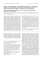
Fig. 3. Remaining activity of D. melanogaster acetylcholinesterase following incubation with mercury. (A) Effect of mercury concentration
with 500 n
M enzyme. (B) Effect of protein concentration with 5 mM mercury.
Fig. 2. Remaining activity of human butyrylcholinesterase following incubation with mercury. (A) Effect of mercury concentration with 15 nM
enzyme. (B) Effect of protein concentration with 2.5 mM mercury. (C) Slow reactivation of inhibited enzyme following dialysis after 30 min of
incubation with mercury. (D) Steady-state analysis of inhibition: enzyme and mercury were incubated for 30 min, after which the substrate
o-nitrophenyl acetate was added to the cuvette at different concentrations, without significant dilution of the sample.
Cholinesterase inhibition by mercury M. F. Frasco et al.
1852 FEBS Journal 274 (2007) 1849–1861 ª 2007 The Authors Journal compilation ª 2007 FEBS
Structure of the HgCl
2
–human
butyrylcholinesterase complex
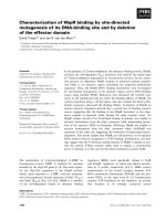
In this complex, four mercury-binding sites were char-
acterized (Fig. 4A). These were attributed on the
basis of very clear anomalous signals of mercury ions
(Fig. 4B–E) at the employed wavelength (i.e. 1.54 A
˚
).
An isomorphous difference map, computed using the
structure factors of the complex and those obtained
from a native crystal (data not shown), confirmed
these positions, displaying four very strong positive
peaks overlapping with the anomalous peaks. In addi-
tion, a pair of positive and negative densities was
found in the active site, next to the catalytic serine
residue (Ser198), on the atypical bond with the bound
butyrate [31]. This feature was interpreted as a dis-
placement of the butyrate upon complexation with
mercury. No mercury ion was found in the active
site.
The first mercury-binding site (occupancy: 75%) was
localized behind the ammonium-binding loci of the
active site (Fig. 4B). At this site, mercury mainly inter-
acts with His77Ne2 and Met81Sd (distances: 2.75 and
3.6 A
˚
, respectively), as well as with surrounding water
molecules (distances: 2.96, 3.09, and 3.81 A
˚
, respect-
ively). The second mercury ion (occupancy: 50%)
binds to His423Nd1, Asn504Od1, and Thr505Oc1 (dis-
tances: 2.33, 2.95, and 3.09 A
˚
, respectively; Fig. 4C).
The third mercury ion (occupancy: 50%) is in close
proximity to, and undergoes electrostatic interaction
with, a sulfate anion (Fig. 4D) from the mother liquor
solution (distance to the closest oxygen atom: 2.3 A
˚
).
The sulfate ion also interacts with His372Nd1 (distance
to the closest oxygen atom: 2.5 A
˚
), as has already been
reported for the native structure. At these three previ-
ously described loci, mercury binding occurs on the
surface of the enzyme but does not involve crystal con-
tacts. At the last binding site (occupancy: 25%), how-
ever, a mercury ion was found at a special position in
the crystal, in close proximity to the two Met511Sd
(distance: 2.6 A
˚
) residues of two symmetry-related
molecules in the crystal (Fig. 4E). Hence, it is involved
in crystal packing interactions.
The structure did not show any mercury ion bound
to a sulfhydryl group, as was observed in the case of
T. californica acetylcholinesterase. However, the only
free and accessible cysteine residue, Cys66, was per-
sulfured (Cys-S-SH) in this batch of enzyme. Soaking
of human butyrylcholinesterase crystals with mother
liquor containing EDTA, dithiothreitol or l-cysteine
did not allow reduction of the per-sulfur. Therefore,
the potential binding of mercury to this cysteine ‘in
solution’ remains an open issue.
DLS assays
At all mercury concentrations, data were fitted as only
one species with a low polydispersity (polydispersity
index ¼ 0.3). An increase in the hydrodynamic radius
of D. melanogaster acetylcholinesterase was observed
with increasing HgCl
2
concentrations (Fig. 5). Under
the experimental conditions used (5 lm enzyme), the
hydrodynamic radius increased linearly with mercury
concentration. Physical changes and protein aggrega-
tion occurred in the first minute after mercury addi-
tion, and the size of the aggregate remained stable for
at least 1 h.
Kinetic model for D. melanogaster
acetylcholinesterase inhibition by mercury
D. melanogaster acetylcholinesterase was incubated
with HgCl
2
for various times, and remaining activities
were measured for 10 s following dilution (10-fold or
100-fold) of the sample. The incubation time varied
from 30 s to 1 h, enzyme concentrations were 50, 100,
300, 500, 700 and 900 nm, and HgCl
2
concentrations
were 1, 2.5, 5 and 10 mm. The 22 experimental curves
were simultaneously analyzed with concurrent models,
taking into account the information obtained from
other experiments: (a) inhibition appears to be noncom-
petitive, binding sites of mercury are located on the pro-
tein surface, and inhibition does not involve residues in
the active site; and (b) mercury binding promotes aggre-
gation, and hence indirectly diminishes the enzyme sen-
sitivity to mercury, most likely because aggregation
reduces accessibility to the second mercury-binding site.
Among all tested possibilities, Scheme 1B appears as
the most simple and appropriate model to describe the
irreversible and slow reversible inhibition of D. melano-
gaster acetylcholinesterase by mercury (see kinetic
constants in Table 1 and curve fitting in supplementary
Fig. S3). According to this model, one mercury ion
binds to the enzyme to form the complex EM (E and
M represent enzyme and mercury molecules, respect-
ively) with an equilibrium constant around 0.2 mm;
this binding is instantaneously reversible and does not
affect enzyme activity. The binding of a second mer-
cury ion to the same enzyme molecule (EM) leads to
an inactive form (EM
2
). This inactivation is slowly
reversible. In addition, this form is not stable and may
result in irreversible enzyme denaturation (D). This
part of the scheme describes the two phases of inhibi-
tion. To describe the effect of protection by enzyme
concentration, we introduced into the model the form
E
2
M
2
, resulting from reversible aggregation of the
form EM without any alteration of the enzymatic
M. F. Frasco et al. Cholinesterase inhibition by mercury
FEBS Journal 274 (2007) 1849–1861 ª 2007 The Authors Journal compilation ª 2007 FEBS 1853
BC
ED
Active-site gorge entrance
A
1
2
3
4
Cholinesterase inhibition by mercury M. F. Frasco et al.
1854 FEBS Journal 274 (2007) 1849–1861 ª 2007 The Authors Journal compilation ª 2007 FEBS
activity. This aggregate form (E
2
M
2
) may either dena-
turate (form D) or dissociate, thereby giving the
reversible inactivated form (EM
2
).
Discussion
Mercury inhibits cholinesterases
The four cholinesterases analyzed in this study are
inhibited by mercury, but through different mecha-
nisms. Mercury in micromolar concentrations inhibits
T. californica acetylcholinesterase, but the other tested
cholinesterases are sensitive only in the millimolar
range. Millimolar concentrations of mercury are irre-
levant both under physiologic conditions and in the
environment; indeed, concentrations up to 300 mgÆL
)1
(% 1mm) have never been reported, even after bio-
accumulation in highly polluted areas.
The initial objective of this study was to evaluate
the inhibition of cholinesterases by mercury, with a
view to using them as biomarkers to survey polluted
areas or for incorporation in biosensors. With regard
to the utilization of cholinesterases as biomarkers, our
work obviously demonstrates that the type and effect-
iveness of inhibition of a cholinesterase by mercury
strongly depend on the species. Therefore, the kinetic
characterization of cholinesterase inhibition in the
selected species would be a prerequisite for field stud-
ies. A biosensor is an alternative to a biomarker, in
that the enzyme is linked to a surface, deep in the sur-
veyed solution, and inhibition of cholinesterase is
recorded. Inhibition occurs in vitro, whereas it occurs
in vivo in biomarkers. Cholinesterases as biological
components were first developed to detect low levels of
insecticides in the environment [34]. Numerous subse-
quent studies have been performed to develop trans-
ducers and to increase enzyme sensitivity and stability
[35]; the biosensor technology for cholinesterases is
therefore available, and permits consideration of their
use for surveying mercury in the environment. Of the
studied enzymes, it appears that only T. californica
acetylcholinesterase could be considered a good candi-
date, as it is the only one that is sensitive enough.
Cholinesterase inhibition is not related
to the active site
Mercury inhibits a large number of enzymes with func-
tional sulfhydryl group(s) in the active site [36–38]. This
does not apply to cholinesterases, as: (a) there is no free
cysteine in the active site; (b) the introduction of a free
cysteine inside the active site of recombinant D. mel-
anogaster acetylcholinesterase (F330C and Y370C) did
not increase the sensitivity to HgCl
2
; and (c) the com-
plex structures of T. californica acetylcholinesterase
Fig. 5. Aggregation of D. melanogaster acetylcholinesterase (5 lM)
as a function of mercury concentration revealed by an increase in
the hydrodynamic radius estimated by DLS.
Fig. 4. Binding of mercury ions in the HgCl
2
–human butyrylcholinesterase complex. (A) Overview of mercury-binding sites on the surface of
the enzyme. (B) First mercury-binding site (numbered ‘1’), next to His77 and Met81 (i.e. on the W-loop, behind the ammonium-binding loci
of the active site ) Trp82). (C) Second mercury-binding site (numbered ‘2’), next to His423, Asn504 and Thr505. (D) Third mercury-binding
site (numbered ‘3¢), next to a sulfate ion bound to His372. (E) Fourth mercury-binding site (numbered ‘4’), at a special position in the crystal,
in proximity to Met511 of two symmetry-related molecules. The omit 2F
o
) F
c
electron density map (contour level 1.5r), as well as the
anomalous map (contour level 4r), are superimposed on the model in (B), (C), (D), and (E).
Table 1. Kinetic constants describing D. melanogaster acetylcholi-
nesterase inhibition by mercury according to Scheme 1B. Binding
of the first mercury ion is treated as an instantaneous step (affinity
is 0.2 m
M). The fit was done simultaneously to all inactivation data
(supplementary Fig. S3) and to the curve of reactivation data (sup-
plementary Fig. S1B). To emphasize the reactivation data, they
were weighted to the same y as the inhibition curves.
K
0
(M) 2.07 ± 0.52 · 10
)4
k
1
(M
)1
Æs
)1
) 681 ± 60
k
2
(s
)1
) 0.121 ± 0.015
k
3
(s
)1
) 0.00564 ± 0.00018
k
4
(M
)1
Æs
)1
) 1.6 ± 0.3 · 10
7
k
5
(s
)1
) 0.0121 ± 0.017
k
6
(M
)1
Æs
)1
) 1.6 · 10
7a
k
7
(s
)1
) 0.0121
a
k
8
(s
)1
) 0.0298 ± 0.0039
a
k
6
was set identical to k
4
, and k
7
to k
5
,ask
7
¼ k
2
· k
5
⁄ (k
2
+ k
5
)
and k
2
) k
5
.
M. F. Frasco et al. Cholinesterase inhibition by mercury
FEBS Journal 274 (2007) 1849–1861 ª 2007 The Authors Journal compilation ª 2007 FEBS 1855
([24] ) Protein Data Bank accession code 2J4F) and
human butyrylcholinesterase (this work ) Protein Data
Bank accession code 2J4C) soaked in HgCl
2
failed to
show mercury binding in their active site, at least with
a dissociation constant lower than 10 mm. This obser-
vation is surprising, considering the strong electrostatic
dipole aligned with the gorge axis, which should attract
positive charges within the active site [39]. The absence
of mercury within the active site may be a consequence
of its large solvation shell (I. Silman, unpublished
results), which could prevent it from entering the gorge.
The quaternary nitrogen of the substrate, on the other
hand, is not hydrated [40], and can thus readily enter
the gorge. The model we propose here for cholinest-
erase inhibition by mercury is a rather general model
for the effect of mercury on proteins; only in cases
where a sensitive free sulfhydryl group is available
should another model be considered, as, for example,
in T. californica acetylcholinesterase.
Mercury-binding sites on cholinesterases
Organic and inorganic mercurials are capable of
forming very tight bonds with functional groups such
as thiolates of cysteine [41,42]. Sulfhydryl groups are
considered to be the main targets of mercury, as they
are the most reactive nucleophilic sites of protein
amino acid side chains. HgCl
2
binds to a single resi-
due to form R-S-Hg-Cl. Results for cholinesterase
suggest that inactivation by mercury involves the free
thiol group of Cys231. This functional group was
shown to be the target of other sulfhydryl reagents,
including organomercurials. Modifications of this resi-
due lead to an irreversible inhibition of the enzyme
that follows pseudo-first-order kinetics [24,33]. How-
ever, introducing a cysteine at the same position in
D. melanogaster acetylcholinesterase did not result in
increased sensitivity to mercury, suggesting an import-
ant role of the surrounding environment in the
Torpedo enzyme.
Mercury may also react with S–S bonds, leading
to their disruption (R-S-Cl + Cl-Hg-S-R) [43]. The
R-S-Cl moiety may later be oxidized by another mer-
cury ion to form the compound R-S-Hg-Cl. The abil-
ity of HgCl
2
to cleave S–S bridges enables it to
disturb the tertiary structure of proteins and hence to
lower their stability. In cholinesterases, three intra-
chain disulfide bonds are conserved. Both site-directed
mutagenesis of the cysteines involved in disulfide
bond formation and cholinesterase treatment with
reducing agents inactivate the enzyme [44], showing
that these disulfides are essential for the protein to
function. However, neither the complex structure with
mercury of human butyrylcholinesterase, nor that
of T. californica acetylcholinesterase [24], showed
evidence of binding at these positions. Most likely,
S–S bridges are too deeply buried inside the protein
and thus are not accessible to the highly hydrophilic
mercury.
Metals are also capable of forming very tight bonds
with histidine and methionine side chains as, for exam-
ple, in metalloenzymes. In cholinesterases, mercurials
were found to be linked to these residues, as evidenced
by the complex structures with mercury of human
butyrylcholinesterase (Fig. 4; Protein Data Bank entry
2J4C) and T. californica acetylcholinesterase (Protein
Data Bank entry 2J4F).
Mercury induces protein aggregation
Aggregation induced by metal ions has been observed
for other protein systems, particularly for proteins
involved in protein deposition diseases [45–48]. DLS
experiments have shown that binding of mercury ions
promotes the aggregation of D. melanogaster acetyl-
cholinesterase, perhaps as a consequence of the cross-
linking of two coordinating residues present at the
surface of the protein. This aggregation depends on
protein concentration, and does not affect the folding
of the protein; it may therefore protect the enzyme
from unfolding, due to the binding of two mercury
ions on the same enzyme molecule.
Putative mechanism of inhibition
A goal of this study was to address the issue of a
clearcut model for cholinesterase inhibition by mer-
cury. It appears that two different mechanisms for
cholinesterase inhibition by mercury may be consid-
ered. The first one, illustrated by the Torpedo enzyme
(Scheme 1A), results from the binding of a mercury
ion to a sensitive free cysteine and leads to irreversible
inactivation. Similar mechanisms have been described
for several proteins, e.g. the Na
+
–K
+
)2Cl
–
cotrans-
porter, cystic fibrosis transmembrane conductance reg-
ulator or urease [49–51].
The three other cholinesterases studied herein illus-
trate the second mechanism of mercury-induced inhibi-
tion. It would probably also be operative for
T. californica acetylcholinesterase if the sensitive
Cys231, which causes irreversible inactivation, was
absent (Scheme 1B). Binding of the first mercury ion is
instantaneous, with an equilibrium constant around
0.2 mm, and does not affect enzyme activity. The bind-
ing of a second mercury ion, however, induces slow,
reversible inactivation of the enzyme (EM
2
), which can
Cholinesterase inhibition by mercury M. F. Frasco et al.
1856 FEBS Journal 274 (2007) 1849–1861 ª 2007 The Authors Journal compilation ª 2007 FEBS
promote irreversible unfolding (D). It may be proposed
that mercury cross-links residues within the same
enzyme molecule, thereby inducing a conformational
change, which can lead to partial unfolding followed by
irreversible denaturation. Mercury may also cross-link
residues belonging to different molecules, leading to
enzyme aggregation. As the model is operative if the
resulting complex (E
2
M
2
) is fully active, one can
assume that these intermolecular cross-links do not
induce conformational changes. Because inhibition
decreases as protein concentration increases (Fig. 3B),
it may be argued that the intermolecular cross-links
protect the enzyme from the inactivating intramolecular
ones. As residues involved in the binding of mercury at
the surface are not conserved in the cholinesterase
family, constants estimated for D. melanogaster acetyl-
cholinesterase should vary for the other cholinesterases;
for example, denaturation rate constants (k
3
and k
8
) are
anticipated to be lower for human butyrylcholinesterase
than for D. melanogaster acetylcholinesterase, as inhibi-
tion reaches a plateau and total reactivation was found
following dilution.
Experimental procedures
Enzymes
D. melanogaster acetylcholinesterase was produced in the
baculovirus system and purified as previously described
[52]. Mutants C290A, A269C, F330C and Y370C were
obtained by site-directed mutagenesis. Mutations outside
the active site (C290A and A269C) did not affect the
enzyme activity, whereas an effect was observed with muta-
tions inside the active site (F330C and Y370C; see supple-
mentary Fig. S4). T. californica acetylcholinesterase was
generously provided by I. Silman (Department of Neuro-
biology, Weizmann Institute of Science, Rehovot, Israel).
The native tetrameric E. electricus acetylcholinesterase and
human butyrylcholinesterase used in the kinetic studies
were obtained from Sigma (St Louis, MO, USA). The
recombinant monomeric human butyrylcholinesterase used
in the crystallographic study was produced and purified as
previously described [53], and was generously provided by
F. Nachon (Centre de Recherche du Service de Sante
´
des
Arme
´
es, La Tronche, France).
Cholinesterase inhibition
Cholinesterases were diluted in Tris buffer (25 mm,
pH 7.0), and ions that could have interfered with the solu-
bility of mercury were removed by exclusion chromatogra-
phy on a Sephadex G25 column (PD10; Amersham, Saclay,
France). In order to ensure cholinesterase stability, BSA
was added to a final concentration of 0.1 mgÆmL
)1
, a con-
dition in which no loss of activity was observed after sev-
eral hours at 25 °C. Preliminary experiments showed that
the impact of albumin on the toxic potency of mercury was
negligible in the experimental conditions described herein,
in accordance with previous reports [54].
For the analysis of inhibition time-courses, enzymes were
incubated with inorganic mercury (HgCl
2
) for different time
periods, and residual activities were measured for 10 s, fol-
lowing 10-fold or 100-fold dilutions. A stock solution of
the substrate o-nitrophenyl acetate (1 m) was prepared in
dimethylsulfoxide, and then diluted to a final concentration
of 1 mm in the reaction buffer. The release of the enzymatic
product o-nitrophenol was monitored spectrophotometrically
by following its absorbance at 405 nm. At the concentra-
tions reported in this study, no significant interference of
mercury occurred with either the substrate o-nitrophenyl
acetate or the product o-nitrophenol [22].
Analysis of equilibrium between human butyrylcholinest-
erase and mercury was performed by incubating the enzyme
with HgCl
2
for 30 min, after which the substrate o-nitro-
phenyl acetate was added to the cuvette at different concen-
trations without significant dilution of the sample. Human
butyrylcholinesterase reactivation experiments were per-
formed by dialysis using Slide-A-Lyzer Dialysis Cassettes of
10 000 molecular weight cutoff, 0.5–3 mL capacity (Pierce,
Rockford, IL, USA).
Experimental data were analyzed by multiple nonlinear
regressions, using the fit program gosa (http://www.
bio-log.biz). Data for Scheme 1A were analyzed using the
solved explicit equations [55]. As integration of the differen-
tial equations was too complex for equations corresponding
to Scheme 1B, numerically solved systems of differential
equations were fitted to the data [56].
Crystallization of human butyrylcholinesterase,
soaking procedure, and data collection for the
HgCl
2
–human butyrylcholinesterase complex
Tetragonal crystals (space group I422) of recombinant
monomeric human butyrylcholinesterase were obtained at
20 °C, using the hanging-drop vapor diffusion method. The
mother liquor solution was composed of 2.1 m ammonium
sulfate and 0.1 m Mes buffer (pH 6.5), and the protein con-
centration was 6 mgÆmL
)1
. As mercury induces protein
aggregation and denaturation, we chose a crystal soaking
procedure rather than cocrystallization to identify mercury-
binding sites. A native human butyrylcholinesterase crystal
was soaked for 30 min at 20 °C, in a mother liquor solu-
tion containing 10 mm HgCl
2
. Prior to the flash cooling,
the crystal was soaked for 20 s in a cryoprotective solution
composed of 2.3 m ammonium sulfate, 0.1 m Mes buffer
(pH 6.5), 10 mm HgCl
2
, and 18% glycerol. After flash-cool-
ing of the crystal to 100 K in a nitrogen gas stream, X-ray
diffraction data were collected on an in-house R-AXIS IV
image plate detector installed on a Rigaku (Sevenoaks,
M. F. Frasco et al. Cholinesterase inhibition by mercury
FEBS Journal 274 (2007) 1849–1861 ª 2007 The Authors Journal compilation ª 2007 FEBS 1857
UK) rotating-anode generator. At the employed wavelength
(k ¼ 1.54 A
˚
), the anomalous signal of mercury ions permit-
ted their unequivocal identification and localization. The
dataset was indexed, merged and scaled using xds ⁄ xscale,
and the amplitude factors were generated using xdsconv
[57]. For further details, see Table 2.
Structure determination and refinement
The native structure of human butyrylcholinesterase (Pro-
tein Data Bank entry code 1POI) without ions, water and
sugar molecules was used as a starting model for rigid body
refinement in the resolution range 20–4 A
˚
. Subsequently,
the dataset underwent simulated annealing to 7500 K, with
cooling steps of 10 K, followed by 250 steps of conjugate-
gradient minimization. Diffraction data from 20 to 2.75 A
˚
were used for refinement, and maps were calculated using
data between 15 and 2.75 A
˚
. All graphic operations, mode-
ling and model building were performed with coot version
0.33 [58]. Minimization and individual B-factor refinement
followed each stage of manual rebuilding. All refinements
and map calculations were done using cns version 1.1 [59].
Structure refinements were evaluated using the procheck
module [60] of the CCP4 suite [61]. Figure 4 was produced
using pymol [62]. A summary of refinement statistics is
shown in Table 2.
DLS assays
Dynamic light scattering (DLS) measurements were
performed to assess aggregate formation in samples of
D. melanogaster acetylcholinesterase incubated with HgCl
2
.
Samples contained 5 lm enzyme prepared in Tris buffer
(25 mm, pH 7.0) and various concentrations of HgCl
2
.
Prior to measurements, enzyme and mercury solutions were
filtered through 0.2 lm polyethersulfone membrane dispo-
sable filters to ensure elimination of dust particles whose
signal would interfere with that of protein molecules. Scat-
tering data were collected for 60 min, at 20 °C, using a
DynaPro MS ⁄ X instrument (Wyatt Technology, Santa
Barbara, CA, USA). Recorded data were analyzed
using dynamics autocorrelation analysis software (ver-
sion 6, Protein Solutions, Wyatt Technology), which
allowed us to obtain the median hydrodynamic radius and
an estimate of the size distribution in the sample (polydis-
perse index).
Acknowledgements
This work was partially supported by ‘Fundac¸ a
˜
o para
a Cieˆ ncia e a Tecnologia’ and EU FEDER funds
(M. F. Frasco PhD grant SFRH ⁄ BD ⁄ 6826 ⁄ 2001; pro-
ject ‘CHOLINEOMANIA’ POCI ⁄ MAR ⁄ 58244 ⁄ 2004)
and by bilateral cooperation projects Portugal ⁄
Slovenia (GRICES ⁄ Ministry of Education, Science and
Sport, 2006) and Portugal ⁄ France (GRICES ⁄ EGIDE,
Pessoa program, 2006). We are grateful to Professor
Israel Silman and Dr Florian Nachon for the generous
gifts of Torpedo californica acetylcholinesterase and
recombinant monomeric human butyrylcholinesterase,
respectively. Financial support by the CEA and the
EMBO (ASTF230-2006) to M. Weik and J. P. Colle-
tier, respectively, is gratefully acknowledged. We grate-
fully acknowledge the ESRF for beamtime under
long-term projects MX387 and MX498.
Table 2. Data collection of the HgCl
2
–human butyrylcholinesterase
complex.
Human butyrylcholinesterase
in complex with
10 m
M HgCl
2
Protein Data Bank accession code 2J4C
Temperature (K) 100
Oscillation step (°)1
Number of frames 120
Exposure time (min per frame) 20
Wavelength (A
˚
) 1.54
Space group I422
Unit cell parameters (A
˚
)
a ¼ b 153.76
c 128.58
Resolution range (A
˚
) 20.00–2.75 (2.80–2.75)
a
Completeness (%) 93.9 (97.8)
R
merge
(%)
b
6.9 (42.1)
I ⁄ rI 20.97 (4.29)
Unique reflections 35 972
Redundancy 4.36
Observations ⁄ parameters ratio 1.90
R
cryst.
(%) 16.26
R
free
(%) 21.95
rmsd bond length (A
˚
) 0.0072
rmsd bond angles (°) 1.5027
rmsd with respect to native
structure (A
˚
)
(Protein Data Bank
accession code 1P0I)
0.1840
Number of atoms 4712
Protein 4176
Carbohydrate 118
Water 365
Ligands and ions 53
Wilson B factor (A
˚
2
) 42.6
Average B factor (A
˚
2
) 49.3
Protein 46.1
Carbohydrate 77.0
Water 63.5
Ligands and ions 78.3
a
Values in paraentheses are for the highest resolution shell.
b
R
merge
¼
R
hkl
R
i
jI
i
ðhklÞÀ<I
ðHKLÞ
>j
R
hkl
R
i
I
i
ðhklÞ
.
Cholinesterase inhibition by mercury M. F. Frasco et al.
1858 FEBS Journal 274 (2007) 1849–1861 ª 2007 The Authors Journal compilation ª 2007 FEBS
References
1 Tchounwou PB, Ayensu WK, Ninashvili N & Sutton D
(2003) Environmental exposure to mercury and its toxi-
copathologic implications for public health. Environ
Toxicol 18, 149–175.
2 Barkay T, Miller SM & Summers AO (2003) Bacterial
mercury resistance from atoms to ecosystems. FEMS
Microbiol Rev 27, 355–384.
3 Zahir F, Rizwi SJ, Haq SK & Khan RH (2005) Low
dose mercury toxicity and human health. Environ Toxi-
col Pharmacol 20, 351–360.
4 Zalups RK (2000) Molecular interactions with mercury
in the kidney. Pharmacol Rev 52, 113–143.
5 Rodgers JS, Hocker JR, Hanas RJ, Nwosu EC &
Hanas JS (2001) Mercuric ion inhibition of eukaryotic
transcription factor binding to DNA. Biochem Pharma-
col 61, 1543–1550.
6 Mirzoian A & Luetje CW (2002) Modulation of neuro-
nal nicotinic acetylcholine receptors by mercury. J Phar-
macol Exp Ther 302, 560–567.
7 Lalancette A, Morin Y, Measures L & Fournier M
(2003) Contrasting changes of sensitivity by lympho-
cytes and neutrophils. Dev Comp Immunol 27 ,
735–747.
8 Gagnaire B, Thomas-Guyon H & Renault T (2004) In
vitro effects of cadmium and mercury on Pacific oyster
Crassostrea gigas (Thunberg), haemocytes. Fish Shellfish
Immun 16, 501–512.
9 Mellerga
˚
rd J, Havarinasab S & Hultman P (2004) Short-
and long-term effects of T-cell modulating agents in
experimental autoimmunity. Toxicology 196, 197–209.
10 Gill TS, Pande J & Tewari H (1990) Use of the fish
enzyme system in monitoring water quality: effects of
mercury on tissue enzymes. Comp Biochem Physiol 97C,
287–292.
11 Suresh A, Sivaramakrishna B, Victoriamma PC & Rad-
hakrishnaiah K (1992) Comparative study on the inhibi-
tion of acetylcholinesterase activity in the freshwater
fish Cyprinus carpio by mercury and zinc. Biochem Int
26, 367–375.
12 Schmidt GH & Ibrahim NM (1994) Heavy metal con-
tent (Hg
2+
,Cd
2+
,Pb
2+
) in various body parts: its
impact on cholinesterase activity and binding glycopro-
teins in the grasshopper Aiolopus thalassinus adults.
Ecotox Environ Safe 29, 148–164.
13 Devi M & Fingerman M (1995) Inhibition of acetylcho-
linesterase activity in the central nervous system of the
red swamp crayfish, Procambarus clarkia, by mercury,
cadmium, and lead. Bull Environ Contam Toxicol 55,
746–750.
14 El-Demerdash FM & Elagamy EI (1999) Biological
effects in Tilapia nilotica fish as indicators of pollution
by cadmium and mercury. Int J Environ Health Res 9,
173–186.
15 El-Demerdash FM (2001) Effects of selenium and mer-
cury on the enzymatic activities and lipid peroxidation
in brain, liver, and blood of rats. J Environ Sci Health
36B, 489–499.
16 Sanz P & Repetto M (1995) Implicaciones toxicologicas
de las enzimas colinesterasas. In Toxicologia Avanzada
(Repetto M, ed) pp. 117–145. Diaz de Santos SA,
Madrid.
17 Basu N, Scheuhammer AM, Rouvinen-Watt K,
Grochowina N, Klenavic K, Evans RD & Chan HM
(2006) Methylmercury impairs components of the choli-
nergic system in captive mink (Mustela vison). Toxicol
Sci 91, 202–209.
18 Abou-Donia MB & Menzel DB (1967) Fish brain choli-
nesterase: its inhibition by carbamates and automatic
assay. Comp Biochem Physiol 21, 99–104.
19 Tomlinson G, Mutus B & McLennan I (1981) Activa-
tion and inactivation of acetylcholinesterase by metal
ions. Can J Biochem 59, 728–735.
20 Huang TL, Obih PO, Jaiswal R, Hartley WR & Thiya-
garajah A (1997) Evaluation of liver and brain esterases
in the spotted gar fish (Lepisosteus oculatus) as biomar-
kers of effect in the lower Mississippi River basin. Bull
Environ Contam Toxicol 58, 688–695.
21 Ellman GL, Courtney KD, Andres V & Featherstone
RM (1961) A new and rapid colorimetric determination
of acetylcholinesterase activity. Biochem Pharmacol 7,
88–95.
22 Frasco MF, Fournier D, Carvalho F & Guilhermino L
(2005) Do metals inhibit acetylcholinesterase (AChE)?
Implementation of assay conditions for the use of
AChE activity as a biomarker of metal toxicity. Biomar-
kers 10, 360–375.
23 S
ˇ
os
ˇ
kic
´
M, Pavlic
˘
MR & Zorko M (1979) Influence of
some metals on acetylcholinesterase. Iugosl Physiol
Pharmacol Acta 15, 71–85.
24 Kreimer DI, Dolginova EA, Raves M, Sussman JL,
Silman I & Weiner L (1994) A metastable state of
Torpedo californica acetylcholinesterase generated by
modification with organomercurials. Biochemistry 33,
14407–14418.
25 Mattes C, Bradley R, Slaughter E & Browne S (1996)
Cocaine and butyrylcholinesterase (BChE): determina-
tion of enzymatic parameters. Life Sci 58 , 257–261.
26 Wilson IB & Harrison MA (1961) Turnover number of
acetylcholinesterase. J Biol Chem 236, 2292–2295.
27 Harel M, Sussman JL, Krejci E, Bon S, Chanal P,
Massoulie J & Silman I (1992) Conversion of acetyl-
cholinesterase to butyrylcholinesterase: modeling and
mutagenesis. Proc Natl Acad Sci USA 89, 10827–10831.
28 Ordentlich A, Barak D, Kronman C, Flashner Y,
Leitner M, Segall Y, Ariel N, Cohen S, Velan B &
Shafferman A (1993) Dissection of the human acetyl-
cholinesterase active center determinants of substrate
specificity. Identification of residues constituting the
M. F. Frasco et al. Cholinesterase inhibition by mercury
FEBS Journal 274 (2007) 1849–1861 ª 2007 The Authors Journal compilation ª 2007 FEBS 1859
anionic site, the hydrophobic site, and the acyl pocket.
J Biol Chem 268, 17083–17095.
29 Vellom DC, Radic Z, Li Y, Pickering NA, Camp S &
Taylor P (1993) Amino acid residues controlling acetyl-
cholinesterase and butyrylcholinesterase specificity.
Biochemistry 32, 12–17.
30 Sussman JL, Harel M, Frolow F, Oefner C, Goldman
A, Toker L & Silman I (1991) Atomic structure of acet-
ylcholinesterase from Torpedo californica: a prototypic
acetylcholine-binding protein. Science 253, 872–879.
31 Nicolet Y, Lockridge O, Masson P, Fontecilla-Camps
JC & Nachon F (2003) Crystal structure of human
butyrylcholinesterase and of its complexes with sub-
strate and products. J Biol Chem 278, 41141–41147.
32 MacPhee-Quigley K, Vedvick TS, Taylor P & Taylor SS
(1986) Profile of the disulfide bonds in acetylcholinester-
ase. J Biol Chem 261, 13565–13570.
33 Steinberg N, Roth E & Silman I (1990) Torpedo acetyl-
cholinesterase is inactivated by thiol reagents. Biochem
Int 21, 1043–1050.
34 Marty J-L, Sode K & Karube L (1992) Biosensor for
detection of organophosphate and carbamate insecti-
cide. Electroanalysis 4, 249–252.
35 Schulze H, Vorlova
´
S, Villatte F, Bachmann TT &
Schmid RD (2003) Design of acetylcholinesterases
for biosensor applications. Biosens Bioelectron 18, 201–
209.
36 Ogawa H, Okamoto M & Fujioka M (1979) Chemical
modification of the active site sulfhydryl group of sac-
charopine dehydrogenase (L-lysine-forming). J Biol
Chem 254, 7030–7035.
37 Todd MJ & Hausinger RP (1991) Reactivity of the
essential thiol of Klebsiella aerogenes urease. Effect of
pH and ligands on thiol modification. J Biol Chem 266,
10260–10267.
38 McCormick SJ & Tunnicliff G (2001) Kinetics of inacti-
vation of glutamate decarboxylase by cysteine-specific
reagents. Acta Biochim Pol 48, 573–578.
39 Ripoll DR, Faerman CH, Axelsen PH, Silman I &
Sussman JL (1993) An electrostatic mechanism for sub-
strate guidance down the aromatic gorge of acetylcholi-
nesterase. Proc Natl Acad Sci USA 90, 5128–5132.
40 Hulme EC, Soper AK, McLain SE & Finney JL (2006)
The hydration of the neurotransmitter acetylcholine in
aqueous solution. Biophys J 91, 2371–2380.
41 Bednar RA, Fried WB, Lock YW & Pramanik B (1989)
Chemical modification of chalcone isomerase by
mercurials and tetrathionate. Evidence for a single
cysteine residue in the active site. J Biol Chem 264,
14272–14276.
42 Cha
´
vez E & Holguı
´
n JA (1988) Mitochondrial calcium
release as induced by Hg
2+
. J Biol Chem 263, 3582–
3587.
43 Brown PR & Edwards JO (1969) Reaction of disulfides
with mercuric ions. Biochemistry 8, 1200–1202.
44 Fremaux I, Maze
`
res S, Brisson-Lougarre A, Arnaud M,
Ladurantie C & Fournier D (2002) Improvement of
Drosophila acetylcholinesterase stability by elimination
of a free cysteine. BMC Biochem 3, 21.
45 Bauer R, Muller A, Richter M, Schneider K, Frey J &
Engelhardt W (1997) Influence of heavy metal ions on
antibodies and immune complexes investigated by
dynamic light scattering and enzyme-linked immunosor-
bent assay. Biochim Biophys Acta 1334, 98–108.
46 Paik SR, Shin HJ, Chang CS & Kim J (1999) Cop-
per(II)-induced self-oligomerization of alpha-synuclein.
Biochem J 340, 821–828.
47 Capanni C, Taddei N, Gabrielli S, Messori L, Orioli P,
Chiti F, Stefani M & Ramponi G (2004) Investigation
of the effects of copper ions on protein aggregation
using a model system. Cell Mol Life Sci 61, 982–991.
48 Lee AM & Singleton SF (2004) Inhibition of the Escher-
ichia coli RecA protein: zinc(II), copper(II) and mercur-
y(II) trap RecA as inactive aggregates. J Inorg Biochem
98, 1981–1986.
49 Jacoby SC, Gagnon E, Caron L, Chang J & Isenring P
(1999) Inhibition of Na(+)-K(+)-2Cl(–) cotransport by
mercury. Am J Physiol 277, C684–C692.
50 Weber GJ, Mehr AP, Sirota JC, Aller SG, Decker SE,
Dawson DC & Forrest JN Jr (2006) Mercury and zinc
differentially inhibit shark and human CFTR ortholo-
gues: involvement of shark cystein 102. Am J Physiol
Cell Physiol 290, C793–C801.
51 Krajewska B, Zaborska W & Chudy M (2004) Multi-
step analysis of Hg
2+
ion inhibition of jack bean
urease. J Inorg Biochem 98, 1160–1168.
52 Chaabihi H, Fournier D, Fedon Y, Bossy JP, Ravallec
M, Devauchelle G & Ce
´
rutti M (1994) Biochemical
characterization of Drosophila melanogaster acetylcholi-
nesterase expressed by recombinant baculoviruses. Bio-
chem Biophys Res Commun 203, 734–742.
53 Nachon F, Nicolet Y, Viguie N, Masson P, Fontecilla-
Camps JC & Lockridge O (2002) Engineering of a
monomeric and low-glycosylated form of human butyr-
ylcholinesterase. Eur J Biochem 269, 630–637.
54 Gu
¨
lden M, Mo
¨
rchel S & Seibert H (2003) Serum albu-
min binding at cytotoxic concentrations of chemicals as
determined with a cell proliferation assay. Toxicol Lett
137, 159–168.
55 Charpentier A, Menozzi P, Marcel V, Villatte F &
Fournier D (2000) A method to estimate acetylcholines-
terase-active sites and turnover in insects. Anal Biochem
285, 76–81.
56 Stojan J (1997) Analysis of progress curves in an acetyl-
cholinesterase reaction: a numerical integration treat-
ment. J Chem Inf Comput Sci 37, 1025–1027.
57 Kabsch W (1993) Automatic processing of rotation
diffraction data from crystals of initially unknown
symmetry and cell constants. J Appl Crystallogr 26,
795–800.
Cholinesterase inhibition by mercury M. F. Frasco et al.
1860 FEBS Journal 274 (2007) 1849–1861 ª 2007 The Authors Journal compilation ª 2007 FEBS
58 Emsley P & Cowtan K (2004) Coot: model-building
tools for molecular graphics. Acta Crystallogr D60,
2126–2132.
59 Bru
¨
nger AT, Adams PD, Clore GM, DeLano WL,
Gros P, Grosse-Kunstleve RW, Jiang J-S, Kuszewski J,
Nilges M, Pannu NS et al. (1998) Crystallography &
NMR system: a new software suite for macromole-
cular structure determination. Acta Crystallogr D54,
905–921.
60 Laskowski RA, MacArthur MW, Moss DS & Thornton
JM (1993) PROCHECK: a program to check the stereo-
chemical quality of protein structures. J Appl Crystal-
logr 26, 283–291.
61 Collaborative Computational Project Number 4 (1994)
The CCP4 Suite: Programs for proteins crystallography.
Acta Crystallogr D50, 760–763.
62 DeLano WL (2002) The PyMOL Molecular Graphics
System. DeLano Scientific, San Carlos, CA.
Supplementary material
The following supplementary material is available
online:
Fig. S1. Enzyme reactivation following dilution. Enzy-
mes were incubated with metal solutions for different
times, and residual activities were measured for 10 s,
following 10-fold or 100-fold dilution; o-nitrophenyl
acetate at a final concentration of 1 mm was used
as substrate. (A) Human butyrylcholinesterase. (B)
D. melanogaster acetylcholinesterase. (C) E. electricus
acetylcholinesterase.
Fig. S2. Inhibition of E. electricus acetylcholinesterase
by mercury. E. electricus acetylcholinesterase (5 nm)
was incubated with metal solutions for different times,
and residual activities were measured for 10 s, follow-
ing 10-fold dilution using o-nitrophenyl acetate at a
final concentration of 1 mm as substrate.
Fig. S3. Curve fitting with the proposed kinetic model
for D. melanogaster acetylcholinesterase inhibition by
mercury. The 22 experimental curves were simulta-
neously fitted using numerical integration, considering
that data possessed a maximum of 10% error. Enzyme
concentrations were 50, 100, 300, 500, 700 and 900 nm,
with four concentrations of HgCl
2
:(A)1mm;(B)2.5mm;
(C) 5 mm; (D) 10 mm. In addition, the reactivation curve
(supplementary Fig. S1B) was included in the fitting
with an appropriate weighting.
Fig. S4. Activity of Drosophila mutants: pS curves of
wild-type and mutants of amino acids in the active site
gorge.
This material is available as part of the online article
from
Please note: Blackwell Publishing is not responsible
for the content or functionality of any supplementary
materials supplied by the authors. Any queries (other
than missing material) should be directed to the corres-
ponding author for the article.
M. F. Frasco et al. Cholinesterase inhibition by mercury
FEBS Journal 274 (2007) 1849–1861 ª 2007 The Authors Journal compilation ª 2007 FEBS 1861