Báo cáo khoa học: The crystal structure of the ring-hydroxylating dioxygenase from Sphingomonas CHY-1 pot
Bạn đang xem bản rút gọn của tài liệu. Xem và tải ngay bản đầy đủ của tài liệu tại đây (1.28 MB, 12 trang )
The crystal structure of the ring-hydroxylating
dioxygenase from Sphingomonas CHY-1
Jean Jakoncic
1
, Yves Jouanneau
2
, Christine Meyer
2
and Vivian Stojanoff
1
1 Brookhaven National Laboratory, National Synchrotron Light Source, Upton, NY, USA
2 Laboratoire de Biochimie et Biophysique des Syste
`
mes Inte
´
gre
´
s, CEA, DSV, DRDC and CNRS UMR 5092, CEA-Grenoble, France
Polycyclic aromatic hydrocarbons (PAHs) are consid-
ered major environmental pollutants due to their cyto-
toxic, mutagenic or carcinogenic character. High
molecular weight PAHs containing four or more fused
benzene rings are of particular concern as they are
more resistant to biodegradation by microorganisms.
Several bacteria, algae and fungi able to degrade PAHs
have been described [1,2], but only a few have been
shown to mineralize four- and five-ring PAHs [3–7].
Recently, a Sphingomonas strain CHY-1 was isolated
for its ability to grow on chrysene [7]. In this strain, a
single ring-hydroxylating dioxygenase (RHD) was found
to catalyze the oxidation of a broad range of PAHs
[8,9]. The dioxygenase has been purified and character-
ized as a three-component enzyme consisting of a
NAD(P)H-dependent reductase, a [2Fe-2S] ferredoxin,
and a terminal oxygenase, PhnI. This dioxygenase
exhibited unique substrate specificity, as it could oxid-
ize half of the 16 PAHs considered to be major pollut-
ants by the US Environmental Protection Agency.
Keywords
bioremediation; crystal structure; heavy
molecular weight polycyclic aromatic
hydrocarbons; Rieske non-heme iron
oxygenase
Correspondence
V. Stojanoff, Brookhaven National
Laboratory, Upton, NY 11973, USA
Fax: +1 631 3443238
Tel: +1 631 3448375
E-mail:
Database
Coordinates and structure factors have been
deposited for PhnI in the Protein Data Bank
under accession code 2CKF
(Received 22 November 2006, revised 24
January 2007, accepted 26 February 2007)
doi:10.1111/j.1742-4658.2007.05783.x
The ring-hydroxylating dioxygenase (RHD) from Sphingomonas CHY-1 is
remarkable due to its ability to initiate the oxidation of a wide range of
polycyclic aromatic hydrocarbons (PAHs), including PAHs containing
four- and five-fused rings, known pollutants for their toxic nature.
Although the terminal oxygenase from CHY-1 exhibits limited sequence
similarity with well characterized RHDs from the naphthalene dioxygenase
family, the crystal structure determined to 1.85 A
˚
by molecular replacement
revealed the enzyme to share the same global a
3
b
3
structural pattern. The
catalytic domain distinguishes itself from other bacterial non-heme Rieske
iron oxygenases by a substantially larger hydrophobic substrate binding
pocket, the largest ever reported for this type of enzyme. While residues in
the proximal region close to the mononuclear iron atom are conserved, the
central region of the catalytic pocket is shaped mainly by the side chains of
three amino acids, Phe350, Phe404 and Leu356, which contribute to the
rather uniform trapezoidal shape of the pocket. Two flexible loops, LI and
LII, exposed to the solvent seem to control the substrate access to the cata-
lytic pocket and control the pocket length. Compared with other naphtha-
lene dioxygenases residues Leu223 and Leu226, on loop LI, are moved
towards the solvent, thus elongating the catalytic pocket by at least 2 A
˚
.
An 11 A
˚
long water channel extends from the interface between the a and
b subunits to the catalytic site. The comparison of these structures with
other known oxygenases suggests that the broad substrate specificity pre-
sented by the CHY-1 oxygenase is primarily due to the large size and par-
ticular topology of its catalytic pocket and provided the basis for the study
of its reaction mechanism.
Abbreviations
LCr, Rieske domain long coil; PAH, polycyclic aromatic hydrocarbons; RHD, ring-hydroxylating dioxygenase.
2470 FEBS Journal 274 (2007) 2470–2481 Journal compilation ª 2007 FEBS. No claim to original US government works
Remarkably, the enzyme was found to be active on
the four-ring chrysene and benz[a]anthracene, and on
the five-ring benzo[a]pyrene, whereas none of the
RHDs isolated so far were able to attack these high
molecular weight PAHs. Sequence comparison of the
oxygenase components of well-characterized RHDs
(Fig. 1) indicated that PhnI is most closely related to
enzymes described as naphthalene dioxygenases [10].
To date the structures of seven RHD terminal oxy-
genases have been reported, including that of the
naphthalene dioxygenases from Pseudomonas sp. strain
NCIB9816-4 (NDO-O
9816-4
) [11–13] and Rhodococcus
sp. strain NCIMB12038 (NDO-O
12038
) [14], the nitro-
benzene dioxygenase from Comamonas sp. strain JS765
(NBDO-O
JS765
) [15], the biphenyl dioxygenase from
Rhodococcus sp. strain RHA1 (BPDO-O
RHA1
) [16], the
cumene dioxygenase from Pseudomonas fluorescens
strain IP01 (CDO-O
IP01
) [17], the 2-oxoquinoline
8-monooxygenase from Pseudomonas putida strain
86 (OMO-O
86
) [18] and the carbazole-1–9 a-dioxy-
genase from Pseudomonas resinovorans strain CA10
(CARDO-O
CA10
) [19]. Except for OMO-O
86
and
CARDO-O
CA10
, which were found to be homotrimers
consisting of a subunits only, all other enzymes exhib-
ited a a
3
b
3
quaternary structure. The a subunit con-
tains a hydrophobic pocket with a mononuclear Fe(II)
center that serves as substrate binding site. As found
for all dioxygenases, the iron atom is coordinated by a
conserved 2-His-1-carboxylate triad [20], and is located
12 A
˚
from the [2Fe )2S] Rieske cluster of the adja-
cent a subunit.
Here, we report the crystal structure of the terminal
oxygenase component from Sphingomonas sp. strain
CHY-1, PhnI, in a substrate-free form. This is the first
crystal structure of a terminal oxygenase that can cata-
lyze the oxidation of a broad range of PAHs including
four- and five-ring PAHs. Based on this structure it is
inferred that the broad specificity of this RHD is due
to the large size and specific topology of its hydropho-
bic substrate-binding pocket.
Results and Discussion
Overall structure
The PhnI crystal structure was determined by mole-
cular replacement using the a subunit structure from
naphthalene dioxygenase NDO-O
9816-4
[11] and the b
subunit from cumene dioxygenase CDO-O
IP01
[17] as
search model. The crystallographic model determined
to 1.85 A
˚
resolution was refined to yield an R factor
of 19.7% and R
free
factor of 23.6% (5% of the reflec-
tions were used for the cross validation calculation),
shown in Table 1. Consistent with biochemical analysis
[9], the PhnI crystal structure can be described by an
a
3
b
3
-type heterohexamer (Fig. 2) with a 454 amino
acid long a subunit and a 174 amino acid long b sub-
unit. (Residues in different subunits will be designated
as, aaa
u
ijk, where u stands for the a or b subunit, aaa
is the three-letter residue denomination and ijk is the
residue number.) In addition to the six polypeptidic
chains, the final model contained three mononuclear
iron atoms, three [2Fe-2S] Rieske clusters and 1096
water molecules. The electron density for one of the a
subunits (chain A) was considerably better than that
found for the other two subunits (chains C and E)
while the electron density for the three b subunits
(chains B, D, and F) was found to be equivalent. Resi-
dues located in flexible regions of the protein where no
electron density was observed were not included in the
final model. These residues include the four initial
amino acids of all three b subunits, the C-termini of
the a subunits, and loop regions located in the vicinity
of the catalytic site. Five water molecules were found
to be in direct contact with the catalytic iron atoms.
Over 88.8% of the residues were found in the most
favorable region of the Ramachandran plot; all of the
11 outliers were located on b-turns in the a subunits
and present well-defined electron density except for
Leu
a
238.
Like other members of the naphthalene dioxygenase
family, PhnI presents a mushroom-like shape [11],
75 A
˚
in height, with the three a subunits forming the
cap (100 A
˚
in diameter) and the three b subunits form-
ing the stem (50 A
˚
in diameter). Each ab heterodimer
is related to the other by a noncrystallographic three-
fold symmetry axis (Fig. 2). No significant structural
differences were observed between the three ab het-
erodimers (average rmsd: 0.26 A
˚
), Fig. 3. The overall
B factor was slightly higher for chains C (32 A
˚
2
) and
E (34 A
˚
2
) than for chain A (22 A
˚
2
), indicating a higher
dynamical disorder, and about the same for the three
b subunits (25 A
˚
2
). Overall, the crystal structure of
PhnI is very similar to that of other RHDs (Fig. 4);
the ab heterodimers rmsd between a carbon chains
being 1.2 A
˚
between PhnI and NDO-O
9816-4
and 1.5 A
˚
between PhnI and BPDO-O
RHA1
. The description that
follows is based on the structure of the ab heterodimer
formed by chains A and B.
b subunit
The PhnI b subunit forms a funnel-shaped conical
cavity that contains in its core a twisted six-stranded
b-sheet surrounded by four a-helices, a short coil
at the N-terminal region (residues 5–10) and an
J. Jakoncic et al. Terminal oxygenase from Sphingomonas CHY-1
FEBS Journal 274 (2007) 2470–2481 Journal compilation ª 2007 FEBS. No claim to original US government works 2471
Fig. 1. Sequence alignment of selected ring hydroxylating dioxygenases. (A) a subunit and (B) b subunit from PhnI (phn1), NDO-O
9816-4
(ndo),
CDO-O
IP01
(cudo), BPDO-O
RHA1
(bpdo) and NBDO-O
JS765
(nbdo). The PhnI a subunit was found to be 40, 31, 34 and 40% identical to ndo,
cudo, bpdo and nbdo, while for the b subunit the identity was found to be lower, 24, 35, 32 and 31%, respectively. Highly conserved resi-
dues are boxed and shown against a red background; boxed residues shown against a yellow background are not totally conserved. The
numbering given above the sequence refers to PhnI. Secondary structural elements are indicated above the alignment. The figure was gen-
erated with
CLUSTALW [36].
Terminal oxygenase from Sphingomonas CHY-1 J. Jakoncic et al.
2472 FEBS Journal 274 (2007) 2470–2481 Journal compilation ª 2007 FEBS. No claim to original US government works
extended loop (residues Pro
b
49 to Ala
b
69). The
C-terminal coil and the third and fourth a-helices
(ba3, ba4) form the 20 A
˚
entrance to the funnel.
(Secondary structure nomenclature is as follows: uvxi,
where u¼a,b stands for a or b subunit, v¼r,c repre-
sents the Rieske or the catalytic domain of the a
subunit and is absent when the structure is related to
the b subunit, x¼a,b stands for a-helix or b-strand,
i¼1,2,3, etc., represents the order following the
sequence.) Together with the extended loop, which
extends 20 A
˚
from the center of the funnel, they form
the base of the b subunit (Fig. 3). The last four resi-
dues in the C-terminal coil (residues 171–174) are
deeply anchored inside the core of the conical shaped
funnel by a hydrogen bond network with strictly
conserved arginine residues among RHDs (residues
126, 140 and 156 in PhnI). Residues in the core
region, mostly those located in the b-sheet, are
mainly involved in interactions between neighboring
b subunits, while the a-helices are located mostly
on the outer part of the stem in contact with the
solvent.
In spite of low amino acid sequence identity between
the b subunits of related RHDs, the PhnI b subunit
shares the global structural pattern (Fig. 4) with
24–35% identical residues and main chain C
a
rmsd ran-
ging between 1.0 and 1.1 A
˚
. The most significant struc-
tural difference between RHDs b subunits is observed
in the extended loop region. In this region the PhnI sec-
ondary structure is closest to the CDO-O
IPO1
and
BPDO-O
RHA1
structures (Fig. 4). Recently it has been
suggested that the b subunit can play different roles in
the various RHDs dioxygenases [31].
a subunit
The a subunit, is composed of two domains: the Rie-
ske domain with the [2Fe-2S] cluster (residues 38–156)
and the catalytic domain (residues 1–37 and 157–454)
with the mononuclear iron (Fig. 3).
The Rieske domain
The Rieske domain presents essentially the same qua-
ternary structure as other RHDs, with three a-helices
(ara1–3) and 11 b-strands (arb1–11). The overall
B factor for this domain is 22 A
˚
2
except for two
flexible and solvent exposed regions for which the
B factor is >35 A
˚
2
. The first region, located on a
b-turn between residues 69–71 is totally exposed to the
solvent and does not interact with other subunits. The
second region located between residues 116–134 forms
a long coil (LCr) that shields the [2Fe-2S] cluster from
the solvent, and interacts with the catalytic domain
from the adjacent a subunit (Fig. 3).
The [2Fe-2S] cluster is located at the edge of the
Rieske domain between two b-turns which form a
gripper-like structure that, with LCr, places the cluster
within 12 A
˚
from the catalytic center of the neighbor-
ing a subunit (Fig. 2). The cluster presents a distorted
lozenge geometry, with planarity ranging from 2.5 to
8.8° for the three centers. As for other RHDs, the clus-
ter is coordinated by the highly conserved Rieske iron–
sulfur motif; Fe1 is coordinated by His
a
82 and
His
a
103, located at the tip of the gripper structure,
while Fe2 is coordinated by Cys
a
80, located on the
b-turn between arb4 and arb5, and Cys
a
100 in the
b-strand, arb7. A far reaching hydrogen network
between highly conserved residues surrounds the
Rieske cluster and its ligands promoting close inter-
actions with the mononuclear iron in the catalytic
domain of the adjacent a subunit.
Table 1. Data processing and refinement statistics. Values in paren-
theses refer to the highest resolution shell.
Crystal data and data processing
Space group P2
1
2
1
2
1
Unit cell parameters a, b, c (A
˚
) 92.64, 112.73, 190.63
a ¼ b ¼ c (°) 90.00
Resolution range (A
˚
) 35.0–1.85 (1.88–1.85)
Measured (unique) reflections 977916 (169583)
Overall redundancy 5.8
Data completeness (%) 99.6 (99.0)
R
sym
a
0.07 (0.59)
I ⁄ rI 22.1 (2.1)
Molecules in asymmetric units 6
Refinement
Resolution limits (A
˚
) 35.0–1.85
R factor ⁄ R free (%) 19.7 ⁄ 23.6
Number of amino acids 1822
Number of protein atoms 14 722
Number of ligand atoms 15
Number of water molecules 1096
Root mean square from ideal values
Bond length (A
˚
) 0.016
Bond angles (degrees) 1.6
Dihedral angles (degrees) 6.9
Temperature factor (A
˚
2
)
Protein atoms 27.5
Ligand atoms 24.2
Water molecules 30.4
Ramachandran plot (%)
Most-favored region 88.8
Additionally allowed 10.3
Generously allowed 0.3
Disallowed region 0.6
a
R
sym
(I) ¼ S
hkl
S
i
|I
hkl,i
-<I
hkl
>|⁄S
hkl
S
i
|I
hkl,i
|, with < I
hkl
> mean
intensity of the multiple I
hkl,i
observations for symmetry-related
reflections.
J. Jakoncic et al. Terminal oxygenase from Sphingomonas CHY-1
FEBS Journal 274 (2007) 2470–2481 Journal compilation ª 2007 FEBS. No claim to original US government works 2473
The catalytic domain
The catalytic domain is composed of 16 a-helices and
11 b-strands (Fig. 1). The core region is dominated by
a nine-stranded antiparallel b-sheet in the center of
the domain with the active site of the enzyme on one
side and the Rieske center on the other side of the
sheet (Fig. 3). Covering one side of the sheet are two
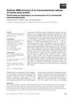
Fig. 2. Crystal structure of PhnI. Ribbon representation of the PhnI a
3
b
3
hexamer along the three-fold symmetry axis (A) and perpendicular
to this axis (B). The three ab units are colored in red, green and blue; the b subunits are represented in lighter tones. Iron atoms are shown
in yellow and sulfur atoms in green. The figures were drawn using the programs
MOLSCRIPT [37] and RASTER 3D [38].
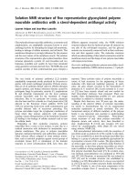
Fig. 3. The PhnI ab heterodimer. Ribbon
representation of the three superposed het-
erodimers in red, green and blue. The a sub-
unit, contains two domains the Rieske
domain with the [2Fe-2S] cluster (residues
38–156) and the catalytic domain (residues
1–37 and 157–454) with the mononuclear
iron. Relevant interactions between domains
and subunits are shown. The figure was
prepared using the programs
MOLSCRIPT [37]
and
RASTER 3D [38].
Terminal oxygenase from Sphingomonas CHY-1 J. Jakoncic et al.
2474 FEBS Journal 274 (2007) 2470–2481 Journal compilation ª 2007 FEBS. No claim to original US government works
consecutive helices, aca10 and aca11 (residues 336–350
and 356–373), which are highly conserved among
RHD structures. Strategically located in the vicinity of
the catalytic iron, aca11 contains residues 356–360 and
carries the totally conserved amino acids Gly
a
354,
Glu
a
357, Asp
a
359 and Asn
a
363, which are part of
a far-reaching hydrogen network surrounding the
catalytic center, as well as Asp
a
360, one of the three
ligands of the catalytic iron atom.
Fully exposed to solvent, the C-terminal region of
the catalytic domain, residues 426–452, containing
a-helices, aca13 and aca14, cover the cap of the catalytic
domain (Fig. 3). Compared with other RHDs, t he C-
terminus is shown to be different in length and amino
acid sequence (Fig. 1). In fact the C-terminal region is
quite different from RHDs of know crystallographic
structure and therefore is not expected to present any
function other than structural.
A large depression, about 20 A
˚
wide, on the surface
of the catalytic domain receives the Rieske domain
from the adjacent a subunit placing the [2Fe-2S] center
in the right conformation with respect to the catalytic
iron. Helix ara2 and the long coil, LCr, anchor the
Rieske domain to the adjacent catalytic domain
between loops acb9 and acb10, acb11 and aca13, and
to loop LI (residues 221–228).
A35A
˚
long cavity extending from the solvent to
the antiparallel b-sheet contains the substrate binding
pocket. With its 12 · 8 · 6A
˚
3
, the PhnI catalytic
pocket is 2A
˚
longer and the largest reported so far
for a RHD. Mostly formed by hydrophobic amino
acids, the pocket is surrounded by two loops exposed
to the solvent, LI (residues 221–238) and LII
(258–265), a-helix, aca6, residues 206–220, containing
two of the mononuclear Fe ligands (His
a
207 and
His
a
212) and helices, aca10 and aca11, which include
Asp
a
360, the third iron ligand. Providing access to the
catalytic pocket loops LI and LII are not completely
represented in the final model. As shown in Fig. 5,
loop LII assumes three different conformations, one
for each of the three a subunits. LI, on the other hand,
could only be partially modeled for one of the three a
subunits, the high flexibility of the loop precluded
modeling for the two other chains.
Interdomain interactions
The a
3
b
3
hexamer is maintained by multiple interdo-
main interactions found in aa, bb and ab interfaces.
Within the same ab heterodimer, strong interactions
give rise to a complex and extended hydrogen network
between residues located at the base of the b subunit
Fig. 4. Superposition of the PhnI ab heterodimer (chains A and B, grey), with NDO-O
9816-4
(blue), CDO-O
IP01
(red), BPDO-O
RHA1
(green) and
NBDO-O
JS765
(yellow). (A) ab heterodimers and (B) catalytic domains. The two solvent exposed loops LI and LII are shown at the entrance
of the catalytic pocket, as well as, the highly conserved helices, aca 10 and aca11. The figure was drawn using the programs
MOLSCRIPT [37]
and
RASTER 3D [38].
J. Jakoncic et al. Terminal oxygenase from Sphingomonas CHY-1
FEBS Journal 274 (2007) 2470–2481 Journal compilation ª 2007 FEBS. No claim to original US government works 2475
and the Rieske and catalytic domains of the a subunit.
In the heterohexamer, the Rieske domain interacts
with the base of the adjacent b subunit and the cata-
lytic domain of the adjacent a subunit. Most of the ab
interactions are conserved at least in the dioxygenases
from the naphthalene family. For instance, the ionic
interaction between Asp
a
91 and Arg
b
163 within one
ab subunit is highly conserved. Another example,
Trp
b
91 at the base of the b subunit (helix ba4)
interacts with Trp
a
210 (helix aca6) from the a subunit
catalytic domain and with Asn
a
101, located on the
gripper structure from the adjacent a subunit Rieske
domain. These and additional numerous interactions
contribute to the cohesion of the a
3
b
3
hexamer and
ultimately favor the catalytic reaction by maintaining
the two redox centers at an appropriate distance from
each other. If multiple a and b interactions are found
in PhnI, the function of the b subunits seem to purely
serve a structural role.
Mononuclear iron
The mononuclear iron is coordinated by a highly con-
served 2-His-1-carboxylate motif [10], His
a
207, His
a
212
and bidentaly by Asp
a
360. The i ron coordination geo-
metry can be described as that of a d istorted octahedron
with the oxygen atom of Asn
a
200, at 4 A
˚
, from the
mononuclear iron atom, occupying the position of a
missing ligand. As observed for other dioxygenases
[16], while the carboxyl oxygen OD1 from Asp
a
360 is
located at 2 A
˚
from the mononuclear iron, the 3 A
˚
coordination distance observed for the Asp
a
360 OD2,
seems rather large compared to the typical 1.9 A
˚
aver-
age distance.
For several dioxygenases the catalytic iron is repor-
ted to be coordinated by one or two water molecules.
In the refined PhnI structure, the three catalytic iron
atoms were found to be coordinated by at least one
water molecule. The crystallographic refinement,
showed a large positive difference in the |Fo|-Fc| elec-
tron density map in two of the three subunits suggest-
ing the existence of an external ligand. The position
of this density is similar to that found for the
NDO-O
98164
crystallographic structure [13] and resem-
bles that of an indole molecule. In the third subunit,
chain E, the refined distance between the two oxygen
atoms, 1.5 A
˚
, suggests the presence of a dioxygen
molecule at the catalytic iron site.
The substrate binding pocket
The PhnI catalytic pocket, the largest reported so far
for RHDs, is at least 2 A
˚
longer, wider and higher at
the entrance when compared to related dioxygenases
[32]. The amino acids lining the PhnI pocket are repre-
sented in Fig. 6 superposed to the NDO-O
98146
cata-
lytic pocket. Only small differences can be observed
between the two structures in the proximal region,
close to the mononuclear iron atom. In the central
region most significant are residues Phe
a
350, Phe
a
404,
Leu
a
356, in PhnI. While Phe
a
404 is replaced by the
smaller residue Ala407 in NDO-O
98146
, Leu
a
356 is
replaced by a bulky aromatic residue (Trp or Phe) in
naphthalene dioxygenases. Together these residues and
the specific conformations of residues Gly
a
205,
Val
a
208, Thr
a
308 contribute to enlarge the PhnI cata-
lytic pocket giving its rather uniform shape without
kinks or torsions as found for other dioxygenases.
Probably the distinctive broad substrate specificity
presented by the dioxygenase from strain CHY-1
toward PAHs [9] can be mostly ascribed to differences
observed in the distal region. Most significant in this
region are residues Leu
a
223 and Leu
a
226 in loop LI,
and Ile
a
253 and Ile
a
260 in loop LII, which most prob-
ably control the access to the catalytic pocket.
To further explore the broad specificity of PhnI
towards high molecular weight PAHs a benz[a]antra-
cene molecule was overlaid to the PhnI substrate bind-
ing pocket. The three most favorable orientations, each
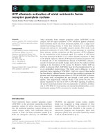
Fig. 5. Surface envelope of the PhnI catalytic pocket. Shown are
the three conformations adopted by loop LII at the entrance of the
catalytic pocket. Loop LI is shown only for chain A as no density
was observed in this region for the two other chains, C and E.
Even for chain A, LI is not fully represented, as no density was
observed for residues 233–236. The figure was made using the
program
PYMOL [39].
Terminal oxygenase from Sphingomonas CHY-1 J. Jakoncic et al.
2476 FEBS Journal 274 (2007) 2470–2481 Journal compilation ª 2007 FEBS. No claim to original US government works
of which corresponded to one of the three dihydrodiol
isomers obtained by enzymatic conversion of this PAH
[9], are shown in Fig. 7. This and several PAHs, known
from enzymatic assays to be dihydroxylated by PhnI,
could be modeled into the PhnI catalytic pocket minim-
izing Van der Waals contacts. These results indicate
that PhnI can bind large substrates made of four or five
rings with minimal or no rearrangement of side chains
[32]. These simulations indicate that amino acids
belonging to loops LI and LII, at the entrance of the
substrate binding pocket, determine the pocket length,
and therefore might play a key role in the substrate
selectivity of the enzyme. Similarly these simulations
showed that Phe
a
350 in the central region of the PhnI
catalytic pocket prevents some specific substrate orien-
tations and therefore is thought to participate in the re-
gio-specificity of the enzyme. Site-specific mutagenesis
of Phe352 in NDO-O
98164
was shown to significantly
alter the regioselectivity of the enzyme [31].
The Asp204 electron transfer bridge
Totally conserved amongst RHDs, Asp
a
204 is buried
in a large depression at the junction of the Rieske
domain and the catalytic domain of neighboring a sub-
unit. In this key position, Asp
a
204 provides a bridge
between the Rieske cluster and the mononuclear iron
center (Fig. 8). In PhnI, Asp
a
204 side chain is located
between His
a
207, ligand to the catalytic iron, and
His
a
103, ligand to the Rieske center in the adjacent a
subunit. Asp
a
204 OD2 is 2.7 A
˚
away from His
a
103
ND2, and OD1 is 3.3 A
˚
from His
a
207 ND1 thus pro-
viding a plausible path for intramolecular electron
transfer. As part of an extended hydrogen network
(Fig. 8) that holds the two redox centers at 12 A
˚
from
each other, Asp
a
204 OD2 is 3.3 A
˚
away from Tyr
a
102
OH (in the adjacent a subunit) and is H-bonded to
Tyr
a
410 OH (2.8 A
˚
). Asp
a
204 OD1 is 3.3 A
˚
from
His
a
207 ND1, and is H bonded to His
a
207 main chain
N atom (2.7 A
˚
). Asp
a
204 main chains atoms O and N
interact with His
a
207 ND1 (2.9 A
˚
) and Asn
a
200 O
(3 A
˚
) atoms, respectively. Specific to this network are
not only highly conserved amino acid side and main
chain interactions, but also interactions with a few
structural waters. The replacement of this aspartic acid
by a Ala, Glu, Gln or Asn in NDO-O
98164
resulted in
a totally inactive enzyme suggesting that it is essential
either directly in electron transfer or in positioning the
two adjacent a subunits to allow effective electron
transfer [33].
Occurrence of a water channel
An 11 A
˚
long channel filled with eight water molecules
extends from the base of the b subunit up to the cata-
lytic site (Fig. 9). The water molecule closest to the
catalytic site is at hydrogen bond distance from
Glu
a
357 and at 4.2 A
˚
from the mononuclear Fe atom.
This channel is also found in other RHDs although
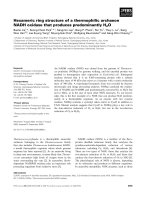
Fig. 6. The superposition of the PhnI and NDO-O
9816-4
catalytic
pocket. The mononuclear Fe ligands are shown in red, PhnI resi-
dues in grey and NDO-O
9816-4
residues in blue. Residues with
similar conformation in both structures are shown in orange. The
largest conformational differences are observed for those residues
at the entrance of the pocket, Leu
a
223, Leu
a
226 and Ile
a
253.
These residues are believed to control the access and the length of
the catalytic pocket while residues in the central region, Phe
a
350,
Leu
a
356 and Phe
a
404 seem to participate in the regio specificity of
the enzyme.
Fig. 7. Superposition of a four ring PAH and the PhnI catalytic
pocket. The molecular surface of a benz[a]antracene molecule, rep-
resented by a mesh, is overlaid on the substrate binding pocket of
PhnI. The three most favorable orientations (A, B and C) shown
requiring minimal rearrangement of residues in the catalytic pocket
correspond to the three dihydrodiol isomers obtained by enzymatic
conversion of this PAH [9].
J. Jakoncic et al. Terminal oxygenase from Sphingomonas CHY-1
FEBS Journal 274 (2007) 2470–2481 Journal compilation ª 2007 FEBS. No claim to original US government works 2477
residues lining the channel are not fully conserved.
Only one of the residues at the entrance of the channel
is conserved throughout the naphthalene dioxygenase
family, Gly
a
354. The function of this channel is not
well understood. Assuming that water molecules serve
as a proton source for the catalytic reaction, the chan-
nel might be a pathway to convey protons to the active
site.
Possible role of Asn
a
200
Located in the vicinity of the mononuclear iron but
further buried in the catalytic pocket, Asn
a
200 is one
of the closest residues to the catalytic iron (4.0 A
˚
) but
not close enough to be a ligand. As Asp
a
204, Asn
a
200
participates in the extended hydrogen network at the
junction of two neighboring a subunits (Fig. 8).
Through Tyr
a
102, in the adjacent a subunit Rieske
domain, Asn
a
200 provides a bridge to Cys
a
100 one of
the Rieske ligands; Asn
a
200 ND2 atom is 2.8 A
˚
away
from Tyr
a
102 hydroxyl group while Cys
a
100, is hydro-
gen bonded through main chains to Trp
a
105, Gly
a
104
and Tyr
a
102.
A theoretical analysis predicts that Asn201 in NDO-
O
98164
would be at hydrogen-bond distance from the
hydroxyl of the enzyme reaction product during a
transition state [34]. In PhnI, the ND2 side chain atom
of Asn
a
200 is 3A
˚
away from one of the water mole-
cules bound to the active site. In the catalytic site of
BPDO-O
RHA1
[16], although the asparagine is replaced
by a glutamine, a hydrogen bond has also been
observed between the side chain atom NE2 and the
water molecule present at the active site. Asn (Gln)
may assist in the stereospecific reaction as it may con-
strain the oxygen through hydrogen bonds. The role of
Asn201 in NDO-O
98164
was tested by substitution of
this residue by Gln, Ser or Ala [35]. The enzyme activ-
ity was significantly reduced but not totally abolished.
It was therefore concluded that Asn201 is not essen-
tial for catalysis, but may be important for maintain-
ing protein–protein interactions between a subunits
Fig. 8. Rieske domain and catalytic domain
of neighboring a subunits. Ligands to the
reaction centers, and residues Asn
a
200 and
Asp
a
204 believed to be involved in the
electron transfer to the catalytic site are
shown in red. Also shown in red are
relevant water molecules in the hydrogen
network. In the background the catalytic
surface envelope of the PhnI pocket
showing the available internal space.
Fig. 9. The PhnI water channel. The channel surface is shown in
blue in the foreground and the surface of the catalytic pocket in
orange in the back. Structural water molecules are shown in red at
the entrance and inside the channel. At the end of the channel a
green mesh represents molecule of benzo[a]pyrene a five ring PAH
superposed into the catalytic pocket. Partial ribbon diagram of the
b subunit, chain B, and a subunit, chain A, are shown in orange and
green, respectively. The figure was made using
PYMOL [39].
Terminal oxygenase from Sphingomonas CHY-1 J. Jakoncic et al.
2478 FEBS Journal 274 (2007) 2470–2481 Journal compilation ª 2007 FEBS. No claim to original US government works
through its H bond with Tyr103 (Tyr
a
102) in the adja-
cent subunit.
In conclusion, the PhnI oxygenase is similar in struc-
ture to the catalytic component of other RHDs, especi-
ally naphthalene dioxygenases. The exceptionally
broad substrate specificity of this enzyme, and in par-
ticular, its ability oxidize large PAH molecules, may be
explained by the large size of its substrate-binding
pocket and the flexibility of residues located at the
entrance. While residues Phe
a
350, Phe
a
404 and
Leu
a
356, shape the pocket and likely influence the reg-
iospecificity of the enzyme, the access to the catalytic
site is most probably controlled by residues in loop LI,
especially Leu
a
223 and Leu
a
226. The present structure
represents a valuable frame to investigate the role
of certain residues on the substrate specificity and ⁄ or
catalytic activity of the enzyme through site-directed
mutagensis.
Experimental procedures
Purification and crystallization of PhnI
The overexpression of recombinant His-tagged PhnI
(ht-PhnI) in P. putida KT2442 and the purification of the
protein were carried out as described by Jouanneau et al.[9].
The oxygenase was further purified by two chromato-
graphic steps under argon. The ht-PhnI preparation was
treated with 0.25 U thrombin ⁄ mg (Sigma-Aldrich, St Louis,
MO, USA) for 16 h at 20 °Cin25mm Tris ⁄ HCl, pH 8.0,
containing 0.15 m NaCl, 2.0 mm CaCl
2
, 0.1 mm
Fe(NH
4
)
2
(SO
4
)
2
and 5% glycerol, then applied to a small
column of TALON affinity chromatography (BD Bio-
sciences, Ozyme, France). The unbound protein fraction
was concentrated on a small DEAE-cellulose column, then
applied to a 2.6 · 110 cm column of gel filtration (AcA34,
Biosepra, Villeneuve, France) eluted at a flow rate of
50 mLÆh
)1
with 25 mm Tris ⁄ HCl, pH 7.5, containing 0.1 m
NaCl, and 5% glycerol. The purified protein was concen-
trated to about 31 mgÆmL
)1
, and frozen as pellets in liquid
nitrogen.
Searches for preliminary crystallization conditions were
carried out using the vapor diffusion method in the hanging
drop configuration. EasyXtal Cryos Suite (Nextal Biotech-
nologies, Montreal, Quebec, Canada) solution number 67
produced small, poorly diffracting crystals within 12 h at
20 °C. Upon refining the crystallization conditions, 250 lm
long crystals were obtained in <8 h in a sitting-drop con-
figuration, by mixing 1 lL of purified PhnI, with 1 l Lof
mother liquor (11% PEG8000, 5% ethanol, 100 mm Hepes
pH 7.0, 15% glycerol, 400 mm (CH
3
COO)
2
Ca and 150 mm
NaCl). To improve the diffraction quality, the nucleation
and crystal growth process were slowed down by covering
each well with 300 lL of mineral oil [21].
Data collection and processing
Diffraction data were recorded at the X6A beam line at the
National Synchrotron Light Source (NSLS; Upton, NY,
USA) [22]. Native crystals directly recovered from the sit-
ting drop, were cooled at 100 K in a cold stream of liquid
nitrogen. A total of 750 frames (oscillation width 0.2°) were
collected on native crystals. Diffraction data were inspected,
indexed, integrated and scaled with the HKL2000 program
suite [23]. Data collection and processing statistics are sum-
marized in Table 1.
Structure solution and refinement
The structure of PhnI was solved by molecular replacement
using molrep [24] after the failure of several experimental
phasing techniques. Based on sequence homology and struc-
tural similarity, the search model for the a subunit consisted
of the naphthalene dioxygenase NDO-O
9816-4
(PDB access
code 1NDO) a subunit while for the b subunit, the cumene
dioxygenase CDO-O
IP01
(PDB access code 1WQL) b sub-
unit was chosen. For both subunits, only main chain atoms
were kept; regions presenting high flexibility and high rmsd
were not considered in the model. Density modification with
noncrystallographic three-fold symmetry (NCS) averaging
[25] was applied according to the solvent content deter-
mined from Matthews Coefficient probability [26]. The ab
heterodimer presenting the best electron density was com-
pleted automatically with arpwarp [27] and manually with
coot [28]; the two other heterodimers were generated using
NCS operators. Restrained refinement was carried out with
refmac [29]. During the final refinement steps, the iron
atom and the [2Fe-2S] cluster were refined with no restrains
on the geometry and coordination. The final model was
analyzed with procheck [30].
Acknowledgements
The authors thank the staff of the National Synchro-
tron Light Source, Brookhaven National Laboratory
(Upton, NY, USA) for their continuous support. This
work was supported by grants from the National Insti-
tute of Health, NIGMS number GM-0080, US
Department of Energy, Bes, number DE-AC02–
98CH10886, and the Centre National de la Recherche
Scientifique, Commisariat a
`
l’Energie Atomique and
Universite
´
Joseph Fourier to UMR5092.
References
1 Juhasz AL & Naidu R (2000) Bioremediation of high
molecular weight polycyclic aromatic hydrocarbons: a
review of the microbial degradation of benzo[a]pyrene.
Int Biodet Biodegr 45, 57–88.
J. Jakoncic et al. Terminal oxygenase from Sphingomonas CHY-1
FEBS Journal 274 (2007) 2470–2481 Journal compilation ª 2007 FEBS. No claim to original US government works 2479
2 Kanaly R & Harayama S (2000) Biodegradation of
high-molecular-weight polycyclic aromatic hydrocarbons
by bacteria,. J Bacteriol 182, 2059–2067.
3 Gibson DT, Mahadevan V, Jerina DM, Yogi H & Yeh
HJ (1975) Oxidation of the carcinogens benzo[a]pyrene
and benzo[a]anthracene to dihydrodiols by a bacterium.
Science 189, 295–297.
4 Dean D & Ross Cerniglia CE (1996) Degradation of
pyrene by Mycobacterium flavescens. Appl Microbiol
Biotechnol 46, 307–312.
5 Boyd DR, Sharma ND, Agarwal R, Resnick SM,
Schocken MJ, Gibson DT, Sayer JM, Yagi H & Jerina
DM (1997) Bacterial dioxygenase-catalysed dihydroxyla-
tion and chemical resolution routes to enantiopure cis-
dihydrodiols of chrysene. J Chem Soc Perkin Trans 1,
1715–1723.
6 Krivobok S, Kuony S, Meyer C, Louwagie M, Willison
JC & Jouanneau Y (2003) Identification of pyrene-
induced proteins in Mycobacterium sp. Strain 6PY1:
evidence for two ring-hydroxylating dioxygenase,.
J Bacteriol 185, 3828–3841.
7 Willison JC (2004) Isolation and characterization of a
novel sphingomonad capable of growth with chrysene
as sole carbon and energy source. FEMS Microbiol Lett
241, 143–150.
8 Demaneche S, Meyer C, Micoud J, Louwagie M,
Willison JC & Jouanneau Y (2004) Identification and
functional analysis of two aromatic-ring-hydroxylating
dioxygenases from a Spingomonas strain that degrades
variuous polycyclic aromatic hydrocarbons. App Environ
Microbiol 70, 6714–6725.
9 Jouanneau Y, Meyer C, Jakoncic J, Stojanoff V &
Gaillard J (2006) Characterization of a ring-hydroxylat-
ing dioxygenase able to oxidize a wide range of poly-
cyclic aromatic hydrocarbons including four and five
ring carcinogens. Biochemistry 45, 12380–12391.
10 Werlen C, Kohler HPE & van der Meer JR (1996) The
broad substrate chlorobenzene dioxygenase and cis-
chlorobenzene dihydrodiol dehydrogenase of Pseudomo-
nas sp. strain P51 are linked evolutionarily to the
enzymes for benzene and toluene degradation,. J Biol
Chem 271, 4009–4016.
11 Kauppi B, Lee K, Carredano E, Parales RE, Gibson
DT, Eklund H & Ramaswamy ( S (1998) Structure of
an aromatic-ring-hydroxylating dioxygenase-naphtha-
lene 1.2-Dioxygenase, Structure 6, 571–586.
12 Carredano E, Karlsson A, Kauppi B, Choudhury D,
Parales RE, Parales JV, Lee K, Gibson DT, Eklund H
& Ramaswamy S (2000) Substrate binding site of
naphthalene 1,2-dioxygenase: functional implications of
indole binding,. J Mol Biol 296, 701–712.
13 Karlsson A, Parales JV, Parales RE, Gibson DT,
Eklund H & Ramaswamy S (2003) Crystal structure of
naphthalene dioxygenase: side-on binding of dioxygen
to iron. Science 299, 1039–1042.
14 Gakhar L, Malik ZA, Allen CC, Lipscomb DA, Larkin
MJ & Ramaswamy S (2005) Structure and increased
thermostability of Rhodococcus sp. naphthalene
1,2-dioxygenase. J Bacteriol 187, 7222–7231.
15 Friemann R, Ivkovic-Jensen MM, Lessner DJ,
Gibson CL, Yu DT, Parales RE, Eklund H &
Ramaswamy S (2005) Structural insight into the
dioxygenation of nitroarene compounds: the crystal
structure of nitrobenzene dioxygenase,. J Mol Biol 348,
1139–1151.
16 Furusawa Y, Nagarajan V, Tanokura M, Masai E,
Fukuda M & Senda T (2004) Crystal structure of the
terminal oxygenase component of biphenyl dioxygenase
derived from Rhodococcus sp. strain RHA1. J Mol Biol
342, 1041–1052.
17 Dong X, Fushinobu S, Fukuda E, Terada T, Nakamura
S, Shimizu K, Nojiri H, Omori T, Shoun H & Wakagi
T (2005) Crystal structure of the terminal oxygenase
component of cumene dioxygenase from Pseudomonas
fluorescens IP01. J Bacteriol 1872483–1872490.
18 Martins BM, Svetlitchnaia T & Dobbek H (2005)
2-Oxoquinoline 8-monooxygenase oxygenase compo-
nent: active site modulation by Rieske-[2Fe)2S] center
oxidation ⁄ reduction,. Structure 13, 817–824.
19 Nojiri H, Ashikawa Y, Noguchi H, Nam JW, Urata M,
Fujimoto Z, Uchimura H, Terada T, Nakamura S,
Shimizu K et al. (2005) Structure of the terminal
oxygenase component of angular dioxygenase, carbazole
1,9a-dioxygenase. J Mol Biol 351, 355–370.
20 Que JL & Ho RYN (1996) Dioxygen activation by
enzymes with mononuclear non-heme iron active sites.
Chem Rev 96, 2607–2624.
21 Chayen NE (1998) Comparative studies of protein
crystallization by vapour-diffusion and microbatch
techniques. Acta Cryst D54, 8–15.
22 Allaire M, Aslantas M, Berntson A, Berman L, Cheung
S, Clay B, Greene R, Jakoncic J, Johnson E, Kao CC
et al. (2003) Modular approach to beam line automa-
tion: the NIGMS facility at the NSLS. Synchr Rad
News 16, 20–25.
23 Otwinowski Z & Minor W (1997) Processing of X-ray
diffraction data collected in oscillation mode. Methods
Enzymol 276, 307–326.
24 Vagin A & Teplyakov A (1997) molrep: an automated
program for molecular replacement. J Appl Cryst 30,
1022–1025.
25 Cowtan K (1994) ‘dm’: An automated procedure for
phase improvement by density modification. Joint
CCP4 ESF-EACBM Newsletter Protein Crystallogr 31,
34–38.
26 Mathews BW (1968) Solvent content of protein crystals.
J Mol Biol 33, 491–497.
27 Perrakis A, Morris RM & Lamzin VS (1999) Auto-
mated protein model building combined with iterative
structure refinement. Nature Struct Biol 6, 458–463.
Terminal oxygenase from Sphingomonas CHY-1 J. Jakoncic et al.
2480 FEBS Journal 274 (2007) 2470–2481 Journal compilation ª 2007 FEBS. No claim to original US government works
28 Emsley P & Cowtan K (2004) coot. Model-building
tools for mol graphics. Acta Cryst D60, 2126–2132.
29 Murshudov GN, Vagin AA & Dodson EJ (1997)
Refinement of macromolecular structures by the
maximum-likelihood method. Acta Cryst D53, 240–255.
30 Laskowski RA, MacArthur MW, Moss DS & Thornton
JM (1993) procheck: a program to check the stereo-
chemical quality of protein structures. J App Cryst 26,
283–291.
31 Parales RE (2003) The role of active site residues in
naphthalene dioxygenase. J Ind Microbiol Biotechnol 30,
271–278.
32 Jakoncic J, Jouanneau Y, Meyer C & Stojanoff V
(2007) The catalytic pocket of the ring-hydroxylating
dioxygenase from Sphingomonas CHY-1. Biochem
Biophys Res Commun 352, 861–868.
33 Parales RE, Parales JV & Gibson DT (1999)
Aspartate 205 in the catalytic domain of naphthalene
dioxygenase is essential for activity,. J Bacteriol 181,
1831–1837.
34 Bassan A, Blomberg MR & Siegbahn PE (2004) A theo-
retical study of the cis-dihydroxylation mechanism in
naphthalene 1,2-dioxygenase,. J Biol Inorg Chem 9,
439–452.
35 Parales RE, Lee L, Resnick SM, Jiang H, Lessner DJ &
Gibson DT (2000) Substrate specificity of naphthalene
dioxygenase: effect of specific amino acids at the active
site of the enzyme. J Bacteriol 182, 1641–1649.
36 Higgins D, Thompson J, Gibson T, Thompson JD,
Higgins DG & Gibson TJ (1994) clustal w: improving
the sensitivity of progressive multiple sequence align-
ment through sequence weighting, position-specific gap
penalties and weight matrix choice. Nucleic Acids Res
224, 673–4680.
37 Kraulis PJ (1991) molscript: a program to produce
both detailed and schematic plots of protein structures.
J Appl Cryst 24, 946–950.
38 Merritt EA & Murphy ME (1994) raster3d, Version
2.0: a program for photorealistic molecular graphics.
Acta Cryst D50, 869–873.
39 DeLano WL (2002) The PyMOL molecular graphics
system. DeLano Scientific, Palo Alto, CA.
J. Jakoncic et al. Terminal oxygenase from Sphingomonas CHY-1
FEBS Journal 274 (2007) 2470–2481 Journal compilation ª 2007 FEBS. No claim to original US government works 2481