Báo cáo khoa học: Biochemical and structural characterization of mammalian-like purine nucleoside phosphorylase from the Archaeon Pyrococcus furiosus pptx
Bạn đang xem bản rút gọn của tài liệu. Xem và tải ngay bản đầy đủ của tài liệu tại đây (875.73 KB, 14 trang )
Biochemical and structural characterization of
mammalian-like purine nucleoside phosphorylase
from the Archaeon Pyrococcus furiosus
Giovanna Cacciapuoti
1
, Sabrina Gorassini
1
, Maria Fiorella Mazzeo
2
, Rosa Anna Siciliano
2
,
Virginia Carbone
2
, Vincenzo Zappia
1
and Marina Porcelli
1
1 Dipartimento di Biochimica e Biofisica ‘F. Cedrangolo’, Seconda Universita
`
di Napoli, Italy
2 Centro di Spettrometria di Massa Proteomica e Biomolecolare, Istituto di Scienze dell’Alimentazione del CNR, Avellino, Italy
Purine nucleoside phosphorylase (PNP) catalyzes the
reversible phosphorolytic cleavage of the glycosidic
bond of purine nucleosides to produce ribose-1-phos-
phate and a free purine base [1–3]. PNPs have been
characterized in a variety of species and may be
grouped into two main groups, PNP-1 and PNP-2.
PNP-1 are found in prokaryotes, are homohexamers
with a subunit of 26 kDa and recognize both 6-oxo
and 6-amino purine nucleosides as substrates. PNP-2
are homotrimers, with a subunit molecular mass of
Keywords
CXC motif; 5¢-deoxy-5¢-methylthioadenosine
phosphorylase; disulfide bonds;
hyperthermostability; purine nucleoside
phosphorylase
Correspondence
G. Cacciapuoti, Dipartimento di Biochimica e
Biofisica ‘F. Cedrangolo’, Seconda Universita
`
di Napoli, Via Costantinopoli 16, 80138,
Napoli, Italy
Fax ⁄ Tel: +39 081 5667519
E-mail:
(Received 2 February 2007, revised 6 March
2007, accepted 12 March 2007)
doi:10.1111/j.1742-4658.2007.05784.x
We report here the characterization of the first mammalian-like purine
nucleoside phosphorylase from the hyperthermophilic archaeon Pyrococcus
furiosus (PfPNP). The gene PF0853 encoding PfPNP was cloned and
expressed in Escherichia coli and the recombinant protein was purified to
homogeneity. PfPNP is a homohexamer of 180 kDa which shows a much
higher similarity with 5¢-deoxy-5¢-methylthioadenosine phosphorylase
(MTAP) than with purine nucleoside phosphorylase (PNP) family mem-
bers. Like human PNP, PfPNP shows an absolute specificity for inosine
and guanosine. PfPNP shares 50% identity with MTAP from P. furiosus
(PfMTAP). The alignment of the protein sequences of PfPNP and PfM-
TAP indicates that only four residue changes are able to switch the specif-
icity of PfPNP from a 6-oxo to a 6-amino purine nucleoside phosphorylase
still maintaining the same overall active site organization. PfPNP is highly
thermophilic with an optimum temperature of 120 °C and is characterized
by extreme thermodynamic stability (T
m
, 110 °C that increases to 120 °C
in the presence of 100 mm phosphate), kinetic stability (100% residual
activity after 4 h incubation at 100 °C), and remarkable SDS-resistance.
Limited proteolysis indicated that the only proteolytic cleavage site is
localized in the C-terminal region and that the C-terminal peptide is not
necessary for the integrity of the active site. By integrating biochemical
methodologies with mass spectrometry we assigned three pairs of intrasub-
unit disulfide bridges that play a role in the stability of the enzyme against
thermal inactivation. The characterization of the thermal properties of the
C254S ⁄ C256S mutant suggests that the CXC motif in the C-terminal
region may also account for the extreme enzyme thermostability.
Abbreviations
hMTAP, human 5¢-deoxy-5¢-methylthioadenosine phosphorylase; MTA, 5¢-deoxy-5¢-methylthioadenosine; MTAP, 5¢-deoxy-5¢-
methylthioadenosine phosphorylase; PfMTAP, 5¢-deoxy-5¢-methylthioadenosine phosphorylase from Pyrococcus furiosus; PfPNP, purine
nucleoside phosphorylase from P. furiosus; PNP, purine nucleoside phosphorylase; SsMTAP, 5¢-deoxy-5¢-methylthioadenosine phosphorylase
from Sulfolobus solfataricus; SsMTAPII, 5¢-deoxy-5¢-methylthioadenosine phosphorylase II from S. solfataricus.
2482 FEBS Journal 274 (2007) 2482–2495 ª 2007 The Authors Journal compilation ª 2007 FEBS
30 kDa and accept only guanosine and inosine as
substrates [3–5]. It is interesting to note that many
organisms that express PNP-1 also express PNP-2 [5].
PNP is a ubiquitous enzyme of purine metabolism
that functions in the salvage pathway of cells. In addi-
tion to the intrinsic biochemical significance, PNP plays
an important biomedical role. In fact, human PNP is a
target for T-cell-related cancers and autoimmune dis-
eases [6]. Moreover, differences in substrate specificity
between Escherichia coli PNP and the human enzyme
have been employed for the development of tumor-
directed gene therapy [5,7–10]. In this strategy, tumor
cells transfected with E. coli PNP gene are able to
convert relatively nontoxic prodrugs into membrane-
permeant cytotoxic compounds. To reduce the toxicity
of prodrugs currently used with E. coli PNP, a good
experimental approach could be the identification of
PNPs with new substrate specificities. In this light,
studies on the molecular and structural characterization
of PNPs from hyperthermophilic Archaea could be
useful to improve the tumor-directed gene therapy
based on the activation of nucleoside analogs prodrugs.
Hyperthermophilic Archaea are of extreme biotechno-
logical interest not only for the exceptional stability of
their biomolecules but also for the peculiar substrate
specificity of their enzymes that provide unique models
for studying and understanding enzyme evolution in
terms of structure, specificity and catalytic properties
[11–15]. In recent years, the increasing number of
solved crystallographic structures has highlighted the
presence of disulfide bonds in several hyperthermo-
philic proteins [16–20], suggesting that disulfide bond
formation represents a significant molecular strategy
adopted by cytosolic hyperthermophilic proteins to
reach higher levels of thermostability.
In Archaea, three enzymes belonging to the PNP fam-
ily have recently been isolated and characterized from
the hyperthermophilic microorganisms Sulfolobus solfa-
taricus (Ss) and Pyrococcus furiosus (Pf). These enzymes
are classified as 5¢-deoxy-5¢-methylthioadenosine phos-
phorylases, as they are able to catalyze the phosphoroly-
tic cleavage of 5¢-deoxy-5¢-methylthioadenosine (MTA),
a natural sulfur-containing nucleoside formed from
S-adenosylmethionine mainly through polyamine bio-
synthesis [21,22]. The three enzymes, 5¢-deoxy-5¢-methyl-
thioadenosine phosphorylase from S. solfataricus
(SsMTAP), 5¢-deoxy-5¢-methylthioadenosine phosphor-
ylase II from S. solfataricus (SsMTAPII) and 5¢-deoxy-
5¢-methylthioadenosine phosphorylase from P. furiosus
(PfMTAP) show features of exceptional thermophilicity
and thermostability with temperature optima and
melting temperatures >100 °C [23–25] and are stabil-
ized by disulfide bonds [16,20,26]. SsMTAP, which
shows a significant sequence identity with E. coli PNP,
is a hexamer consisting of six identical subunits of
26.5 kDa and utilizes inosine, guanosine, adenosine,
and MTA as substrates [23]. The crystal structure of
SsMTAP reveals that it contains three intermonomer
disulfide bridges in each hexamer [16]. SsMTAP II is a
homohexamer (subunit 30 kDa), characterized by extre-
mely high affinity towards MTA. SsMTAPII shares
51% identity with human 5¢-deoxy-5¢ -methylthioadeno-
sine phosphorylase (hMTAP) and is able to recognize
adenosine [24] in contrast to hMTAP, which is highly
specific for MTA. The crystal structure of SsMTAPII
indicates a dimer of trimers with two pairs of intrasub-
unit disulfide bridges [20]. Finally, PfMTAP is a hexa-
meric protein that, like SsMTAPII, shares 50% identity
with hMTAP. PfMTAP is characterized by a broad sub-
strate specificity with 20-fold higher catalytic efficacy for
adenosine and MTA than for inosine and guanosine
[25]. PfMTAP is stabilized by two intrasubunit disulfide
bridges [26].
The analysis of the complete genomic sequence of
P. furiosus shows, beside PfMTAP, a second enzyme
that, on the basis of the high identity with PfMTAP is
annotated as MTAPII. We renamed this enzyme as
PNP as it is completely unable to cleave MTA while, in
analogy with human PNP, it is characterized by a strict
substrate specificity towards inosine and guanosine.
This paper describes the cloning, recombinant
expression and structural and functional characteri-
zation of purine nucleoside phosphorylase from the
hyperthermophilic archaeon P. furiosus (PfPNP) aimed
to elucidate the structure ⁄ function ⁄ stability relation-
ship in this enzyme and to explore its biotechnological
applications. By integrating classical biochemical meth-
odologies with mass spectrometry, we assigned three
intrasubunit disulfide bridges important for the enzyme
stability. Finally, the characterization of the thermal
properties of the C254S ⁄ C256S mutant allowed us to
propose that the CXC motif in the C-terminal region
of PfPNP may also account for the extreme thermo-
stability of the enzyme. PfPNP, on the basis of its
substrate specificity is the first example of a mamma-
lian-like PNP reported in Archaea.
Results and Discussion
Analysis of PfPNP gene, primary sequence
comparison and expression
The analysis of the complete sequenced genome of
P. furiosus revealed an open reading frame (PF0853)
encoding a 265-amino acid protein homologous to
hMTAP. This enzyme is annotated as hypothetical
G. Cacciapuoti et al. Purine nucleoside phosphorylase from P. furiosus
FEBS Journal 274 (2007) 2482–2495 ª 2007 The Authors Journal compilation ª 2007 FEBS 2483
MTAPII and has been renamed by us PfPNP. The
putative molecular mass of the protein predicted from
the gene was 29 208 Da. The coding region starts with
an ATG triplet at the position 826577 of the P. furio-
sus genome. The first stop codon TAG is encountered
at the position 827374. Upstream from the coding
region 24 bp before the starting codon there is a
stretch of purine-rich nucleosides (CCTCC) that may
function as the ribosome-binding site [27]. Putative
promoter elements, which are in good agreement with
the archaeal consensus [27] designed box A and box B
are found close to the transcription start site. A hexa-
nucleotide with the sequence TATTATA similar to the
box A is located 19 bp upstream from the start codon
and resembles the TATA box which is involved in
binding the archaeal RNA polymerase [27]. A putative
box B (ATGC) overlaps the ATG codon. Finally, a
pyrimidine-rich region (TTTTTAT) strictly resembling
the archaeal terminator signal [27], is localized 8 bp
downstream from the translation stop codon.
To overproduce PfPNP, the gene was amplified by
PCR and cloned into pET-22b(+) under the T
7
RNA
polymerase promoter. The gene sequence was found to
be identical with the published sequence [28] except for
a single mutation at the third codon, where A was sub-
stituted with G resulting in Arg instead of Gly. Since
in repeated gene amplification experiments carried out
utilizing different preparation of the same primers we
always obtained the same result, it is possible to hypo-
thesize that a mistake is present in GenBank at level
of the third codon of PfPNP gene. Comparison of
the deduced primary sequence of PfPNP with enzy-
mes present in GenBank Data Base reveals a much
higher similarity of PfPNP with members of MTAP
family, such as MTAP from Pyrococcus abyssi (87%
identity), MTAP from Pyrococcus horikoshii (84%
identity), MTAP from Thermococcus kodakarensis
(76% identity), than with members of PNP family such
as PNP from Methanopyrus kandlery AV19 (51% iden-
tity) and PNP from Aquifex aeolicus (47% identity).
This evidence could also be noted by comparing the
amino acid sequence of PfPNP with related enzymes
characterized from various sources, that indicated a
high sequence identity with PfMTAP (50%), SsMTAP-
II (48%) and hMTAP (40%) while a lower identity
was observed with E. coli PNPII (30%) and hPNP
(27%). No significant similarity was found with E. coli
PNP, SsMTAP, and PNP from Thermus thermophilus.
The recombinant PfPNP was produced in a soluble
form in E. coli BL21 cells harboring the plasmid pET-
PfPNP at 37 °C in the presence of isopropyl-b-d-thio-
galactoside. Under the experimental conditions selected
for the expression, about 10 g of wet cell paste was
obtained from 1 L of culture. The PfPNP activity of
recombinant E. coli BL21 cells harboring pET-PfPNP,
was 17.9 unitsÆmg
)1
at 80 °C, confirming that PfPNP
gene had been cloned and expressed.
Enzyme purification and properties
Recombinant PfPNP was purified to homogeneity by a
fast and efficient two-step procedure that utilizes a
heat treatment and affinity chromatography on MTI-
Sepharose (Table 1). SDS⁄ PAGE of PfPNP reveals a
single band with a molecular mass of 29 ± 1 kDa,
which is in fair agreement with the expected mass cal-
culated from the amino acid sequence. The identity of
the protein was checked by N-terminal sequencing
which also revealed that the initial methionine was
post-translationally removed. This result was con-
firmed by MALDI-MS analysis of the HPLC purified
protein. The experimental mass value (m ⁄ z 28 966.23)
was in good agreement with the theoretical average
molecular mass of the full length gene product without
the N-terminal methionine (28 977.39 Da), being the
observed mass difference partly due to the presence of
disulfide bridges.
The molecular mass of PfPNP was estimated to be
180 ± 9 kDa by size exclusion chromatography, which
indicated a hexameric structure in solution. Therefore,
on the basis of its quaternary structure PfPNP is a mem-
ber of the hexameric group of PNPs (PNP-1) together
with the structurally characterized PNPs from Archaea,
including SsMTAP [16,23], SsMTAPII [20,24], and
PfMTAP [25,26] and from Bacteria, such as PNP
from E. coli (EcPNP) [29], PNP from T. thermophilus
(TtPNP) [30], and E. coli uridine phosphorylase [31].
Substrate specificity and comparative kinetic
characterization
To elucidate the physiological role of PfPNP and
its functional relationships with PfMTAP, we carried
Table 1. Purification of recombinant purine nucleoside phosphory-
lase from P. furiosus. A typical purification from 10 g of wet cells is
shown.
Total
protein
(mg)
Total
activity
(units)
Specific
activity
a
(unitsÆmg
)1
)
Yield
(%)
Purification
(n-fold)
Crude extract 134.0 126.9 0.95 100 1
Heat treatment 15.9 114.25 7.18 90.1 7.5
MTI-Sepharose 3.3 59.0 17.9 46.5 18.8
a
Specific activity is expressed as nmol of hypoxanthine formed per
min per mg of protein at 80 °C.
Purine nucleoside phosphorylase from P. furiosus G. Cacciapuoti et al.
2484 FEBS Journal 274 (2007) 2482–2495 ª 2007 The Authors Journal compilation ª 2007 FEBS
out a detailed kinetic characterization of PfPNP and a
comparative kinetic analysis of the two enzymes.
Initial velocity studies carried out with increasing
concentrations of purine nucleosides in the presence of
saturating concentration of phosphate gave typical
Michaelis–Menten kinetics. While PfMTAP showed a
broad substrate specificity being able to phosphorolyti-
cally cleave both 6-amino and 6-oxo purine nucleosides
[25], PfPNP, in analogy with mammalian enzyme, is
specific for guanosine and inosine with K
m
values of
122 and 322 lm, respectively. Moreover, the relative
efficiency of the nucleoside substrates was determined
by comparing the respective k
cat
⁄ K
m
ratios. As shown
in Table 2, the substrate activity of PfPNP with ino-
sine and guanosine gave comparable k
cat
⁄ K
m
values
(2.61 · 10
7
and 2.2 · 10
7
, respectively) that are four
orders of magnitude higher than those of PfMTAP for
the same substrates, indicating that PfPNP is the
enzyme physiologically involved in the 6-oxo-purine
nucleoside catabolism in P. furiosus. When phosphate
concentration was varied at fixed saturating concentra-
tion of inosine, non-Michaelis–Menten kinetics were
observed with two different K
m
values for phosphate
of 6.2 and 259 lm. This result is in agreement with the
data reported in the literature on the complexity of
phosphate binding for PfMTAP [25] and for PNPs
from various sources [32,33].
The results of substrate specificity studies are
supported by the analysis of the sequence alignment
of PfPNP, PfMTAP, hMTAP and hPNP reported
in Fig. 1. The amino acid residues of PfPNP and
Table 2. Kinetic parameters of PfPNP and PfMTAP. Activities were
determined at 80 °C as described in Experimental procedures.
K
mapp
(lM) k
cat
(s
)1
) k
cat ⁄
K
m app
(s
)1
ÆM
)1
)
PfPNP
Inosine 322 84.19 2.61 · 10
7
Guanosine 122 28.05 2.20 · 10
7
PfMTAP
a
MTA 147 24.46 1.66 · 10
5
Adenosine 109 22.79 2.09 · 10
5
Inosine 963 9.38 9.74 · 10
3
Guanosine 916 7.31 7.98 · 10
3
a
The data for PfMTAP have already been published [25].
Fig. 1. Multiple sequence alignment of PfPNP, PfMTAP, hMTAP, and hPNP. The phosphate (w) ribose, (m) and base (d) binding sites of
hMTAP (above the sequence) and of hPNP (below the sequence) are indicated. Identical residues between PfPNP and PfMTAP at the hypo-
thetical active sites are highlighted in a grey box. PfPNP cysteine residues are shown in white lettering on a black background.
G. Cacciapuoti et al. Purine nucleoside phosphorylase from P. furiosus
FEBS Journal 274 (2007) 2482–2495 ª 2007 The Authors Journal compilation ª 2007 FEBS 2485
PfMTAP corresponding to those present at the active
sites of hPNP [34] and hMTAP [35], respectively, were
compared with highlight the changes that may account
for the difference in substrate specificity among the
two P. furiosus enzymes. As expected on the basis of
the very high sequence identity (50%), the hypothetical
active sites of PfPNP and PfMTAP are very similar
and only few key residue changes are observable.
Three important substitutions are localized at the level
of the base binding site where Glu169, Asn211, and
Ala213 of PfPNP replace Ser163, Asp204 and Asp206
of PfMTAP, respectively. It is important to note that
these substitutions are exactly those that are respon-
sible for the different substrate specificity of hPNP and
hMTAP (Glu201, Asn243 and Val245 of hPNP instead
of Ser178, Asp220 and Asp222 of hMTAP, respect-
ively). The last important substitution is observable at
the ribose pocket where His223 of PfPNP substitutes
Ala215 of PfMTAP. Also in this case, the same substi-
tution takes place in mammalian enzyme where the
change of His257 of hPNP with Val233 of hMTAP
makes hydrophilic the hydrophobic pocket, preventing
the binding of the 5-methylthioribose moiety. As for
the remaining differences between the hypothetical
active sites of the two P. furiosus enzymes, they are all
conservative substitutions except for the change of
Ile56 of PfPNP with Phe57 of PfMTAP. It is interest-
ing to note in this respect that the corresponding resi-
due Tyr88 of hPNP is not determinant since the
interactions between PNP and sugar ring are primarily
hydrophobic [34]. In conclusion, only four substitu-
tions are able to switch the specificity of the enzyme
from 6-oxo to 6-amino purine nucleoside phosphory-
lase still maintaining the same overall active site organ-
ization. On the basis of the reported results, PfPNP
shows peculiar structural and functional properties.
The enzyme, in fact, although characterized by the
hexameric quaternary structure distinctive of bacterial
PNP, exhibits a substrate specificity that makes it the
first archaeal mammalian-like PNP.
Thermal properties and limited proteolysis
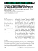
The temperature dependence of the activity of PfPNP in
the range from 30 °C to 140 °C is shown in Fig. 2. The
enzyme is highly thermoactive; its activity increased
sharply up to the optimal temperature of 120 °C and a
50% activity was still observed at 133 °C. This behavior
led to a discontinuity in the Arrhenius plot at about
84 °C, with two different activation energies.
To study the thermodynamic stability of PfPNP we
measured the residual activity after 10 min incubation
at increasing temperature. The corresponding diagram
reported in Fig. 3A is characterized by a sharp trans-
ition that allowed us to calculate an apparent melting
temperature of 110 °C. This value increases to 120 °C
in the presence of 100 mm phosphate indicating that
this substrate is able to stabilize the enzyme toward
temperature. A similar substrate protection against
thermal denaturation was also observed for the homol-
ogous enzymes SsMTAP [23], PfMTAP [25], SsMTAP-
II [24], and hMTAP [36].
The resistance of PfPNP to irreversible heat inacti-
vation processes was monitored by subjecting the
enzyme to prolonged incubations in a temperature
range from 100 to 115 °C and by measuring the resid-
ual activity under standard conditions. As observed in
Fig. 3B, the enzyme decay obeys first-order kinetics.
The results obtained indicate that PfPNP is character-
ized by a notably high kinetic stability retaining full
activity after 4 h incubation at 100 °C (inset in
Fig. 3B) and showing half-lives of 69, 12, and 5 min at
105, 110, and 115 °C, respectively. Kinetic stability has
been reported as a property of some naturally occur-
ring proteins that are trapped in their native conforma-
tions by an high energy barrier that slows down the
unfolding processes. It has also been reported in the
literature that kinetically stable proteins are extremely
resistant to SDS-induced denaturation [37]. Therefore,
we incubated PfPNP in the presence of 2% SDS at
increasing temperature and then we measured the cata-
lytic activity under standard conditions. As shown
in Fig. 4A, PfPNP remains fully active after 30 min
02
0
4
06
08
001
0510210906030
)C°( erutarepmeT
Residual activity %
01 x T/
1
5
2
3
4
5
053
003
052
log V
Fig. 2. The effect of temperature on PfPNP activity. The activity
observed at 120 °C is expressed as 100%. The assay was per-
formed as indicated under Experimental procedures. Arrhenius plot
is reported in the inset; T is measured in Kelvin.
Purine nucleoside phosphorylase from P. furiosus G. Cacciapuoti et al.
2486 FEBS Journal 274 (2007) 2482–2495 ª 2007 The Authors Journal compilation ª 2007 FEBS
incubation at 50 °C and still retains 60% residual
activity after 5 min incubation at 90 °C. Phosphate is
able to increase the already high stability of PfPNP
toward the detergent. In fact, after 15 min incubation
at 100 °C with 2% SDS and 100 m m phosphate, the
enzyme still shows about 20% residual activity
(Fig. 4B) while in the same experimental conditions
but in the absence of phosphate, it appears completely
inactive. It is interesting to note that no protective
effect against SDS inactivation has been observed in
the presence of inosine indicating that only phosphate
is able to form a binary complex with the enzyme.
These results suggest that PfPNP, in analogy with
PfMTAP [26], could act via an ordered Bi-Bi mechan-
ism with the phosphate binding preceding the nucleo-
side binding in the phosphorolytic direction.
The high kinetic stability of PfPNP is indicative of a
compact and rigid structure that allows the protein to
retain its native state in extreme experimental condi-
tions. It has been proposed that kinetic stability, by lim-
iting the access of the protein to partially and globally
unfolded conformations could be responsible not only
for the extreme resistance to SDS-induced denaturation
but also for the stability against proteolytic degradation
[37]. To verify this hypothesis and to obtain information
about the flexible regions of PfPNP exposed to the sol-
vent and susceptible to proteolytic attack we subjected
the enzyme to limited proteolysis. PfPNP resulted com-
pletely resistant to several proteases, such as trypsin,
chymotrypsin, proteinase K and subtilisin. Only ther-
molysin was able to cleave the enzyme. Therefore, pro-
teolytic degradation of PfPNP was investigated by
measuring the residual activity after incubation with
thermolysin at 60 °C followed by SDS ⁄ PAGE of the
digested material. A protein band with an apparent
molecular mass of about 2.6 kDa less than that of
PfPNP appears as the proteolysis proceeds while no
concomitant decrease of catalytic activity was observed.
The analysis of the proteolytic fragment by Edman deg-
radation showed that the amino terminus was preserved
AB
Fig. 3. Thermostability of PfPNP. (A) Resid-
ual PfPNP activity after 5 min of incubation
at temperatures shown in the absence (d)
or in the presence of 100 m
M phosphate
(j). Apparent Tms are reported in the inset.
(B) Kinetics of thermal inactivation of PfPNP
as a function of incubation time. The
enzyme was incubated at 100 °C (see
inset), 105 °C(j), 110 °C(m), and 115 °C
(d) for the time indicated. Aliquots were
then withdrawn and assayed for the activity
as described under Experimental proce-
dures.
Fig. 4. Effect of phosphate on the thermostability of PfPNP in the presence of 2% SDS. (A) The enzyme was incubated at 50 °C(s), 70 °C
(m), 80 °C(j), and 90 °C(d) with 2% SDS. (B) The enzyme was incubated at 80 °C(j), 90 °C(d), and 100 °C(D) with 2% SDS in the
presence of 100 m
M phosphate. At the time indicated, aliquots were withdrawn and assayed for PfPNP activity as described under Experi-
mental procedures. Activity values are expressed as percentage of the time-zero control (100%).
G. Cacciapuoti et al. Purine nucleoside phosphorylase from P. furiosus
FEBS Journal 274 (2007) 2482–2495 ª 2007 The Authors Journal compilation ª 2007 FEBS 2487
thus indicating that the proteolytic cleavage site is locali-
zed in the C-terminal region. Moreover, the observation
that no decrease of enzymatic activity occurred during
proteolysis suggests that the C-terminal peptide of
PfPNP is not necessary for the integrity of the active
site. No substrate protection against proteolysis was
observed, confirming the conclusions drawn from the
analysis of the sequence alignment reported in Fig. 1
that highlights the absence of hypothetical substrate-
binding sites in the C-terminal region of PfPNP.
Effect of reducing agent and disulfide bond
assignment
In recent years, it has becoming evident that, in spite
of their susceptibility to oxidative degradation, cysteine
residues are abundant in genomes of various hyper-
thermophilic Archaea and Bacteria [38]. Moreover,
disulfide bonds are now known to occur in many
hyperthermophilic and intracellular archaeal proteins
[16–20], where they are thought to represent an
important structural mechanism to obtain higher sta-
bility. The unusual stability features of PfPNP and the
elevated content of cysteine residues deduced from the
gene (six per subunit) prompted us to investigate on
the presence of stabilizing disulfide bonds. Therefore,
the thermal stability of PfPNP was investigated by
heating the enzyme in the presence of reducing agents.
As reported in Fig. 5, after 1 h incubation at temper-
atures until 70 °C, the enzyme remains completely
stable even at high concentrations of dithiothreitol
(0.8 m) whereas it becomes susceptible to the effect of
the reducing agent as the temperature raises. In fact, in
the presence of 0.4 m dithiothreitol, PfPNP retains
only 20% activity after 1 h incubation at 100 °C.
These results offer convincing evidence that PfPNP, in
analogy with the homologous PfMTAP, contains disul-
fide bonds important for the stability against thermal
unfolding and denaturation. This hypothesis is suppor-
ted by the observation that (a) five out of six cysteine
residues of PfPNP are well conserved with respect to
PfMTAP (Fig. 1), and (b) in PfMTAP four of these
cysteine residues are involved in disulfide bonds [26].
To elucidate the S–S bridge arrangement, PfPNP
was initially subjected to CNBr reaction and analyzed
by MALDI-TOF-MS both in linear and in reflectron
positive-ion mode. The signal at m ⁄ z 3761.25 generated
from the C-terminal peptide 231–265 (monoisotopic
molecular mass 3762.14 Da), occurred two mass units
lower than expected on the basis of its amino acid
sequence, thus indicating the presence of an intrapep-
tide disulfide bond joining Cys254 and Cys256. More-
over, the signal at m ⁄ z 13893.61 was assigned to a
three peptides cluster, consisting of peptides 92–187
(average molecular mass 10838.37 Da), 188–201 (aver-
age molecular mass 1555.87 Da) and 202–216 (average
molecular mass 1499.81 Da) held together by two
disulfde bonds (Table 3).
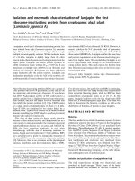
In order to confirm the presence of the Cys254–
Cys256 bridge, the peptide mixture originated from
CNBr reaction was subjected to enzymatic digestion
with Endoproteinase Glu-C. In the MALDI-TOF mass
spectrum the signal at m ⁄ z 3160.80 corresponded to
the peptide 236–265 containing the S–S bridge (mono-
isotopic molecular mass 3159.81 Da). Nevertheless,
isotope distribution of the signal could suggest the
presence of a low percentage (10%) of the peptide
having the cysteine residues in the reduced form
(monoisotopic molecular mass 3161.80 Da), as can be
deduced from the lower intensity of the peak at
m ⁄ z 3161.83 and the higher intensity of peaks from
m ⁄ z 3162.86 to m ⁄ z 3165.79 compared with the theor-
etical isotope distribution expected for the peptide with
the S–S bridge (Fig. 6).
The S–S pattern of the other cysteine residues (136,
162, 190, 202) was determined cleaving the peptide
chain between Cys136 and Cys162, by means of tryptic
digestion of the protein. In the MALDI-TOF mass
spectra the signal at m ⁄ z 3022.39 could be assigned
to the pairing of the two peptides 158–167 (monoiso-
topic molecular mass 1081.48 Da) and 179–197
02
04
06
08
001
8.06.04.02.00
[lotierhtoihtiD
M]
Residual activity(%)
Fig. 5. Effect of reducing agents on PfPNP thermostability. The
enzyme (2 lg) was incubated for 60 min in 20 m
M Tris ⁄ HCl pH 7.4
containing dithiothreitol at indicated concentrations at 70 °C(d),
80 °C(j), 90 °C(m), and 100 °C(s). Aliquots were then withdrawn
and assayed for PNP activity as described under Experimental
procedures.
Purine nucleoside phosphorylase from P. furiosus G. Cacciapuoti et al.
2488 FEBS Journal 274 (2007) 2482–2495 ª 2007 The Authors Journal compilation ª 2007 FEBS
(monoisotopic molecular mass 1942.00 Da) thus indi-
cating that Cys162 is linked to Cys190. Similarly, the
signal at m ⁄ z 4371.87 could be generated by the pep-
tides 125–140 (average molecular mass 1923.17 Da)
and 198–220 (average molecular mass 2450.87 Da)
linked by a disulfide bond between Cys136 and Cys202
(Table 3). The S–S arrangement was further confirmed
by submitting the tryptic peptide mixture to tandem
mass spectrometric experiments. As an example, the
MS ⁄ MS analysis of the peptide containing the S–S
bond between Cys162 and Cys190 is reported in detail.
The triply charged ion at m ⁄ z 1008.14, generated from
disulfide-containing peptide (158–167) + (179–197),
was selected for CID experiments and Fig. 7 reports
the MS⁄ MS spectrum and the peptide amino acid
sequence. Fragment ions belonging to series b (con-
taining the N-terminal region of the peptide) and
y (containing the C-terminal region) were originated
from the entire sequence of both peptides 158–167 and
179–197. Diagnostic fragment ions of the S–S pairing
resulted to be the singly charged ion y
7
(m ⁄ z 769.44)
originated from the fragment 191–197 and its comple-
mentary doubly charged ion b
12
(m ⁄ z 1127.50) origin-
ated from the fragment 179–190 linked to the intact
peptide 158–167. This is further demonstrated by the
singly charged ion y
5
(m ⁄ z 559.27) produced from the
fragment 163–167 and by the complementary doubly
charged ion b
5
(m ⁄ z 1232.51) originated from the frag-
ment 158–162 linked to the intact peptide 179–197. It
is interesting to note that the disulfide bonds 136–202
and 254–256 are conserved in PfMTAP and SsMTAP-
II confirming the disulfide arrangement of PfPNP.
The presence of three disulfide bonds justify the
extreme stability features of PfPNP. These covalent
links, in fact, lowering the entropy of the unfolded poly-
peptide and introducing at the same time new molecular
relative intensity %
z/
m
0
05
001
0713561306
1
3
relative intensity %
z/m
68.2613
08.3613
38.1613
08.4613
97.5613
08.0613
0913
0413
0
05
001
n
o
it
u
birtsid epot
os
il
a
c
i
ter
o
e
h
T
1.6508
.
0
6
1
3
0010
8
.1613
9.7918.2613
6.861
8
.3613
1.8318.4613
7
.
711
8
.5613
noitu
birtsid epotosilatnemirepxE
2
.3
5
0
8
.0
6
1
3
1.
6
838.16
1
3
00168.2613
7.
9
708.3613
9.8408.4613
2
.5
297.5613
z/m
z/
m )%(
y
tisnetni
e
v
italer
)%(y
tisne
t
ni
evit
al
e
r
B
A
Fig. 6. Isotope distribution of the signal at
m ⁄ z 3160.80 originated from the peptide
236–265 with a disulfide bridge. Experimen-
tal (A) and theoretical (B) isotope distribu-
tions are shown.
Table 3. Disulfide arrangement of PfPNP. The solid lines indicate S–S bridges exactly assigned, while dashed lines refer to S–S bridges
which could not be assigned in the experiment.
Experimental m ⁄ z-values Amino acid sequence of disulfide-containing peptides
Disulfide pattern obtained from CNBr reaction
3761.25
231
QKKSEDIVKLILAAIPLIPKERRCGCKDALKGATG
265
13893.61
92
KPGDFVILDQIIDFTVSRPRTFYDGEESPHERKFVAHVDFTEPY
CPEIRKALITAARNLGLPYHPRGTYVCTEGPRFETAAEIRAYRILGGDVVGM
187
188
TQCPEAILARELEM
201
202
CYATVAIVTNYAAGM
216
Disulfide pattern obtained from tryptic digestion
3022.39
158
167
140
GTYVCTEGPR ILGGDVVGMTQCPEAILAR
197
4371.87
125
FVAHVDFTEPYCPEIR ELEMCYATVAIVTNYAAGMSGKK
220
G. Cacciapuoti et al. Purine nucleoside phosphorylase from P. furiosus
FEBS Journal 274 (2007) 2482–2495 ª 2007 The Authors Journal compilation ª 2007 FEBS 2489
interactions into the protein structure could be respon-
sible for increasing the kinetic stability that is in turn
responsible for trapping the protein in its native state
also in the extreme environmental conditions.
Characterization of C254S ⁄ C256S mutant and role
of the CXC motif
To elucidate if the disulfide CGC localized at the
C-terminus of PfPNP, in spite of its unusual structural
features, could play a role in the stabilization of the
protein we utilized site-directed mutagenesis to substi-
tute Cys254 and Cys256 with serine. The large-scale
preparation of the C254S ⁄ C256S mutant was per-
formed as described above for recombinant PfPNP.
Purified mutant protein showed, under either native
(gel filtration) or denaturing (SDS ⁄ PAGE) conditions
M
r
values identical to the wild-type PfPNP and proved
to be fully active indicating the compatibility of the
substitutions with the native state of the protein. We
then carried out the characterization of the thermal
properties of the mutant in comparison with those of
PfPNP. The results obtained indicate that the substitu-
tion of Cys254 and Cys256 with serine significantly
affect both thermodynamic stability (T
m
, 102 °C) and
kinetic stability (38% residual activity after 4 h incuba-
tion at 100 °C, half-life of 35.5 min at 105 °C) of the
enzyme suggesting an important role of the pair
Cys254-Cys256 in the thermal stabilization of the
enzyme.
Disulfide bonds between cysteine residues separated
by a single amino acid are extremely rare in nature. In
addition to the disulfide CGC in PfMTAP [26] and
CSC in SsMTAPII [24], the two highly PfPNP homol-
ogous enzymes, only few examples are present in the
literature [39–43]. The following considerations allowed
us to hypothesize that the presence of a conserved
unusual CXC disulfide in PfPNP, PfMTAP and SsM-
TAPII would be not casual. Firstly, a CGC motif in a
mutant of E. coli thioredoxin reductase [43] displays a
disulfide reduction potential that is close to that of
protein disulfide isomerase. This soluble eukaryotic
protein is the most efficient known catalyst of the
formation and isomerization of disulfide bonds [44],
especially those within kinetically trapped, structured
folding intermediates [45]. Second, a strict analogy
may be observed between the CSC motif in SsMTAPII
and the CGC motif in the thiol oxidase Erv2p from
yeast, a FAD-dependent protein that can promote
disulfide bond formation during the protein biosynthe-
sis in the yeast endoplasmic reticulum [42]. In fact, as
demonstrated by the elucidation of the three-dimen-
sional structure, either in SsMTAPII [20] or in Erv2p
[42] the CXC motif is part of a flexible C-terminal
segment that can swing into the vicinity of another
cysteine pair. In particular, in Erv2p the CGC motif
was found to be involved in a disulfide relay that may
help to shuttle electrons between dithiols of the sub-
strate protein and the FAD-proximal disulfide [42].
Third, in analogy with Erv2p, the CGC motif of
PfPNP is localized in the C-terminus of the enzyme
that, as indicated by the protease sensitivity of the
polypeptide chain at neighboring residues, is a flexible
region. All these considerations and the results indica-
ting a reduced thermodynamic and kinetic stability of
the mutant C254S⁄ C256S with respect to the wild-type
PfPNP, suggest that, as already hypothesized for SsM-
TAPII [20,24], the two cysteines of the CGC motif in
Fig. 7. MS ⁄ MS spectrum of the peptides
158–167 and 179–197 linked by S–S brid-
ges. Diagnostic fragment ions b
5
and y
5
originated from the peptide 158–167, while
ions b* and y* were from the peptide
179–197.
Purine nucleoside phosphorylase from P. furiosus G. Cacciapuoti et al.
2490 FEBS Journal 274 (2007) 2482–2495 ª 2007 The Authors Journal compilation ª 2007 FEBS
PfPNP can undergo reversible oxidation-reduction to
rescue the possible damage of the other two disulfide
bonds. The presence of a low percentage of the protein
with Cys254 and Cys256 in the reduced form further
supports this hypothesis.
It has been recently demonstrated that specific pro-
tein disulfide oxidoreductases, structurally and functio-
nally related to eukaryotic protein disulfide isomerase,
play a key role in intracellular disulfide-shuffling in
hyperthermophilic proteins [46–48]. In addition to
protein disulfide oxidoreductases, the oxidized CXC
motif in hyperthermophilic enzymes with intrasubunit
disulfide bonds, such as PfPNP, PfMTAP, and SsM-
TAPII, could represent an ingenious strategy adopted
by these proteins to preserve their folded state in the
extreme conditions.
Experimental procedures
Bacterial strains, plasmid, enzymes
and chemicals
MTA was prepared from AdoMet [23]. Thermolysin and
Endoproteinase Glu-C were obtained from Boehringer
(Mannheim, Germany). O-Bromoacetyl-N-hydroxysuccini-
mide, cytochrome c, trypsin, cyanogen bromide (CNBr),
angiotensin, adrenocorticotropic hormone fragment 18–39;
nucleosides, purine bases and standard proteins used in
molecular mass studies were obtained from Sigma
(St Louis, MO, USA). Dithiothreitol and isopropyl-b -d-
thiogalactoside were from Applichem (Darmstadt, Ger-
many). Sephacryl S-200 and AH-Sepharose 4B were
obtained from Amersham Pharmacia Biotech; polyvinyli-
dene fluoride membranes (0.45 mm pore size) were obtained
from Millipore (Bedford, MA, USA.). Specifically synthes-
ized oligodeoxyribonucleotides were obtained from MWG-
Biotech (Ebersberg, Germany). Plasmid pET-22b(+) and
the NucleoSpin Plasmid kit for plasmid DNA preparation
were obtained from Genenco (Duren, Germany). E. coli
strain BL21(kDE3) was purchased from Novagen (Darms-
tadt, Germany). P. furiosus chromosomal DNA was kindly
provided by C. Bertoldo (Technical University, Hamburg-
Harburg, Germany). Restriction endonucleases and DNA-
modifying enzymes were obtained from Takara Bio, Inc.
(Otsu, Shiga, Japan). Pfu DNA polymerase was purchased
from Stratagene (La Jolla, CA, USA). Nonspecific adeno-
sine deaminase was purified 200-fold from Aspergillus
oryzae powder (Sanzyme, Calbiochem, Los Angeles, CA,
USA) according to Wolfenden et al. [49].
Enzyme assay
Purine nucleoside phosphorylase activity was determined
following the formation of purine base from the corres-
ponding nucleoside by HPLC using a Beckman system
Gold apparatus. The assay was carried out as already
reported [25]. Unless otherwise stated, the standard incuba-
tion mixture contained the following: 20 lmol potassium
phosphate buffer, pH 7.4, 400 nmol of the nucleoside and
the enzyme protein in a final volume of 200 lL. The incu-
bation was performed in sealed glass vials for 5 min at
80 °C, except where indicated otherwise. Control experi-
ments in the absence of the enzyme were performed in
order to correct for nucleoside hydrolysis. When the assays
were carried out at temperatures above 80 °C, the reaction
mixture was preincubated for 2 min without the enzyme
that was added immediately before starting the reaction.
An Ultrasphere ODS RP-18 column was employed and the
elution was carried out with 5 : 95 (v ⁄ v) mixture of 95%
methanol and 0.1% trifluoroacetic acid in H
2
O. The retent-
ion times of inosine and hypoxantine, guanosine and guan-
ine were 10.5 min and 4.7 min, and 11.5 min and 4.3 min,
respectively. The amount of purine base formed is deter-
mined by measuring the percentage of the absorbance
integrated peak area of purine base formed with respect to
the total (nucleoside + purine base) absorbance integrated
peak areas. In all of the kinetic and purification studies the
amounts of the protein was adjusted so that no more than
10% of the substrate was converted to product and the
reaction rate was strictly linear as a function of time and
protein concentration. One unit of enzyme activity was
defined as the amount of enzyme that catalyzes the cleavage
of 1 lmol of inosine per minute at 80 °C.
Determination of kinetic constants
Homogeneous preparations of PfPNP were used for kinetic
studies. The purified enzyme gave a linear rate of reaction
for at least 10 min at 80 °C, thus, an incubation time of
5 min was employed for kinetic experiments. All enzyme
reactions were performed in triplicate. Kinetic parameters
were determined from Lineweaver–Burk plots of initial
velocity data. K
m
and V
max
values were obtained from
linear regression analysis of data fitted to the Michaelis–
Menten equation. Values given are the average from at
least three experiments with standard errors. The k
cat
value
was calculated by dividing V
max
by the total enzyme con-
centration. Calculations of k
cat
were based on an enzyme
molecular mass of 180 kDa.
Analytical methods for protein
Protein concentration was determined by means of the
Bradford method [50] using bovine serum albumin as the
standard. The molecular mass of the native protein was
determined by gel filtration on a calibrated Sephacryl S-200
column as already reported [24]. The molecular mass under
dissociating conditions was determined by SDS polyacryla-
mide gel electrophoresis, as described by Weber et al. [51].
G. Cacciapuoti et al. Purine nucleoside phosphorylase from P. furiosus
FEBS Journal 274 (2007) 2482–2495 ª 2007 The Authors Journal compilation ª 2007 FEBS 2491
Samples were heated at 100 °C for 5 min in 2% SDS and
2% 2-mercaptoethanol and run in comparison with molecu-
lar weight standards. Protein homogeneity was assessed by
SDS ⁄ PAGE. N-terminal sequence analysis of the purified
enzyme was performed by Edman degradation on an
Applied Biosystem 473 A sequencer. The sample was sub-
jected to SDS polyacrylamide gel electrophoresis and elec-
troblotted on a polyvinylidene fluoride membrane prior to
analysis.
Stability and thermostability studies
The stability of PfPNP activity in the presence of SDS and
dithiothreitol was examined at the indicated temperatures
as reported in [24]. Immediately after the addition of the
compound (time-zero control) and at different time inter-
vals, aliquots were removed from each sample and analyzed
for activity in the standard assay. Activity values are
expressed as a percentage of the zero-time control (100%).
Enzyme thermostability was tested by incubating the pro-
tein in sealed glass vials at temperatures between 100 °C
and 115 °C in an oil bath. Samples (2 lg) were taken at
time intervals and residual activity was determined by the
standard assay at 80 °C.
Cloning and expression of the PfPNP-encoding
gene
The putative PfPNP gene PF0853 (GenBank
TM
accession
number AE010200) from P. furiosus was cloned into the
pET-22b(+) expression vector via two engineered restric-
tion sites (Nde I and EcoR I) introduced by PCR with the
following primers 5¢-GCTGGTGGT
CATATGCCCAGG-3¢
sense, and 5 ¢-GTTAATCCGTTTG
GAATTCGTC-3¢, anti-
sense (the introduced restriction sites are underlined). Iso-
lated genomic P. furiosus DNA (20 ng), hydrolyzed by
EcoR I was used as a template. PCR amplification was per-
formed with P. furiosus DNA polymerase and a Minicycler
(Genenco) programmed for 30 cycles, each cycle consisting
of denaturation at 92 °C for 1 min, annealing at 55 °C for
2 min and extension at 68 °C for 2 min plus 5 s Æcycle
)1
, fol-
lowed by an extension final step of 15 min at 68 °C. The
amplified gene (25 ng), hydrolyzed by Nde I and EcoRI
was inserted into pET22b(+) (150 ng). The recombinant
plasmid was named pET-PfPNP. The nucleotide sequence
of the inserted gene was determined by MWG BIOTECH
to ensure that no mutations were present in the gene.
pET-PfPNP recombinant plasmid was transformed into
E. coli BL21 (kDE3) cells. Single recombinant colonies
were used to inoculate 1 L of LB medium containing
100 lgÆmL
)1
ampicillin at 37 °C for 22 h. When the culture
reached an optical density of 3.0 isopropyl-b-d-thiogalacto-
side was added at 1 mm final concentration and the induction
was prolonged for 5 h. Cells were harvested by centrifugation
and lysed as described by Sambrook et al. [52].
Preparation of MTI-Sepharose
The preparation of 5¢-methylthioinosine (MTI)-Sepharose
was performed as described by Kim et al. [53] by treating
AH-Sepharose 4B (5 g) with 1 mmol of O-bromoacetyl-
N-hydroxysuccinimide and then coupling the resin with
10 mg of MTI. MTI was pepared by enzymatic deamina-
tion of MTA utilizing nonspecific adenosine deaminase
(adenosine aminohydrolase, EC 3.5.4.4) purified from
Aspergillus oryzae. MTA (30 lmol) was incubated with
adenosine deaminase at 37 °C for 16 h in Tris ⁄ HCl 0.1 m,
pH 7.4 as described in [54] and the reaction was stopped
by the addition of trichloroacetic acid 10%. The formation
of MTI was checked by reverse-phase HPLC on a
4.6 · 250 mm Ultrasphere ODS (5 lm particle size) col-
umn (Beckman) using an Agilent 1100 series cromato-
graph. The column, was equilibrated and eluted with a
20 : 80 (v ⁄ v) mixture of 95% methanol and 0.1% trifluoro-
acetic acid in H
2
O at a flow rate of 1 mLÆmin
)1
. The chro-
matogram showed that, after the enzymatic reaction, the
peak of MTA (retention time 10 min) was replaced by a
new peak, with a lower retention time (7 min) thus indica-
ting the complete conversion, in these experimental condi-
tions, of MTA to MTI.
Purification of recombinant PfPNP
Recombinant PfPNP was purified in two steps. The cell-free
extract of BL21 E. coli cells expressing PfPNP was heated
at 100 °C for 10 min and centrifuged at 20 000 g for
60 min. After dialysis overnight against 10 mm Tris ⁄ HCl
pH 7.4, the enzyme was applied to an affinity column of
MTI-Sepharose (2 · 12 cm) equilibrated with 20 mm
Tris ⁄ HCl pH 7.4. The column was washed stepwise with
50 mL of the equilibration buffer and then with the
same buffer containing 0.07 m NaCl until the absorbance
at 280 nm reached the baseline. PNP activity was then
eluted with 20 mm Tris ⁄ HCl pH 7.4 containing 0.1 m NaCl
and 3 mm inosine. Active fractions were pooled, concen-
trated and dialyzed extensively against 10 mm Tris ⁄ HCl
pH 7.4.
Limited proteolysis experiments
Limited proteolysis experiments were carried out by incuba-
ting recombinant PfPNP with thermolysin as already des-
cribed [23]. To follow the degradation of the intact protein
over 2 h of incubation the digested material was submitted
to SDS/PAGE followed by staining with Coomassie blue
R-250.
For the amino sequence analysis, samples of the digested
recombinant PfPNP after SDS ⁄ PAGE, were electrophoreti-
cally blotted onto a polyvinylidene fluoride membrane as
already reported [24], utilizing a Bio-Rad Mini trans-blot
transfer cell apparatus (Bio-Rad, Hercules, CA, USA).
Purine nucleoside phosphorylase from P. furiosus G. Cacciapuoti et al.
2492 FEBS Journal 274 (2007) 2482–2495 ª 2007 The Authors Journal compilation ª 2007 FEBS
Multiple sequence alignment
Protein similarity searches were performed using the data
from Swiss-Prot and Protein Identification Resource (PIR)
data banks. The multiple alignment was constructed using
the clustal method [55].
Site-directed mutagenesis
The construct pET-PfPNP was used as a template for
site-directed mutagenesis by the Quik-Change procedure
(Stratagene). The primers: 5¢-CCAAAGGAGAGGAGG
AGCGGGAGCAAAGATGCT-3¢ and 5¢-AGCATCTTT
GCTCCCGCTCCTCCTCTCCTTTGG-3¢ (nucleotide sub-
stitutions are underlined) were used to make two silent
mutations which replaced Cys254 and Cys256 with Ser.
pET-PfPNP (100 ng) was used for PCR amplification. The
resulting PCR product was checked by DNA sequence ana-
lysis and then used to transform E. coli BL21 (kDE3) for
overproduction of the protein utilizing the same protocol
used for the expression of recombinant PfPNP.
HPLC analyses
Samples containing 100 lg of protein were purified by
reverse-phase HPLC as already reported [56] using a flow
rate of 1 mLÆmin
)1
and a linear gradient from 10% to 80%
solvent B over 35 min.
Chemical and enzymatic reactions
CNBr protein cleavage was performed in 70% trifluoroace-
tic acid, overnight at room temperature under an inert
atmosphere in the dark. Digestion with endopeproteinase
Glu-C was carried out in 50 mm ammonium acetate,
pH 5.0, overnight at 37 °C, using an enzyme to substrate
ratio of 1 : 50 w ⁄ w. Tryptic digestion was performed in
50 mm ammonium bicarbonate, pH 8.5, at 37 ° C for 4 h,
using an enzyme to substrate ratio of 1 : 100 w ⁄ w.
MALDI-TOF-MS analyses
Protein samples and peptide mixtures obtained from
enzymatic or chemical digestion were analyzed on a
MALDI-TOF mass spectrometer Voyager DE
TM
PRO
(Applied Biosystems, Foster City, CA, USA), as already
described [57]. In the m ⁄ z range 4000–40000 mass spectra
were acquired in linear positive-ion mode and calibrated
using as internal standards the average double and singly
charged peaks originated from cytochrome c (m ⁄ z 6181.05
and 12 361.10, respectively). Mass spectra in the m ⁄ z range
700–4000 were acquired in reflectron positive-ion mode and
calibrated using the monoisotopic peaks of angiotensin
(m ⁄ z 931.5154) and adrenocorticotropic hormone fragment
18–39 (m ⁄ z 2465.1989). All the signals are singly charged
ions (MH
+
) and the m ⁄ z-values recorded in linear positive-
ion mode were reported as average values, while the
m ⁄ z-values recorded in reflectron positive-ion mode were
reported as monoisotopic values. Theoretical isotopic distri-
bution of the peptide 236–265 was calculated by means of
the MS-isotope program of protein prospector [58].
Tandem mass spectrometric experiments
(ESI-MS
⁄
MS)
Tandem mass spectrometric analyses performed on a
hybrid quadruple ⁄ orthogonal time of flight instrument
(QStar Pulsar, Applied Biosystems) equipped with nano-
spray source, were carried out as already described [57].
Acknowledgements
This research was supported by grant from ‘Minis-
tero dell’Universita
`
e della Ricerca scientifica’ PRIN
2004, and Regione Campania L.R. n. 5/2002.
References
1 Parks RE Jr & Agarwal RP (1972) Purine nucleoside
phosphorylase. In The Enzymes (Boyer, PD, ed.), Vol.
7, pp. 483–514. Academic Press, New York, NY.
2 Bzowska A, Kulikowska E & Shugar D (2000) Purine
nucleoside phosphorylases: properties, functions
and clinical aspects. Pharmacol Therapeutics 88,
349–425.
3 Pugmire MJ & Ealick SE (2002) Structural analysis
reveals two distinct families of nucleoside phosphory-
lases. Biochem J 361, 1–25.
4 Stoeckler JL (1984) Purine nucleoside phosphorylase a
target for chemotherapy. In Developments in Cancer
Chemotherapy (Glazer, RI, ed.), pp. 35–60. CRC Press
Inc., Boca Raton, FL.
5 Zhang Y, Parker WB, Sorscher EJ & Ealick SE (2005)
PNP anticancer gene therapy. Current Topics Med Chem
5, 1259–1274.
6 Schramm VL (2002) Development of transition state
analogues of purine nucleoside phosphorylase as
anti-T-cell agents. Biochim Biophys Acta 1587,
107–117.
7 Critchley RJ, Jezzani S, Radford KJ, Goussard S,
Lemoine NR, Grillot-Courvalin C & Vassaux G (2004)
Potential therapeutical applications of recombinant
invasive E. coli. Gene Ther 11, 1224–1233.
8 Deharvengt S, Wack S, Uhring M, Aprahmian M &
Hajri A (2004) Suicide gene ⁄ prodrug therapy for
pancreatic adenocarcinoma by E. coli purine nucleoside
phosphorylase and 6-methylpurine 2¢-deoxyriboside.
Pancreas 28, 54–64.
G. Cacciapuoti et al. Purine nucleoside phosphorylase from P. furiosus
FEBS Journal 274 (2007) 2482–2495 ª 2007 The Authors Journal compilation ª 2007 FEBS 2493
9 Sorscher EJ, Peng S, Bebok Z, Allan PW, Bennett LL
& Parker WB (1994) Tumor cell bystander killing in
colonic carcinoma utilizing Escherichia coli DeoD gene
to generate toxic purines. Gene Ther 1, 233–238.
10 Parker WB, King SA, Allan PW, Bennett LL, Secrist
3rd JA, Montgomery JA, Gilbert KS, Waud WR, Wolls
AH, Gillespie GY et al. (1997) In vivo gene therapy of
cancer with E. coli purine nucleoside phosphorylase.
Hum Gene Ther 8, 1637–1644.
11 Zeikus JG, Vieille C & Savchenko A (1998) Thermo-
zymes: biotechnology and structure-function relation-
ships. Extremophiles 2, 179–183.
12 Kumar S & Nussinov R (2001) How do thermophilic
proteins deal with heat? Cell Mol Life Sci 58,
1216–1233.
13 Ladenstein R & Antranikian G (1998) Proteins from
hyperthermophiles: stability and enzymatic catalysis
close to the boiling point of water. Advances in Bio-
chemical Engineering ⁄ Biotechnology (Sheper, T, ed.),
Vol. 612. Springer-Verlag, Berlin Heidelberg.
14 Sterner R & Liebl W (2001) Thermophilic adaptation of
proteins. Crit Rev Biochem Mol Biol 36, 39–106.
15 Vieille C & Zeikus GJ (2001) Hyperthermophilic
enzymes: sources, uses and molecular mechanisms for
thermostability. Microbiol Mol Biol Rev 65, 1–43.
16 Appleby TC, Mathews II, Porcelli M, Cacciapuoti G &
Ealick SE (2001) Three-dimensional structure of a
hyperthermophilic 5¢-deoxy-5¢-methylthioadenosine
phosphorylase from Sulfolobus solfataricus. J Biol Chem
42, 39232–39242.
17 Choi I-G, Bang W-G, Kim S-H & Yu YG (1999)
Extremely thermostable serine-type protease from
Aquifex pyrophilus. Molecular cloning, expression and
characterization. J Biol Chem 274, 881–888.
18 Cort JR, Mariappan SV, Kim C-Y, Park MS, Peat TS,
Waldo GS, Terwillger TC & Kennedy MA (2001) Solu-
tion structure of Pyrobaculum aerophilum DsrC, an
archaeal homologue of the c-subunit of dissimilatory
sulfite reductase. Eur J Biochem 268, 5842–5849.
19 Toth EA, Worby C, Dixon JE, Goedken ER, Marqusee
S & Yeates TO (2000) The crystal structure of adenylo-
succinate lyase from Pyrobaculum aerophilum reveals an
intracellular protein with three disulfide bonds. J Mol
Biol 301, 433–450.
20 Zhang Y, Porcelli M, Cacciapuoti G & Ealick SE
(2006) The crystal structure of 5¢-deoxy-5¢-methylthioa-
denosine phosphorylase II from Sulfolobus solfataricus,
a thermophilic enzyme stabilized by intramolecular
disulfide bonds. J Mol Biol
357, 252–262.
21 Pegg AE & Williams-Ashman HG (1969) Phosphate-sti-
mulated breakdown of 5¢-methylthioadenosine by rat
ventral prostate. Biochem J 115, 241–247.
22 Williams-Ashman HG, Seidenfeld J & Galletti P (1982)
Trends in the biochemical pharmacology of 5¢-deoxy-
5¢-methylthioadenosine. Biochem Pharmacol 31, 277–288.
23 Cacciapuoti G, Porcelli M, Bertoldo C, De Rosa M &
Zappia V (1994) Purification and characterization of
extremely thermophilic and thermostable 5 ¢-methylthio-
adenosine phosphorylase from the archaeon Sulfolobus
solfataricus. Purine nucleodide phosphorylase activity
and evidence for intersubunit disulfide bonds. J Biol
Chem 269, 24762–24769.
24 Cacciapuoti G, Forte S, Moretti MA, Brio A, Zappia V
& Porcelli M (2005) A novel hyperthermostable
5¢-deoxy-5¢-methylthioadenosine phosphorylase from
the archaeon Sulfolobus solfataricus. FEBS J 272,
1886–1899.
25 Cacciapuoti G, Bertoldo C, Brio A, Zappia V &
Porcelli M (2003) Purification and characterization of
5¢-methylthioadenosine phosphorylase from the
hyperthermophilic archaeon Pyrococcus furiosus.
Substrate specificity and primary structure analysis.
Extremophiles 7 , 159–168.
26 Cacciapuoti G, Moretti MA, Forte S, Brio A,
Camardella L, Zappia V & Porcelli M (2004)
Methylthioadenosine phosphorylase from the archaeon
Pyrococcus furiosus. Mechanism of the reaction and
assignment of disulfide bonds. Eur J Biochem 271,
4834–4844.
27 Van der Oost J, Ciaramella M, Moracci M, Pisani FM,
Rossi M & de Vos WM (1998) Molecular biology of
hyperthermophilic Archaea. Adv Biochem Eng Biotechnol
61, 87–115.
28 Robb F, Maeder DL, Brown JR, Di Ruggiero J, Stump
MD, Yeh RK, Weiss RB & Dunn DM (2001) Genomic
sequence of hyperthermophile Pyrococcus furiosus:
implications for physiology and enzymology. Methods
Enzymol 330, 134–157.
29 Koellner GL, Luic M, Shugar D, Saenger W &
Bzowska A (1998) Crystal structure of the ternary com-
plex of E. coli purine nucleoside phosphorylase with for-
mycin B, a structural analogue of the substrate inosine,
and phosphate (sulphate) at 2.1 A
˚
resolution. J Mol
Biol 280, 153–166.
30 Tahirov TH, Inagaki E, Ohshima N, Kitao T, Kuroishi
C, Ukita Y, Takio K, Kobayashi M, Kiramitsu S et al.
(2004) Crystal structure of purine nucleoside phosphory-
lase from Thermus thermophilus. J Mol Biol 337 (1149),
1160.
31 Caradoc-Davies TT, Cutfield SM, Lamont IL &
Cutfield JF (2004) Crystal structure of Escherichia coli
uridine phosphorylase in two native and three com-
plexed forms reveal basis of substrate specificity,
induced conformational changes and influence of
potassium. J Mol Biol 337, 337–354,.
32 Koellner G, Bzowska A, Wielgus-Kutrowska B, Luic
M, Steiner T, Saenger W & Stepinski J (2002) Open and
closed conformation of E. coli purine nucleoside phos-
phorylase active center and implications for catalytic
mechanism. J Mol Biol 18, 351–371.
Purine nucleoside phosphorylase from P. furiosus G. Cacciapuoti et al.
2494 FEBS Journal 274 (2007) 2482–2495 ª 2007 The Authors Journal compilation ª 2007 FEBS
33 Tebbe J, Bzowska A, Wielgus-Kutrowska B, Schroeder
W, Kazimierczuk Z, Shugar D, Saenger W & Koellner
G (1999) Crystal structure of the purine nucleoside
phosphorylase (PNP) from Cellulomonas sp. and its
implication for the mechanism of trimeric PNPs. J Mol
Biol 294, 1239–1255.
34 Mao C, Cook WJ, Zhou M, Federov AA, Almo SC &
Ealick SE (1998) Calf spleen purine nucleoside phos-
phorylase complexed with substrates and substrate
analogues. Biochemistry 37, 7135–7146.
35 Appleby TC, Erion MD & Ealick SE (1999) The struc-
ture of human 5¢-deoxy-5¢ -methylthioadenosine phos-
phorylase at 1.7 A
˚
resolution provides insights into
substrate binding and catalysis. Structure 7, 629–641.
36 Della Ragione F, Oliva A, Gragnianiello V, Russo GL,
Palumbo R & Zappia V (1990) Physicochemical and
immunological studies on mammalian 5¢-deoxy-
5¢-methylthioadenosine phosphorylase. J Biol Chem 265,
6241–6246.
37 Manning M & Colon W (2004) Structural basis of pro-
tein kinetic stability: resistance to sodium dodecylsulfate
suggests a central role for rigidity and a bias toward
b-sheet structure. Biochemistry 43, 11248–11254.
38 Mallick P, Boutz DR, Eisenberg D & Yeates TO (2002)
Genomic evidence that the intracellular proteins of
archaeal microbes contain disulfide bonds. Proc Natl
Acad Sci USA 99, 9679–9684.
39 Krishnaswamy S & Rossmann MG (1990) Structural
refinement and analysis of Mengo virus. J Mol Biol 211,
803–844.
40 Smith CA, Toogood HS, Baker HM, Daniel RM &
Baker EN (1999) Calcium-mediated thermostability in
the subtilisin superfamily: the crystal structure of Bacil-
lus Ak.1 protease at 1.8 A
˚
resolution. J Mol Biol 294,
1027–1040.
41 Jacob U, Muse W, Eser M & Bardwen JCA (1999)
Chaperone activity with a redox switch. Cell 96,
341–352.
42 Gross E, Sevier CS, Vala A, Kaiser CA & Fass D (2002)
A new FAD-binding fold and intersubunit disulfide
schuttle in the thiol oxidase Erv2p. Nat Struct Biol 9,
61–67.
43 Woycechowsky KJ & Raines RT (2003) The CXC
motif: a functional mimic of protein disulfide isomerase.
Biochemistry 42, 5387–5394.
44 Frand AR, Cuozzo JW & Kaiser CA (2000) Pathways
for protein disulphide bond formation. Trends Cell Biol
10, 203–210.
45 Weissman JS & Kim PS (1993) Efficient catalysis of
disulphide bond rearrangements by protein disulphide
isomerase. Nature 365, 185–188.
46 Pedone E, Ren B, Ladenstein R, Rossi M & Bartolucci
SF (2004) Functional properties of the protein disulfide
oxidoreductase from the archaeon Pyrococcus furiosus.
A member of a novel protein family related to protein
disulfide-isomerase. Eur J Biochem
271, 3437–3448.
47 Ladenstein R & Ren B (2006) Protein disulfides and
protein disulfide oxidoreductases in hyperthermophiles.
FEBS J 273, 4170–4185.
48 Pedone E, Limauro D, D’Alterio R, Rossi M &
Bartolucci S (2006) Characterization of a multifunc-
tional protein disulfide oxidoreductase from Sulfolobus
solfataricus. FEBS J 273 , 5407–5420.
49 Wolfenden R, Sharpless TK & Allan R (1967) Substrate
binding by adenosine deaminase. Specificity, pH depen-
dence, and competition by mercurials. J Biol Chem 242,
977–983.
50 Bradford MM (1976) A rapid and sensitive method for
the quantitation of microgram quantities of protein
utilizing the principle of protein-dye binding. Anal
Biochem 72, 248–254.
51 Weber K, Pringle JR & Osborn M (1972) Measurement
of molecular weight by electrophoresis on SDS-acryla-
mide gel. Methods Enzymol 260, 3–27.
52 Sambrook J, Fritsch EF & Maniatis T (1989) Molecular
cloning: a laboratory manual. Cold Spring Harbor
Laboratory Press, Cold Spring Harbor, NY.
53 Kim S, Nochumson S, Chin W & Paik WK (1978) A
rapid method for the purification of S-adenosylmethio-
nine: protein-carboxyl O-methyltransferase by affinity
chromatography. Anal Biochem 84, 415–422.
54 Schlenk F, Zydek-Cwick CR & Hutson NK (1971)
Enzymatic deamination of adenosine sulfur compounds.
Arch Biochem Biophys 142, 144–149.
55 Higgins DG & Sharp PM (1998) clustal: a package
for performing multiple sequence alignment of a micro-
computer. Gene 73, 237–244.
56 Siciliano R, Rega B, Marchetti M, Seganti L, Antonimi
G & Valenti P (1999) Bovine lactoferrin peptidic
fragments involved in inhibition of herpes simplex virus
type 1 infection. Biochem Biophys Res Commun 264,
19–23.
57 Mazzeo MF, De Giulio B, Senger S, Rossi M, Malorni
A & Siciliano RA (2003) Identification of transglutami-
nase-mediated deamidation sites in a recombinant
alpha-gliadin by advanced mass-spectrometric meth-
odologies. Protein Sci 12, 2434–2442.
58 Clauser KR, Baker P & Burlingame AL (1999) Role
of accurate mass measurement (+ ⁄ – 10 ppm) in
protein identification strategies employing MS or
MS ⁄ MS and database searching. Anal Chem 71,
2871–2882.
G. Cacciapuoti et al. Purine nucleoside phosphorylase from P. furiosus
FEBS Journal 274 (2007) 2482–2495 ª 2007 The Authors Journal compilation ª 2007 FEBS 2495