Nghiên cứu thành phần hóa học của lá cây ráng Tây Sơn Dicranopteris linearis (Burm. F.) Underw.
Bạn đang xem bản rút gọn của tài liệu. Xem và tải ngay bản đầy đủ của tài liệu tại đây (272.87 KB, 6 trang )
TẠP CHÍ KHOA HỌC
TRƯỜNG ĐẠI HỌC SƯ PHẠM TP HỒ CHÍ MINH
Vol. 19, No. 10 (2022): 1593-1598
Tập 19, Số 10 (2022): 1593-1598
ISSN:
2734-9918
HO CHI MINH CITY UNIVERSITY OF EDUCATION
JOURNAL OF SCIENCE
Website:
/>
Research Article *
STUDY ON CHEMICAL CONSTITUENTS OF THE LEAVES
OF DICRANOPTERIS LINEARIS (BURM. F.) UNDERW.
Nguyen Thu Anh1, Nguyen Thi Hoai Thu2, Nguyen Ngoc Khanh Van3, Duong Thuc Huy1*
1
Ho Chi Minh City Universiry of Education, Vietnam
University of Medicine and Pharmacy at HCMC, Vietnam
3
Gia Dinh High School, Ho Chi Minh City, Vietnam
*
Corresponding author: Duong Thuc Huy – Email:
Received: March 03, 2022; Revised: April 06, 2022; Accepted: October 06, 2022
2
ABSTRACT
Dicranopteris linearis (Burm. F.) Underw. has many popular traditional uses in Asian
countries, such as treating ulcers and boils, allergic symptoms, or respiratory troubles. This study
investigated the phytochemical of D. linearis grown in Lam Dong Province. The leaf powder of D.
linearis was used to prepare a crude extract. This extract was then applied to the liquid-liquid
partition to give different polar fractions. The EA fraction was thus applied to silica gel column
chromatography to obtain four compounds. Their chemical structures were elucidated by using
Nuclear Magnetic Resonance spectroscopy, as well as by the comparison of their NMR data with
reported ones. Four compounds consisted of three flavonols, isoquercetin (1), quercetin (2),
kaempferol (3), and a sterol, stigmast-5,22-dien-3β-ol-3-O-β-D-glucopyranoside (4).
Keywords: Dicranopteris linearis, isoquercetin; kaempferol; quercetin; stigmast-5-en-3β-ol3-O-β-D-glucopyranoside
1.
Introduction
Dicranopteris linearis (Burm. F.) Underw. is a common fern and belongs to the
Gleicheniaceae family. They are distributed in Africa and Asia and grow widely in Vietnam,
especially in low, hot, and dry mountainous areas (Chi, 2002). The D. linearis extracts
showed antidiabetic, anticancer, antibacterial, antioxidant, analgesic, and anti-HIV activities
(Li et al., 2006; Li et al., 2008; Chen et al., 2014; Ponnusamy et al., 2015; Zakaria et al.,
2016; Duy et al., 2019). In particular, they are widely used as a folk medicine to treat fever
(Malaysia), intestinal worms (Indochina), asthma, infertility in women (India), wound
(Papua New Guinea) (Kamisan et al., 2014), cough, allergic, and respiratory disorders
(Mymensingh) (Sarker et al., 2009).
Cite this article as: Nguyen Thu Anh, Nguyen Thi Hoai Thu, Nguyen Ngoc Khanh Van, & Duong Thuc Huy
(2022). Study on chemical constituents of the leaves of Dicranopteris linearis (Burm. F.) Underw.. Ho Chi
Minh City University of Education Journal of Science, 19(10), 1593-1598.
1593
HCMUE Journal of Science
Nguyen Thu Anh et al.
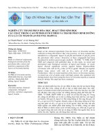
This study reported the isolation and structural elucidation of four compounds,
including isoquercetin (1), quercetin (2), kaempferol (3), and β-sitosterol 3-O-β-Dglucopyranoside (4) from the leaves of D. linearis collected in Lam Dong Province,
Vietnam.
OH
OH
29
4'
HO
7
19
1'
O
HO
2
5
OH
4
O
(1)
HO
O
O
O
O
HO
2''
OH
OH
OH
OH
OH
O
OH
(2)
O
(3)
OH
HO
HO
26
20
27
18
OH
1''
22
21
OH
OH
OH
1
O
3
1'
OH
7
O
(4)
Figure 1. Chemical structures of isolated compounds 1-4
2.
Experiment
2.1. General experimental procedures
The NMR spectra were recorded on a Bruker Avance 500 spectrometer (500 MHz for
1
H–NMR and 125 MHz for 13C–NMR). n-Hexane, ethyl acetate (EtOAc), methanol
(MeOH), and acetone were used to prepare extracts and to elute column chromatography
and thin-layer chromatography. Thin-layer chromatography was carried out on silica gel 60
(Merck, 40-63 μm), and spots were visualized by spraying with 10% H2SO4 solution,
followed by heating.
2.2. Plant material
The leaves of D. linearis (Burm. F.) Underw. were collected in Lam Dong Province,
Vietnam, from January to March 2021. The specimen was identified by Prof. Tran Cong
Luan, Tay Do University and deposited at the herbarium in the laboratory of the Faculty of
Chemistry, Ho Chi Minh City University of Education, Vietnam.
2.3. Extraction and isolation
The leaf powder of D. linearis (5.0 kg) was macerated with methanol three times (3 x
30 L) at room temperature. The solvent was removed from the filtrated solution at reduced
pressure to obtain a crude extract (280.0 g). This extract was successively applied to the
liquid-liquid partition with increasing polarity of solvents: n-hexane, n-hexane: EtOAc (1:1,
v/v), EtOAc to give H (27.44 g), HEA (34.79 g), and EA (23.00 g) extracts. The watersoluble layer was dried to obtain the MeOH extract (60.03 g).
The EA extract (23.0 g) was subjected to a silica gel column chromatography with a
mobile phase of n-hexane-EtOAc-acetone (1:4:4, v/v/v) to give 13 fractions (EA1-EA13).
Fraction EA2 (3.6 g) was subjected to a silica gel column chromatography with a gradient
solvent of n-hexane-EtOAc-acetone (1:4:4, v/v/v) to yield seven subfractions (EA2.1EA2.7). Subfraction EA2.2 (1.12 g) was rechromatographed on a silica gel eluting with nhexane: EtOAc (1:1) to afford 1 (12.0 mg). Subfraction EA2.3 (0.93 g) was
rechromatographed on a silica gel eluting with n-hexane: EtOAc: acetone (2:19:9, v/v/v) to
afford 2 (40.0 mg). Subfraction EA2.4 (0.62 g) was rechromatographed on a silica gel eluting
1594
HCMUE Journal of Science
Vol. 19, No. 10 (2022): 1593-1598
with n-hexane: EtOAc: acetone (1:4:2, v/v/v) to afford 3 (16.0 mg). Subfraction EA4 (3.3 g)
was chromatographed on a silica gel using n-hexane-EtOAc-acetone (1:4:4) to yield 12
subfractions (EA4.1-EA4.12). Subfraction EA4.6 (0.33 g) was chromatographed on a silica
gel using n-hexane-EtOAc (1:3) as eluent to give 4 (8.3 mg).
• Isoquercetin (1). Yellow powder. The 1H–NMR data (500 MHz, DMSO-d6, δ ppm, J
in Hertz): 12.64 (1H, s, 5-OH), 10.87 (1H, s, 7-OH), 9.75 (1H, s, 3'-OH), 9.17 (1H, s, 4'OH), 7.66 (1H, brd, 8.5 Hz, H-6'), 7.52 (1H, brs, H-2'), 6.81 (1H, d, 8.5 Hz, H-5'), 6.40 (1H,
brs, H-8), 6.20 (1H, brs, H-6), 5.37 (1H, d, 8.0 Hz, H-1''), 5.29 (1H, s, 2''-OH), 5.14 (1H, s,
3''-OH), 4.87 (1H, s, 4''-OH), 4.44 (1H, s, 6''-OH), 3.28-3.56 (m, H-2'' to H-6''), 3.24 (1H, m,
H-2''), 3.23 (1H, m, H-3''), and 3.08 (1H, m, H-5''). 13C-NMR (125 MHz, DMSO-d6 ): 60.0
(C-6''), 67.8 (C-4''), 71.0 (C-2''), 73.0 (C-3''), 75.7 (C-5''), 93.3 (C-8), 98.5 (C-6), 101.6 (C1''), 103.7 (C-10), 115.0 (C-2'), 116.0 (C-5'), 120.9 (C-6'), 121.8 (C-1'), 133.3 (C-3), 144.6
(C-4'), 148.3 (C-3'), 156.1 (C-2), 161.1 (C-9), 164.0 (C-5), 165.2 (C-7), and 177.3 (C-4).
• Quercetin (2). Yellow powder. The 1H–NMR data (Acetone-d6, δ ppm, J in
Hertz): 12.49 (1H, s, 5-OH), 10.84 (1H, s, 7-OH), 9.69 (1H, s, 4'-OH), 9.35 (1H, s, 3'-OH),
8.09 (1H, s, 3-OH), 7.67 (1H, d, 2.5 Hz, H-2'), 7.54 (1H, dd, 8.5, 2.5 Hz, H-6'), 6.88 (1H, d,
8.5 Hz, H-5'), 6.40 (1H, d, 2.0 Hz, H-8), and 6.18 (1H, d, 2.0 Hz, H-6).
• Kaempferol (3). Yellow powder. The 1H–NMR data (Acetone-d6, δ ppm, J in Hertz):
8.16 (2H, d, 8.5 Hz, H-2', H-6'), 7.02 (2H, d, 8.5 Hz, H-3', H-5'), 6.54 (1H, d, 2.0 Hz, H-8),
and 6.27 (1H, d, 2.0 Hz, H-6).
• Stigmast-5,22-dien-3β-ol-3-O-β-D-glucopyranoside (4). Yellow powder. The 1H–
NMR data (DMSO-d6, δ ppm, J in Hertz): 5.33 (1H, d, 3.2 Hz, H-6), 4.84-4.88 (3H, 2'-OH,
3'-OH, 4'-OH), 4.42 (1H, t, 5.8 Hz, 6'-OH), 4.22 (1H, d, 7.8 Hz, H-1'), 3.93 (1H, m, H-3),
3.01-3.56 (6H, H-2', H-3', H-4', H-5', H-6'), 0.90 (3H, d, 6.4 Hz, H-21), 0.86 (3H, s, H-19),
0.82 (3H, t, 6.5 Hz, H-29), 0.81 (3H, d, 5.0 Hz, H-26), 0.79 (3H, d, 5.0 Hz, H-27), and 0.65
(3H, s, H-18). 13C–NMR (125 MHz, DMSO-d6): 12.2 (C-18), 12.3 (C-29), 19.1 (C-21), 19.4
(C-19), 19.6 (C-27), 20.2 (C-26), 21.1 (C-11), 23.1 (C-28), 24.3 (C-15), 25.9 (C-23), 28.3
(C-16), 29.2 (C-25), 29.7 (C-7), 31.9 (C-2), 31.9 (C-8), 33.8 (C-22), 36.0 (C-20), 36.7 (C10), 37.3 (C-1), 38.8 (C-12), 40.0 (overlap, C-4), 42.4 (C-13), 45.6 (C-24), 50.1 (C-9), 55.9
(C-17), 56.7 (C-14), 61.6 (C-6'), 70.6 (C-4'), 74.0 (C-2'), 77.2 (C-3'), 77.3 (C-5'), 77.4 (C3), 101.3 (C-1'), 121.7 (C-6), and 140.9 (C-5).
3.
Results and discussion
Compound 1 was obtained as a yellow powder. The 1H-NMR spectrum of 1 displayed
signals of a flavanone skeleton. This spectrum showed a chelated hydroxyl proton signal at
δH 12.64 (1H, s) of a 5-OH as normal, along with other hydroxy proton signals at δH 10.87
(1H, s, 7-OH), 9.75 (1H, s, 3'-OH), and 9.17 (1H, s, 4'-OH). At the higher magnetic field, it
revealed a set of three aromatic protons at δH 7.66 (1H, brd, 8.5 Hz, H-6'), 7.52 (1H, brs, H2'), and 6.81 (1H, d, 8.5 Hz, H-5'), identifying the presence of a 1,3,4-trisubstituted benzene
1595
HCMUE Journal of Science
Nguyen Thu Anh et al.
ring. The proton spectrum also showed a pair of broad-singlet aromatic protons with a small
coupling constant at δH 6.40 (1H, brs, H-8) and 6.20 (1H, brs, H-6). These signals were
characteristic markers for the flavonol backbone. On the other hand, the proton spectrum of
1 showed signals of a β-glucose unit, including an anomeric proton at δH 5.37 (1H, d, 8.0 Hz,
H-1'') and five oxymethines in the range δH 3.08-3.56. These corresponded to the presence
of twenty-one carbon signals on the 13C-NMR spectrum of a flavonol glucoside, including
eight quaternary aromatic carbons, six aromatic methine carbons, an anomeric carbon (δC
101.6, C-1''), one carbonyl carbon (δC 177.34), four oxygenated methine carbons at δC 67.8
(C-4''), 71.0 (C-2''), 73.0 (C-3''), 75.7 (C-5'') and an oxygenated methylene signal at δC 60.0
(C-6''). Additionally, in comparison of 1H- and 13C-NMR spectral data of compound 1 with
those reported in the literature (Kazuma et al., 2003; Napolitano et al., 2012), compound 1
was assigned to be isoquercetin.
Compound 2 was obtained as a yellow powder. The 1H-NMR spectrum of 2 displayed
five hydroxy proton signals at δH 12.49 (1H, s, 5-OH), 10.86 (1H, s, 7'-OH), 9.69 (1H, s, 4'OH), 9.35 (1H, s, 3'-OH), and 8.09 (1H, s, 3-OH). The 1,3,4–trisubstituted benzene ring in
2 was determined by the displaying of three proton signals at δH 7.67 (1H, d, 2.5 Hz, H-2'),
7.54 (1H, dd, 8.5 Hz, 2.5 Hz, H-6'), and 6.88 (1H, d, 8.5 Hz, H-5'). Moreover, its 1H-NMR
showed two meta-coupling proton signals at δH 6.40 (1H, d, 2.0 Hz, H-8) and 6.18 (1H, d,
2.0 Hz, H-6). All the above signals were characterized for a flavone skeleton. The good
compatibility between its NMR data and those in the literature (Zhang et al., 2008) suggested
the structure of 2 to be quercetin.
Compound 3 was obtained as a yellow powder. The comparison NMR data of 2 and 3
showed many similar signals of a flavone. However, the proton spectrum of 2 possessed
signals at δH 8.16 (2H, d, 8.5 Hz, H-2′ and H-6′) and 7.02 (2H, d, 8.5 Hz, H-3′ and H-5′) of
a para–disubstituted benzene ring, instead of a 1,3,4–trisubstituted benzene ring as in 2.
Based on the above analysis and the good correspondence of NMR data of 3 with those
reported in the literature (Xiao et al., 2006), 3 were thus assigned as kaempferol.
Compound 4 was obtained as a yellow powder. The 1H-NMR spectrum of compound
4 displayed an olefin proton signal at δH 5.33 (1H, d, 3.2 Hz, H-6), an oxymethine group at
δH 3.93 (1H, m, H-3), six methyl proton signals including two tertiary methyl groups
resonating at δH 0.86 (3H, s, CH3-19) and 0.65 (3H, s, CH3-18); three secondary methyl
groups resonating at δH 0.90 (3H, d, 6.4 Hz, CH3-21), 0.81 (3H, d, 5.0 Hz, CH3-26), and 0.79
(3H, d, 5.0 Hz, CH3-27); and one primary methyl group resonating at δH 0.82 (3H, t, 6.5 Hz,
CH3-29). These were the characteristics of a stigmastane-type steroid (Cayme & Ragasa,
2004). The 1H-NMR spectrum also revealed an anomeric proton signal at δH 4.22 (1H, d, 7.8
Hz, H-1′), and the oxygenated methylene and methines in the range 3.01-3.56 ppm of H-1′
to H-6′), which was typical of a glucose moiety. These corresponded to the 13C-NMR
spectrum exhibiting 35 carbons, consisting of 1 quaternary olefinic carbon (δC 140.9), an
1596
HCMUE Journal of Science
Vol. 19, No. 10 (2022): 1593-1598
olefin methine carbon (δC 121.7), one hydroxymethyl carbon (δC 77.4) at C-3 and signals of
a sugar unit comprising 1 anomeric carbon (δC 101.3, C-1'), 4 oxygenated methine carbons
(δC 70.6, 74.0, 77.2, 77.3), and an oxygenated methylene carbon (61.6, C-6'). The 1H and
13
C-NMR spectral features of 4 resembled those of stigmast-5,22-dien-3β-ol-3-O-β-Dglucopyranoside (Farabi et al., 2017), which was common phytosterol. Therefore, 4 was
determined as stigmast-5-en-3β-ol-3-O-β-D-glucopyranoside. This compound was isolated
for the first time from leaves of D. linearis.
4.
Conclusions
From the leaves of D. linearis in Lam Dong Province, four compounds, including three
flavonols isoquercetin (1), quercetin (2), and kaempferol (3), and one sterol stigmast-5,22dien-3β-ol-3-O-β-D-glucopyranoside (4) were isolated. Their chemical structures were
determined by using NMR spectroscopic method as well as comparison with the literature.
Further studies on this species are in progress.
Conflict of Interest: Authors have no conflict of interest to declare.
REFERENCES
Cayme, J. M., & Ragasa, C. (2004). Structure elucidation of stigmasterol and β-sitosterol from
Sesbania grandiflora Linn Pers. and β-carotene from Heliotropium indicum Linn. by NMR
spectroscopy. Kimika, 20(1), 5-12.
Chen, J., Chen, J., & Gao, K. (2014). Chemical constituents and biological activities of Dicranopteris
linearis. Chem Nat Compd, 49(6), 970-972.
Farabi, K., Harneti, D., Nurlelasari, Maharani, R., Hidayat, A. T., Supratman, U., Awang, K., &
Shiono, Y. (2017). Cytotoxic steroids from the bark of Aglaia argentea (Meliaceae). CMU J
Nat Sci., 16(4), 293-306.
Kazuma, K., Noda, N., & Suzuki, M. (2003). Malonylated flavonol glycosides from the petals of
Clitoria ternate. Phytochemistry, 62(2), 229-237.
Le, Q. D., Nguyen, M. C., & Nguyen, P. T. (2016). Screening α-amylase and α-glucosidase inhibitor
activities of traditional medicinal plants in diabetes treatment. Vietnam Agric Fish, 22,
139-147.
Li, X., Cheng, X., Yang, L., Wang, R., & Zheng, Y. (2006). Dichotomains A and B: two new highly
oxygenated phenolic derivatives from Dicranopteris dichotoma. Org Lett, 38(3), 679-682.
Li, X., Tu, L., Zhao, Y., Peng, L., Xu, G., Cheng, X., & Zhao, Q. 2008. Terpenoids from two
Dicranopteris Species. Helv Chim Acta. 91, 856-861.
Napolitano, J. G., Lankin, D. C., Chen, S. N., & Pauli, G. F. (2012). Complete 1H-NMR spectral
analysis of ten chemical markers of Ginkgo biloba. Magn Reson Chem, 50(8), 569-575.
1597
HCMUE Journal of Science
Nguyen Thu Anh et al.
Ponnusamy, Y., Chear, N. J., Ramanathan, S., & Lai, C. (2015). Polyphenols rich fraction of
Dicranopteris linearis promotes fi broblast cell migration and proliferation in vitro.
J Ethnopharmacol, 168, 305-314.
Sarker, S. K., & Hossain, A. E. (2009). Pteridophytes of greater Mymensingh district of Bangladesh
used as vegetables and medicines. Bangladesh J Plant Taxon, 16(1), 47-56.
Vo, V. C. (2002). Dictionary of medical plants in Vietnam. Med Publshing House, 1083-1084.
Xiao, Z. P., Wu, H. K., Wu, T., Shi, H., Hang, B., & Aisa. H. A. (2006). Kaempferol and quercetin
flavonoids from Rosa rugosa. Chem Nat Compd, 42(6), 736-737.
Zakaria, Z. A., Mohamed, A. M., Jamil, N. S. M., Somchit, M. N., Zuraini, A., Rofiee, M. S., Arifah,
A. K., & Sulaiman, M. R. (2011). In vitro cytotoxic and antioxidant properties of the aqueous,
chloroform and methanol extracts of Dicranopteris linearis leaves. African J Biotechnol.
10(2), 273-282.
Zhang, Y., Wang, D., Yang, L., Zhou, D., & Zhang, J. (2014). Purification and characterization of
flavonoids from the leaves of Zanthoxylum bungeanum and correlation between their structure
and antioxidant activity. PloS One, 9(8), e105725.
NGHIÊN CỨU THÀNH PHẦN HÓA HỌC CỦA LÁ CÂY RÁNG TÂY SƠN
DICRANOPTERIS LINEARIS (BURM. F.) UNDERW.
Nguyễn Thu Anh1, Nguyễn Thị Hoài Thu2, Nguyễn Ngọc Khánh Vân3, Dương Thúc Huy1*
Trường Đại học Sư phạm Thành phố Hồ Chí Minh, Việt Nam
Trường Đại học Y Dược Thành phố Hồ Chí Minh, Việt Nam
3
Trường THPT Gia Định, Thành phố Hồ Chí Minh, Việt Nam
*
Tác giả liên hệ: Dương Thúc Huy – Email:
Ngày nhận bài: 03-3-2022; ngày nhận bài sửa: 06-4-2022; ngày duyệt đăng: 06-10-2022
1
2
TÓM TẮT
Dicranopteris linearis (Burm. F.) Underw. được ứng dụng rộng rãi trong y học cổ truyền ở
các nước châu Á với các công dụng như điều trị loét, nhọt, các triệu chứng dị ứng và rối loạn hô
hấp… Nghiên cứu này được thực hiện nhằm khảo sát hóa thực vật của lá cây Dicranopteris linearis
mọc ở Lâm Đồng. Bột lá khô Dicranopteris linearis được điều chế thành cao thơ. Sau đó, thực hiện
chiết lỏng – lỏng cao thơ thu được các cao có độ phân cực khác nhau. Cao EA được tiến hành sắc
kí cột để cơ lập bốn hợp chất. Cấu trúc hóa học của các hợp chất được xác định bằng các phương
pháp phổ nghiệm kết hợp so sánh với tài liệu tham khảo. Bốn hợp chất trên bao gồm ba hợp chất
flavonols, isoquercetin (1), quercetin (2) và kaempferol (3) và một hợp chất sterol, stigmast-5,22dien-3β-ol-3-O-β-D-glucopyranoside (4). Hợp chất 4 lần đầu tiên được biết có hiện diện trong lá
cây Dicranopteris linearis.
Từ khóa: Dicranopteris linearis;isoquercetin; kaempferol; quercetin; stigmast-5-en-3β-ol-3O-β-D-glucopyranoside
1598