Báo cáo khoa học: Characterization of a second proliferating cell nuclear antigen (PCNA2) from Drosophila melanogaster docx
Bạn đang xem bản rút gọn của tài liệu. Xem và tải ngay bản đầy đủ của tài liệu tại đây (583.72 KB, 12 trang )
Characterization of a second proliferating cell nuclear
antigen (PCNA2) from Drosophila melanogaster
Tatsushi Ruike
1
, Ryo Takeuchi
1
, Kei-ichi Takata
1,2
, Masahiko Oshige
1,3
, Nobuyuki Kasai
1
,
Kaori Shimanouchi
1
, Yoshihiro Kanai
1
, Ryoichi Nakamura
1
, Fumio Sugawara
1
and
Kengo Sakaguchi
1
1 Department of Applied Biological Science, Faculty of Science and Technology, Tokyo University of Science, Japan
2 University of Pittsburgh Cancer Institute, Hillman Cancer Center, PA, USA
3 Department of Biochemistry and Molecular Biology, Indiana University School of Medicine, IN, USA
In eukaryotes, proliferating cell nuclear antigen
(PCNA), a trimeric and ring-shaped protein, is involved
in various nuclear events. During chromosomal DNA
replication, PCNA is loaded onto DNA by the repli-
cation factor C complex and acts as a DNA sliding
clamp for DNA polymerase d [1] and DNA polymerase
e [2]. PCNA also participates in the DNA repair
machinery with multiple binding partners such as Xero-
derma pigmentosum G (XP-G), apurinic ⁄ apyrimidinic
(AP) endonucleases, DNA glycosylases, and translesion
DNA synthesis polymerases [3]. Other studies have
demonstrated the interaction of PCNA with proteins
that contribute to cell cycle regulation [4], DNA methy-
lation [5] and chromatin remodeling [6].
In Drosophila melanogaster, DmPCNA1 is encoded
by the gene mus209 [7]. Most mus209 mutants show
nonconditional lethality. However, some mus209
mutant alleles show a temperature-sensitive phenotype,
and are hypersensitive to methyl methanesulfonate
(MMS) and ionizing radiation, reflecting the participa-
tion of DmPCNA1 in DNA repair [8]. In addition,
DmPCNA1 gene expression is controlled by the DNA
replication-related element (DRE) ⁄ DRE-binding factor
(DREF), or DRE ⁄ DREF system [9], which is respon-
sible for activating the promoters of the 180 and
73 kDa subunits of DNA polymerase a and cyclin A,
among others. Therefore, like other eukaryotic
PCNAs, DmPCNA1 is also closely linked to chromo-
somal DNA replication and cell cycle progression.
Recently, two or three types of PCNA have been
identified in archaeans such as Aeropyrum pernix,
Pyrococcus furiosus and Sulfolobus solfataricus [10–12]
Keywords
DNA repair; Drosophila melanogaster;
proliferating cell nuclear antigen; sliding
clamp
Correspondence
K. Sakaguchi, Department of Applied
Biological Science, Faculty of Science and
Technology, Tokyo University of Science,
2641 Yamazaki, Noda-shi, Chiba-ken 278,
Japan
Fax: +81 471 23 9767
Tel: +81 471 24 1501
E-mail:
(Received 09 August 2006, revised 14 Sep-
tember 2006, accepted 18 September 2006)
doi:10.1111/j.1742-4658.2006.05504.x
The eukaryotic DNA polymerase processivity factor, proliferating cell nuc-
lear antigen, is an essential component in the DNA replication and repair
machinery. In Drosophila melanogaster, we cloned a second PCNA cDNA
that differs from that encoded by the gene mus209 (for convenience called
DmPCNA1 in this article). The second PCNA cDNA (DmPCNA2) enco-
ded a 255 amino acid protein with 51.7% identity to DmPCNA1, and was
ubiquitously expressed during Drosophila development. DmPCNA2 was
localized in nuclei as a homotrimeric complex and associated with
Drosophila DNA polymerase d and e in vivo. Treatment of cells with
methyl methanesulfonate or hydrogen peroxide increased the amount of
both DmPCNA2 and DmPCNA1 associating with chromatin, whereas
exposure to UV light increased the level of association of only DmPCNA1.
Our observations suggest that DmPCNA2 may function as an independent
sliding clamp of DmPCNA1 when DNA repair occurs.
Abbreviations
GST, glutathione-S-transferase; MMS, methyl methanesulfonate; PCNA, proliferating cell nuclear antigen; S2 cells, Drosophila Schneider 2
cells.
5062 FEBS Journal 273 (2006) 5062–5073 ª 2006 The Authors Journal compilation ª 2006 FEBS
and in higher plants such as carrots [13]. In contrast, it
is known that several sequences homologous to PCNA
are present in mammalian genomes, although they are
reportedly pseudogenes [14,15].
In the present study, we identified and characterized
a second Drosophila PCNA (DmPCNA2). DmPCNA2
was present as a 29 kDa protein product and localized
in nuclei as a homotrimer in a similar fashion to
DmPCNA1. Both MMS and hydrogen peroxide
(H
2
O
2
) treatments increased the level of DmPCNA2
associating with chromatin, suggesting that
DmPCNA2 may be another sliding clamp involved in
the repair of MMS- and H
2
O
2
-induced DNA lesions.
Results
Molecular cloning of Drosophila PCNA2
The studies on PCNA in archaeans and higher plants
suggest that some organisms may have several PCNA
proteins [10–13]. A search of the Drosophila genome
sequence database [16] identified a gene, listed as
CG10262 in FlyBase, that has homology to
DmPCNA1, the original Drosophila PCNA encoded by
mus209 [7]. According to the genome sequence data-
base, this PCNA-like gene is located at 37F2 on the
long arm of chromosome 2 and is composed of two
exons and one intron. In comparison, the DmPCNA1
gene is located at 56F11 on the short arm of chromo-
some 2 and is composed of two exons and one intron.
We cloned the cDNA of this PCNA-like gene, design-
ated as DmPCNA2 in this study, by RT-PCR ampli-
fication and determined the 5¢- and 3¢-termini of the
gene by 5¢- and 3¢-RNA ligase mediated rapid amplifi-
cation of cDNA elements (RLM-RACE) analysis. The
cDNA of the DmPCNA2 gene was 1019 bp in length,
and had a Drosophila consensus sequence for transla-
tion initiation, 5¢-(C ⁄ A)AA(A ⁄ C)ATG, and a putative
poly(A) addition signal sequence, 5¢-AATAAA [17,18].
It encoded a predicted product of 255 amino acids
with a molecular mass of 28.5 kDa, and showed
51.7% sequence identity with DmPCNA1 . Addition-
ally, it showed 44.7% sequence identity to the human
PCNA and 40.7% to Schizosaccharomyces pombe
PCNA, whereas DmPCNA1 shows 70.7% and 49.6%
identity, respectively. The nucleotide sequence data
of DmPCNA2 cDNA have been submitted to the
DDBJ ⁄ EMBL ⁄ GenBank nucleotide sequence data-
bases (accession number: AB195794).
We carried out a multiple sequence alignment of
DmPCNA2 and DmPCNA1 to identify conserved
structural domains in the two proteins (Fig. 1).
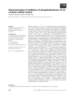
Fig. 1. Amino acid sequence alignment of Drosophila melanogaster proliferating cell nuclear antigen 2 (DmPCNA2) and DmPCNA1. Identical
and similar amino acid residues are boxed in black and gray, respectively. The interdomain connecting loop and the C-terminal tail, known to
be important for interaction of PCNA-binding proteins, are indicated. The DmPCNA2 polypeptide lacked amino acid residues from positions
190 to 194 of DmPCNA1, which corresponds to part of the D
2
E
2
loop.
T. Ruike et al. Second PCNA in Drosophila melanogaster
FEBS Journal 273 (2006) 5062–5073 ª 2006 The Authors Journal compilation ª 2006 FEBS 5063
Eukaryotic PCNA proteins have an interdomain
connecting loop that interacts with proteins such as
DNA polymerase d, flag endonuclease 1 (FEN-1), and
XP-G, and also a C-terminal tail that interacts with
DNA polymerase e and replication factor C [3]. These
regions are conserved in both DmPCNA2 and
DmPCNA1, but a small region of the D
2
E
2
loop is
absent in DmPCNA2 [19]. According to previous stud-
ies, PCNAs from several archaeans (A. pernix,
P. furiosus and S. solfataricus) also lack the D
2
E
2
loop
[10–12]. The biophysical role of this loop is still
unknown.
Analysis of DmPCNA2 expression during
Drosophila development
In Drosophila, DmPCNA1 is highly transcribed in pro-
liferating tissues [20], and its transcription is controlled
by the transcription factors DREF and early region 2
transcription factor (E2F) [9,21]. These transcription
factors regulate the expression of DNA replication- and
cell proliferation-related genes. To investigate the biolo-
gical role of DmPCNA2, we first performed northern
hybridization experiments on a range of Drosophila
developmental stages. A high level of expression of
DmPCNA1 was detected in embryos at 0–2 and 8–12 h
of development; moderate expression was present in
unfertilized eggs, 4–8 and 12–16 h embryos, and adult
females, and in Kc cells (Fig. 2A). Similar results were
found in a previous study [20]. In contrast to the expres-
sion pattern of DmPCNA1, it was difficult to detect a
DmPCNA2 signal at any developmental stage (Fig. 2A).
We therefore carried out an RT-PCR screen for
DmPCNA2 expression in the different stages of Dro-
sophila development. Expression levels were compared
in the linear range of RT-PCR amplification. As shown
in Fig. 2B, a DmPCNA2 cDNA-specific band was vis-
ible ubiquitously throughout Drosophila development.
To determine whether the putative DRE and E2F
sites found in DmPCNA1 were present in the
5¢-upstream region of DmPCNA2, we isolated genomic
DNA from adult Drosophila and cloned the
5¢-upstream region (approximately 1000 bp) of the
DmPCNA2 gene. We searched this 1000 bp nucleotide
sequence using genetyx-mac v. 9 processing software
but did not find either a DRE site (5¢-TATCGATA-3¢)
or an E2F site (5¢-TATCCCGC-3¢) in the 5¢-upstream
region of the DmPCNA2 gene.
Next, we sought to detect endogenous DmPCNAs
by antibodies raised against peptides unique to either
DmPCNA2 or DmPCNA1. Using specific antibodies
for DmPCNA2 or DmPCNA1, protein bands of
approximately 29 or 35 kDa, respectively, were
observed in western blots of Drosophila Schneider 2
(S2) cells (Fig. 2C). In contrast, no significant staining
was detectable with preimmune rabbit IgG. Further-
more, using anti-Flag serum, protein bands of approxi-
mately 30 or 36 kDa, respectively, were also observed
in western blots of S2 cells that have stable expression
of Flag-tagged DmPCNA2 or Flag-tagged DmPCNA1
(Fig. 2C).
Analysis of trimer formation by DmPCNA2 and
DmPCNA1
As PCNA forms a ring-shaped homotrimer with a
central cavity [19], we analyzed the homotrimeri-
zation of DmPCNA2 and its heterotrimerization
with DmPCNA1. We first produced recombinant
proteins and purified these to near homogeneity. The
purified DmPCNA2 ⁄ T7-(His)
6
eluted as a single peak
with a calculated molecular mass of 100 kDa in a
Sephacryl S-300 gel filtration column, as also did
DmPCNA1 ⁄ T7-(His)
6
(Fig. 3A). This result suggests
that DmPCNA2 is able to form a homotrimer. We
therefore simulated the three-dimensional structures of
DmPCNA2 and DmPCNA1, using the data from
human PCNA (Fig. 3B). The possible structures of
the DmPCNA2 homotrimer resemble that of the
DmPCNA1 homotrimer, except for the small region of
the D
2
E
2
loop. This loop in DmPCNA2 was shorter
than that in DmPCNA1.
The simulation of the possible structure of the
DmPCNA2 homotrimer suggests that the sizes of the
homotrimers of DmPCNA2 and DmPCNA1 are sim-
ilar. We therefore investigated whether DmPCNA2
could form a heterotrimer complex with DmPCNA1.
We performed a glutathione-S-transferase (GST)
pull-down experiment using DmPCNA2 ⁄ GST or
DmPCNA1 ⁄ GST and DmPCNA2 ⁄ T7-(His)
6
or
DmPCNA1 ⁄ T7-(His)
6
. The indicated GST fusion pro-
teins and T7-(His)
6
-tagged proteins of DmPCNAs were
mixed in NaCl ⁄ P
i
(lanes 1–5 in Fig. 4A), incubated at
4 °C for 12 h, and precipitated with GST Sepharose-
4B beads. Under these conditions, however, we were
unable even to find an interaction of DmPCNA1 with
itself (lane 3 in Fig. 4A). Unsurprisingly, therefore, we
could not detect an interaction between DmPCNA2
and DmPCNA1 (lanes 4 and 5 in Fig. 4A). It is poss-
ible that both DmPCNA2 and DmPCNA1 might
already have been present as homotrimers prior to
mixing and that they could not exchange monomers
under the experimental conditions used. Therefore,
we sought to reconstitute the trimeric forms of
DmPCNAs. When the proteins were forced to disso-
ciate to the monomeric state by incubation at 4 °C for
Second PCNA in Drosophila melanogaster T. Ruike et al.
5064 FEBS Journal 273 (2006) 5062–5073 ª 2006 The Authors Journal compilation ª 2006 FEBS
6 h in NaCl ⁄ P
i
containing 0.1% Tween-20, followed
by dialysis at 4 °C for 6 h in NaCl ⁄ P
i
, DmPCNA2
could form a heterotrimeric complex with DmPCNA1
in vitro (lanes 9 and 10 in Fig. 4A).
Next, we used an immunoprecipitation assay to
determine whether DmPCNA2 and DmPCNA1 could
form a heterotrimer in vivo. As we could not detect
endogenous DmPCNA2 in the crude extracts from S2
cells unless SuperSignal West Femto Maximum was
used as the chemiluminescence reagent (Fig. 2C), we
considered that further analyses of endogenous
DmPCNA2 with anti-DmPCNA serum were impracti-
cal. Therefore, using the Drosophila Expression Sys-
tem, we carried out an immunoprecipitation using S2
cells that stably expressed V5-tagged DmPCNA1 and
Flag-tagged DmPCNA1, V5-tagged DmPCNA2 and
AB
C
Fig. 2. Expression of Drosophila melanogaster PCNA2 (DmPCNA2) during Drosophila development. (A) Northern hybridization analysis.
3¢-UTRs of DmPCNA2 and DmPCNA1 cDNAs (33.6% nucleotide sequence homology) were used as specific probes. RP-49 mRNA served
as a loading control. (B) RT-PCR analysis of DmPCNA2. Expression of Act5C was used as an internal control; expression of DmPCNA1 was
also analyzed to ensure that RT-PCR reflected the results of the northern hybridization. The cycle numbers used are indicated. NC is the neg-
ative control. (C) Western blotting analysis of endogenous DmPCNAs. Crude extracts from Drosophila Schneider 2 (S2) cells were separated
by 12.5% SDS ⁄ PAGE and blotted with preimmune rabbit IgG (left panel) serum, anti-DmPCNA2 serum (second panel from the left), or anti-
DmPCNA1 serum (middle panel). The 29 kDa protein band of DmPCNA2 and the 35 kDa protein band of DmPCNA1 are indicated by arrows.
Crude extracts from S2 cells expressing Flag-tagged DmPCNA2 (second panel from the right) or Flag-tagged DmPCNA1 (right panel) were
separated by 12.5% SDS ⁄ PAGE and blotted with anti-Flag serum. The 30 kDa protein band of Flag-tagged DmPCNA2 and the 36 kDa pro-
tein band of Flag-tagged DmPCNA1 are indicated by arrows. The sizes of the molecular mass markers are indicated on the left.
T. Ruike et al. Second PCNA in Drosophila melanogaster
FEBS Journal 273 (2006) 5062–5073 ª 2006 The Authors Journal compilation ª 2006 FEBS 5065
Flag-tagged DmPCNA2, or V5-tagged DmPCNA1
and Flag-tagged DmPCNA2. We detected interactions
between DmPCNA1 molecules and between
DmPCNA2 molecules with V5 and Flag tags, but
found no evidence of an interaction between
DmPCNA2 and DmPCNA1 (Fig. 4B). These data sug-
gest that DmPCNA2 can only form a homotrimer
in vivo, and that the heterotrimerization in vitro may
be an artificial event.
Association of DmPCNA2 with Drosophila DNA
polymerases d and e
PCNA was originally identified as a DNA sliding
clamp for DNA polymerases [22]. In humans, PCNA
associates with the p120 catalytic subunit of DNA
polymerase d through interaction with the p66 third
subunit [23]. In Schizosaccharomyces pombe, PCNA
interacts with the Pol2p catalytic subunit of DNA
polymerase e [24]. We therefore tested whether
DmPCNA2 could associate with the catalytic subunits
of Drosophila DNA polymerase d and DNA poly-
merase e. We carried out an immunoprecipitation
assay using crude extract from S2 cells that had stable
expression of V5-tagged DmPCNA2. As shown in
Fig. 5, both DNA polymerase d and DNA polymerase
e are precipitated with anti-V5 serum, indicating that
DmPCNA2 can associate with DNA polymerase d and
DNA polymerase e in vivo.
Properties of binding of DmPCNA2 and
DmPCNA1 to chromatin damaged by various
mutagens
As described earlier, PCNA is involved in DNA repair
[3]. In humans, the amount of PCNA binding to
A
B
Fig. 3. Homotrimer formation of Drosophila
melanogaster proliferating cell nuclear anti-
gen 2 (DmPCNA2). (A) Gel filtration chroma-
tography analysis. Two hundred micrograms
of purified DmPCNA2 ⁄ T7-(His)
6
or
DmPCNA1 ⁄ T7-(His)
6
was loaded onto a
Sephacryl S300 gel filtration column. The cir-
cle indicates the position of the maximum
peak at which DmPCNA2 (left panel) or
DmPCNA1 (right panel) was found. Molecu-
lar mass standards (open squares) used
were ferritin (440 kDa), aldolase (158 kDa),
albumin (67 kDa), ovalbumin (43 kDa) and
ribonuclease A (13.7 kDa). (B) Building of a
model of a ring-shaped, three-dimensional
structure of DmPCNA2 (left panel) and
DmPCNA1 (right panel): upper panel, back
view; lower panel, side view. In the dia-
grams for DmPCNA1, purple balls represent
amino acid residues from 190 to 194 that
are present in DmPCNA1 but absent from
DmPCNA2.
Second PCNA in Drosophila melanogaster T. Ruike et al.
5066 FEBS Journal 273 (2006) 5062–5073 ª 2006 The Authors Journal compilation ª 2006 FEBS
chromatin increases in cells treated with mutagens such
as MMS [25], H
2
O
2
[26] and UV light [27]. We there-
fore examined the association of DmPCNA2 and
DmPCNA1 with chromatin following treatment with
DNA-damaging agents. First, we visualized the sub-
cellular localization of V5-tagged DmPCNA2 and
Flag-tagged DmPCNA1 in S2 cells using immunofluo-
rescence microscopy. We found that both DmPCNA2
and DmPCNA1 were localized in the nucleus
(Fig. 6A). Next, we prepared fractions from the cyto-
plasm, the whole chromatin, a high-salt wash and the
core nuclear matrix from S2 cells. As controls for the
fractionation procedure, we used western blotting with
antibodies against b-tubulin (a nonchromatin-bound
protein) and histone H4 (a chromatin-bound protein).
DmPCNA2 and DmPCNA1 were present in the cyto-
plasmic and the whole chromatin fractions (Fig. 6B).
Following exposure to DNA-damaging agents, both
DmPCNA2 and DmPCNA1 showed increased levels
of association with chromatin, with a time-dependent
relationship (Fig. 6C). The level of DmPCNA2 in the
whole chromatin fraction reached a maximum at 5–8 h
after MMS treatment and at 3 h after H
2
O
2
treatment
(Fig. 6C). In contrast, the amount of DmPCNA1 in
this fraction continued to increase up to 8 h after
MMS treatment and 5 h after H
2
O
2
treatment
(Fig. 6C). UV light treatment increased the level
of DmPCNA1 associating with chromatin but not of
DmPCNA2. Mitomycin C did not alter the levels
of either DmPCNA2 or DmPCNA1 associating with
chromatin. We also investigated the binding of
DmPCNA2 to chromatin after treatment with various
doses of DNA-damaging agents (Fig. 6D). MMS-trea-
ted S2 cells were collected 5 h after treatment, and S2
cells treated with H
2
O
2
, UV light or mitomycin C were
harvested at 3 h. The amounts of DmPCNA2 in the
whole chromatin fractions increased in a dose-depend-
ent fashion after MMS and H
2
O
2
treatments, but were
A
B
Fig. 4. Interaction of Drosophila melano-
gaster proliferating cell nuclear antigen 2
(DmPCNA2) and DmPCNA1. (A) In vitro
interaction of DmPCNA2 and DmPCNA1.
Lanes 1–5: the indicated proteins were
mixed in NaCl ⁄ P
i
at 4 °C for 12 h. Lanes
6–10: the indicated proteins were mixed in
NaCl ⁄ P
i
containing 0.1% Tween-20 at 4 °C
for 6 h, and this was followed by dialysis in
NaCl ⁄ P
i
at 4 °C for 6 h. The proteins bound
to GST Sepharose-4B beads were analyzed
by western blotting with anti-T7 or anti-GST
serum. (B) In vivo interaction between
DmPCNA2 and DmPCNA1. Drosophila
Schneider 2 (S2) cells expressing the indica-
ted DmPCNAs were harvested and lysed.
The lysates were immunoprecipitated (IP)
with anti-V5 serum. The washed immuno-
precipitates were separated by 12.5%
SDS ⁄ PAGE and blotted for either Flag or V5
(left panel). The lysates were immunoprecip-
itated with anti-Flag serum and blotted
sequentially for V5 or Flag (right panel).
T. Ruike et al. Second PCNA in Drosophila melanogaster
FEBS Journal 273 (2006) 5062–5073 ª 2006 The Authors Journal compilation ª 2006 FEBS 5067
not influenced by UV light or mitomycin C treatments
(Fig. 6D).
Discussion
In this study, we identified a second PCNA cDNA
from Drosophila melanogaster. This PCNA, which we
call here DmPCNA2, had two conserved regions, an
interdomain connecting loop and a C-terminal tail.
DmPCNA2 formed homotrimers and associated with
DNA polymerase d and DNA polymerase e in vivo.In
addition, DmPCNA2, as well as DmPCNA1, was
present in the whole chromatin fraction of cellular
proteins. Taken together, these results suggest that
DmPCNA2 can act as a DNA sliding clamp for these
DNA polymerases.
Yamaguchi and colleagues reported that the expres-
sion of DmPCNA1 is controlled by the transcription
factors DREF and E2F, which are abundant in tissues
such as the ovary and in unfertilized eggs and early
embryos [9,21]. Thus, DmPCNA1 mRNA is highly
expressed in proliferating tissues and decreases rapidly
during development [20]. In contrast to DmPCNA1,
there was no evidence for putative binding sites for
DREF and E2F in the 5¢-upstream region of
DmPCNA2. Moreover, DmPCNA2 was constantly
expressed even in pupae in which few cells are pro-
liferating. These data suggest that expression of
DmPCNA2 might not be related to cell proliferation.
We found different patterns of binding to chromatin
between DmPCNA2 and DmPCNA1 in S2 cells trea-
ted with DNA-damaging agents. MMS and H
2
O
2
induced a more rapid association of DmPCNA2 with
chromatin than of DmPCNA1. UV light induced the
association of DmPCNA1 with chromatin, but not of
DmPCNA2. These results suggest that each DmPCNA
functions independently when DNA is damaged. It has
been reported that PCNA cannot load itself onto
DNA in vitro and requires a clamp loader protein to
achieve this association [28,29]. Therefore, the patterns
of association of DmPCNA2 and DmPCNA1 with
chromatin might reflect differential loading onto dam-
aged DNA by clamp loaders. DmPCNA1 probably
functions in the repair of MMS-, H
2
O
2
- and UV light-
induced lesions in a similar manner to other eukaryotic
PCNAs. In eukaryotes, base excision repair is known
to be the major pathway for repair of MMS- and
H
2
O
2
-induced DNA lesions and is often initiated by
several DNA glycosylases [30]. In S. pombe, PCNA
and Rad9 ⁄ Rad1 ⁄ Hus1 differentially participate in base
excision repair through interaction with the DNA gly-
cosylase MutY homolog [31]. Although the precise
function of DmPCNA2 remains unclear, one hypothe-
sis is that DmPCNA2 might participate in the base
excision repair pathway through interaction with some
of the Drosophila DNA glycosylases. Another possibil-
ity is that DmPCNA2 might simply support
DmPCNA1 in the repair of MMS- and H
2
O
2
-induced
DNA damage.
Our next task in the near future will be to elucidate
how DmPCNA2 functions in the DNA repair system.
The analysis of flies with mutation of DmPCNA2 will
help us to understand its biophysiologic roles as well
as enable identification of the DmPCNA2 binding
partners.
Experimental procedures
Cloning of DmPCNA2
Total RNA from Kc cells was reverse transcribed using the
SuperScript First-Strand Synthesis System (Invitrogen, Car-
lsbad, CA) with an oligo-(dT)
12)18
primer. Amplification of
the DmPCNA2 cDNA was performed using ExTaq thermo-
stable DNA polymerase (TaKaRa, Ohtsu, Japan) and the
following primers: forward, 5¢-ATGCTCGAGGCGCGTT
Fig. 5. Association of Drosophila melanogaster proliferating cell
nuclear antigen 2 (DmPCNA2) with DNA polymerase d (Dmpol d)
and Dmpol e. Drosophila Schneider 2 (S2) cells expressing
V5-tagged DmPCNA2 were harvested and lysed. The lysates were
immunoprecipitated (IP) with anti-V5 serum. The washed immuno-
precipitates were separated by 5% SDS ⁄ PAGE and analyzed by
western blotting with anti-Dmpol d, anti-Dmpol e or anti-V5 serum.
Second PCNA in Drosophila melanogaster T. Ruike et al.
5068 FEBS Journal 273 (2006) 5062–5073 ª 2006 The Authors Journal compilation ª 2006 FEBS
AB
DC
Fig. 6. Chromatin-binding patterns of Drosophila melanogaster proliferating cell nuclear antigen 2 (DmPCNA2) and DmPCNA1 in response to
DNA-damaging agents. (A) Immunofluorescent analysis of the localization of V5-tagged DmPCNA2 and Flag-tagged DmPCNA1. DmPCNA2 is
shown in red, DmPCNA1 in green, and DNA in blue after DAPI staining. Bar represents 5 lm. (B) Fractionation of DmPCNA2 and
DmPCNA1. Drosophila Schneider 2 (S2) cells expressing V5-tagged DmPCNA2 and Flag-tagged DmPCNA1 were extracted to obtain cyto-
plasmic, whole chromatin, high-salt-wash and core nuclear matrix fractions. The fractions were analyzed by western blotting with the indica-
ted antibodies. (C) Chromatin binding of DmPCNA2 and DmPCNA1 in response to DNA-damaging agents [0.02% methyl methanesulfonate
(MMS), 1.5 m
M H
2
O
2
,35JÆm
)2
UV light and 0.02% mitomycin C (MMC)]. S2 cells were collected at the indicated post-treatment intervals.
(D) Chromatin binding of DmPCNA2 after various doses of DNA-damaging agents. S2 cells were treated with MMS (concentration range
0.01–0.1%), H
2
O
2
(concentration range 0.5–2.5 mM), UV light (dose range 15–70 JÆm
)2
) or MMC (concentration range 0.01–0.1%). The chro-
matin fractions were analyzed by western blotting with anti-V5 serum.
T. Ruike et al. Second PCNA in Drosophila melanogaster
FEBS Journal 273 (2006) 5062–5073 ª 2006 The Authors Journal compilation ª 2006 FEBS 5069
TGAG-3¢; and reverse, 5¢-CTAGAAATCGGGGTCATT
CA-3¢. The amplified cDNA was cloned into the pGEM-T
vector (Promega, Madison, WI). To identify the 5¢- and
3¢-termini of the gene, 5¢- and 3¢-RLM-RACE was per-
formed in accordance with the manufacturer’s recom-
mended protocol (FirstChoice RLM-RACE kit; Ambion,
Austin, TX).
Northern hybridization and RT-PCR analysis
Total RNAs were extracted using Trizol (Invitrogen) from
unfertilized Drosophila eggs, embryos, larvae, adult flies
and from Kc cells. Northern hybridization was carried out
as described previously [32]. The 3¢-UTR of DmPCNA2
cDNA (nucleotides 863–1019) or that of DmPCNA1 (nucle-
otides 873–997) was used as the specific probe. Full-length
ribosomal protein 49 (Rp-49) cDNA was used as a control.
For RT-PCR analysis, total RNAs (tissue and cell
sources described above) were treated with DNase I
(TaKaRa) to remove traces of genomic DNA contamin-
ation, and purified with phenol ⁄ chloroform. First-strand
cDNA was synthesized from 1 lg of total RNA using
the SuperScript First-Strand Synthesis System (Invitrogen)
with random hexamers, and then amplified using the
following primers: DmPCNA2 ) forward, 5¢-ATGCTCGA
GGCGCGTTTGAG-3¢, and reverse, 5¢-CTAGAAATC
GGGGTCATTCA-3¢; DmPCNA1 – forward, 5¢-ATGTTC
GAGGCACGCCT-3¢, and reverse, 5¢-TTATGTCTCGTT
GT CCTCGA-3¢; Act5c ) forward, 5¢-TGTGGATACTCC
TCCCGACA-3¢, and reverse, 5¢-ATCCCGATCCTGAC
TCTT-3¢. The PCR conditions were: DmPCNA2 – 94 °C
for 5 min, 94 °C for 45 s, 55 °C for 45 s, 72 °C for 1 min,
24 cycles, 5 min extension at 72 °C; DmPCNA1 ) 94 °C
for 5 min, 94 °C for 45 s, 55 °C for 45 s, 72 °C for 1 min,
21 cycles, 5 min extension at 72 °C; Act5c ) 94 °C for
5 min, 94 °C for 45 s, 55 °C for 45 s, 72 °C for 1 min 30 s,
17 cycles, 5 min extension at 72 °C. PCR products were
visualized by staining with SYBR Gold nucleic acid gel
stain (Molecular Probes, Eugene, OR) after agarose gel
electrophoresis.
Generation of antibodies to DmPCNA2 and
anti-DmPCNA1
A keyhole limpet haemocyanin (KLH)-conjugated syn-
thetic peptide with an extra cysteine on the N-terminus
(CKKDYTCFIQLPSS, amino acids 129–142 of
DmPCNA2) or (CKLAQTGSVDKEEEA, amino acids
181–194 of DmPCNA1) was used for inoculation into rab-
bits (Bio Matrix Research, Kashiwa, Japan). For detection
of endogenous DmPCNA2, anti-DmPCNA2 serum or pre-
immune rabbit IgG diluted to 0.5 lgÆmL
)1
served as pri-
mary antibodies. Horseradish peroxidase-conjugated goat
anti-(rabbit IgG) (Vector Laboratories, Burlingame, CA)
diluted to 2 ng Æ mL
)1
served as the secondary antibody.
Chemiluminescence was detected with SuperSignal West
Femto Maximum (Pierce, Rockford, IL). For detection of
endogenous DmPCNA1, anti-DmPCNA1 serum diluted
to 1 lgÆmL
)1
and horseradish peroxidase-conjugated goat
anti-(rabbit IgG) diluted to 50 ngÆmL
)1
served as primary
and secondary antibodies, respectively. Chemiluminescence
was detected with enhanced chemiluminescence (ECL)
western blotting detection reagents (Amersham Pharmacia
Biotech, Piscataway, NJ).
Animals were fed water and standard rabbit food and
maintained on a 12 h light/dark cycle. Polyclonal antiserum
to the peptide was raised in rabbits by subcutaneous injec-
tion of 0.15 mg of the peptide emulsified in Freund’s com-
plete adjuvant. Two weeks after the primary injection,
boosts of 0.3 mg of the peptide in Freund’s incomplete adju-
vant were injected every 2 weeks. The rabbits were bled one
week after the final boost under anesthesia. The rabbits were
treated in accordance with procedures approved by the Ani-
mal Ethics Committee of the Science University of Tokyo.
Purification of recombinant DmPCNA2 or
DmPCNA1 proteins
The DmPCNA2 coding region was cloned into pET21a
(Novagen, Darmstadt, Germany) or pGEX-6P-1 vectors
(Amersham Pharmacia Biotech). T7-(His)
6
-tagged
DmPCNA2 [DmPCNA2 ⁄ T7-(His)
6
] protein was over-
expressed in Escherichia coli BL21 (DE3) (Novagen) and
purified with His-Bind Resin according to the manufacturer’s
protocol (Novagen). GST fusion DmPCNA2 (DmPCNA2 ⁄
GST) protein was overexpressed in E. coli BL21 (DE3)
and purified with Glutathione Sepharose-4B (Amersham
Pharmacia Biotech). Production and purification of
DmPCNA1 ⁄ T7-(His)
6
protein and DmPCNA1 ⁄ GST protein
were carried out as described above for DmPCNA2.
Gel filtration column chromatography
Samples of purified DmPCNA2 ⁄ T7-(His)
6
and
DmPCNA1 ⁄ T7-(His)
6
proteins were dialyzed against
TEMG buffer (50 mm Tris ⁄ HCl, pH 7.9, 1 mm EDTA,
pH 8.0, 5 mm 2-mercaptoethanol, 10% glycerol) containing
0.2 m NaCl. A 200 lg sample of each protein was sepa-
rately loaded onto a gel filtration column (Sephacryl S-300
gel column; Amersham Pharmacia Biotech) equilibrated
with the same buffer. The molecular mass was estimated
from a calibration curve using ferritin (440 kDa), aldolase
(158 kDa), albumin (67 kDa), ovalbumin (43 kDa) and
ribonuclease A (13.7 kDa).
Three-dimensional structure model building
The predicted structure of the human PCNA protein
was used to set the parameters for constructing models
Second PCNA in Drosophila melanogaster T. Ruike et al.
5070 FEBS Journal 273 (2006) 5062–5073 ª 2006 The Authors Journal compilation ª 2006 FEBS
of DmPCNA2 and DmPCNA1. We used the swiss-model
program [33–35] to generate three-dimensional models of
the DmPCNA2 and DmPCNA1 proteins.
GST pull-down assay
Equal amounts of purified GST fusion proteins and puri-
fied T7-(His)
6
-tagged proteins were mixed in NaCl ⁄ P
i
and
incubated at 4 °C for 12 h, or mixed in NaCl ⁄ P
i
containing
0.1% Tween-20 and incubated at 4 °C for 6 h. The mix-
tures were then dialyzed in NaCl ⁄ P
i
at 4 °C for 6 h. GST
Sepharose-4B beads (Amersham Pharmacia Biotech) were
added to the samples, which were then incubated at 4 °C
for 1 h. After being washed six times with 0.8 mL of
NaCl ⁄ P
i
, the bound proteins were eluted with TEMG buf-
fer containing 10 mm reduced glutathione and analyzed by
western blotting with mouse monoclonal antibody T7
(Novagen) and rabbit polyclonal anti-GST serum.
Cell culture, plasmid construction, and
transfection
S2 cells were cultured in Schneider’s Drosophila Medium
(Invitrogen) containing 10% heat-inactivated fetal bovine
serum at 25 °C. The expression vector for V5-tagged
DmPCNA2 was constructed by cloning the DmPCNA2
coding region into pAc5.1 ⁄ V5-His C (Invitrogen). Flag-
tagged DmPCNA2 was constructed by cloning the N-ter-
minally Flag-tagged DmPCNA2 coding region into
pAc5.1 ⁄ V5-His C from which the V5-His tag had been
removed. Expression vectors for V5-tagged DmPCNA1 and
Flag-tagged DmPCNA1 were constructed as described
above for DmPCNA2. All transfections and establishment
of the stable cell lines were performed in accordance with
the manufacturer’s protocols (Invitrogen).
Immunoprecipitation experiments
Aliquots of 1 · 10
7
S2 cells were washed in NaCl ⁄ P
i
and
suspended in TEMG buffer containing 0.15 m NaCl, 0.01%
NP-40, and the protease inhibitors phenylmethanesulfonyl
fluoride (1 mm), leupeptin (1 mm) and pepstatin A (1 mm).
After sonication, the lysates were rocked at 4 °C for
30 min, and then centrifuged at 10 000 g for 10 min (MX-
201; TOMY; TMA-29 rotor). The supernatants were pre-
cleared by treatment with protein G Sepharose beads
(Amersham Pharmacia Biotech) at 4 °C for 1 h. Cleared
lysates were immunoprecipitated with protein G Sepharose
beads and a mouse monoclonal V5 antibody (Invitrogen)
or anti-Flag serum (Sigma, St Louis, MO) at 4 °C for 2 h.
Immunoprecipitates were washed three times with the same
buffer, solubilized in SDS ⁄ PAGE sample buffer, and ana-
lyzed by western blotting. For generation of antibodies
to DNA polymerase d, the purified recombinant DNA
polymerase d fragment (amino acid residues 104–445) was
used for inoculation into rabbits. The generation of anti-
DNA polymerase e was described in a previous report [36].
Immunofluorescence analysis
S2 cells were placed on poly-(l-lysine)-coated coverslips
and fixed with 4% paraformaldehyde in NaCl⁄ P
i
for
10 min at room temperature. After several washes with
NaCl ⁄ P
i
, the cells were treated with methanol for permeabi-
lization. The samples were incubated with primary antibod-
ies, mouse monoclonal anti-V5 serum and rabbit polyclonal
anti-Flag serum, at 4 °C overnight, and then treated for 1 h
with the secondary antibodies Alexa546 anti-(mouse IgG)
and Alexa488 anti-(rabbit IgG) (Molecular Probes). They
were also counterstained with 4¢,6-diamidine-2-phenylindole
(DAPI). The preparations were observed under a fluore-
scence microscope and the data were collected using a
CCD camera (Nikon, Chiyoda, Japan).
Fractionation of cellular proteins
S2 cells were exposed to MMS or mitomycin C for 1 h
or to H
2
O
2
for 15 min. The cells were then washed once
and incubated prior to sampling. UV-irradiated S2 cells
were incubated in the dark in order to distinguish the
effects of UV irradiation from those of the photoreacti-
vating mechanism. After incubation, S2 cells were washed
three times with ice-cold NaCl ⁄ P
i
. Aliquots of 1 · 10
7
S2
cells were lysed in 500 lL of cytoskeleton buffer (CSK
buffer: 10 mm Hepes, pH 7.4, 100 mm NaCl, 300 mm
sucrose, 3 mm MgCl
2
,1mm EGTA, 5 mm 2-mercapto-
ethanol, 1 mm phenylmethanesulfonyl fluoride, 1 mm leu-
peptin, 1 mm pepstatin A, 0.5% Triton X-100) at 4 °C
for 5 min and centrifuged at 3000 g for 5 min (MX-201;
TOMY; TMA-29 rotor). The soluble cytoplasmic fraction
was removed, and the pellet was washed once with
500 lL of CSK buffer. The pellet was then resuspended
in 200 lL of CSK buffer containing 100 U of RNase-free
DNase I (TaKaRa). After 30 min at 37 °C, ammonium
sulfate was added to a final concentration of 0.25 m. The
samples were incubated for 5 min at 4 °C and centrifuged
as above. The soluble chromatin fraction was removed,
and the pellet was extracted in CSK buffer with 2 m
NaCl for 5 min at 4 °C. After another centrifugation, the
2 m NaCl wash was removed, and the nuclear matrix pel-
let was resuspended in 50 lL of SDS ⁄ PAGE sample buf-
fer. For western blot analysis, equal cell equivalents from
each fraction were subjected to SDS ⁄ PAGE and probed
with appropriate antibodies: mouse monoclonal anti-V5
serum, rabbit polyclonal anti-Flag serum (Sigma), mouse
monoclonal anti-b -tubulin serum (Chemicon, Temecula,
CA), or rabbit polyclonal anti-Histone H4 (Imgenex, San
Diego, CA).
T. Ruike et al. Second PCNA in Drosophila melanogaster
FEBS Journal 273 (2006) 5062–5073 ª 2006 The Authors Journal compilation ª 2006 FEBS 5071
References
1 Zhang P, Mo JY, Perez A, Leon A, Liu L, Mazloum
N, Xu H & Lee MY (1999) Direct interaction of proli-
ferating cell nuclear antigen with the p125 catalytic sub-
unit of mammalian DNA polymerase delta. J Biol
Chem 274, 26647–26653.
2 Eissenberg JC, Ayyagari R, Gomes XV & Burgers PM
(1997) Mutations in yeast proliferating cell nuclear anti-
gen define distinct sites for interaction with DNA poly-
merase delta and DNA polymerase epsilon. Mol Cell
Biol 17, 6367–6378.
3 Maga G & Hubscher U (2003) Proliferating cell nuclear
antigen (PCNA): a dancer with many partners. J Cell
Sci 116, 3051–3060.
4 Waga S, Hannon GJ, Beach D & Stillman B (1994) The
p21 inhibitor of cyclin-dependent kinases controls DNA
replication by interaction with PCNA. Nature 369, 574–
578.
5 Chuang LS, Ian HI, Koh TW, Ng HH, Xu G & Li BF
(1997) Human DNA-(cytosine-5) methyltransferase–
PCNA complex as a target for p21WAF1. Science 277,
1996–2000.
6 Hasan S, Hassa PO, Imhof R & Hottiger MO (2001)
Transcription coactivator p300 binds PCNA and may
have a role in DNA repair synthesis. Nature 410, 387–
391.
7 Henderson DS, Banga SS, Grigliatti TA & Boyd JB
(1994) Mutagen sensitivity and suppression of position-
effect variegation result from mutations in mus209, the
Drosophila gene encoding PCNA. EMBO J 13, 1450–
1459.
8 Henderson DS, Bailey DA, Sinclair DA & Grigliatti TA
(1987) Isolation and characterization of second chromo-
some mutagen-sensitive mutations in Drosophila melano-
gaster. Mutat Res 177 , 83–93.
9 Yamaguchi M, Hayashi Y, Nishimoto Y, Hirose F &
Matsukage A (1995) A nucleotide sequence essential for
the function of DRE, a common promoter element for
Drosophila DNA replication-related genes. J Biol Chem
270, 15808–15814.
10 Daimon K, Kawarabayasi Y, Kikuchi H, Sako Y &
Ishino Y (2002) Three proliferating cell nuclear antigen-
like proteins found in the hyperthermophilic archaeon
Aeropyrum pernix: interactions with the two DNA poly-
merases. J Bacteriol 184, 687–694.
11 Cann IK, Ishino S, Hayashi I, Komori K, Toh H,
Morikawa K & Ishino Y (1999) Functional inter-
actions of a homolog of proliferating cell nuclear anti-
gen with DNA polymerases in Archaea. J Bacteriol
181, 6591–6599.
12 De Felice M, Sensen CW, Charlebois RL, Rossi M &
Pisani FM (1999) Two DNA polymerase sliding clamps
from the thermophilic archaeon Sulfolobus solfataricus.
J Mol Biol 291, 47–57.
13 Hata S, Kouchi H, Tanaka Y, Minami E, Matsumoto
T, Suzuka I & Hashimoto J (1992) Identification of car-
rot cDNA clones encoding a second putative proliferat-
ing cell-nuclear antigen, DNA polymerase delta
auxiliary protein. Eur J Biochem 203, 367–371.
14 Taniguchi Y, Katsumata Y, Koido S, Yoshimura S,
Suemizu H & Moriuchi T (1992) Isolation of a new
human pseudogene for proliferating cell nuclear antigen.
Nucleic Acids Symp Ser 27, 147–148.
15 Yamaguchi M, Hayashi Y, Hirose F, Matsuoka S,
Moriuchi T, Shiroishi T, Moriwaki K & Matsukage A
(1991) Molecular cloning and structural analysis of
mouse gene and pseudogenes for proliferating cell
nuclear antigen. Nucleic Acids Res 19, 2403–2410.
16 Celniker SE, Wheeler DA, Kronmiller B, Carlson JW,
Halpern A, Patel S, Adams M, Champe M, Dugan SP,
Frise E et al. (2002) Finishing a whole-genome shotgun:
release 3 of the Drosophila melanogaster euchromatic
genome sequence. Genome Biol
3, RESEARCH0079.1–
0079.14.
17 Cavener DR (1987) Comparison of the consensus
sequence flanking translational start sites in Drosophila
and vertebrates. Nucleic Acids Res 15, 1353–1361.
18 Proudfoot NJ & Brownlee GG (1976) 3¢ Non-coding
region sequences in eukaryotic messenger RNA. Nature
263, 211–214.
19 Krishna TS, Kong XP, Gary S, Burgers PM & Kuriyan
J (1994) Crystal structure of the eukaryotic DNA poly-
merase processivity factor PCNA. Cell 79, 1233–1243.
20 Yamaguchi M, Nishida Y, Moriuchi T, Hirose F, Hui
CC, Suzuki Y & Matsukage A (1990) Drosophila prolif-
erating cell nuclear antigen (cyclin) gene: structure,
expression during development, and specific binding of
homeodomain proteins to its 5¢-flanking region. Mol
Cell Biol 10, 872–879.
21 Yamaguchi M, Hayashi Y & Matsukage A (1995)
Essential role of E2F recognition sites in regulation of
the proliferating cell nuclear antigen gene promoter dur-
ing Drosophila development. J Biol Chem 270, 25159–
25165.
22 Prelich G, Kostura M, Marshak DR, Mathews MB &
Stillman B (1987) The cell-cycle regulated proliferating
cell nuclear antigen is required for SV40 DNA replica-
tion in vitro. Nature 326, 471–475.
23 Pohler JR, Otterlei M & Warbrick E (2005) An in vivo
analysis of the localisation and interactions of human p66
DNA polymerase delta subunit. BMC Mol Biol 6: 17.
24 Dua R, Levy DL, Li CM, Snow PM & Campbell JL
(2002) In vivo reconstitution of Saccharomyces cerevisiae
DNA polymerase epsilon in insect cells. Purification and
characterization. J Biol Chem 277, 7889–7896.
25 Shibata Y & Nakamura T (2002) Defective flap endonu-
clease 1 activity in mammalian cells is associated with
impaired DNA repair and prolonged S phase delay.
J Biol Chem 277, 746–754.
Second PCNA in Drosophila melanogaster T. Ruike et al.
5072 FEBS Journal 273 (2006) 5062–5073 ª 2006 The Authors Journal compilation ª 2006 FEBS
26 Balajee AS, Dianova I & Bohr VA (1999) Oxidative
damage-induced PCNA complex formation is efficient
in xeroderma pigmentosum group A but reduced in
Cockayne syndrome group B cells. Nucleic Acids Res
27, 4476–4482.
27 Toschi L & Bravo R (1988) Changes in cyclin ⁄ prolife-
rating cell nuclear antigen distribution during DNA
repair synthesis. J Cell Biol 107, 1623–1628.
28 Tan CK, Castillo C, So AG & Downey KM (1986) An
auxiliary protein for DNA polymerase-delta from fetal
calf thymus. J Biol Chem 261, 12310–12316.
29 Ellison V & Stillman B (2003) Biochemical characteri-
zation of DNA damage checkpoint complexes: clamp
loader and clamp complexes with specificity for 5¢
recessed DNA. Plos Biol 1: E33.
30 Ide H & Kotera M (2004) Human DNA glycosylases
involved in the repair of oxidatively damaged DNA.
Biol Pharm Bull 27, 480–485.
31 Chang DY & Lu AL (2005) Interaction of checkpoint
proteins Hus1 ⁄ Rad1 ⁄ Rad9 with DNA base excision
repair enzyme MutY homolog in fission yeast,
Schizosaccharomyces pombe. J Biol Chem 280,
408–417.
32 Takata K, Ishikawa G, Hirose F & Sakaguchi K (2002)
Drosophila damage-specific DNA-binding protein 1
(D-DDB1) is controlled by the DRE ⁄ DREF system.
Nucleic Acids Res 30, 3795–3808.
33 Schwede T, Kopp J, Guex N & Peitsch MC (2003)
SWISS-MODEL: an automated protein homology-mod-
eling server. Nucleic Acids Res 31, 3381–3385.
34 Guex N & Peitsch MC (1997) SWISS-MODEL and the
Swiss-PdbViewer: an environment for comparative pro-
tein modeling. Electrophoresis 18, 2714–2723.
35 Peitsch MC, Wells TN, Stampf DR & Sussman JL
(1995) The Swiss-3DImage collection and PDB-Browser
on the World-Wide Web. Trends Biochem Sci 20, 82–84.
36 Aoyagi N, Oshige M, Hirose F, Kuroda K, Matsukage
A & Sakaguchi K (1997) DNA polymerase epsilon from
Drosophila melanogaster. Biochem Biophys Res Commun
230, 297–301.
T. Ruike et al. Second PCNA in Drosophila melanogaster
FEBS Journal 273 (2006) 5062–5073 ª 2006 The Authors Journal compilation ª 2006 FEBS 5073