Báo cáo khoa học: IMP1 interacts with poly(A)-binding protein (PABP) and the autoregulatory translational control element of PABP-mRNA through the KH III-IV domain pdf
Bạn đang xem bản rút gọn của tài liệu. Xem và tải ngay bản đầy đủ của tài liệu tại đây (887.06 KB, 13 trang )
IMP1 interacts with poly(A)-binding protein (PABP) and
the autoregulatory translational control element of
PABP-mRNA through the KH III-IV domain
Gopal P. Patel and Jnanankur Bag
Department of Molecular and Cellular Biology, University of Guelph, Ontario, Canada
Regulation of gene expression is fundamental to
almost all biological activities. Multiple layers of regu-
latory mechanisms control essentially every step of
gene expression in eukaryotes. It was thought that
regulation of transcription is the master switch of gene
expression in eukaryotes [1]; however, it is becoming
increasingly evident that the majority of regulatory
mechanisms are employed at the post-transcriptional
and translational levels [2,3]. In order to be functional,
cellular mRNA associates with a wide array of RNA-
binding proteins to form a messenger ribonucleopro-
tein particle (mRNP). The constituent of the mRNP
dictates the fate of mRNA [4]. It is therefore not sur-
prising that functionally related eukaryotic genes may
represent ‘post-transcriptional operons’ because they
are regulated coordinately at post-transcriptional levels
by unique combinations of mRNA-binding proteins
that recognize common cis-elements among the
mRNAs [5].
The poly(A)-tail is one of the most common cis-
acting sequence elements found in the 3¢ UTR of
eukaryotic mRNAs, which predominantly binds to
poly(A)-binding protein (PABP). The 3¢ poly(A)-tail
and PABP, together, influence almost every aspect of
mRNA metabolism including maturation, transporta-
tion, localization, translation and stability [6–8]. Given
the significant function of PABP in mRNA biology, its
cellular level is tightly regulated at the translational level
Keywords
autoregulation; IMP1; PABP; poly(A)-binding
protein; translational control
Correspondence
J. Bag, Department of Molecular and
Cellular Biology, University of Guelph,
Guelph, Ontario, N1G 2W1, Canada
Fax: +1 519 837 2075
Tel: +1 519 824 4120 Ext. 53390
E-mail:
(Received 12 June 2006, revised 1 October
2006, accepted 25 October 2006)
doi:10.1111/j.1742-4658.2006.05556.x
Repression of poly(A)-binding protein (PABP) mRNA translation involves
the formation of a heterotrimeric ribonucleoprotein complex by the binding
of PABP, insulin-like growth factor II mRNA binding protein-1 (IMP1)
and the unr gene encoded polypeptide (UNR) to the adenine-rich autoregu-
latory sequence (ARS) located at the 5¢ untranslated region of the PABP-
mRNA. In this report, we have further characterized the interaction
between PABP and IMP1 with the ARS at the molecular level. The dissoci-
ation constants of PABP and IMP1 for binding to the ARS RNA were
determined to be 2.3 nm and 5.9 nm, respectively. Both PABP and IMP1
interact with each other, regardless of the presence of the ARS, through
the conserved C-terminal PABP-C and K-homology (KH) III-IV domains,
respectively. Interaction of PABP with the ARS requires at least three out
of its four RNA-binding domains, whereas KH III-IV domain of IMP1 is
necessary and sufficient for binding to the ARS. In addition, the strongest
binding site for both PABP and IMP1 on the ARS was determined to
be within the 22 nucleotide-long CCCAAAAAAAUUUACAAAAAA
sequence located at the 3¢ end of the ARS. Results of our analysis suggest
that both proteinÆprotein and proteinÆRNA interactions are involved in
forming a stable ribonucleoprotein complex at the ARS of PABP mRNA.
Abbreviations
ARC, autoregulatory ribonucleoprotein complex; ARS, autoregulatory sequence; IMP1, insulin-like growth factor II mRNA binding protein-
1KH, K-homology; mRNP, messenger ribonucleoprotein particle; PABP, poly(A)-binding protein; RBD, RNA-binding domain; REMSA, RNA
electrophoretic mobility shift assay; RRM, RNA-recognition motif; TOP, terminal oligopyrimidine tract; UNR, unr gene encoded polypeptide.
5678 FEBS Journal 273 (2006) 5678–5690 ª 2006 The Authors Journal compilation ª 2006 FEBS
by two repressible cis-acting sequence elements, the ter-
minal oligopyrimidine tract (TOP) [9] and an adenine-
rich autoregulatory sequence (ARS) [10]. The TOP
element encompasses the first 31 nucleotides, whereas
the ARS spans nucleotides 71–131 in the 5¢ UTR of the
PABP mRNA. The TOP element regulates PABP trans-
lation in growth-dependent and tissue-specific manners
[11,12], whereas the ARS functions constitutively in all
types of cells [9,10]. It has been generally accepted that
at elevated cellular levels, PABP binds to the ARS
region of its own mRNA and represses translation by
stalling the movement of the 40S ribosomal subunit
along the 5¢ UTR [13,14]. Recent studies in our laborat-
ory have shown that, besides PABP, the ARS binds to
insulin-like growth factor II mRNA binding protein-1
(IMP1) and the unr gene encoded polypeptide (UNR) to
form a heterotrimeric autoregulatory ribonucleoprotein
complex (ARC) [15]. Mutational analyses of the ARS
have shown a strong correlation between the formation
of the heterotrimeric complex and repression of a repor-
ter gene expression. UNR showed lesser affinity for the
ARS, and its presence in the ARC required association
with PABP. However, IMP1 is capable of binding to the
ARS with high affinity independently, and can also
interact with PABP [15].
There are several functional similarities between
PABP and IMP1. Both polypeptides have been impli-
cated in mRNA localization, turnover, and transla-
tional control. No enzymatic activity has been
associated with either PABP or IMP1, and it seems
that their functions are attributed to their ability to
bind to specific RNA sequences and to act as a scaf-
fold for protein–protein interactions. PABP contains
four RNA-binding domains (RBD I to IV) arranged
in tandem at its N-terminus and a protein-binding aux-
iliary domain at its C-terminus. Concurrently, PABP
exhibits preferential affinity for poly(A) stretches and
also interacts with several cytosolic polypeptides such
as Paip1 [16,17], Paip2 [18,19], eIF4B [20], poly(C)-
binding proteins [21], UNR [22], eIF4G [23], Rna15
[24], eRF3 [25], and TcUBP-1 [26].
IMP1 belongs to the conserved valine-isoleucine-
cystine-lysine-glutamine containing (VICKZ) family of
mRNA-binding proteins consisting of two RNA-recog-
nition motifs (RRM I and II) at its N-terminus and
four K-homology (KH) domains arranged in tandem
at its C-terminus [27]. Interestingly, associations of
IMP1 with both RNAs and proteins are primarily
mediated by the KH-domains [28]. The full repertoire
of RNA-sequence targets and polypeptide partners of
IMP1 has not yet been defined. The RNA targets of
IMP1 include Igf-II [27], c-myc [29], tau [30], FMR1
[31], and PABP [15] mRNAs; whereas its known
polypeptide partners consist of G3 BP, HuD [30],
FMRP [31], and PABP [15].
In the present study, we have further characterized
the interaction between PABP and IMP1 on the ARS
RNA for a better understanding of their role in trans-
lational regulation of PABP expression. The results of
our studies show that both PABP and IMP1 bind
strongly to nucleotides between 110 and 131 of the
ARS RNA. Binding of PABP to the ARS requires a
minimum of three RBDs (RBD I to III or RBD II to
IV), whereas binding of IMP1 to the ARS is predom-
inantly mediated by the KH III–IV domains. In addi-
tion, protein interaction analyses confirmed that
PABP-C and KH III–IV domains are essential and
sufficient for both homo- and hetrodimerization
between PABP and IMP1. Taken together, these
results indicate that IMP1 and PABP may form a plat-
form for the formation of a large ARC on the ARS
through further protein–protein interactions.
Results
The minimal RBD requirement for the interaction
between PABP and the ARS
As the A-rich autoregulatory translational control ele-
ment of PABP mRNA is not a perfect poly(A) tract,
and binds less efficiently to PABP than a comparable
size poly(A) tract [15], we set out to examine whether
there is a difference in how the ARS and a poly(A)
RNA binds PABP. We investigated the relative import-
ance of individual RBDs of PABP in binding the ARS,
and compared it to that of a poly(A) RNA. Various
[
35
S]methionine labeled PABP peptides containing one
or more RBDs were synthesized in vitro (Fig. 1), and
allowed to bind to the ARS RNA coupled agarose
beads as described previously [15]. Analyses of the elut-
ed bound proteins from these beads were performed by
SDS ⁄ PAGE. The results (Fig. 2) show that PABP pep-
tides containing a single RBD domain failed to bind
the ARS RNA (Fig. 2A: lanes 1, 5, 8 and 10).
Although RBD I-II peptide showed a weak binding to
the ARS (Figs 2.A: lane 2), other combinations of two
RBDs did not show any detectable binding to the ARS
RNA (Fig. 2A: lanes 6 and 9). The presence of at least
three of the four RBD domains was required in the
PABP peptide for efficient binding to the ARS RNA
(Fig. 2A: lanes 3 and 7). PABP peptides containing
either RBDs I-II-III or II-III-IV were almost equally
effective as the full length PABP (Fig. 2A: lane 12) or a
PABP peptide containing all four RBDs (Fig. 2A:
lane 4). In addition, as expected the PABP-C domain
showed no RNA binding activity (Fig. 2A: lane 11).
G. P. Patel and J. Bag Binding of IMP1 to PABP and PABP-mRNA
FEBS Journal 273 (2006) 5678–5690 ª 2006 The Authors Journal compilation ª 2006 FEBS 5679
Similar studies were performed to examine how PABP
peptides bind to a 50 nucleotide long poly(A) RNA. In
contrast to the ARS RNA, the poly(A)
50
only required
the presence of just two of the four RBDs (Fig. 2A:
lanes 2, 6, 9), or only the RBD II (Fig. 2B: lane 5), for
efficient binding. PABP peptides containing a combina-
tion of three (Fig. 2B: lanes 3 and 7) or all four RBDs
(Fig. 2B: lane 4) showed binding similar to the full
length PABP (Fig. 2B: lane 12). These results are in
agreement with a previous report that the RBD II is
responsible for most of the poly(A) binding activity of
PABP [32]. In these studies we used the radiolabelled
in vitro translated luciferase as a negative control,
which showed no binding to either the ARS or the
poly(A) RNA (Fig. 2, lane 13). We also used the vector
derived unrelated pGEM-RNA as an additional negat-
ive control for the binding assays (Fig. 2C). In our
assays using the RBD I-IV, RBD II-IV, and the full
length PABP, a number of lower kDa bands (than the
corresponding peptide) were found to bind both ARS
and the poly(A) RNAs. These peptides are most likely
the premature translation termination products from
longer mRNAs, which is a common problem with the
rabbit reticulocytes lysate cell-free system used in our
studies to synthesize the PABP peptides.
PABP and IMP1 binding region of the ARS
We have shown earlier that at least three polypeptides
PABP, IMP1, and UNR bind to the ARS element of
PABP mRNA, and among these polypeptides only
PABP and IMP1 can bind to the ARS independently
[15]. Therefore, we wanted to examine whether PABP
and IMP1 bind to distinct subregions of the ARS. Dif-
ferent ARS RNA fragments were used for RNA elec-
trophoretic mobility shift assay (REMSA) and UV
cross-linking studies with purified PABP and IMP1. In
addition, we performed RNase footprinting studies to
examine the IMP1 binding region of the ARS RNA.
The result of our REMSA studies show that the pres-
ence of the two terminal short stretches of adenines at
the 5¢ and the 3¢ ends of the ARS were not essential
for binding to IMP1 (Fig. 3B: lanes 2 and 3, and the
sequence of ARS and DARS-4 in Fig. 3A). The 20
nucleotide long region of the 5¢ end of the ARS
(DARS-L; Fig. 3B: lane 4) and the A and U rich
region located at the middle segment of the ARS
(DARS-C; Fig. 3B: lane 5) were unable to form a sta-
ble complex with the IMP1. We found that an A, U
and C rich region located at the 3¢ end of ARS
(DARS-R; Fig. 3B: lane 6) was sufficient for binding
to IMP1. Because both DARS-4 and DARS-R binds to
IMP1, we tested whether a 14 nucleotide long common
sequence 5¢-CCCCAAAAAAAUUU-3¢ between the
two constructs, is the minimal IMP1-binding sequence.
Results of REMSA (Fig. 3C) show that the 14 nucleo-
tide long RNA was able to bind both PABP and
IMP1, albeit, less efficiently than the 22 nucleotide
long DARS-R RNA. Therefore, the presence of addi-
tional nucleotides either at the 5¢ or the 3¢ (as in the
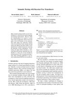
Fig. 1. Architecture of PABP and IMP1 constructs used in the present study. Protein expression constructs containing various portions of
IMP1 and PABP open reading frames were created using primers given in Table 2. The constructs prepared using pQE primers were cloned
into pQE80L plasmid vector for the expression of 6· His tag fusion protein in E. coli. The constructs prepared using pDU primers were
cloned into pDUAL-GC plasmid vector for the in vitro expression of proteins in the rabbit reticulocytes lysate cell-free system.
Binding of IMP1 to PABP and PABP-mRNA G. P. Patel and J. Bag
5680 FEBS Journal 273 (2006) 5678–5690 ª 2006 The Authors Journal compilation ª 2006 FEBS
DARS-4) ends of the minimal RNA sequence has a
significant stimulatory effect on the binding of PABP
and IMP1.
Experiments using UV cross-linking between radio-
labelled RNA and purified IMP1 (Fig. 3D) yielded
results similar to those observed by REMSA (Fig. 3B).
Interestingly, both IMP1 and PABP showed similar
preference for the 3¢ end of the ARS (Fig. 3D: lane 6;
and Fig. 3E: lane 6). Furthermore, to examine whether
the same region of the ARS is involved in binding
IMP1 when it is present in the sequence context of the
entire ARS, we performed RNase footprinting analyses
using the ARS RNA and purified 6· His-IMP1
(Fig. 3F). The results show protection of the sequence
at the 3¢ end of the ARS in presence of IMP1 (Fig. 3F:
compare lanes 4 and 5). Comparison of this region
(Fig. 3F: lane 4) with the RNA ladder (Fig. 3F: lane 1)
suggest that the IMP1 binding site of the ARS falls
within the nucleotide sequence shown in the DARS-R.
We further investigated the ability of both
PABP and IMP1 to bind to the DARS-R RNA
simultaneously using REMSA. The results (Fig. 4)
show that both PABP and IMP1 formed similar size
complexes with the ARS when used separately in bind-
ing assays. Presence of equimolar concentration of
both PABP and IMP1 in the binding reaction pro-
duced a significant level of a slower migrating com-
plex, which indicates the formation of a heterodimeric
complex with the ARS.
Comparison of binding affinity of the ARS
to PABP and IMP1
In the previous UV cross-linking assays, PABP showed
a slightly higher binding ability to the ARS than what
was observed for the IMP1 (Fig. 3D,E). Therefore, we
compared the binding affinities of IMP1 and PABP
for the ARS in detail (Fig. 5). We measured the per-
centage of bound RNA at various protein concentra-
tions by REMSA [33]. The results show that PABP
binds to the ARS approximately two times more
efficiently than the IMP1 (Fig. 5C). The calculated
A
B
C
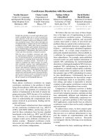
Fig. 2. Binding of the different RBD of
PABP with the ARS and poly(A)
50
RNA.
[
35
S]methionine labelled different RBD-
domains of PABP (Fig. 1), prepared by
in vitro translation in a cell-free rabbit reticu-
locytes lysate, were incubated with the ARS
(A), poly(A)
50
(B) or pGEM (C) conjugated
agarose beads in the chromatography
buffer, washed extensively with the same
buffer, and the bound proteins were eluted
by boiling the beads in a protein sample
loading buffer. The samples were analyzed
by 13% SDS ⁄ PAGE, the gel was impregna-
ted in 1
M sodium salicylate, vacuum dried
and visualized by autofluorography.
G. P. Patel and J. Bag Binding of IMP1 to PABP and PABP-mRNA
FEBS Journal 273 (2006) 5678–5690 ª 2006 The Authors Journal compilation ª 2006 FEBS 5681
A
B
D
E
F
C
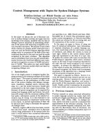
Fig. 3. PABP and IMP1 binding region of the ARS. (A) Different regions of the ARS RNA used in the gel-shift and UV cross-linking assays.
(B) RNA gel-shift analyses of the binding of IMP1 with the different regions of the ARS RNA. REMSA was performed using 5 ng purified
6· His-IMP1 and 0.1 ng (10 000 c.p.m.) RNA. Lane 1, radioactive DARS-R RNA only; lanes 2–6, purified 6· His-IMP1 was incubated with
radiolabelled ARS, DARS-4, DARS-L, DARS-C, and DARS-R RNAs, respectively. Samples were analyzed on a 5% polyacrylimide gel under
nondenaturing conditions, vacuum dried, and visualized by autoradiography. (C) RNA gel-shift analyses of the binding of IMP1 to DARS-R
and DARS-S RNA was performed as described in (B). Lane 1, radioactive DARS-R RNA only; lanes 2 and 4, purified 6· His-PABP was incuba-
ted with DARS-R and DARS-S RNA, respectively; lanes 3 and 5, purified 6· His-IMP1 was incubated with DARS-R and DARS-S RNA. (D) and
(E) RNA-protein UV cross-linking studies. Purified, 5 ng 6· His-IMP1 (D) and 5 ng 6· His-PABP (E) were used for these studies. One sample
in both panels containing protein and ARS RNA was analyzed without UV treatment (lane 1). Lanes 2–6, 6· His-IMP1 (D) and 6· His-PABP
(E) were incubated with 1 ng radiolabelled (100 000 c.p.m.) ARS, DARS-4, DARS-L, DARS-C, and DARS-R RNAs. After the UV treatment,
the samples were treated with RNase A ⁄ RNase T1, fractionated on a 13% SDS ⁄ PAGE and visualized by autoradiography. (F) RNase foot-
printing analysis. IMP1 interacting domain of the ARS RNA was analyzed by RNase footprinting as described in the Experimental procedures.
Lane 1, RNA ladder was prepared by partially hydrolyzing the 5¢ end radiolabelled ARS RNA with 0.1
M NaOH. Lane 2 and 3, 5¢ end radiola-
belled ARS RNA with or without purified 6· His-IMP1, respectively. Lanes 4 and 5, 5¢ end radiolabelled ARS RNA with or without purified
6· His-IMP1 was partially digested with RNase One (Promega). The samples were analyzed by 13% PAGE in presence of 8% urea as a
denaturing agent and the bands were visualized by autoradiography.
Binding of IMP1 to PABP and PABP-mRNA G. P. Patel and J. Bag
5682 FEBS Journal 273 (2006) 5678–5690 ª 2006 The Authors Journal compilation ª 2006 FEBS
dissociation constants for PABP–ARS and IMP1–ARS
interactions were found to be approximately 2.3 nm
and 5.9 nm, respectively.
The IMP1 domain responsible for binding to
the ARS
IMP1 is a modular protein with two RRM type and
four KH RNA binding domains. To examine which of
the six RNA binding domains are necessary for the
ability of IMP1 to bind ARS, we expressed various
portions of IMP1 as His-tagged peptides, and purified
by affinity chromatography. These peptides were ana-
lyzed for complex formation with the radiolabelled
ARS RNA by UV cross-linking assay. The results
show that the RRM I-II domain binds to the ARS
very inefficiently (Fig. 6, compare lanes 2 and 3),
whereas the KH I-II peptide did not show any detect-
able binding to the ARS (Fig. 6, lane 4). The ability to
bind ARS was present within the KH III-IV region of
IMP1. The ability of the KH III-IV domain containing
peptide to bind ARS was similar to what was observed
for both the full length IMP1 and KH I-IV peptide.
Interaction between PABP and IMP1
In a previous study, we reported that IMP1 is a novel
PABP partner [15]. Therefore, we further investigated
how these two polypeptides interact with each other.
Different PABP and IMP1 domains were synthesized
in vitro as [
35
S]methionine labeled peptides, and their
ability to bind matrix bound IMP1 or PABP was
A
B
C
Fig. 5. Binding affinity of the ARS RNA to PABP and IMP1 (A) and
(B) Gel-shift assays of binding of PABP and IMP1 to the ARS RNA.
Uniformly radiolabelled ARS RNA was incubated with an increasing
amount of purified PABP or IMP1 for 5 min at room temperature
as described in the legend of Fig. 3. The samples were fractionated
on a 5% PAGE under nondenaturing conditions, and visualized by
autoradiography. Lane 1, samples without protein; lanes 2–9, sam-
ples with an increasing amount of protein (0.9 ng increment). (C)
The radioactive bands corresponding to the bound and free ARS
RNA in (A) and (B) were excised by superimposing the radiograph,
and the level of radioactivity was measured by scintillation counter.
The average ratio of the RNP complex ⁄ free RNA in each lane from
three separate experiments was plotted against the amount of the
protein. The binding constant was calculated by determining the
protein molar concentration at 50% binding efficiency [33].
Fig. 4. Simultaneous binding of PABP and IMP1 with the DARS-R
RNA. Approximately 0.1 ng (10 000 c.p.m.) uniformly radiolabelled
DARS-R RNA was incubated with purified 6· His-PABP and 6· His-
IMP1, either individually or simultaneously, for 5 min at room tem-
perature. The samples were analyzed by 5% PAGE. Lane 1, RNA
only; lane 2, RNA + 5 ng 6· His-PABP; lane 3, RNA + 5 ng 6· His-
PABP and 4.5 ng 6· His-IMP1; lane 4, RNA + 4.5 ng 6· His-IMP1.
G. P. Patel and J. Bag Binding of IMP1 to PABP and PABP-mRNA
FEBS Journal 273 (2006) 5678–5690 ª 2006 The Authors Journal compilation ª 2006 FEBS 5683
examined. The results of our studies show that the
PABP-C domain (Fig. 7A: lane 5) alone was capable
of interacting with IMP1 as efficiently as the full
length PABP (Fig. 7A: lane 1). None of the RBDs of
PABP showed any binding to IMP1 (Fig. 7A: lanes
2–4). Similar studies using IMP1 peptides showed that
the ability of IMP1 to dimerize resides within the
KH III-IV domains (Fig. 7A: lanes 6, 9 and 10), and
other domains of IMP1 did not contribute towards its
homodimerization (Fig. 7A: lanes 7 and 8).
When we used the full length PABP-matrix as the
bait, only the PABP-C domain showed ability to
homodimerize PABP (Fig. 7B: lanes 1 and 5). In addi-
tion, our results show that the ability of IMP1 to bind
PABP resides within its KH III-IV domains (Fig. 7B:
lane 9). The IMP1 peptide containing the KH III-IV
domains was able to bind PABP as efficiently as the
peptide containing all four KH domains or the full
length IMP1 (Fig. 7B: lanes 6 and 10). We also per-
formed binding assays using matrix-bound PABP-C
and IMP1 KH III-IV peptides (Fig. 7D,E) to examine
whether the short peptides could pull down the inter-
acting peptide partners. We show here that PABP-C
alone can pull down the full length PABP and IMP1
(Fig. 7D: lanes 1 and 3), and also the protein interact-
ing domains of PABP and IMP1 (Fig. 7D: lanes 2, 4,
and 5). In similar studies using IMP1 KH III-IV pep-
tide as bait, we found that it can pull down both the
Fig. 6. The IMP1 domain responsible for binding to the ARS. Trun-
cated IMP1 polypeptides were expressed and purified from E. coli
for UV cross-linking analysis with the radiolabelled ARS RNA. Full
length IMP1 (lane 2), RNA recognition motifs RRM I-II (lane 3), KH
domains KH I-II (lane 4), KH III-IV (lane 5), and KH I-IV (lane 6) were
used for these studies. One sample containing IMP1 (lane 1) was
analyzed without UV treatment. Samples were incubated at room
temperature for 5 min. After the UV treatment, the samples
were treated with RNase A ⁄ RNase T1, fractionated on a 13%
SDS ⁄ PAGE and visualized by autoradiography.
A
B
C
D
Fig. 7. Interaction between PABP and IMP1. [
35
S]methionine
labelled full length or truncated version of PABP and IMP polypep-
tides were incubated with IMP1 (A), PABP (B), b-Gal (C), PABP-C
(D: lanes 1–5), and KH III-IV (D: lanes 6–10) conjugated agarose
beads in the chromatography buffer, washed extensively with the
same buffer, and the bound proteins were eluted by boiling the
beads in the chromatography buffer containing 300 m
M imidizole.
The samples were analyzed by 13% SDS ⁄ PAGE, the gel was
impregnated in 1
M sodium salicylate, vacuum dried and visualized
by autofluorography.
Binding of IMP1 to PABP and PABP-mRNA G. P. Patel and J. Bag
5684 FEBS Journal 273 (2006) 5678–5690 ª 2006 The Authors Journal compilation ª 2006 FEBS
full length PABP and the PABP-C peptide (Fig. 7D:
lanes 6 and 7). Furthermore, the IMP1 KH III-IV pep-
tide was also able to pull down the full length IMP1,
and IMP1 peptides containing the KH III-IV domains
(Fig. 7D: lanes 3–5). These results confirmed that the
interaction between PABP and IMP1 is mediated by
the PABP-C and KH III-IV domains of these polypep-
tides, respectively.
Discussion
We have shown in these studies that the ARS, an
A-rich translational control element in the 5¢ UTR of
PABP mRNA, interacts with PABP differently than a
comparable size RNA consisting exclusively of the
adenine base [poly(A)
50
]. While RBD II is the main
poly(A) interacting domain [32], at least three RBDs
of PABP are required for efficient binding to the ARS.
The combinations of either the RBDs I-II-III or RBDs
II-III-IV have similar affinities for the ARS. It is
known that RBDs I and II have specific affinity
towards the poly(A), and RBDs III and IV bind to the
nonpoly(A) sequences [32]. As such, it is not unex-
pected that the presence of at least one nonspecific
RBD is necessary to bind to the ARS, which consists
of stretches of A, C and U bases.
We have shown earlier that in addition to PABP,
the ARS binds to IMP1. In these studies we have com-
pared the binding of IMP1 and PABP, and surpris-
ingly we have found that both polypeptides bind
strongly to a 22 nucleotide long CCCAAAAAAA
UUUACAAAAAA sequence located at the 3¢ end
of the ARS. Furthermore, CCCAAAAAAAUUU was
found to be the minimal sequence required for binding
to both PABP and IMP1. However, this short RNA
did not bind either protein as strongly as RNAs with
additional adenine nucleotides either at the 5¢ or 3¢
ends. It is possible that the flanking sequences provide
a suitable landing place for PABP and IMP1 on the
RNA. In vitro RNAÆprotein binding studies showed
that other regions of the ARS do not posses a strong
affinity for either PABP or the IMP1. It is possible
that the other short regions of the ARS on their own
could form a different secondary structure, than when
they are present as a part of the entire ARS. There-
fore, these short RNAs on their own may not interact
with PABP and ⁄ or IMP1. We however, consider this
possibility unlikely because the RNase footprinting
analyses using the full length ARS also showed bind-
ing of IMP1 to the 3¢ end of the ARS. Whether the 22
nucleotide long region of the ARS could repress trans-
lation of a reporter mRNA in vivo has not been stud-
ied yet.
The results of our studies suggest that both IMP1
and PABP bind to the same region of the ARS, which
implies that they could compete with each other for
binding to the ARS. Our results showed that in the
presence of both PABP and IMP1, a heterodimeric
complex was formed on the DARS-R RNA. As PABP
binds to the ARS more tightly than what was observed
for the IMP1, it is possible that PABP may first bind
to the ARS, and interact with IMP1. In future studies
it will be interesting to examine whether the PABP
peptide lacking the C-terminal IMP1 interacting
domain can form a heterodimeric complex with IMP1
on the ARS RNA.
How IMP1 and PABP will bind to the PABP
mRNA in vivo may depend on their relative abun-
dance. In HeLa cells, we found that both polypeptides
are almost equally abundant (results not shown).
Because a large amount of cellular PABP is already
bound to the 3¢ poly(A) tract of mRNA, the free
IMP1 could be more abundant than the free PABP. In
addition, PABP and IMP1 interact with each other; as
such, it is also possible that binding of either PABP or
IMP1 to the ARS could attract the other partner
through protein–protein interaction to form a hetero-
dimeric RNAÆprotein complex. Another possibility that
needs further investigation is whether dimerization
between PABP and IMP1 prior to binding the RNA
could alter their binding site on the ARS.
To understand the molecular nature of the interac-
tion of IMP1 with the ARS and PABP, we examined
the IMP1 domain involved in binding to the ARS and
PABP. We have shown here that among the two RNA
binding and four KH domains of IMP1 only the
KH III-IV domain is necessary to bind both ARS
RNA and PABP. Because the same domain of IMP1
is involved in binding to its polypeptide partner, and
the ARS, it is likely that a dynamic conformational
change occurs during the formation of the heterodi-
meric ARS RNAÆprotein complex. Earlier studies have
shown that the KH III-IV domain of IMP1 is also
involved in binding to the translational control element
of insulin like growth factor mRNA [28] to repress its
translation. Therefore, these two domains of IMP1
have the bonafide translational repressor activity. In
addition, the earlier studies by Nielsen et al. [34]
showed that the same IMP1 domains were involved in
forming mRNA granules, and localizing the repressed
insulin like growth factor mRNA to specific sub-
cytoplasmic domains. Whether IMP1 is involved in
localizing the repressed PABP mRNA to a distinct
subcytoplasmic region has not been studied yet.
Whether the ARS-bound IMP1 interacts with PABP
through the ARS has not been directly tested. Never-
G. P. Patel and J. Bag Binding of IMP1 to PABP and PABP-mRNA
FEBS Journal 273 (2006) 5678–5690 ª 2006 The Authors Journal compilation ª 2006 FEBS 5685
theless, indirect evidence suggests that IMP1 is capable
of binding directly to PABP. We have shown here that
the RNA-binding domains of PABP cannot bind
IMP1, which would be expected should the ARS RNA
be involved in the interaction between PABP and
IMP1. Therefore, we suggest that both PABP and
IMP1 bind to the ARS independently, and then mutu-
ally stabilize the RNAÆprotein complex through pro-
tein–protein interaction. Moreover, it is unlikely that
any contaminating ARS-like RNA derived from
Escherichia coli in our agarose-PABP beads during its
purification from the bacterial cell extract, could have
been indirectly involved in binding IMP1, as E. coli
RNA did not show any competition with the ARS
RNA in gel shift assays (results not shown).
Additional studies to characterize the IMP1 inter-
acting domain of PABP showed that it resides exclu-
sively within the PABP-C domain. Although RBD II
of PABP could interact with several polypeptide part-
ners including eIF4G [23], PAIP1 [16,17], and PAIP2
[18,19], the PABP-C is the main protein–protein inter-
acting domain of PABP. In a previous study PABP-C
domain was shown to be indispensable for the auto-
regulation of PABP mRNA translation [35]. Our
results suggest that the main function of the PABP-C
domain in translational repression may be to interact
with IMP1 to form a heterodimeric RNAÆprotein com-
plex. Whether IMP1 can bind to the 3¢ poly(A) track
of all mRNA by interacting with the PABP-C domain
and plays a role in all mRNA metabolism remains to
be examined.
Both in vitro and in vivo studies in our lab [15] have
shown that the ARS forms a heterotrimeric complex
with three known RNA-binding proteins, PABP,
IMP1, and UNR. However, in the studies reported
here we have focused on the interaction of ARS with
PABP and IMP1, because only the IMP1 and PABP
bind to the ARS independently. As UNR is a known
PABP binding protein, its presence in the heterotri-
meric ARS RNAÆprotein complex is probably through
its binding to PABP. The individual role of the poly-
peptides of the heterotrimeric complex is not known.
It is possible that each polypeptide participates at a
distinct step of translational control. There is more to
translational control than simply preventing the ribo-
some from binding to the mRNA. For a foolproof
mechanism to prevent unwanted mRNA translation,
the decision to repress translation of a specific mRNA
may be made by tagging the mRNA while it is in the
nucleus. IMP1 is a known shuttle protein; therefore, it
may bind to the ARS containing mRNA in the nuc-
leus, and tag the mRNA for repression. PABP may
then bind to the tagged mRNA by binding to both
ARS and IMP1. UNR is a member of the cold-shock
domain containing protein family. These proteins can
act as ‘RNA histone’, and protect the repressed
mRNA from degradation [36]. Finally, the ARS–
IMP1–PABP–UNR complex could form even a larger
multisubunit autoregulatory complex through a series
of protein–protein interactions. This multimeric com-
plex would provide a stronger roadblock to stall the
scanning of the mRNA by 40S ribosomal subunits
than a monomeric ARS–PABP complex. It is conceiv-
able that a multi subunit RNAÆprotein complex
needs to be formed with the translational repressor cis-
element to prevent the large molecular machine such
as the 40S ribosomal subunit to read-through the
translational control element.
Experimental procedures
Plasmid construction
Double stranded oligodeoxynucleotides encoding either
poly(A)
50
or various regions of the ARS (nucleotides 71–
131 of the human PABP cDNA, GeneBank ID: Y00345)
were generated by annealing complementary synthetic
oligonucleotide sets (Table 1; only sense sequences are
given). The annealed products were digested with respective
restriction enzymes (MBI Fermentas; Amherst, NY, USA),
purified from a 2.5% agarose gel using the QIAquick gel
extraction kit (Qiagen; Mississauga, ON, Canada), and
cloned into pEGFP-N3 (Clontech-BD Biosciences; Burling-
ton, ON, Canada) plasmid vectors.
Protein expression constructs containing various por-
tions of IMP1 (GenBank ID: NM_006546) and PABP
(GeneBank ID: Y00345) open reading frames were gener-
ated by using appropriate primers (Table 2 and Fig. 1).
The PCR products were digested with appropriate restric-
tion enzymes (MBI Fermentas), purified from a 1% ag-
arose gel by QIAquick gel extraction kit (Qiagen), and
cloned into pDUAL-GC (Stratagene, La Jolla, CA, USA)
or pQE80L (Qiagen) expression vectors. All plasmids were
Table 1. Primers used to create various ARS constructs.
Primer Sense sequence
ARS EcoRI-T
7
-aaaaaatccaaaaaaaatctaaaaaaatcttttaaaaaa
ccccaaaaaaatttacaaaaaa-BamHI
A
¨
ARS-4 EcoRI-T
7
-tccaaaaaaaatctaaaaaaatcttttaaaaaa
ccccaaaaaaattt-BamHI
A
¨
ARS-L EcoRI-T
7
-aaaaaatccaaaaaaaatct-BamHI
A
¨
ARS-C EcoRI-T
7
-tctaaaaaaatcttttaaaaaacccc-BamHI
A
¨
ARS-R EcoRI-T
7
-ccccaaaaaaatttacaaaaaatc-BamHI
A
¨
ARS-S EcoRI-T
7
-ccccaaaaaaattt-BamHI
Poly(A)
50
EcoRI-T
7
-aaaaaaaaaaaaaaaaaaaaaaaaaaaaaa
aaaaaaaaaaaaaaaaaaaa-BamHI
Binding of IMP1 to PABP and PABP-mRNA G. P. Patel and J. Bag
5686 FEBS Journal 273 (2006) 5678–5690 ª 2006 The Authors Journal compilation ª 2006 FEBS
propagated in E. coli DH5a (Invitrogen, Carlsbad, CA,
USA), isolated using GenElute plasmid maxi-prep kit (Sig-
ma, Oakville, ON, Canada) and confirmed to be correct
by DNA sequencing.
In vitro synthesis and radiolabelling of RNA
pEGFP-N3 plasmids containing either oligo(A)
50
or various
ARS region under the control of the T
7
RNA polymerase
promoter were linearized with BamHI, and pGEM-T vector
(Promega, Madison, WI, USA) was linearized with SalI
restriction enzyme for in vitro run-off transcription. Tran-
scription reactions were usually performed at 37 °C for 2 h
in a final volume of 100 lL containing 10 lg of a DNA
template, 2.5 mm of each NTP, and 100 units of T
7
RNA
polymerase (Promega). Uniformly radiolabelled RNA was
synthesized under similar conditions in a final reaction vol-
ume of 25 lL containing 150 lCi [
32
P]ATP[aP] (MP Bio-
medicals, Irvine,CA, USA) and the final concentration of
cold ATP reduced to 25 lmol. The 5¢ end radiolabelled
RNA was prepared by first dephosphorylating cold RNA
using calf intestine phosphatase, followed by phosphoryla-
tion using T
4
polynucleotide kinase (T
4
-PNK) in presence
of [
32
P]ATP[cP]. The contaminating nucleotides, incom-
pletely transcribed products and the DNA template were
removed by fractionating transcription reaction mixtures on
13% polyacrylamide gels under denaturing conditions [37].
The amount of RNA and its specific radioactivity were
determined using a spectrophotometer and scintillation
counter, respectively.
Expression and purification of 6
·
His-tag fusion
protein
Escherichia coli DH5a transformed with pQE80L expression
vector (Qiagen) containing various portions of IMP1 or PABP
open reading frames (Fig. 1) were grown to an early log phase
and induced for 4 h with isopropyl thio-b-d-galactoside. The
bacterial cells were harvested and lysed with 1 mgÆmL
)1
of
lysozyme in a lysis buffer (50 mm NaH
2
PO
4
, 500 mm NaCl,
30 mm imidizole, 13 mm 2-mercaptoethanol, 2 mm MgCl
2
,
1mm phenylmethanesulfonyl fluoride, 0.5% IgepalCA-630,
and 5% glycerol [pH 8.0]) at 0 °C for 30 min. The lysate was
cleared by centrifugation at 12 000 g for 5 min and the
supernatant was mixed with Ni-NTA agarose beads
(Qiagen). After shaking at 4 °C for 30 min, the beads were
washed extensively with a washing buffer (50 mm NaH
2
PO
4
,
500 mm NaCl, 50 mm imidizole, 13 mm 2-mercaptoethanol,
2mm MgCl
2
,1mm phenylmethanesulfonyl fluoride, 0.5%
IgepalCA-630, and 5% glycerol [pH 8.0]) and the bound pro-
teins were eluted in the elution buffer (50 mm NaH
2
PO
4
,
500 mm NaCl, 300 mm imidizole, 13 mm 2-mercaptoethanol,
2mm MgCl
2
,1mm phenylmethanesulfonyl fluoride, 0.5%
IgepalCA-630, and 5% glycerol [pH 8.0]).
The protein concentration of the eluted fraction was deter-
mined by a protein assay kit (Bio-Rad, Burlington, ON,
Canada), and equilibrated with a storage buffer (10 mm
Hepes-KOH [pH 7.5], 3 mm MgCl
2
, 140 mm KCl, 5%
glycerol, 1 mm dithiothreitol, 0.02% Igepal CA-630, 0.5 mm
phenylmethanesulfonyl fluoride, 10 lgÆmL
)1
leupeptin, and
2 lgÆmL
)1
aprotinin) using the Microcon YM-30 concentra-
tion column (Millipore, Etobicoke, ON, Canada) and stored
at )80 °C in small aliquots. The integrity and purity of the
affinity purified polypeptide was examined by SDS ⁄ PAGE.
The desired polypeptide band was quantified by scanning the
stained gel. Preparations containing more than 80% unde-
graded IMP1 and PABP were used for further studies.
In vitro synthesis of radiolabelled protein pDUAL-GC
vector (Stratagene) containing various portions of IMP1 or
PABP open reading frames (Fig. 1) was linearized with
KpnI and transcribed using T
7
RNA polymerase system as
described. The contaminating nucleotides were removed by
centrifugation using the Microcon YM-30 concentration
column (Millipore). Approximately 0.1 lg of RNA was
translated using rabbit reticulocytes lysate (Promega)
containing 0.02 mm amino acids mixture and 30 l Ci
[
35
S]methionine in a total reaction volume of 100 lL (70%
retic lysate) for 90 min at 30 °C. The specific radioactivity
was determined using trichloroacetic acid precipitation and
the quality of translated product was analyzed on 13%
SDS ⁄ PAGE followed by autoradiography.
RNA electrophoretic mobility shift assay
For REMSA, 1–10 ng of purified protein was incubated
with 0.1 ng (1 · 10
4
c.p.m.) of radiolabelled RNA for
Table 2. Primers used to create truncated PABP and IMP1 protein
expression vectors.
Number Primer Sequence
1 pQE-PABP(s) BamHI-aaccccagtgccccc
2 pQE-PABPC(s) BamHI-gagcgccaggctcac
3 pQE-IMP1(s) BamHI-aacaagctttacatcggc
4 pQE-KH1(s) BamHI-gtggacatcccccttcgg
5 pQE-KH3(s) BamHI-gctgctccctatagctcc
6 pDU-PABP(s) EarI-catgaaccccagtgcc
7 pDU-RBDII(s) EarI-catggatgttataaagggc
8 pDU-RBDIII(s) EarI-catgggacgatttaagtct
9 pDU-RBDIV(s) EarI-catggaacagatgaaacaa
10 pDU-PABPC(s) EarI-catggagcgccaggctcac
11 pDU-IMP1(s) EarI-catgaacaagctttacatcg
12 pDU-KH1(s) EarI-catggtggacatcccccttcgg
13 pDU-KH3(s) EarI-catggctgctccctatagctcc
14 PABP(as) KpnI-ttaaacagttggaacaccgg
15 RBDI(as) KpnI-ttagcctactccacttttgcg
16 RBDII(as) KpnI-ttaagcttctcgttctttacg
17 RBDIII(as) KpnI-ttagcgcttaagttccgtct
18 RBDIV(as) KpnI-ttactggttagtgaggagagc
19 IMP1(as) KpnI-ttacttcctccgtgcctg
20 RRM2(as) KpnI-ttactgctgcttggctgg
21 KH2(as) KpnI-ttagctggatgaagctgg
G. P. Patel and J. Bag Binding of IMP1 to PABP and PABP-mRNA
FEBS Journal 273 (2006) 5678–5690 ª 2006 The Authors Journal compilation ª 2006 FEBS 5687
10 min at 22 °C in a total reaction volume of 18 lL in the
binding buffer (10 mm Hepes-KOH [pH 7.5], 3 mm MgCl
2
,
140 mm KCl, 5% glycerol, 1 mm dithioreitol, 0.02% Igepal
CA-630, 10 lgofE. coli tRNA and 0.02% bromophenol
blue). Subsequently, the sample was analyzed by 5% non-
denaturing PAGE in 0.5· TBE buffer (45 mm Tris-borate,
1mm EDTA [pH 8.0]), 100 V, at 4 °C. The gel was then
vacuum dried and autoradiographed.
UV cross-linking assay
For UV-induced cross-linking assays, approximately 5 ng
of purified protein was incubated with 1ng (1· 10
5
c.p.m.) of radiolabelled RNA at 22 °C for 10 min in a total
reaction volume of 27 lL in the binding buffer. The sample
was irradiated by UV-light (254 nm, 4000 lwÆcm
)2
)at4°C
for 5 min, and treated with RNase T1 (25 units) and
RNase A (1 l g) at 37 °C for 5 min. Finally, the sample
was boiled in a protein sample loading buffer (6% glycerol,
2% SDS, 100 mm dithioreitol, and 0.02% bromophenol
blue in 60 mm Tris ⁄ HCl [pH 6.6]) for 5 min and analyzed
by 13% SDS ⁄ PAGE.
Protein pull-down assay
To analyze RNA–protein interactions, in vitro synthesized
and gel purified RNA was oxidized by sodium periodate
treatment and covalently linked to adipic acid hydrazide ag-
arose (Sigma) as described previously [38]. The unbound
RNA was removed by washing the beads twice with 2 m
NaCl followed by equilibrating the beads with the chroma-
tography buffer (10 mm Hepes-KOH [pH 7.5], 3 mm MgCl
2
,
140 mm NaCl, 5% glycerol, 1 mm dithioreitol, 0.01% Triton
X-100). The RNA conjugated beads were incubated with
in vitro synthesized [
35
S]methionine labeled protein at 4 °C
for 15 min. The beads were washed extensively with the
chromatography buffer. The RNA-bound protein was eluted
by boiling in protein sample loading buffer and analyzed by
13% SDS ⁄ PAGE followed by an autoradiography.
To analyze protein–protein interactions, Ni-NTA agarose
beads were conjugated with 6· His-tag fusion protein
expressed in E. coli as described. The beads were equili-
brated with the chromatography buffer and incubated with
in vitro synthesized [
35
S]methionine labeled protein at 4 °C
for 15 min. The beads were washed extensively with the
chromatography buffer and the affinity-bound protein was
eluted in the chromatography buffer supplemented with
300 mm imidizole. The sample was analyzed by 13%
SDS ⁄ PAGE and visualized by an autoradiography.
RNase protection assay
To identify protein binding sites in the RNA, the 5¢ end
radiolabelled RNA (1 · 10
4
c.p.m.) was first incubated with
50 ng of purified protein for 10 min on ice in a total reac-
tion volume of 18 lL in the binding buffer. The reaction
was then treated with 1 unit (as defined by the supplier) of
RNase One (Promega) at 22 °C for 5 min. The RNase was
inactivated by incubation at 75 °C in RNA loading buffer
(50% formamide, 2% SDS final concentration) and ana-
lyzed by 13% PAGE in the presence of 8% urea as a dena-
turing agent. Finally, the gel was fixed (5% methanol, 5%
acetic acid), dried under vacuum, and subjected to autora-
diography.
Acknowledgements
This work was supported by a grant from The
Canadian Institutes of Health Research (CIHR) and
The National Science and Engineering Research
Council (NSERC). We are thankful to Dr J. Chris-
tiansen for providing the IMP1 clone. We also thank
Mrs S. Ma for her help in the preparation of the
revised manuscript.
References
1 Mendez R & Richter JD (2001) Translational control
by CPEB: a mean to the end. Nat Rev Mol Cell Biol 2,
521–529.
2 Gygi SP, Rochon Y, Franza BR & Aebersold R (1999)
Correlation between protein and mRNA abundance in
yeast. Mol Cell Biol 19, 1720–1730.
3 Ideker T, Thorsson V, Ranish JA, Christmas R, Buhler
J, Eng JK, Bumgarner R, Goodlett DR, Aebersold R &
Hood L (2001) Integrated genomic and proteomic ana-
lyses of a systematically perturbed metabolic network.
Science 292, 929–934.
4 Moore MJ (2005) From birth to death: the complex
lives of eukaryotic mRNAs. Science 309, 1514–1518.
5 Keene JD & Tenenbaum SA (2002) Eukaryotic mRNPs
may represent posttranscriptional operons. Mol Cell 9,
1161–1167.
6 Mangus DA, Evans MC & Jacobson A (2003) Poly(A)-
binding protein: multifunctional scaffolds for the post-
transcriptional control of gene expression. Genome Biol
4, 223–238.
7 Gorgoni B & Gray NK (2004) The roles of cytoplasmic
poly(A)-binding proteins in regulating gene expression:
a developmental perspective. Brief Funct Genomic
Proteomic 3, 125–141.
8 Kuhn U & Wahle E (2004) Structure and function of
poly(A) binding protein. Biochim Biophys Acta 1678,
67–84.
9 Hornstein E, Git A, Nraunstein I, Avni D & Meyuhas
O (1999) The expression of poly(A)-binding protein
gene is translationally regulated in a growth-dependent
Binding of IMP1 to PABP and PABP-mRNA G. P. Patel and J. Bag
5688 FEBS Journal 273 (2006) 5678–5690 ª 2006 The Authors Journal compilation ª 2006 FEBS
fashion through a 5¢-terminal oligopyrimidine tract
motif. J Biol Chem 274, 1708–1714.
10 Wu J & Bag J (1998) Negative control of the poly(A)
binding protein mRNA translation is mediated by the
adenine-rich region of its 5¢ UTR. J Biol Chem 273,
34535–34542.
11 Avni D, Shama S, Loreni F & Meyuhas O (1994) Verte-
brate mRNAs with a 5¢-terminal pyrimidine tract are
candidates for translational repression in quiescent cells:
characterization of the translational cis-regulatory ele-
ment. Mol Cell Biol 14, 3822–3833.
12 Shama S & Meyuhas O (1996) The translational cis-
regulatory element of mammalian ribosomal protein
mRNAs is recognized by the plant translational appa-
ratus. Eur J Biochem 236, 383–388.
13 Bag J (2001) Feedback inhibition of poly(A)-binding
protein mRNA translation. A possible mechanism of
translation arrest by stalled 40S ribosomal subunits.
J Biol Chem 276, 47352–47360.
14 de Melo Neto OP, Standart N & Martins de SaC (1995)
Autoregulation of poly(A)-binding protein synthesis
in vitro. Nucleic Acids Res 23, 2198–2205.
15 Patel GP, Ma S & Bag J (2005) The autoregulatory
translational control element of poly(A)-binding protein
mRNA forms a heteromeric ribonucleoprotein complex.
Nucleic Acids Res 33, 7074–7089.
16 Gray NK, Coller JM, Dickson KS & Wickens M (2000)
Multiple portions of poly (A) -binding protein stimulate
translation in vivo. EMBO J 19, 4723–4733.
17 Craig AWB, Haghighat A, Yu ATK & Sonenberg N
(1998) Interaction of polyadenylate-binding protein with
the eIF4G homologue PAIP enhances translation.
Nature 392, 520–523.
18 Khaleghpour K, Kahvejian A, de Crescenzo G, Roy G,
Svitkin YV, Imataka H, O’Connor-McCourt M &
Sonenberg N (2001) Dual interaction of the transla-
tional repressor Paip2 with poly (A)-binding protein.
Mol Cell Biol 21, 5200–5213.
19 Khaleghpour K, Svitkin YV, Craig AW, DeMaria CT,
Deo RC, Burley SK & Sonenberg N (2001) Transla-
tional repression by a novel partner of human poly(A)-
binding protein, Paip2. Mol Cell 7, 205–216.
20 Le H, Tanguay RL, Balasta ML, Wei CC, Browning
KS, Metz AM, Gross DJ & Gallie DR (1997) Transla-
tion initiation factors eIF-iso4G and eIF4B interact with
the poly(A)-binding protein and increase its RNA bind-
ing activity. J Biol Chem 272, 16247–16255.
21 Wang Z, Panayiota T & Kiledjian M (1999) An mRNA
stability complex functions with poly(A)-binding protein
to stabilize mRNA in vitro. Mol Cell Biol 19, 4552–
4560.
22 Cheng TC, Yamashita A, Chen CYA, Yamashita Y,
Zhu W, Durdan S, Kahvejian A, Sonenberg N & Shyu
AB (2004) UNR, a new partner of poly(A)-binding pro-
tein, plays a key role in translationally coupled mRNA
turnover mediated by the c-fos major coding-region
determinant. Genes Dev 18, 2010–2023.
23 Tarun SJ Jr & Sachs AB (1996) Association of the yeast
poly (A) tail binding protein with translation initiation
factor eIF-4G. EMBO J 15, 7168–7177.
24 Amrani N, Minet M, Le Gouar M, Lacroute F &
Wyers F (1997) Yeast Pab1 interacts with Rna15 and
participates in the control of the poly(a) tail length
in vitro. Mol Cell Biol 17, 3694–3701.
25 Cosson B, Berkova N, Couturier A, Chabelskaya S,
Philippe M & Zhouravleva G (2002) Poly(A)-binding
protein and eRF3 are associated in vivo in human and
Xenopus cells. Biol Cell 94, 205–216.
26 D’Orso I & Frasch AC (2002) TcUBP-1, an mRNA
destabilizing factor from Trypanosomes, homodimerizes
and interacts with novel AU-rich element and poly(A)-
binding proteins forming a ribonucleoprotein complex.
J Biol Chem 277, 50520–50528.
27 Nielsen J, Christiansen J, Lykke-Andersen J, Johnsen
AH, Wewer UM & Nielsen FC (1999) A family of insu-
lin-like growth factor II mRNA-binding proteins
represses translation in late development. Mol Cell Biol
19, 1262–1270.
28 Nielsen J, Kristensen MA, Willemoes M, Nielsen FC &
Christiansen J (2004) Sequential dimerization of human
zipcode-binding protein IMP1 on RNA: a cooperative
mechanism providing RNP stability. Nucleic Acids Res
32, 4368–4376.
29 Bernstein PL, Herrick DJ, Prokipcak RD & Ross J
(1992) Control of c-myc mRNA half-life in vitro by a
protein capable of binding to a coding region stability
determinant. Genes Dev 6, 642–654.
30 Atlas R, Behar L, Elliott E & Ginzburg I (2004) The
insulin-like growth factor mRNA binding-protein IMP-1
and the Ras-regulatory protein G3 BP associate with tau
mRNA and HuD protein in differenciated P19 neuronal
cells. J Neurochem 89, 613–626.
31 Rackham O & Brown CM (2004) Visualization of
RNA–protein interactions in living cells: FMRP and
IMP1 interact on mRNAs. EMBO J 23, 3346–3355.
32 Burd CG, Matunis EL & Dreyfuss G (1991) The mul-
tiple RNA-binding domains of the mRNA poly(A)-
binding protein have different RNA-binding activities.
Mol Cell Biol 11, 3419–3424.
33 Wieland G, Hemmerich P, Koch M, Stoyan T, Hege-
mann J & Diekmann S (2001) Determination of the
binding constants of the centromere protein Cbf1 to all
16 centromere DNAs of Saccharomyces cerevisiae.
Nucleic Acids Res 29, 1054–1060.
34 Nielson FC, Nielson J, Kristensen M, Koch G & Chris-
tiansen J (2002) Cytoplasmic trafficking of IGF-II
mRNA-binding protein by conserved KH domains.
J Cell Sci 115, 2087–2097.
35 Melo EO, de Dhalia RSaMC, Standart N & de Melo
Neto OP (2003) Identification of a C-terminal
G. P. Patel and J. Bag Binding of IMP1 to PABP and PABP-mRNA
FEBS Journal 273 (2006) 5678–5690 ª 2006 The Authors Journal compilation ª 2006 FEBS 5689
poly(A)-binding protein (PABP) –PABP interaction
domain. J Biol Chem 278, 46357–46368.
36 Coles LS, Bartley MA, Bert A, Hunter J, Polyak S,
Diamond P, Vadas MA & Goodall GJ (2004) A multi-
protein complex containing cold shock domain (Y-box)
and polypyrimidine tract binding proteins forms on the
vascular endothelial growth factor mRNA. Eur J Bio-
chem 271, 648–660.
37 Wildeman AG & Nazar RN (1981) Studies on the
secondary structure of 5.8S rRNA from a thermo-
phile, Thermomyces lanuginosus. J Biol Chem 256,
5675–5682.
38 Caputi M & Zahler AM (2001) Determination of the
RNA binding specificity of the heterogeneous nuclear
ribonucleoprotein (hnRNP) H ⁄ H ⁄ F ⁄ 2H9 family. J Biol
Chem 276, 43850–43859.
Binding of IMP1 to PABP and PABP-mRNA G. P. Patel and J. Bag
5690 FEBS Journal 273 (2006) 5678–5690 ª 2006 The Authors Journal compilation ª 2006 FEBS