Báo cáo khoa học: Cold stress defense in the freshwater sponge Lubomirskia baicalensis pot
Bạn đang xem bản rút gọn của tài liệu. Xem và tải ngay bản đầy đủ của tài liệu tại đây (1.28 MB, 14 trang )
Cold stress defense in the freshwater sponge
Lubomirskia baicalensis
Role of okadaic acid produced by symbiotic dinoflagellates
Werner E. G. Mu
¨
ller
1,2
, Sergey I. Belikov
2
, Oxana V. Kaluzhnaya
1,2
, Sanja Perovic
´
-Ottstadt
1
,
Ernesto Fattorusso
3
, Hiroshi Ushijima
4
, Anatoli Krasko
1
and Heinz C. Schro
¨
der
1
1 Institut fu
¨
r Physiologische Chemie, Abteilung Angewandte Molekularbiologie, Universita
¨
t Mainz, Germany
2 Limnological Institute of the Siberian Branch of Russian Academy of Sciences, Irkutsk, Russia
3 Dipartimento di Chimica delle Sostanze Naturali, Universita
`
di Napoli ‘Federico II’, Italy
4 Department of Developmental Medical Sciences, Institute of International Health, The University of Tokyo, Japan
The taxon sponges (phylum Porifera) has been surpris-
ingly successful during evolutionary development. This
metazoan phylum is the only one to have survived the
severe Varanger–Marinoan ice age (605–585 million
years ago) of the Neo-Proterozoic eon (1000–520
million years ago), during which the earth was almost
Keywords
dinoflagellates; heat shock protein;
Lubomirskia baicalensis; okadaic acid;
protein phosphatase
Correspondence
W. E. G. Mu
¨
ller, Institut fu
¨
r Physiologische
Chemie, Abteilung Angewandte
Molekularbiologie, Universita
¨
t,
Duesbergweg 6, 55099 Mainz, Germany
Fax: +49 6131 3925243
Tel: +49 6131 3925910
E-mail:
Website: />Database
The sequences from Lubomirskia baicalen-
sis reported here have been deposited in
the GenBank database under the accession
numbers AM392283 (protein phosphatase
LUBAIHSP70PP1) and AM392284 (heat
shock protein-70 LUBAIHSP70)
Note
This article is dedicated to Professor Michele
Sara
`
, Professor of Zoology at the University
of Genova, for his outstanding contributions
to marine biology, 1926–2006
(Received 16 August 2006, revised 21
October 2006, accepted 27 October 2006)
doi:10.1111/j.1742-4658.2006.05559.x
The endemic freshwater sponge Lubomirskia baicalensis lives in Lake Bai-
kal in winter (samples from March have been studied) under complete ice
cover at near 0°C, and in summer in open water at 17 °C (September). In
March, specimens show high metabolic activity as reflected by the produc-
tion of gametes. L. baicalensis lives in symbiosis with green dinoflagellates,
which are related to Gymnodinium sanguineum. Here we show that these
dinoflagellates produce the toxin okadaic acid (OA), which is present as a
free molecule as well as in a protein-bound state. In metazoans OA inhibits
both protein phosphatase-2A and protein phosphatase-1 (PP1). Only
cDNA corresponding to PP1 could be identified in L. baicalensis and sub-
sequently isolated from a L. baicalensis cDNA library. The deduced poly-
peptide has a molecular mass of 36 802 Da and shares the characteristic
domains known from other protein phosphatases. As determined by west-
ern blot analysis, the relative amount of PP1 is almost the same in March
(under ice) and September (summer). PP1 is not inhibited by low OA con-
centrations (100 nm); concentrations above 300 nm are required for inhibi-
tion. A sponge cell culture system (primmorphs) was used to show that at
low temperatures (4 °C) expression of hsp70 is strongly induced and hsp70
synthesis is augmented after incubation with 100 nm OA to levels measured
at 17 °C. In the enriched extract, PP1 activity at 4 °C is close to that meas-
ured at 17 °C. Immunoabsorption experiments revealed that hsp70 contri-
butes to the high protein phosphatase activity at 4 °C. From these data we
conclude that the toxin OA is required for the expression of hsp70 at low
temperature, and therefore contributes to the cold resistance of the sponge.
Abbreviations
hsp70, heat shock protein-70; OA, okadaic acid; PP1, protein phosphatase-1; PP2A, protein phosphatase-2A.
FEBS Journal 274 (2007) 23–36 ª 2006 The Authors Journal compilation ª 2006 FEBS 23
completely covered by ice; most organisms became
extinct during this period [1]. As ‘living fossils’ [2], spon-
ges represent evolutionarily the oldest extant taxon, and
thus allow insight into the genome organization of ani-
mals that lived prior to the ‘Cambrian Explosion’. At
that time, sponges existed exclusively in the marine envi-
ronment, whereas later some taxa also occupied fresh-
water biotopes (during the Cenozoic period). From
cosmopolitan freshwater species, e.g. Ephydatia fluvia-
tilis, endemic species branched off in ‘old lakes’. Lake
Baikal (Siberia), for example, harbors many prominent
endemic sponges [3]. Major reasons for the recent rapid
evolution of the endemic sponge fauna in some areas,
that still continues, are: (a) successful adaptation to
environmental conditions, (b) dominance of sexual
reproduction over asexual reproduction in sponges, and
(c) differences in the habitats (littoral on rocks or on
calcifying algae, e.g. Chara sp.) [4].
In Lake Baikal the dominant endemic sponge species
Lubomirskia baicalensis livesinacoldenvironment;in
March at an ambient temperature of )0.5 °CandinSep-
tember at around 17 °C [5]. These animals, which grow
at depths of 1–20 m, maintain constant metabolic activit-
ies with pumping rates similar to those of species that live
at 15–20 °C [6,7]. Surprisingly, L. baicalensis produces
gametes and embryos in March when the lake is com-
pletely covered by 1 m of ice. One major source of essen-
tial organic carbon for the animals during this season is
their ecological, symbiotic relationship with chlorophyll-
containing dinoflagellates [5]. It has been reported previ-
ously that these symbiotic ‘Zoochlorellae’ exist in a 3-mm
thick external layer of the sponge [8]. Field observations
revealed that, in the absence of light, the dinoflagellates
die and are removed from the sponge specimens which
likewise die (W. E. G. Mu
¨
ller, University of Mainz,
unpublished results). In L. baicalensis these protists,
which are closely related to G ymnodinium sanguineum,
produce glycerol and transfer this intermediate metabo-
lite via an aquaporin channel into the sponge cells
(W. E. G. Mu
¨
ller, University of Mainz, submitted). This
raises two questions: how do the sponges live in the cold
environment; or, more specifically, (a) how do they solve
the problem of protein folding at low temperature, and
(b) how do they overcome the barrier of the required acti-
vation energy for the enzyme-mediated catalysis?
This study mainly addresses the first question. It is
known that the growth of Escherichia coli at low tem-
perature is facilitated by chaperonins [9]. The question
is, which sensor in these microorganisms regulates the
expression of the respective heat shock proteins? Here
we tested the hypothesis that secondary metabolites
produced by the symbiotic ⁄ commensalic organisms in
sponges contribute to the cold stress response.
The dinoflagellates of the taxon Gymnodinium identi-
fied in L. baicalensis are related to G. sanguineum (Alve-
olata; Dinophyceae; Gymnodiniales; Gymnodiniaceae;
Gymnodinium), which has been described as a compo-
nent of harmful algal blooms and has been found to be
hemolytic and ichthyotoxic [10]. One toxin often
produced in these algae [11] is okadaic acid (OA), a
polyether C38 fatty acid, originally isolated from Hali-
chondria okadaii [12]. The major targets of OA in all
metazoans hitherto studied are the catalytic subunits of
the proteins phosphatase-1 (PP1) and protein phos-
phatase-2A (PP2A) which are sensitively inhibited at
nanomolar concentrations (the 50% inhibitory con-
centration for PP1 is 3–150 nm and that for PP2A is
0.03–0.2 nm) [11].
Here we show that OA is present in L. baicalensis,
where it is synthesized by the dinoflagellates. In order
to perform functional studies between OA and PP1, the
cDNA coding for PP1 had to be identified in L. baical-
ensis. This polypeptide shares high sequence similarity
with mammalian PP1. Antibodies against PP1 allowed
its assessment during temperature-dependent expression
in vitro (cell culture) and in vivo (animals). At lower
concentrations (< 100 nm), OA has no effect on the
level and activity of the protein phosphatase(s) but
induces the expression of heat shock protein-70
(hsp70). From earlier studies it is known that OA can
trigger the expression of heat shock proteins in tissues
[13]. We applied the in vivo sponge cell culture system,
the primmorphs [2], to demonstrate that in primmorphs
at 4 °C, OA upregulates both the expression of hsp70
transcripts and the amount of hsp70 protein to levels
found at the ambient temperature of 17 °C. Subsequent
depletion experiments with antibodies against hsp70
showed that functionally active chaperon ⁄ hsp70 mole-
cules are required for full protein phosphatase activity
at low temperature. From the data we conclude that at
lower concentrations (< 100 nm) the secondary meta-
bolite OA mediates ⁄ controls in L. baicalensis the cold
stress defense, whereas higher concentrations are
required to inhibit the protein phosphatase(s). It has
been established that sponges, like Suberites domuncula
[14] or L. baicalensis, which contain symbiotic micro-
organisms, display a stronger ‘resistance’ to OA; with
regard to S. domuncula only concentrations > 300 nm
have a significant effect on protein synthesis.
Results
L. baicalensis specimens in winter and summer
Animals were collected during September 2005 and
March 2006 (Fig. 1A,B). During these seasons, the
Cold stress defense in sponges W. E. G. Mu
¨
ller et al.
24 FEBS Journal 274 (2007) 23–36 ª 2006 The Authors Journal compilation ª 2006 FEBS
animals have a bright green color, which is due to the
high abundance of dinoflagellates (Fig. 1C,D) related
to the taxon G. sanguineum (Alveolata; W. E. G. Mu
¨
ller,
University of Mainz, submitted). Interestingly, during
winter the sponges form sexual propagation bodies,
reflecting an active metabolism. Figure 1E shows one
spermatogenic cyst.
Presence of OA in L. baicalensis
The OA concentration in L. baicalensis was determined
using HPLC ⁄ MS analysis to be 83 ± 9 ngÆg
)1
wet
weight (100 nm; March), whereas tissue from speci-
mens collected in September had a lower OA content
of 57 ± 6 ngÆg
)1
(70 nm). These values were confirmed
by competitive ELISA giving concentrations of
75 ngÆg
)1
(March) and 45 ngÆg
)1
(September).
Protein-coupled OA in L. baicalensis
Protein extracts were prepared from specimen tissue
collected in March. This preparation was subjected to
SDS ⁄ PAGE (10% gel; Fig. 2, lane a). After transfer,
blots were incubated with anti-OA sera (pAb-OA). This
revealed one prominent protein band of 14 kDa (Fig. 2,
lane b). The specificity of the reaction was proven using
antibodies that had been adsorbed with OA bound to
the FID-33 peptide; under these conditions the immu-
noreaction of the 14 kDa band was strongly suppressed
(Fig. 2, lane c). In contrast, the signal at 14 kDa was
of the same strength when membranes were incubated
with pAb-OA, which had been pretreated with FID-33
prior to use in the western blots (not shown).
Identification dinoflagellates in tissue using
anti-OA sera
Slices of sponge tissue were prepared and reacted with
pAb-OA. The antibodies stained the dinoflagellates
very brightly, whereas the sponge cells did not react
A
B
F
C
D
E
I
G
J
H
K
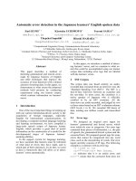
Fig. 1. L. baicalensis specimens during late summer (September) (A)
and the ice cover season (March) (B). In both seasons sponges con-
tain associated dinoflagellates (taxon Gymnodinium). Cross-sections
were prepared and examined using transmission electron microsco-
py. (C, D) Sections through a branch from a September specimen
show the abundantly present dinoflagellates (d) that are assembled
at the rim of the green branches. (E) Frequently the specimens dur-
ing the March season contain spermatogenic cysts (sc). Identifica-
tion of OA-producing dinoflagellates in L. baicalensis (F–K). (F, I)
Slices were prepared from tissue of L. baicalensis and the cells were
visualized Nomarsky interference contrast optics; dinoflagellates
(d) as well as sponge cells (s) are marked. (G) In one series, the
slices were reacted with pAb-OA and then with Cy3-conjugated goat
anti-(rabbit IgG) and finally inspected by immunofluorescence
(wavelength ¼ 546 nm). (H) Autofluorescence of the chlorophyll in
the dinoflagellates was detected at a wavelength of 490 nm. (J) In
parallel, the slices were reacted with pAb-OA, which had been
pretreated with OA, coupled to the FID-33 oligopeptide and then
with the labeled secondary antibodies. (K) The same area was also
analyzed with a wave-length of 490 nm. Scale bar ¼10 lm.
W. E. G. Mu
¨
ller et al. Cold stress defense in sponges
FEBS Journal 274 (2007) 23–36 ª 2006 The Authors Journal compilation ª 2006 FEBS 25
(Fig. 1G); using Nomarsky interference contrast optics
the granule-containing dinoflagellates could be identi-
fied (Fig. 1F). As further evidence for the localization
of the dinoflagellates, slices were illuminated with
green fluorescent light (490 nm) to identify dinoflagel-
lates based on the autofluorescence of their chlorophyll
(Fig. 1H). Again, the dinoflagellates were highlighted
in areas positive for pAb-OA. In a parallel series, sec-
tions were treated with adsorbed antibodies against
OA; this preparation showed a slight signal only very
occasionally (Fig. 1J). Dinoflagellates could again be
visualized by their autofluorescence (Fig. 1K).
L. baicalensis PP1 catalytic subunit
Complete cDNA encoding the L. baicalensis PP1 pro-
tein (LBPP1) was obtained from a cDNA library using
a degenerate primer against the Ser⁄ Thr-specific pro-
tein phosphatase signature of mammalian protein
phosphatases. The ORF between nucleotides 60–62
and 1057–1059(stop) codes for a 319 amino acid
polypeptide (PP1_LUBAI) with a predicted size of
36 802 Da (Fig. 3A); the sequence was termed
PP1_LUBAI. Like the related mammalian sequences,
the sponge protein comprises a characteristic Ser⁄ Thr-
specific protein phosphatase signature (amino acids
121 and 126) and the conserved matallophosphoest-
erase (amino acids 57 and 252). The calcineurin-like
phosphoesterase (amino acids 57 and 252) overlaps
with the latter region. Similarity between the sponge
molecule and other metazoan PP1 sequences is high;
the sponge PP1 shares 288 similar and 271 identical
amino acids with human PP1 (length: 323 amino
acids), known to bind to OA (Fig. 3A). The overall
similarity ⁄ homology to metazoan sequences is
80% ⁄ 70%. For the alignment in Fig. 3A, the human
protein with the highest similarity score was used
(‘Expect value [E]’ 2e-162) [15]; this phosphatase is
known to function as PP1 (catalytic subunit, gamma
isoforms).
A phylogenetic tree was computed after alignment
of the metazoan sequences with yeast and plant-related
PP1 (Fig. 3B). The tree was rooted with the highest
similar phosphatases from Arabidopsis thaliana (pro-
tein phosphatase-type 1; NP_181514.1) and Saccharo-
myces cerevisiae (type 1 Ser ⁄ Thr protein phosphatase;
NP_011059.1). Among the metazoan proteins, the
sponge phosphatase clusters together with that of Dro-
sophila melanogaster (Ser ⁄ Thr protein phosphatase;
CAA49594.1), while the Caenorhabditis elegans
protein (even-like phosphatases family member
(NP_001022616.1) forms a separate branch together
with the human sequence. Both branches are separated
only with low significance.
Relative PP1 content
Because of the high sequence similarity between L. bai-
calensis PP1 and the corresponding mammalian phos-
phatases it was possible to use a commercial antibody
for the western blots. Extracts were size-separated by
SDS ⁄ PAGE and either stained with Coomassie Brilli-
ant Blue (Fig. 3C, lane a) or the proteins were trans-
ferred to poly(vinylidene difluoride) membranes and
reacted with antibodies against PP1, as described in
Experimental procedures. Extracts from animals
obtained in both March (Fig. 3, lane b) and September
(Fig. 3, lane c) show a strong signal at 37 kDa, cor-
responding to the size of sponge PP1. In contrast, if
the blots were reacted with adsorbed antibodies, the
signals were strongly reduced (lanes d and e).
Phosphatase activity in the extract
Extracts were prepared from sponge tissue specimens
collected in March or September and subjected to the
protein phosphatase assay described in Experimental
procedures. The specific enzyme activities in tissues
from winter and summer animals were almost identical
(22.6 ± 4.7 versus 23.4 ± 4.2 nmolÆmin
)1
Æmg
)1
). If OA
was added, the reactions in the two series of experi-
ments were inhibited dose-dependently; at 100 nm the
activity differed from that seen in the controls (March,
17.3 ± 3.9 nmolÆmin
)1
Æmg
)1
; September, 19.2 ± 4.3
nmolÆmin
)1
Æmg
)1
). However, at 300 nm the activity
Fig. 2. Identification of protein-coupled OA in L. baicalensis.
Extracts were prepared and the protein size separated by
SDS ⁄ PAGE (10% gel). Lane a, the gel was stained with Coomassie
Brilliant Blue. The proteins were blot transferred and the filters were
either incubated with anti-OA sera (lane b) or with the antibody pre-
paration, which had been preincubated with FID-33-OA which had
been adsorbed with FID-33-OA (lane c). M, marker proteins.
Cold stress defense in sponges W. E. G. Mu
¨
ller et al.
26 FEBS Journal 274 (2007) 23–36 ª 2006 The Authors Journal compilation ª 2006 FEBS
decreased to 80% (March, 3.7 ± 1.9 nmolÆmin
)1
Æ
mg
)1
; September, 5.2 ± 1.6 nmolÆmin
)1
Æmg
)1
).
Expression level of hsp70 in animals and
primmorphs
The semiquantitative steady-state level of hsp70 tran-
scripts in animals was in the same range, regardless of
whether the RNA had been isolated from specimens
collected in March or in September. For these Nor-
thern blot studies the EST probe for hsp70 from
L. baicalensis (LUBAIHSP70 ) was used (Fig. 4A).
In order to assess the expression level under con-
trolled laboratory conditions, in vivo primmorphs were
incubated for 24 h at 4 and 17 °C. Primmorphs were
incubated in the dark to suppress the metabolic activ-
ity of the remaining symbiotic algae. Setting the
expression level at 4 °C to onefold, the amount of
hsp70 transcripts in the primmorph cells incubated at
17 °C was much higher (10-fold; Fig. 4A). However,
if primmorphs were incubated at 4 °C together with
100 nm OA the amount of hsp70 transcripts was the
same as that measured for cultures maintained at
17 °C. The toxin had no strong effect on hsp70 expres-
sion in cells at 17 °C (Fig. 4A). In controls, a-tubulin
expression was determined using the L. baicalensis
probe LUBAITUB in a parallel Northern blotting
experiment. Almost identical signal intensities were
seen, confirming that the same amount of RNA was
loaded onto the gels. From these results, we conclude
that OA induces hsp70 expression at the lower incuba-
tion temperature.
To support these studies, comparative Northern and
western blot experiments were performed using hsp70
(LUBAIHSP70) or antibodies (mAb-HSP70) as the
respective probes (Fig. 4B). Again, the Northern blot
A
B
C
Fig. 3. PP1 from L. baicalensis. (A, B) The PP1 sequence from L. baicalensis. (A) Alignment of the sponge PP1 protein (PP1_LUBAI) with
the human PP1 which binds to OA (PP1_HUMAN; accession number 1JK7_A). Amino acids, identical in both sequences, are in inverted type
and those similar in both sequences are shaded. The characteristic Ser ⁄ Thr-specific protein phosphatase signature (S ⁄ Tp) and the conserved
metallophosphoesterase (MetPhoEsterase) regions are marked. (B) The phylogenetic tree is constructed from the two abovementioned
sequences as well as the PP1 from D. melanogaster (PP1_DROME; CAA49594.1), from C. elegans (seven-like phosphatases family mem-
ber) (PP1 °CAEEL; NP_001022616.1), from S. cerevisiae (PP1_YEAST; NP_011059.1) as well as from A. thaliana (PP1_ARATH;
NP_181514.1), which was used as outgroup to root the tree. After alignment the tree was built. Scale bar indicates an evolutionary distance
of 0.1 amino acid substitutions per position in the sequence. (C) Identification of PP1 in tissue from L. baicalensis. In all lanes 10 lg of pro-
tein were separated. Lane a, the separated proteins were identified with Coomassie Brilliant Blue. Western blot experiments: lanes b and c,
the membranes were reacted with antibodies against PP1 (PcAb-PP1) and then with labelled secondary antibodies to visualize the immuno-
complexes with the BM chemoluminescence substrate kit. Samples from March (lane b) and September (lane c) were analyzed. In parallel,
membranes with the extracts were treated with adsorbed PcAb-PP1 (lanes d and e). The size markers are given.
W. E. G. Mu
¨
ller et al. Cold stress defense in sponges
FEBS Journal 274 (2007) 23–36 ª 2006 The Authors Journal compilation ª 2006 FEBS 27
studies showed low expression of hsp70 in primmorphs
incubated at 4 °C or in the absence of OA, in compar-
ison with those incubated with OA or at 17 °C
(Fig. 4B,a). Steady-state expression of the a-tubulin
gene is shown using the same amount of RNA for ana-
lysis. The data from western blot experiments showed a
similar expression pattern; low levels of hsp70 in cul-
tures incubated at 4 °C and without OA, in comparison
with those incubated with the toxin and at higher tem-
perature (Fig. 4B,b). From these data, we conclude that
the level of hsp70 is controlled in primmorphs at both
a transcriptional and translation level.
Effect of depletion of hsp70 from extracts on the
activity of protein phosphatase(s)
An immunodepletion study was performed as described
in Experimental procedures. Extracts were prepared
from animals collected in September and incubated at 4
or 17 °C. Unexpectedly, enzyme activity at 4 °C was
only 20% lower (18.1 ± 4.2 nmolÆmin
)1
Æmg
)1
) than
that measured at 17 °C (22.7 ± 5.1 nmolÆmin
)1
Æmg
)1
).
However, after incubation of the extracts for 60 min
with antibodies against hsp70 the activity of the protein
phosphatase was reduced at 4 °C to 5.3 ± 2.9 nmolÆ
min
)1
Æmg
)1
, whereas the antibodies had no effect on
activity at 17 °C (Fig. 5). The adsorbed mAb-HSP70
A
Ba
b
Fig. 4. Expression of heat shock protein hsp70 transcripts in ani-
mals and primmorphs. (A) Northern blot analysis. RNA was extrac-
ted both from animals, collected in March or September, and from
primmorphs cultivated in vitro. As indicated, the primmorphs were
cultivated either 4 °Corat17°C for 24 h in the absence (–) or
presence of 100 n
M of OA (+). Then, total RNA was isolated and
fractionated by electrophoresis, transferred to nylon membrane,
and hybridized with the respective labeled probes; hsp70
(LUBAIHSP70)ora-tubulin (LUBAITUB). 2 lg of total RNA were
loaded into each slot. The relative degree of expression was corre-
lated with that seen for the minimal expression in primmorphs at
4 °C (set to onefold). (B) Comparison of the level of hsp70 tran-
scripts and hsp70 protein in primmorphs, incubated at 4 or 17 °C
in the absence (–) or presence of OA (+). As marked, the
primmorphs were incubated at these two temperatures for 24 h
and - ⁄ +OA. Then extracts were prepared for Northern blotting
(RNA) or western blotting (protein) (a) Northern blot: after size
fraction and transfer the filter was hybridized with the hsp70
probe: N1, incubation at 4 °C in the absence of OA; N2, at 4 °Cin
the presence of OA; N3, incubation at 17 °C in the absence of
OA; N4, incubation at 17 °C in the presence of OA. In parallel, a
filter was hybridized with a a-tubulin probe. (b) From the same
samples the proteins were extracted and subjected to western
blot analysis. Samples from cultures incubated at 4 °C in the
absence (lane W1) or presence of OA (W2) or at 17 °C without
(lane W3) or with OA (lane W3) are loaded onto the gel, and after
separation and transfer probed with the antibodies mAb-HSP70.
The relative expression levels, correlated to the values assessed
for cultures at 4 °C and without OA (set to onefold), are given.
Fig. 5. Effect of antibodies against hsp70 on the activity of the pro-
tein phosphatase in extracts from animals. Extracts were prepared
from animals, collected in summer and tested for protein phospha-
tase activity at 4 or 17 °C, as described in Experimental proce-
dures. Where indicated the samples were pretreated either with
mAb-HSP70 or with adsorbed mAb-HSP70.
Cold stress defense in sponges W. E. G. Mu
¨
ller et al.
28 FEBS Journal 274 (2007) 23–36 ª 2006 The Authors Journal compilation ª 2006 FEBS
preparation did not show a significant effect on the
enzyme activities. From these results we conclude
that: (a) hsp70 proteins, which are supposedly func-
tionally active, are present in sponge extracts together
with the enzyme; and (b) hsp70 is required for the
full enzyme activity during incubation at lower
temperature.
Discussion
OA is a secondary metabolite produced by free-living
microalgae, primarily by Prorocentrum lima [16]. Other
dinoflagellates, e.g. Gymnodinium sp., are also consid-
ered to be producers [17]. Secondary metabolites are
surely not without metabolic function for the producer
or the host, because then they would have been elimin-
ated during evolution. They have, however, no direct
role in the growth of the producing organism and are
considered not to play a key role in the maintenance
of cellular function but in defense [18]. It remains
unexplained, however, why some secondary metabo-
lites, like OA, are synthesized not only by one taxon,
but by a whole range of microorganisms. These
dinoflagellates are harbored in a series of hosts, e.g.
mussels [11] or sponges such as H. okadaii [12],
S. domuncula [14] and Geodia cydonium (W. E. G.
Mu
¨
ller, University of Mainz, unpublished results), or,
as shown here, in L. baicalensis. This latter finding is
surprising, because L. baicalensis is a freshwater
sponge, in contrast to the others which are marine ani-
mals. These findings suggest that OA has a crucial role
in the maintenance of a symbiotic relationship between
algae and host. With S. domuncula it has been shown
that OA augments the concentration-dependent
immune defense system against bacteria [14], and as
described recently, kills symbiotic ⁄ parasitic annelids
[19]. The dinoflagellates present in L. baicalensis are
related to G. sanguineum, a species that has been found
worldwide, especially in coastal waters [20].
We have shown that the dinoflagellates (G. sangui-
neum) produce OA in L. baicalensis. For identification,
we applied antibodies raised against OA, which have
been previously qualified as specific for this secondary
metabolite [14,19]. In cross-sections though L. baical-
ensis these antibodies reacted specifically with the dino-
flagellates; their signals could be suppressed by
adsorption with free OA. Analytical measurements
revealed that the concentration of OA in L. baicalensis
is 50 ngÆg
)1
of tissue (70 nm), a level comparable
with that found in other sponges [14]. In addition,
western blot analysis has shown that, like in extracts
prepared from S. domuncula and L. baicalensis,OA
exists in a covalent linkage with a protein of 14 kDa.
This finding, first described in S. domuncula [19], can
be explained as a depot⁄ storage form of the free OA.
It is not known whether OA is released from the
sponge into the surrounding aqueous habitat. Previous
studies with S. domuncula have shown that in this
sponge OA accumulates in the epithelial layers of the
aquiferous system within the animals, suggesting that
OA is involved in defense against microbial invaders
[21]. Because L. baicalensis ingests ⁄ feeds on microor-
ganisms and plankton [7] it is very likely that OA is
accumulated in the aquiferous canal system and acts
as a protecting metabolite.
Based on existing data, it is increasingly evident that
OA in the symbiotic bacteria also affects the primary
cell metabolism of the host. Previously, the main focus
of research has been on the effect of OA on attacking
or commensalic organisms, via inhibition of enzymes
(protein phosphatases) [11]. In view of the data, the
corresponding cDNA for one of these enzymes (PP1)
needed to be identified first. cDNA coding for PP1
was completely isolated and the corresponding protein
deduced. This polypeptide contains all the characteris-
tic domains of other enzymes in this group, e.g. the
characteristic Ser ⁄ Thr-specific protein phosphatase
signature and the conserved metallophosphoesterase
region. Based on the high sequence similarity between
the sponge protein phosphatase and mammalian
enzymes an antibody against the latter could be used
here. Signals obtained by western blot analysis,
37 kDa, matched the expected size. Enzyme activity in
the prepared extract was 20 nmol inorganic phos-
phate released per min and mg of protein in tissue
extracts. Inhibition studies with OA were performed
which revealed that the toxin blocks the enzyme(s)
significantly at OA concentrations > 300 nm;at
100 nm no inhibition was seen. The sensitivity of the
enzyme to OA was in the range published previously
[11,14]. In the plant Medicago sativa it could be shown
that at low temperature hyperphosphorylation of
proteins occurs and this is the result of inhibition of
protein phosphatase(s) [22]. It has been established
that OA is a more sensitive inhibitor of PP2 than of
PP1 [11]. Therefore, we screened our EST database
from L. baicalensis, which comprises over 4000
sequences, and also performed extensive screening
studies using degenerate primers, designed against the
conserved regions within the PP2 nucleotide sequence,
to identify PP2 transcripts in the cDNA library from
L. baicalensis. However, these attempts were without
success. Therefore, we focused our studies at the
protein and cellular level on PP1 only. Nevertheless,
the data presented here do not exclude that PP2 is
involved in thermoregulation in L. baicalensis.
W. E. G. Mu
¨
ller et al. Cold stress defense in sponges
FEBS Journal 274 (2007) 23–36 ª 2006 The Authors Journal compilation ª 2006 FEBS 29
As outlined earlier, L. baicalensis lives in a biotope
with ambient temperatures between )0.5 °C (March)
and 17 °C (August–September). To date, no data have
been available that could help in understanding which
protection system allows these animals to maintain
high metabolic activity during these extreme situations.
The first series of experiments now demonstrates that
the relative level of enzyme(s) (protein phosphatase)
in the animals is the same in March or in September
Also, the sensitivities of the enzymes towards OA are
very similar. Based on these results, we conclude that
OA has an inhibitory effect in the animals during both
seasons at concentrations > 300 nm.
In multicellular organisms one major protection sys-
tem against temperature stress is provided by the heat
shock proteins [23], with hsp70 being the most thor-
oughly studied example. In contrast to the related con-
stitutively expressed cognate hsc70, which changes its
level only slightly upon differing stresses, hsp70 is
strongly upregulated upon exposure to stress [24]. For
the marine sponges S. domuncula or G. cydonium we
were able to show that at both the gene-expression level
[25] and the protein level [26] expression of hsp70 increa-
ses strongly after temperature change and also after
exposure to xenobiotics [27]. The induction of the gene
with respect to the stressors proceeds with the same kin-
etics in the sponge and in fish [28]. Focusing on Lake
Baikal sponges, hsp70 proved to be a suitable biomarker
for xenobiotics and temperature stress [29].
Few publications are available that describe the
effect of OA on the expression of heat shock proteins
[13,30]. These authors demonstrated that injection of
300 ng of OA into 250 g rats resulted in notable
expression of hsp70 ⁄ 72 after an incubation of 72 h.
More importantly, Joyeux et al. [30] showed that OA
treatment results in a potentiation of hsp72 mRNA
expression. In this study, we tested whether OA chan-
ges the level of hsp70 expression at both the gene
expression level and the protein level. For these stud-
ies, the primmorph system was used, which allowed
the study of this toxin under controlled laboratory
conditions. In earlier studies, primmorphs have proved
to be suitable for measuring these effects [31].
A hsp70 cDNA probe was used to measure semiquan-
titatively the steady-state expression hsp70 in animals
collected in March and September; there were no signifi-
cant changes. As outlined, the sponges contain OA in
March and September. However, if primmorphs, kept in
the dark to suppress the photosynthetic activity of
the algae, were cultivated at 4 °C the level of hsp70
transcripts was very low compared with primmorphs
cultivated at 17 °C, or animals in the biotope, irrespect-
ive of whether they were collected at )0.5 °Corat
12 °C. If primmorphs were cultivated at 4 °C together
with 100 nm OA the hsp70 level reached values seen in
animals or primmorphs incubated at 17 °C. Interest-
ingly, the level of hsp70 protein also followed this pat-
tern. From these results we conclude that OA causes, at
both the gene- and the protein level, increased expres-
sion of this heat shock protein in primmorphs at 4 °C.
Interestingly, expression of a-tubulin in primmorphs is
low during incubation at 4 °C, and is upregulated in the
presence of OA or at higher temperatures. This suggests
that OA plays an inducer role for hsp70 and tubulin in
primmorphs under cold stress conditions.
A set of immunodepletion experiments was per-
formed. Extracts from animals collected during the
summer were prepared that, according to the above-
mentioned data, contained greater amounts of hsp70
protein. They were subjected to protein phosphatase
activity determination in the presence or absence of
antibodies raised against hsp70. Hsp70 is known to
bind to target protein(s) in the presence of ATP
[23,32,33], which was therefore added. If the extracts
were assayed for protein phosphatase(s) activity it was
found that at lower temperatures (4 °C) enzyme activ-
ity was close to that seen after incubation at 17 °C.
However, if antibodies were added to the mixture and
incubation was performed at 4 °C the activity was
almost completely abolished. At 17 °C the antibodies
had no effect on the high expression level of hsp70.
These results strongly suggest that native hsp70 binds
at lower temperature to the enzyme(s) and restored the
activity to values seen at higher temperature.
OA is a toxin present in dinoflagellates that coexist
with marine animals ⁄ sponges and, as described here, in
freshwater sponges. Quantitative determinations
showed that the level of this toxin in the freshwater
sponge L. baicalensis was as high as in the marine
sponge S. domuncula. As reported here, in these fresh-
water animals OA is involved in processes that result
in a high steady-state expression of the chaperon–
hsp70 system. In addition, the data suggest that the
hsp system contributes to the cold thermotolerance
seen in L. baicalensis, because the secondary metabo-
lite OA functions as an inducer for hsp70. Therefore,
it can be postulated that OA contributes markedly to
the survival strategies of these animals during unfavo-
rable environmental conditions. This view is outlined
in Fig. 6. In animals, collected during winter and
summer the level of hsp70 is high. If extracts
from these specimens were used for immunodepletion
experiments the ‘activating’ effect of hsp70 on protein
phosphatase activity could be demonstrated in extracts
prepared from animals collected during the cold
season. This suggests that at low temperature ATP-
Cold stress defense in sponges W. E. G. Mu
¨
ller et al.
30 FEBS Journal 274 (2007) 23–36 ª 2006 The Authors Journal compilation ª 2006 FEBS
dependent hsp70 molecules are required for the pro-
motion of folding, transport and ⁄ or assembly of target
proteins participating in the primary metabolism or,
perhaps also, in the composition of the fluid mem-
branes. Energetically, the dinoflagellates supply their
host with primary metabolites via their photosynthetic
activity, glycerol being the major compound. We
recently identified that the dinoflagellates synthesize
glycerol which is then taken up by cells of L. baicalensis
through the aquaporin channel (W. E. G. Mu
¨
ller,
manuscript submitted). In conclusion, our data suggest
that OA causes induction of hsp70 at low ⁄ cold tem-
perature stress; in turn, hsp70 contributes to the proper
activity of the protein phosphatase at low temperature.
Experimental procedures
Chemicals and enzymes
Restriction enzymes, SNAP ‘Total RNA Isolation Kit’,
Superscript II and reagents for RACE procedure were pur-
chased from Invitrogen (Carlsbad, CA), TriplEx2 vector
from BD (Palo Alto, CA), TRIzol Reagent from Gib-
coBRL (Grand Island, NY), Hybond-N
+
nylon membrane
from Amersham (Little Chalfont, UK), mAb against hsp70
(bovine; H 5147) was obtained from Sigma (St Louis, MO),
polyclonal antibodies raised against PP1 were from Santa
Cruz Biotechnology (Santa Cruz, CA), CDP from Roche
(Mannheim; Germany), Technovit 8100 from Heraeus Kul-
zer (Wehrheim, Germany), Sephadex G-20 from Pharmacia
(Uppsala, Sweden), Lake Baikal water was obtained from
‘Lake’ Comp. (Irkutsk; Russia), and the protein phospha-
tase assay kit from Upstate Biotechnology (Lake Placid,
NY). Okadaic acid was purchased from Alexis Biochemi-
cals (Gru
¨
nberg; Germany), the toxin was dissolved in
dimethylsulfoxide.
Sponges, cells/primmorphs and cDNA libraries
Specimens of L. baicalensis (Porifera, Demospongiae, Haplo-
sclerida) were collected in Lake Baikal (Russia) near the
village Bolshiye Koty (51°58 N, 105°21¢E) from depths
between 7 and 12 m during September 2005 and March 2006.
Primmorphs were prepared by immerging sponge tissue
into natural Lake Baikal water, supplemented with 50 mm
EDTA. Lake Baikal water was used to guarantee a suitable
mineral composition [34]. After gentle squeezing and subse-
quent shaking for 30 min at 16 °C on a rotatory shaker,
the solution was discarded and new Baikal water ⁄ EDTA
was added. After 40 min the supernatant was collected and
filtered through a 40-lm mesh nylon net; shaking in Baikal
water ⁄ EDTA and filtration were repeated once. Single cells
Fig. 6. Proposed function of OA in L. baicalensis. The toxin OA is produced by the dinoflagellates from the taxon Gymnodinium. In both win-
ter and summer the dinoflagellates produce primary metabolites via their photosynthetic activity; the metabolite is taken up by the sponge
cells. Using the primmorph system it could be shown that the level of hsp70 is low at the lower temperature (in primmorphs at 4 °C). How-
ever, the abundance of hsp70 increases in response to low concentrations of OA (100 n
M) to levels which are seen in primmorphs incubated
at a higher temperature (17 °C) or in animals collected during March and September. It is postulated that a sufficiently high level of hsp70 is
one prerequisite for the promotion of folding, transport and ⁄ or assembly of target proteins participating in the primary metabolism or in the
composition of the fluid membranes during unfavorable environmental conditions (in nature, at )0.5 °C). Only at higher concentrations
(> 300 n
M) does OA inhibit protein phosphatase(s).
W. E. G. Mu
¨
ller et al. Cold stress defense in sponges
FEBS Journal 274 (2007) 23–36 ª 2006 The Authors Journal compilation ª 2006 FEBS 31
were harvested by centrifugation (500 g for 5 min, Eppen-
dorf centrifuge 5702 with rotor A-8-17) and washed once.
The cells of this pellet were resuspended in Baikal water,
supplemented with 5 lgÆmL
)1
penicillin and 100 lgÆmL
)1
streptomycin. A cell suspension of 10
7
cells was added to
6 mL (final volume) of medium in 10 mL flasks (Nuclon
surface; #136196; Nunc Wiesbaden, Germany). Primmorphs
were obtained from these single cells; they reached sizes of
3–7 mm after two days in the dark. The primmorphs were
incubated at 4 or 17 °C; during the summer season the
ambient water environment of L. baicalensis can reach a
temperature of up 22 °C [35].
The cDNA library from L. baicalensis was prepared in
TriplEx2 vector.
Preparation of antibodies against OA
Polyclonal antibodies against OA (pAb-OA) were raised in
female rabbits (White New Zealand) as described previously
[14]. The antigen (OA) was covalently coupled via its C-ter-
minus to the FID-33 oligopeptide (sequence: NH
2
-FIDA-
VWKCVTPFIDAVWKTKFICVTPFIDAVWK-COOH),
using EDC (1-ethyl-3-(3-dimethylaminopropyl)carbodi-
imide; Sigma) as described previously [14]. This OA conju-
gate was injected at 4-week intervals into the animals; after
three boosts serum was collected and the antibodies were
prepared [36]. The titer of the antibodies was determined to
be 1 : 2000. Where indicated the antibodies (pAb-OA;
0.3 mL of undiluted serum) were adsorbed with FID-33
coupled to OA (1 mg) for 30 min at room temperature.
The studies with animals (antibody production) have been
approved by the respective state authorities.
Histological analysis
Tissue was fixed in paraformaldehyde, embedded in Tech-
novit 8100 and sectioned [37]. The 4-lm thick slices were
incubated with pAb-OA (1 : 250 dilution) overnight. Then
the slides were treated with Cy3-conjugated goat anti-(rab-
bit IgG) for 2 h. Subsequently, the sections were inspected
by immunofluorescence with an Olympus AHBT3 micro-
scope, using an excitation light wavelength of 546 nm. In
addition, the slices were illuminated with light of a wave-
length of 490 nm, which detects the autofluorescence of
chlorophyll in the dinoflagellates. In parallel, the slices were
inspected directly using Nomarsky interference contrast
optics. In controls, the pAb-OA (10 lg) were preincubated
with OA, coupled to the FID-33 oligopeptide (10 lg).
Competitive ELISA
The ELISA was performed similar to the procedure des-
cribed earlier [14]. OA, coupled to the FID-33 oligopeptide
was linked to 96-well plates (Covalink-primary amine;
Nunc) [14]. After three washing steps with NaCl ⁄ P
i
(containing 0.05% Tween-20) the plates were blocked prior
to use with 3% of fatty acids-free bovine serum albumin in
NaCl ⁄ P
i
. Then 100 lL of pAb-OA were added at different
dilutions to each well for 90 min. Subsequently, the immu-
nocomplexes were visualized using secondary antibodies,
coupled to horseradish peroxidase (1 : 1000; Sigma, Dei-
senhofen, Germany) under application of o-phenylenedi-
amine as substrate. The plates were read at 492 nm. In the
competitive ELISA procedure, OA was added at different
concentrations (1 ngÆmL
)1
to 1 lgÆmL
)1
NaCl ⁄ P
i
) from a
stock solution of 1 mgÆmL
)1
, dissolved in methanol. Within
the range of 10 ngÆmL
)1
to 1 lgÆmL
)1
the change of the
absorbance was linear (logarithmic plot). For the determin-
ation in the tissue of the sponge, extracts were prepared
from the tissue with 80% methanol. The values for the
absorbance were extrapolated using the calibration curve
obtained with the free toxin.
Identification of OA-bound protein
Extracts were prepared from tissue samples; they were
homogenized in lysis buffer (1· Tris-buffered saline,
pH 7.5, 1 mm EDTA, 10 mm NaF, 0.1 lm aprotinin, 1 mm
sodium orthovanadate). Total cell extracts (10 lgÆlane
)1
)
were subjected to electrophoresis in 10% polyacrylamide
gels containing 0.1% SDS ⁄ PAGE as described previously
[37]. The gels were stained with Coomassie Brilliant Blue.
Subsequently, western-blotting experiments were performed
after transfer of the proteins onto poly(vinylidene difluo-
ride) membranes (Millipore-Roth) using pAb-OA (1 : 300
dilution). After incubation for 3 h, the blots were incubated
with goat anti-(rabbit IgG), peroxidase-coupled (1 : 5000
dilution; New England Biolabs). Detection of the immuno-
complex was carried out using the BM Chemoluminescence
Blotting Substrate kit. Where indicated the pAb-OA had
been adsorbed with OA coupled to the FID-33 oligo-
peptide.
Relative PP1 content (western blotting)
The relative content of PP1 in the extracts was determined
by western blotting. First, the tissue extracts (see previous
section) were size separated by electrophoresis (SDS ⁄
PAGE ⁄ 12% polyacrylamide); samples of 10 lg protein
extract were loaded onto the gels. In addition, the proteins
were blot transferred and reacted with polyclonal antibodies
raised against PP1 (PcAb-PP1; 1 : 2000 dilution). After incu-
bation for 3 h, blots were incubated with peroxidase-coupled
goat anti-(rabbit IgG); and the immunocomplexes were
visualized using the BM Chemoluminescence Blotting
Substrate kit. In control experiments 10 lL of undiluted
PcAb-PP1 was adsorbed with 10 lL of cell extract
(1 mgÆmL
)1
; 30 min; 4 °C) prior to the use onto the blots.
Cold stress defense in sponges W. E. G. Mu
¨
ller et al.
32 FEBS Journal 274 (2007) 23–36 ª 2006 The Authors Journal compilation ª 2006 FEBS
Heat shock protein content (western blotting)
The effect of OA on hsp70 expression was determined in
primmorphs. Extracts were prepared (see above) and sub-
jected to western blot analysis using 10% polyacrylamide
gels (SDS ⁄ PAGE). Ten micrograms of protein extract per
lane were applied and after blot transfer reacted with anti-
HSP70 mAb (mAb-HSP70; 1 : 5000 dilution) in Tris-
buffered saline containing 0.1% milk powder protein and
0.05% Tween-20 for 1.5 h at room temperature, followed
by alkaline phosphatase-conjugated anti-mouse IgG for 1 h
at room temperature. The immunoblots were stained using
5-bromo-4-chloro-3-indolyl phosphate ⁄ nitroblue tetrazo-
lium.
Phosphatase activity in the extract
The relative protein in the extract phosphatase content was
determined in crude extract following the described proce-
dure [38]. Cell extracts were prepared by homogenization
using a 20 mm Hepes buffer (pH 7.5; containing 150 mm
NaCl, 1.5 mm MgCl
2
,1mm EDTA, 1 mm phenyl-
methylsulfonyl fluoride, 1 mgÆmL
)1
aprotinin, 1 mg ÆmL
)1
leupeptin, 10% v ⁄ v glycerol and 1 mm Na-orthovanadate).
After standing for 10 min at 0 °C the suspension was cen-
trifuged for at 22 000 g (20 min, Eppendorf centrifuge 5702
with rotor A-8-17) to remove the spicules and the tissue
particles. The supernatant was collected and passed though
a Sephadex G-20; the loading volume of the extract was
5–10% of the total volume of the column. The column was
eluted with the Hepes buffer. Fractions were collected and
protein phosphatase activity was determined.
The protein phosphatase activities (both PP1 and PP2A)
were determined by applying the protein phosphatase assay
kit and the synthetic phosphorylated substrate KRpTIRR
[38]. The cell extract was added and the reaction (incuba-
tion temperature at 17 °C) terminated after 20 min by
addition of Malachite Green solution. Absorbance was
determined at 660 nm to determine the inorganic phosphate
release and correlated to the amount of protein (1 mg).
Where indicated, assays were supplemented with 100 and
300 nm of OA.
Immunodepletion studies
Extracts were prepared from animals collected during the
summer season (September), using the buffer system des-
cribed above. Extracts (100–150 lL) containing 120 lgof
protein and supplemented with 100 lm ATP were incubated
in the absence or presence of 10 lL of hsp70 antibodies
(mAb-HSP70) for 60 min at room temperature. Subse-
quently, the activity of the protein phosphatase(s) was
determined at 4 and 17 °C, respectively, applying the des-
cribed procedure and the synthetic phosphorylated sub-
strate. In controls, 10 lL of undiluted mAb-HSP70 were
adsorbed with 10 lL of cell extract (1 mgÆmL
)1
; 30 min;
4 °C) prior to the use onto the blots.
Transmission microscopy analysis
For transmission microscopy analysis samples were cut into
pieces (2 mm
3
), incubated in 0.1 m phosphate buffer (sup-
plemented with 2.5% glutaraldehyde, 0.82% NaCl, pH 7.4)
and washed in 0.1 m phosphate buffer (1.75% NaCl) at
room temperature. After treating the samples with 1.25%
NaHCO
3
, 2% OsO
4
and 1% NaCl, they were dehydrated
with ethanol. Dried samples were incubated with propylene
oxide, fixed in propylene oxide ⁄ araldite (2 : 1), covered with
pure araldite and hardened at 60 °C for two days prior to
cutting to 60 nm ultrathin slices (Ultracut S; Leica, Wetz-
lar; Germany). The samples were transferred onto coated
copper grids and analyzed with a Tecnai 12 device (FEI
Electron Optics, Eindhoven; Netherlands).
Isolation of cDNA encoding PP1
PCR was applied to identify the complete cDNA encoding
for L. baicalensis PP1. Degenerate primers were designed
against the conserved Ser ⁄ Thr-specific protein phosphatase
signature of mammalian PP1s, e.g. the human PP1 which
binds to OA (accession number 1JK7_A) [39]. This segment
is present in the human protein sequence between amino
acids 123 and 130. PCR was carried out with the forward
primer, 5¢-GGIAAC ⁄ TCAC ⁄ TGAA ⁄ GTGT ⁄ CGCIAGC ⁄
TAT-3¢ and the vector primer at an initial denaturation at
94 °C for 5 min, followed by 30 amplification cycles at
94 °C for 30 s, 52 °C for 45 s, 75 °C for 1.5 min, and a
final extension step at 75 °C for 10 min. Fragments were
isolated and cloned into the pCRII-TOPO vector in E. coli
TOP10 cells (Invitrogen). Sequencing was performed with
primers directed to the SP6 promoter and the T7 promoter.
The sequence was completed with insert-specific primers in
combination with 5¢-RACE primer or with 3¢-RACE pri-
mer using the CapFishing Full-length cDNA Premix Kit
(Seegene Inc., Rockville, MD, USA). The final sequence
was confirmed by an additional PCR using primers directed
against the nontranslated region of the cDNA, followed by
sequencing. The clone encoding the PP1 molecule from
L. baicalensis LBPP1 is 1359 nucleotides long, excluding
the poly(A) tail.
EST sequence: heat shock protein
In the cDNA ⁄ EST (expressed sequence tag) database from
L. baicalensis, which comprises 4000 sequences, more than
70 tags encoding heat shock proteins exist. The dominant
sequences code for hsp70. One sequence was selected
which has a length of 416 nucleotides and was termed
LUBAIHSP70.
W. E. G. Mu
¨
ller et al. Cold stress defense in sponges
FEBS Journal 274 (2007) 23–36 ª 2006 The Authors Journal compilation ª 2006 FEBS 33
Sequence analysis
The deduced protein sequence of L. baicalensis PP1
(PP1_LUBAI) was compared with those of the most closely
related proteins, especially those from human, Drosophila
melanogaster, Caenorhabditis elegans, Saccharomyces cere-
visiae and Arabidopsis thaliana using the neighbor-joining
method [40]. The degree of support for internal branches
was further assessed by bootstrapping [41]. Accurate mul-
tiple protein sequence alignments were made using the soft-
ware clustal w [42].
Northern blotting
RNA was isolated from primmorphs incubated for 24 h in
the absence or presence of OA. Samples were frozen, pul-
verized in liquid nitrogen and RNA was extracted using the
TRIzol Reagent. Total RNA (2 lg) was fractionated by
electrophoresis, transferred to a Hybond-N+ nylon mem-
brane, and hybridized overnight at 50 °C. The hsp70 probe
(LUBAIHSP70), labeled using the PCR-DIG-Probe-Synthe-
sis Kit, was used. As a control to assure that the same
amount of RNA was loaded onto the gels, the housekeep-
ing gene a-tubulin (LUBAITUB; AJ971711) from L. baical-
ensis was used. After washing, DIG-labeled nucleic acid
was detected with anti-DIG Fab fragments conjugated to
alkaline phosphatase, and visualized by chemiluminescence
technique using CDP-star. The screens were scanned with
the GS-525 Molecular Imager (Bio-Rad; Hercules, CA).
Analytical determinations
Protein concentrations were determined as described previ-
ously [43] using BSA as standard. The free level of OA was
quantified in tissue from animals collected in March and
September by coupled HPLC ⁄ MS as described [14,44]. The
concentrations were correlated with the weight of fresh tis-
sue used for the analysis (n ¼ 5; mean values ± SD are
given].
Statistics
For the statistical evaluation Student’s t-test was applied;
the means and the standard errors (± SE) are given [45].
Acknowledgements
We thank Academician Dr Michael A. Grachev (Lim-
nological Institute, Irkutsk; Russia) very much for very
important discussions. This work was supported by
grants from the Bundesministerium fu
¨
r Bildung und
Forschung Germany [projects: Joint German-Russian
Laboratory for Biology of Sponges at the Limnologi-
cal Institute RAS in Irkutsk; and Center of Excellence
BIOTECmarin], the European Commission, the Deut-
sche Forschungsgemeinschaft and the International
Human Frontier Science Program; as well as by a
grant from the Presidium of the Russian Academy of
Science (no. 25.5) and from RFBR (no. 03-04-4985).
References
1 Hoffman PF, Kaufman AJ, Halverson GP & Schrag
DP (1998) A neoproterozoic snowball earth. Science
281, 1342–1346.
2Mu
¨
ller WEG, Wiens M, Batel R, Steffen R, Borojevic
R & Custodio MR (1999) Establishment of a primary
cell culture from a sponge: primmorphs from Suberites
domuncula. Mar Ecol Progr Series 178, 205–219.
3 Schro
¨
der HC, Efremova SM, Itskovich VB, Belikov S,
Masuda Y, Krasko A, Mu
¨
ller IM & Mu
¨
ller WEG
(2003) Molecular phylogeny of the freshwater sponges
in Lake Baikal. J Zool Syst Evol Res 41, 80–86.
4Mu
¨
ller WEG, Mu
¨
ller IM & Schro
¨
der HC (2006)
Evolutionary relationship of Porifera within the
eukaryotes. Hydrobiologia 568, 167–176.
5 Kozhova OM & Izmest’eva LR (1998) Lake Baikal –
Evolution and Biodiversity. Backhuys, Leiden.
6 Savarese M, Patterson MR, Chernykh VI & Fialkov
VA (1997) Trophic effects of sponge feeding within
Lake Baikal’s littoral zone. 1. In situ pumping rate.
Limnol Oceanogr 42 , 171–178.
7 Pile AJ, Patterson MR, Savarese M, Chernykh VI &
Fialkov VA (1997) Trophic effects of sponge feeding
within Lake Baikal’s littoral zone. 2. Sponge abundance,
diet, feeding efficiency, and carbon flux. Limnol Ocea-
nogr 42, 178–184.
8 Efremova SM (1981) The structure and embryonial deve-
lopment of the Baikalian sponge Lubomirskia baicalensis
(Pallas) and relationships of Lubomirskiidae with other
sponges. In Morphogenesis in Sponges (Efremova SM,
ed), pp. 93–107. Leningrad State University, Leningrad.
9 Ferrer M, Lu
¨
nsdorf H, Chernikova TN, Yakimov M,
Timmis KN & Golyshin PN (2004) Functional conse-
quences of single: double ring transitions in chapero-
nins: like in the cold. Mol Microbiol 53, 167–182.
10 Garcia-Herna
´
ndez J, Garcia-Rico L, Jara-Marini ME,
Barraza-Guardado R & Weaver AH (2005) Concentra-
tion of heavy metals in sediment and organisms during
a harmful algal bloom (HAB) at Kun Kaak Bay,
Sonora, Mexico. Mar Poll Bull 50, 733–739.
11 Vieytes MR, Louzao MC, Alfonso A, Cabado AG &
Botana LM (2000) Mechanism of action and toxicology.
In Seafood and Freshwater Toxins: Pharmacology, Phy-
siology and Detection (Botana LM, ed.), pp. 239–256.
Marcel Dekker, New York, NY.
12 Tachibana K, Scheuer PJ, Tsukitani Y, Kikuchi H, Van
Engen D, Clardy J, Gopichand Y & Schmitz J (1981)
Cold stress defense in sponges W. E. G. Mu
¨
ller et al.
34 FEBS Journal 274 (2007) 23–36 ª 2006 The Authors Journal compilation ª 2006 FEBS
Okadaic acid, a cytotoxic polyether from two marine
sponges of the genus Halichondria. J Am Chem Soc 103,
2469–2471.
13 Arias C, Becerra-Garcia F, Arrieta I & Tapia R (1996)
The protein phosphatase inhibitor okadaic acid induces
heat shock protein expression and neurodegeneration in
rat hippocampous in vivo. Exp Neurol 153, 242–254.
14 Wiens M, Luckas B, Bru
¨
mmer F, Ammar MSA, Steffen
R, Batel R, Diehl-Seifert B, Schro
¨
der HC & Mu
¨
ller WEG
(2003) Okadaic acid: a potential defense molecule for the
sponge Suberites domuncula. Mar Biol 142, 213–223.
15 Coligan JE, Dunn BM, Ploegh HL, Speicher DW &
Wingfield PT (2000) Current Protocols in Protein
Science. Wiley, Chichester.
16 Murakami Y, Oshima Y & Yasumoto T (1982) Identifi-
cation of okadaic acid as a toxic component of a marine
dinoflagellate Prorocentrum lima. Nihon Sisan Gakkaishi
48, 69–72.
17 Luckas B & Meixner B (1988) Vorkommen und Bestim-
mung von Okadasa
¨
ure in Muscheln der deutschen
Nordseeku
¨
ste. Z Lebensmitteluntersuchung –Forsch A
187, 421–424.
18 Proksch P (1994) Defensive role for secondary metabo-
lites from marine sponges and sponge-feeding nudi-
branchs. Toxicon 32, 639–655.
19 Schro
¨
der HC, Breter HJ, Fattorusso E, Ushijima H,
Wiens M, Steffen R, Batel R & Mu
¨
ller WEG (2006)
Okadaic acid: an apoptogenic toxin for symbiotic ⁄ para-
sitic annelids in the demosponge Suberites domuncula.
Appl Environ Microbiol 72, 4907–4916.
20 Steidinger KA & Tangen K (1996) Dinoflagellates. In
Identifying Marine Diatoms and Dinoflagellates (Tomas
CR, ed.), pp. 387–584. Academic Press, San Diego, CA.
21 Bo
¨
hm M, Hentschel U, Friedrich A, Fieseler L, Steffen
R, Gamulin V, Mu
¨
ller IM & Mu
¨
ller WEG (2001) Mole-
cular response of the sponge Suberites domuncula to
bacterial infection. Mar Biol 139, 1037–1045.
22 Monroy AF, Labbe
´
E & Dhindsa RS (1997) Low tem-
perature perception in plants: effects of cold protein
phosphorylation in cell-free extracts. FEBS Lett 410,
206–209.
23 Parsell DA & Lindquist S (1994) Heat shock proteins
and stress tolerance. In The Biology of Heat Shock
Proteins and Molecular Chaperones (Morimoto RI,
Tissie
`
res A & Georgopoulos C, eds), pp. 457–494.
Cold Spring Harbor Laboratory Press, Cold Spring
Harbor, NY.
24 Geething MJ & Sambrook J (1992) Protein folding in
the cell. Nature 355, 33–45.
25 Koziol C, Wagner-Hu
¨
lsmann C, Mikoc A, Gamulin V,
Kruse M, Pancer Z, Scha
¨
cke H & Mu
¨
ller WEG (1996)
Cloning of the heat-inducible biomarker, the cDNA
encoding the 70-kDa heat shock protein, from the mar-
ine sponge Geodia cydonium: response to natural stres-
sors. Mar Ecol Progr Series 136, 153–161.
26 Schro
¨
der HC, Hassanein HMA, Lauenroth S,
Koziol C, Mohamed TAAA, Lacorn M, Steinhart H,
Batel R & Mu
¨
ller WEG (1999) Induction of DNA
strand breaks and expression of HSP70 and GRP78
homolog by cadmium in the marine sponge
Suberites domuncula. Arch Environ Contam Toxicol 36,
47–55.
27 Mu
¨
ller WEG, Batel R, Lacorn M, Steinhart H, Simat
T, Lauenroth S, Hassanein H & Schro
¨
der HC (1998)
Accumulation of cadmium and zinc in the marine
sponge Suberites domuncula and its potential conse-
quences on single-strand breaks and on expression of
heat-shock protein: a natural field study. Mar Ecol
Progr Series 167, 127–135.
28 Schro
¨
der HC, Batel R, Hassanein HMA, Lauenroth S,
Jenke H-S, Simat T, Steinhart H & Mu
¨
ller WEG (2000)
Correlation between the level of the potential biomar-
ker, heat-shock protein, and the occurrence of DNA
damage in the dab Limanda limanda: a field study in the
North Sea and the English Channel. Mar Env Res 49,
201–215.
29 Efremova SM, Margulis BA, Guzhova IV, Itskovich
VB, Lauenroth S, Mu
¨
ller WEG & Schro
¨
der HC (2002)
Heat shock protein Hsp70 expression and DNA damage
in Bakalian sponges exposed to model pollutants and
wastewater from Baikalsk pulp and paper plant. Aquat
Toxicol 57, 267–280.
30 Joyeux M, Arnaud C, Richard MJ, Yellon DM, Dem-
enge P & Ribuot C (2000) Effect of okadaic acid, a
protein phosphatase inhibitor, on heat stress-induced
HSP72 synthesis and thermotolerance. Cardiovasc Drugs
Ther 14, 441–446.
31 Mu
¨
ller WEG & Custodio MR (2000) Primary cell cul-
ture from a sponge (2000) Primmorphs. In Aquatic
Invertebrate Cell Culture (Mothersill C & Austin, B,
eds), pp. 205–291. Springer, New York, NY.
32 Kim D, Lee YL & Corry PM (1993) Employment of a
turbidimetric assay system to study the biochemical role
of hsp70 in heat-induced protein aggregation. J Therm
Biol 18, 165–175.
33 Vogel M, Bukau B & Mayer MP (2006) Allosteric regu-
lation of hsp70 chaperones by a proline switch. Mol Cell
21, 359–367.
34 Mu
¨
ller WEG, Schro
¨
der HC, Wrede P, Kaluzhnaya OV
& Belikov SI (2006) Speciation of sponges in Baikal–
Tuva region (an outline). J Zool Syst Evol Res 44, 105–
117.
35 Kozhov M (1963) Lake Baikal and its Life. Junk, The
Hague.
36 Harlow E, Lane D (1988) Antibodies, A Laboratory
Manual. Cold Spring Harbor Laboratory, Cold Spring
Harbor, NY.
37 Bo
¨
hm M, Mu
¨
ller IM, Mu
¨
ller WEG & Gamulin V
(2000) The mitogen-activated protein kinase p38 path-
way is conserved in metazoans: cloning and activation
W. E. G. Mu
¨
ller et al. Cold stress defense in sponges
FEBS Journal 274 (2007) 23–36 ª 2006 The Authors Journal compilation ª 2006 FEBS 35
of p38 of the SAPK2 subfamily from the sponge Suber-
ites domuncula. Biol Cell 29, 95–104.
38 Rastogi S, Sentex E, Elimban V, Dhalla NS &
Netticadan T (2003) Elavated levels of protein
phosphatase-1 and phosphatase 2A may contribute to
cardiac dysfunction in diabetes. Biochim Biophys Acta
1638, 273–277.
39 Maynes JT, Bateman KS, Cherney MM, Das AK, Luu
HA, Holmes CF & James MN (2001) Crystal structure
of the tumor-promoter okadaic acid bound to protein
phosphatase-1. J Biol Chem 276, 44078–44082.
40 Saitou N & Nei M (1987) Neighbor-joining method: a
new method for reconstructing phylogenetic trees. Mol
Biol Evol 4, 406–425.
41 Felsenstein J (1993) P
HYLIP, Version 3.5. University of
Washington, Seattle, WA.
42 Thompson JD, Higgins DG & Gibson TJ (1994) CLUS-
TAL W: improving the sensitivity of progressive multi-
ple sequence alignment through sequence weighting,
positions-specific gap penalties and weight matrix
choice. Nucleic Acids Res 22, 4673–4680.
43 Lowry OH, Rosebrough NJ, Farr AL & Randall RJ
(1951) Protein measurement with the folin phenol
reagent. J Biol Chem 193, 265–275.
44 Hummert C, Kastrup S, Reinhardt K, Reichelt M &
Luckas B (2000) Use of gel permeation chromatography
for automatic and rapid extract clean-up for the deter-
mination of diarrhetic shellfish toxins (DSP) by liquid
chromatography–mass spectrometric. Chromatographia
51, 397–403.
45 Sachs L (1984) Angewandte Statistik. Springer Verlag,
Berlin.
Cold stress defense in sponges W. E. G. Mu
¨
ller et al.
36 FEBS Journal 274 (2007) 23–36 ª 2006 The Authors Journal compilation ª 2006 FEBS