Báo cáo khoa học: Novel dissociation mechanism of a polychaetous annelid extracellular haemoglobin pptx
Bạn đang xem bản rút gọn của tài liệu. Xem và tải ngay bản đầy đủ của tài liệu tại đây (759.73 KB, 15 trang )
Novel dissociation mechanism of a polychaetous annelid
extracellular haemoglobin
Morgane Rousselot, Dominique Le Guen, Christine Chabasse and Franck Zal
Equipe Ecophysiologie: Adaptation et Evolution Mole
´
culaires, UMR 7144, CNRS-UPMC, Station Biologique, 29682 Roscoff, France
The giant extracellular hexagonal bilayer haemoglobins
(HBL-Hbs), found in most terrestrial, aquatic, shallow-
water and deep-sea annelids (including vestimentifer-
ans) are complexes of globin and nonglobin linker
chains, of 3.6 MDa. They represent a summit of
complexity for oxygen-binding haem proteins [1,2] and
a remarkable hierarchical organization, as evidenced
by the crystal structure of Lumbricus Hb [3]. A model
of the quaternary structure of Arenicola marina
HBL-Hb has been proposed by Zal and collaborators
based on electrospray ionization (ESI)-MS analysis
and multiangle laser light scattering (MALLS) meas-
urements [4]. The authors provided an inventory of the
constituting polypeptide chains and identified the exist-
ence of 10 subunits (eight of which are globins), inclu-
ding two monomers (a
1
and a
2
)of 15 kDa, and five
disulfide-bonded trimers ( 49 kDa). The remaining
two chains are linkers that are disulfide bonded to
form homo- and heterodimers ( 50 kDa). These
latter polypeptide chains are essential for maintaining
the integrity of the HBL-Hb molecule [5,6]. Three and
six copies of each of the two monomer subunits, and
one copy of the trimer, form a dodecamer subunit
[(a
1
)
3
(a
2
)
6
T], of a mean mass close to 200 kDa. The
molecular mass of the dodecamer subunit has been
determined, by ESI-MS, to be 204 ± 0.08 kDa [7],
which is in good agreement with the model of the qua-
ternary structure proposed by Zal and collaborators
[4]. Twelve such complexes of globin chains are linked
together by 42 linker chains to reach a total mass of
3648 ± 24 kDa. Therefore, each of the 12 subunits
of the whole molecule is then associated to an aver-
age of 3.5 linkers, leading to the overall formula
[(a
1
)
3
(a
2
)
6
T]L
3.5
.
Keywords
dissociation; ESI-MS; hemoglobin; MALLS;
polychaete
Correspondence
M. Rousselot, Place Georges Teissier,
BP 74, 29682 Roscoff, Cedex, France
Fax: +33 298292324
Tel: +33 298292323
E-mail:
(Received 30 November 2005, revised 18
January 2006, accepted 23 January 2006)
doi:10.1111/j.1742-4658.2006.05151.x
The extracellular haemoglobin of the marine polychaete, Arenicola marina,
is a hexagonal bilayer haemoglobin of 3600 kDa, formed by the covalent
and noncovalent association of many copies of both globin subunits
(monomer and trimer) and nonglobin or ‘linker’ subunits. In order to ana-
lyse the interactions between globin and linker subunits, dissociation and
reassociation experiments were carried out under whereby Arenicola hexag-
onal bilayer haemoglobin was exposed to urea and alkaline pH and the
effect was followed by gel filtration, SDS ⁄ PAGE, UV-visible spectropho-
tometry, electrospray-ionization MS, multiangle laser light scattering and
transmission electron microscopy. The analysis of Arenicola haemoglobin
dissociation indicates a novel and complex mechanism of dissociation com-
pared with other annelid extracellular haemoglobins studied to date. Even
though the chemically induced dissociation triggers partial degradation of
some subunits, spontaneous reassociation was observed, to some extent.
Parallel dissociation of Lumbricus haemoglobin under similar conditions
shows striking differences that allow us to propose a hypothesis on the nat-
ure of the intersubunit contacts that are essential to form and to hold such
a complex quaternary structure.
Abbreviations
ESI, electrospray ionization; Hb, haemoglobin; HBL, hexagonal bilayer; MALLS, multiangle laser light scattering; RI, refractive index;
RW, average gyration radius; TEM, transmission electron microscopy.
1582 FEBS Journal 273 (2006) 1582–1596 ª 2006 The Authors Journal compilation ª 2006 FEBS
Polymerization is needed in extracellular respirat-
ory proteins for retention in the vascular system and
for adequate oxygen capacity at a manageable osmo-
tic pressure, but this size requirement poses issues
for spontaneous assembly. The in vivo association of
such complex proteins remains unclear in polychaete
annelids. The pathway of folding of HBL-Hbs has
been reported to involve independent folding of indi-
vidual domains, followed by domain interaction for
the oligochaete, L. terrestris Hb [5]. Moreover, it
was found that oligomeric proteins might require the
presence of molecular chaperones to promote the
assembly of the functional units [8]. However, to
date, such proteins have not been described for the
in vivo assembly of HBL-Hb. Since 1996, significant
efforts have been devoted, by several laboratories, to
elucidate, in greater detail, the arrangements between
the subunits from a structural point of view [3,9].
The stability of the quaternary structure of annelid
HBL-Hb has been studied by changing the chemical
composition of the medium, as follows (a) by vary-
ing pH, (b) incubation in the presence of chaotropic
salts or (c) incubation in the presence of denaturat-
ing agents. The dissociation–reassociation process of
Arenicola Hb has never been investigated in detail
and remains poorly understood despite several elec-
trophoretic and gel-filtration studies [10,11]. There is
an increasing interest in understanding the dissoci-
ation and association process of this Hb because it
provides useful information about subunit interac-
tions necessary to maintain the quaternary structure.
Moreover, Arenicola Hb has been proposed as a use-
ful model system for developing therapeutic extracel-
lular blood substitutes [12] and requires a detailed
study of subunit interactions in order to identify the
optimal composition of storage and transfusion
buffer.
This article reports the results of an in-depth study
of the dissociation of Arenicola Hb followed by gel fil-
tration, SDS ⁄ PAGE, spectrophotometry, light scatter-
ing and ESI-MS. Two different dissociation techniques
were employed: alkaline pH and addition of urea at
pH 7.0. In this investigation, our attention was focused
on the mechanism of subunits dissociation and on the
reassociation of the subunits after dissociation. This
was accomplished, in part, by comparison with the
well-studied extracellular Hb of the oligochaete,
L. terrestris [3,5,6,13–16].
Results
Subunit composition of native Arenicola Hb, and
dissociation products
Native Arenicola Hb
The subunit composition of freshly prepared samples
of native Arenicola Hb was re-examined by SDS ⁄
PAGE and ESI-MS to permit comparison with previ-
ous data (Fig. 1) [4]. The deconvoluted ESI-MS spec-
tra (Fig. 1A) and the SDS ⁄ PAGE pattern (Fig. 1B) of
10 000
20 000 30 000
40 000 50 000
60 000
Mass (Da)
%
100
0
15 975
15 952
23 122
24 065
24 219
49 581
49 612
49 657
49 708
49 750
50 323
15 950
23 500 24 000
49 500
49 750
50 000 50 250 50 500 52 000
//
III
II
I
14.4 kD
a
20.1 kD
a
30 kDa
45 kDa
66 kDa
97 kDa
B
I II III
A
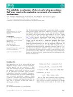
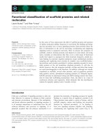
Fig. 1. Subunit composition of native Arenicola haemoglobin (Hb). (A) MaxEnt-processed electrospray ionization (ESI)-MS spectrum of dena-
turated Arenicola Hb. The insets show the details of monomeric chains (I), linker subunits unobserved previously (II), trimeric globin complex
T and the homodimer D
1
(III). (B) Left lane: SDS ⁄ PAGE of unreduced Arenicola Hb which confirms the presence of the three groups of sub-
units: I, II and III. (B) Right lane: migration of low molecular weight standards (Amersham). Results of a single representative experiment are
presented.
M. Rousselot et al. Self-assembling properties of A. marina haemoglobin
FEBS Journal 273 (2006) 1582–1596 ª 2006 The Authors Journal compilation ª 2006 FEBS 1583
the unreduced Arenicola Hb revealed three groups of
subunits: I, II and III. Group I consists of the two
monomeric globin chains a
1
and a
2
(15 952 ± 1.0
and 15 975 ± 1.0 Da); a new linker subunit group
(group II) was observed, which is composed of three
constant monomeric chains (23 122 ± 1.0, 24 065 ±
1.0 and 24 219 ± 1.0 Da); and group III is composed
of the five disulfide-bonded globin trimers (49 581 ±
4.0, 49 612 ± 4.0, 49 657 ± 4.0, 49 708 ± 4.0 and
49 750 ± 4.0 Da) and the linker homodimer, D
1
(50 323 ± 4.0 Da).
Spectrophotometric titration of Arenicola Hb
In order to investigate the presence of any pH- or
urea-dependent change surrounding the haem pocket,
the optical spectra (300–700 nm) of Arenicola Hb were
recorded between pH 2.0 and 12 and exposed to an
increasing concentration of urea (1–8 m) for 48 h
(Fig. 2). The absorption spectrum of oxyhaemoglobin
over the range 300–700 nm is not significantly altered
at pH 7.0 over 48 h (Fig. 2A). At acidic pH (Fig. 2B),
the spectrum gradually changes from that of oxyhae-
moglobin to that of methaemoglobin: the Soret band
becomes broader and slightly less intense, with a shift
to a lower wavelength, a decrease in the intensity of
the a (574 nm) and b (540 nm) bands, and the forma-
tion of a distinct absorption at 630 nm and near
500 nm. Spectrophotometric data showed an import-
ant decrease in the intensity of the Soret band, charac-
teristic of haem loss, for pH values of < 3.0 (data not
shown), > 8.0 (Fig. 2C) and in the presence of an
increasing concentration of urea (Fig. 2D).
Gel filtration and SDS ⁄ PAGE patterns of the
dissociated subunits
Figure 3 shows typical gel filtration elution profiles of
partially dissociated Arenicola Hb and Lumbricus Hb
at alkaline pH 8 (Fig. 3A,B, respectively) and in the
presence of 4 m urea at pH 7 (Fig. 3C,D, respectively).
The elution profile of Lumbricus Hb (Fig. 3B,D) is in
agreement with results published previously [14]. In
addition to the undissociated Hb (Fig. 3, HBL), three
peaks corresponding to the dodecamer subunit (D), the
trimer + linker (T+L) subunits, and the monomer
(M) subunit are observed. The Arenicola Hb profile
is different (Fig. 3A,C) because only two peaks are
AU
0.5
1.0
t0h
t3h
t6h
t24h
t48h
500 600400 700
λ (nm)
C
D
t0h
t3h
t6h
t24h
t48h
1
M
2
M
5
M
8
M
500 600400300
0.5
1.0
t0h
t48h
A
B
β
β
β
β
α
α
α
α
Fig. 2. Spectrophotometric titration of Arenicola haemoglobin (Hb). Overlay of UV-visible spectra of Arenicola Hb, dissociated under various
conditions for 48 h (A–C) at ambient temperature: (A) 0.1
M Tris ⁄ HCl buffer at pH 7.0; (B) 0.1 M Tris ⁄ HCl buffer at pH 5.0; and (C) 0.1 M
Tris ⁄ HCl buffer at pH 9.0. (D) Arenicola Hb immediately after exposure to increasing concentrations (1–8 M) of urea at pH 7.0. The arrows
indicate the evolution of the absorbance with time (A–C) or with an increasing concentration of urea (D). AU, absorbance unit. Results are
presented for a single representative experiment.
Self-assembling properties of A. marina haemoglobin M. Rousselot et al.
1584 FEBS Journal 273 (2006) 1582–1596 ª 2006 The Authors Journal compilation ª 2006 FEBS
observed. The nonreduced SDS ⁄ PAGE on collected
fractions (Fig. 3, inset) showed that the initial subunit
content of the first peak (Fig. 3, lanes 1 and 4) is sim-
ilar to that of native Arenicola Hb, corresponding to
undissociated Hb (I
HBL
) (the concentration of each
sample loaded on the gel are slightly different). Peak
I
D
which has the size expected for a putative one-
twelfth of the whole molecule of Arenicola Hb compri-
ses the trimers and the monomers (Fig. 3, lanes 2 and
5), confirming that it corresponds to the dodecamer.
Two additional, less intense, bands are also observed
and they are present in all the other lanes in the mid-
dle of the gel [17]. These bands have previously been
reported for Arenicola Hb as polymerization of the
monomer or partial dissociation of the disulphide-
bounded trimers, during the preparation of the sam-
ples before migration on the gel [18]. Moreover, no
corresponding polypeptide chains were observed dur-
ing MS analysis (see below, Fig. 4A). After the dissoci-
ation of Lumbricus Hb, all the subunits (trimer, linker
and monomer) are present in the dissociated fractions
(lane 7 and 8). The pattern corresponding to dissoci-
ated fractions of Arenicola Hb (Fig. 3, lane 3 and 6)
exhibits alterations with the absence of the bands cor-
responding to the linker subunits. Control experiments
were carried out in the presence of reducing agent or
protease inhibitor and revealed similar gel filtration
and SDS ⁄ PAGE patterns, indicating that the differ-
ences are not the result of degradation by a protease.
Dissociated subunits observed by ESI-MS
Figure 4 shows ESI-MS spectra for dissociated Areni-
cola Hb at alkaline pH. The spectra are similar for the
dissociation in the presence of urea. The deconvoluted
mass spectrum of undissociated Arenicola Hb (Fig. 4A)
is similar to that for the native Arenicola Hb (Fig. 1A).
The dodecamer subunit (Fig. 4A), was found to con-
tain all the subunits T and M, and a small amount of
the linker homodimer D
1
, which had not dissociated
from the dodecamer. The deconvoluted spectrum of
fully dissociated Arenicola Hb (Fig. 4A) reveals the
absence of the linker subunits at 50 319 Da and at
23 122, 24 065 and 24 219 Da and the less intense
Fig. 3. Dissociation patterns of Arenicola haemoglobin (Hb) and Lumbricus Hb. Comparison between the dissociation patterns of Arenicola
Hb and Lumbricus Hb were performed by gel filtration on a Superose 6-C column and followed at 280 nm (broken line) and 414 nm (solid
line), and by unreduced SDS ⁄ PAGE electrophoresis. Arenicola Hb and Lumbricus Hb were analysed immediately after incubation in 0.1
M
Tris ⁄ HCl buffer. (A) Arenicola Hb at pH 8.0; (B) Lumbricus Hb at pH 8.0; (C) Arenicola Hb in 4 M urea at pH 7.0; (D) Lumbricus Hb in 4 M
urea at pH 7.0. The inset shows the unreduced SDS ⁄ PAGE of Arenicola HBL-Hb (lane AmHb)andLumbricus hexagonal bilayer-Hb (HBL-Hb)
(lane LtHb) and of the numbered fractions. The concentrations of each sample loaded on the gel are slightly different. The undissociated
peak is labeled HBL, and the dissociated peaks are the dodecamer D, the trimer and the linker subunits T+L, and the monomer subunit M.
p indicates additional artefactual bands caused by the polymerization of monomers (see the text for details). AU, absorbance unit. Results
are presented for a single representative experiment.
M. Rousselot et al. Self-assembling properties of A. marina haemoglobin
FEBS Journal 273 (2006) 1582–1596 ª 2006 The Authors Journal compilation ª 2006 FEBS 1585
relative intensity of the trimers. These observations are
in agreement with the observation of the disappearance
of linker subunits on the SDS ⁄ PAGE patterns (Fig. 3,
lanes 2, 3, 5 and 6). Moreover, the multicharged
spectra for fully dissociated Arenicola Hb (Fig. 4B)
revealed several new peaks for m ⁄ z < 900, indicating
possible degradation of the protein.
Kinetic of dissociation of Arenicola Hb
Dissociation of Arenicola Hb followed by gel filtration
The extent of dissociation of purified Arenicola Hb
over the pH range 2–12 and at increasing concentra-
tions of urea (from 1 m to 8 m in 0.1 m Tris ⁄ HCl buf-
fer, pH 7.0), at 4 °C for 25 h, was investigated by gel
filtration (Fig. 5). The pH stability curves at three dif-
ferent incubation times is represented in Fig. 5A and
is divided into four sections (a) h, pH < 3.0 and
pH > 12.0: spontaneous release of the haem from the
pocket and simultaneous protein unfolding, (b) d,pH
3.0–4.0 and pH 7.0–12.0: Arenicola Hb dissociation,
(c) p, pH around the isoelectric point (4.0–5.0): Areni-
cola Hb precipitate, and (d) s, pH 5.5–7.0: the quater-
nary structure of Arenicola Hb is maintained. The
dissociation of Arenicola HBL-Hb is a rapid time- and
pH-dependent process at alkaline pH, as revealed by
the slope of the percentage HBL curve (Fig. 5A).
Between 1 and 4 m urea, the dissociation of HBL-Hb
is faster within the first 2 h and slows down to reach
an equilibrium at 20 h (Fig. 5B). The HBL-Hb is
fully dissociated immediately after exposure to 6 m
urea. Figure 6 represents the overlaid chromatograms
of typical elution profiles of dissociated Arenicola Hb
at alkaline pH (Fig. 6A,B) and in 4 m urea (Fig. 6C,D)
at three incubation times. The profiles are similar and
even if the formation of dodecamer is less rapid in
M
L
T
D
1
Undissociated AmHb
Dodecamer
Fully dissociated AmHb
A
Mass (Da)
B
Fully dissociated AmHb
Undissociated AmHb
%
m/z
800
1000 1200 1400 1600 1800 2000 2200 2400
0
100
0
100
%
16000
15953
15976
23000 24000
24065
23122
24219
49600 50000 50400
49659
49612
49559
50319
49709
49753
50274
mass
20000 30000 40000 50000
0
100
%
Fig. 4. ESI-MS profile of dissociation products of Arenicola haemo-
globin (Hb). Electrospray ionization-MS (ESI-MS) analysis of the dis-
sociated subunits of Arenicola Hb (AmHb) after dissociation at
alkaline pH (pH 8.0). The dissociation products were isolated by gel
filtration and prepared as described in the Experimental procedures.
(A) Overlay of the MaxEnt-processed ESI-MS spectrum of undisso-
ciated Arenicola Hb, the dodecamer and of fully dissociated Areni-
cola Hb. The enlarged regions show details of monomeric chains
(M ), trimeric complex (T ) with the homodimer D
1
, and linkers (L )
for undissociated Arenicola Hb. (B) Multicharged spectra of native
and of fully dissociated Arenicola Hb. The degradation products are
framed. Results are presented for a single representative experi-
ment.
0
20
40
60
80
100
2 3 4 5 6 7 8 9 10 11 12 13
pH
Percent of total
h
d
p
s
d
h
isoelectric point
% HBL
t0h
% HBL
t5h
% HBL
t25h
% D
t0h
% D
t5h
% D
t25h
A
0
20
40
60
80
100
01234 756
Urea concentration (M).
Percent of total
% HBL t0h
% HBL t2h
% HBL t20h
% D t0h
% D t2h
% D t20h
% HBL t10h
% D t10h
B
Fig. 5. Kinetics of dissociation of Arenicola haemoglobin (Hb). Time
course of the dissociation of Arenicola Hb (solid line) and of the
dodecamer (D) (broken line) at different incubation time-points, fol-
lowed by gel filtration on a Superose 6-C column. The Hb was
dissociated, as described in the Experimental procedures. The per-
centage of undissociated hexagonal bilayer (HBL) and of the
dodecamer are determined by integrating the chromatogram at
414 nm using the
MILLENIUM software. (A) Dissociation of Arenicola
Hb over the pH range 2–12. h, d, p and s indicate four different
states of Arenicola Hb dissociation as a function of pH (see the text
for details). (B) Dissociation of Arenicola Hb in urea from 1
M to
7
M. Results are presented for a single representative experiment.
Self-assembling properties of A. marina haemoglobin M. Rousselot et al.
1586 FEBS Journal 273 (2006) 1582–1596 ª 2006 The Authors Journal compilation ª 2006 FEBS
urea, its dissociation is faster. As soon as Arenicola
HBL-Hb is fully dissociated, the dodecamer dissociates
slowly (Figs 5 and 6) into smaller subunits containing
haem (absorbance at 414 nm) but also nonhaem-con-
taining fragments, with retention times corresponding
to molecular masses of < 15 kDa (framed Fig. 6B,D).
The ratio of the absorbance A
414
: A
280
of the dode-
camer peak increases during the first hour of the disso-
ciation from 2.75 to 2.85 (A
414
: A
280
native Arenicola
Hb ¼ 2.23). Then, it remains constant to decrease pro-
gressively with time. The same variation is observed
for the two other peaks at alkaline pH and in the pres-
ence of urea.
Effect of divalent cations at alkaline pH
Figure 7 reveals the effect of divalent cations on the
dissociation of Arenicola Hb at alkaline pH immedi-
ately after exposure to the buffer. No dissociation is
observed when Arenicola Hb is diluted in seawater (pH
7.8), while it is almost completely dissociated upon
dilution in 0.1 m Tris ⁄ HCl buffer at pH 7.8. The pres-
ence of Ca
2+
and Mg
2+
either prevents (Fig. 7) or
decreases the extent of dissociation of these molecules
at alkaline pH. While slightly further dissociation is
observed in the presence of EDTA at alkaline pH
(presumably by competitive complexing of the divalent
cations), no significant dissociation occurs at neutral
and acidic pH. A similar experiment was carried out
for the dissociation of Arenicola Hb in 4 m urea in the
Time (min)
t4h
t0h
t24h
t4h
t0h
t24h
30
20
40
10
50
I
D
30
20
40
t5h
t25h
I
HBL
10
t0h
t5h
t25h
t0h
0.00
0.04
0.08
0.00
0.10
0.20
A
414
50
A
B
C
D
pH 8.0
Urea 4
M
A
280
I
D
I
HBL
Fig. 6. Formation of disrupted apoglobin induced by dissociation. Gel filtration elution profile on a Superose 6-C column of dissociated Areni-
cola haemoglobin (Hb) in 0.1
M Tris ⁄ HCl buffer showing the formation of disrupted apoglobins at 280 nm (framed). The haemoglobin is dis-
sociated, as described in the Experimental procedures. Elution profile of Arenicola Hb at (A) 414 nm and (B) 280 nm, immediately after
exposure at pH 8.0 (solid line), after 5 h (broken line) and 25 h (dotted line). Elution profile of Arenicola Hb at (C) 414 nm and (D) 280 nm,
immediately after exposure in 4
M urea at time zero (solid line), after 4 h (broken line) and 24 h (dotted line). The undissociated peak is labe-
led HBL, and the major dissociated peak is the dodecamer D. Results are presented for a single representative result.
0
20
40
60
80
100
120
5,5 6 6,5 7 7,5 8 8,5
pH
% undissociated HBL
Fig. 7. Structure stabilization induced by divalent cations at alkaline
pH. Dissociation of Arenicola haemoglobin (Hb) immediately after
exposure to the buffer, under different conditions over the pH
range 6.0–8.0, followed by gel filtration on a Superose 6-C column.
The percentage of undissociated Hb was determined by integrating
the chromatogram at 414 nm using the
MILLENIUM software and is
represented as a function of pH. The dissociation, expressed as
percentage of undissociated Arenicola Hb, in different conditions,
was shown as follows: diamonds, 0.1
M Tris ⁄ HCl buffer; crosses,
0.1
M Tris ⁄ HCl buffer and 5 mM EDTA; triangles, 0.1 M Tris ⁄ HCl
buffer and 50 m
M Mg
2+
; squares, 0.1 M Tris ⁄ HCl buffer and 50 mM
Ca
2+
and asterisks, sea water (pH 7.8). Results are the means ±
SD for three individual experiments at each point.
M. Rousselot et al. Self-assembling properties of A. marina haemoglobin
FEBS Journal 273 (2006) 1582–1596 ª 2006 The Authors Journal compilation ª 2006 FEBS 1587
presence and absence of Ca
2+
, and no stabilizing effect
was observed (data not shown).
Dissociation pattern followed by MALLS
MALLS analysis of partially dissociated Arenicola Hb
at pH 7.8 yielded profiles shown in Fig. 8, with
molecular mass (Fig. 8A) and gyration radius (RW)
(Fig. 8B) estimated during the elution profile at three
different incubation time-points. The estimated
molecular mass (Fig. 8A) decreases during incubation,
and the polydispersity (estimated by the molar mass
slopes) assumes a downward curvature shape, partic-
ularly for peaks I
HBL
,I
1
and I
2
, characteristic of a less
homogenous population. The polydispersity of the
peak I
HBL
indicates that it includes intermediates of
dissociation which are truncated HBL Arenicola Hb
(Fig. 8). Truncated HBL-Hbs (partially dissociated
HBL-Hb particles lacking one-sixth to one-half of the
HBL structure) are also observed on the transmission
electron microscopy (TEM) images of the I
HBL
frac-
tion purified by gel filtration (Fig. 9A). Even if the
estimated average RW values (Fig. 8B) are close to the
angular variation detection limit of 10 nm, the RW
decreases after 2 h with an important scattering and
increase observed, after 24 h of incubation, for I
1
and I
2
.
Reassembly of HBL structure
The extent of reassociation of Arenicola Hb was
investigated by MALLS after dissociation at alkaline
pH. As scattering intensity is strongly dependent on
particle radius, a small amount of large particules in
the sample would give a large response with the light
scattering detector, although their amount, as meas-
ured by the refractive index (RI) response, is low.
These interesting properties allowed us to observe a
reassembly of Arenicola Hb, which was not so easily
observed using gel filtration only. Figure 10 shows
MALLS representative results obtained with the reas-
sembly of HBL-Hb structures from dissociated Areni-
cola HBL-Hb, immediately after exposure to alkaline
pH 8.0 and 9.0 (Fig. 10A,B respectively) and after 1 h
at pH 8.0 (Fig. 10C). Similar results were observed in
the presence of 4 m urea. While different ionic com-
position buffers at pH 7.0 were tested, the reassembly
was only observed in a buffer containing an ionic
composition similar to that of A. marina blood (see
Experimental procedures), at pH 7.0, and after a very
short dissociation incubation time (< 5 min). The
reassociation is limited, as revealed by the RI profiles
of the I
HBL
peak after reassociation and the propor-
tion of reassociated HBL-Hb (Fig. 10A,B). The obser-
vation of the reassociation is characterized by the
differences of the light scattering signals for the I
HBL
peak, before and after the reassociation (Fig. 10A,B).
The reassociation is not observed after 1 h of dissoci-
ation at alkaline H (Fig. 10C) and is less important
as pH increases (Fig. 10B) and coincides with the
absence of truncated HBL-Hbs (retention time
between 20 and 25 min), as revealed by the MALLS
profile (Fig. 10C). Control experiments using a redu-
cing agent or protease inhibitors during the dissoci-
ation process, did not improve the reassociation. The
reassociation is confirmed by the TEM images of
I
HBL
isolated by gel filtration after the reassociation
15
20
25
30
35
40
Time (min)
0
10
100
Rw (nm)
100
10
1000
Mw (kDa)
I
HBL
I
HBL
I
D
I
D
I
1
I
1
I
2
I
2
A
B
Fig. 8. Evaluation of the molecular weight and gyration radius (RW)
during the dissociation process of Arenicola haemoglobin (Hb). Dis-
sociation of Arenicola Hb in 0.1
M Tris ⁄ HCl buffer followed by multi-
angle laser light scattering (MALLS) during the elution from a gel
exclusion column (Superose 6-C). The solid curve represents the
refractive index (RI) profile overlaid with the dotted curve which
represents the light scattering profile at 90° (LS) versus the retent-
ion time. The RI and LS data have been scaled to make the com-
parison easier. (A) Distribution of the molecular weight values at
different incubation times: molecular mass profiles of Arenicola Hb
are shown immediately after exposure at pH 7.8 (red crosses),
after 2 h of dissociation (black squares) and after 24 h of dissoci-
ation (blue triangles). The RI and LS profiles correspond to a disso-
ciation time of 2 h. (B) Distribution of the gyration radius (RW)
values at different incubation times: RW profiles of Arenicola Hb
immediately after exposure at pH 7.8 (red crosses), after 2 h of dis-
sociation (black squares) and after 24 h of dissociation (blue trian-
gles). Results are presented for a single representative experiment.
Self-assembling properties of A. marina haemoglobin M. Rousselot et al.
1588 FEBS Journal 273 (2006) 1582–1596 ª 2006 The Authors Journal compilation ª 2006 FEBS
process (Fig. 9B). We can distinguish truncated HBL-
Hbs in a more structured conformation than before
reassociation (Fig. 9A) and structured HBL-Hb sim-
ilar to native Arenicola HBL-Hb (Fig. 9C).
Discussion
The structural data (Fig. 1) confirmed published data
on native Arenicola Hb to some extent [4], but also
revealed some differences. One difference is the
absence of the heterodimer D
2
(51981 ± 4.0 Da) and
the observation of smaller chains, of 24 kDa, which
might correspond to the putative linker L
2
or to lin-
kers that were not previously observed [4]. The linkers
are cysteine-rich proteins which, in A. marina [19] as in
Riftia pachyptila [20], were found to bind H
2
Sat
slightly alkaline pH, resulting in the formation of per-
sulfides for detoxification purpose in nonsymbiotic spe-
cies. However, the role of cysteines in binding H
2
S
appears to be controversial, as revealed by recent stud-
ies [21,22], and is still under active investigation. In an
acidic environment, as used for ESI-MS analysis under
denaturing conditions, H
2
S is released and some rear-
rangement could occur, resulting in a possible cleavage
of the heterodimer, D
2
, into smaller subunits. More-
over, the animals used to collect blood were obviously
different from those used in previous studies, and it is
possible that different alleles exist in different popula-
tions of A. marina.
A complex mechanism of dissociation
Dissociation profile of Arenicola Hb
The dissociation of Arenicola Hb was investigated in
detail at alkaline pH and in the presence of urea. Our
results are in agreement with studies by Daniel and
collaborators [23] who found that Arenicola Hb is less
stable at alkaline pH than Lumbricus Hb. Extracellular
annelid Hbs usually dissociate at pH ‡ 8.0 [13,24]
according to an equilibrium process, as observed in
L. terrestris and Tubifex tubifex Hbs [24]. The peculi-
arity of A. marina extracellular Hb is that the dissoci-
ation occurs even at pH values between 7.0 and 8.0, in
a buffer that does not contain any other ions, such as
alkaline earth cations (Figs 5A and 7). In addition, this
is not an equilibrium process. Indeed, the dissociation
is almost immediately complete at pH 8.0 and is time-
dependent (Figs 5 and 8). The dissociation profiles of
Arenicola Hb in urea are similar (Figs 6 and 8), sug-
gesting that the mechanism of dissociation is common
to both denaturing treatments, even if the kinetics are
different. The formation of the dodecamer is faster at
alkaline pH and its dissociation occurs more rapidly
in the presence of urea (Fig. 6). This reveals the
importance of hydrogen bonds in the structure of the
dodecamer. Several simultaneous dissociations of an
HBL-Hb structure can be envisioned, as proposed for
Lumbricus Hb dissociation [14]. However, the dissoci-
ation process of Arenicola Hb is more complex to
AB
C
Fig. 9. Electron micrographs of Arenicola haemoglobin (Hb), before and after reassociation. Electron micrographs of Arenicola Hb, negatively
stained showing self-association properties of Arenicola Hb. (A) View of truncated HBL (I
d
) and dodecamers (D)ofArenicola Hb isolated by
gel filtration after dissociation. (B) View of reassociated Arenicola Hb (peak I
HBL
) isolated by gel filtration; top (t) and side views (s) and of par-
tially reassociated Arenicola Hb (Ir) isolated by gel filtration. (C) View of native Arenicola Hb; top (t) and side (s) views. Scale bar, 100 nm.
Results are presented for a single representative experiment after dissociation at alkaline pH.
M. Rousselot et al. Self-assembling properties of A. marina haemoglobin
FEBS Journal 273 (2006) 1582–1596 ª 2006 The Authors Journal compilation ª 2006 FEBS 1589
interpret. The quaternary structure is rapidly affected
at alkaline pH or in the presence of urea (Fig. 5). The
dissociation leads to the rapid formation of the one-
twelth protomers (D+L) through truncated HBLs.
Indeed, results from gel filtration, MALLS and TEM
analyses reveal the presence of a small amount of trun-
cated HBLs at the early stage of the dissociation pro-
cess (Figs 8, 9A and 10) and the formation of one
major peak, I
D
(Figs 3A,B and 6), interpreted as the
dodecamer, according to structural analysis (Fig. 3,
lanes 2 and 5, Fig. 4A). However, the higher molecular
mass of peak I
D
(MALLS results, Fig. 8A), the pres-
ence of D
1
on the ESI-MS spectra of the dodecamers
(Fig. 4A), and the A
414
: A
280
value, which increases
during the first incubation hour for peak I
D
(Fig. 6),
all indicate that the dodecamer is still associated with
linkers at the start of the dissociation. Then, the lin-
kers dissociate from the dodecamer, resulting in a
decrease of molecular mass (peak I
D,
Fig. 8A). The do-
decamer does not dissociate into stable trimers and
monomers, as observed for Lumbricus Hb [14], but
into higher molecular mass units (peaks I
1
and I
2
,
Fig. 8A), in low abundance and transitory. The dena-
turation of these subunits is evident from the variation
of the RW value (Fig. 8B). The RW increases for I
1
and I
2
, while the molecular mass decreases after 24 h
of dissociation. These variations of RW are character-
istic of an extended unfolded conformation during the
dissociation process. The decrease of RW after 2 h of
dissociation is explained by the formation of smaller
subunits with smaller radius. The important scatter is
caused by the presence of a mix of small structured
subunits and small destructed subunits, which have a
higher RW value. After 24 h of dissociation, most of
these dissociated subunits are denaturated, so the scat-
ter is less important.
Structural alterations of Arenicola Hb
UV-visible spectroscopy around the Soret band provi-
ded information about the haem environment. An
observation by Ascoli and collaborators [25] suggested
that oxidation of earthworm Hb affected its quaternary
structure, leading to dissociation. In Arenicola Hb,
however, by comparing the dissociation profiles at alka-
line and acidic pH (Fig. 5A) and the light absorp-
tion spectrum (especially between 500 and 700 nm)
(Fig. 2B,C), it appears that the spectral changes are
only partially related to the dissociation. Indeed, at
pH 8.0 and above, the extensive dissociation of Arenico-
la Hb was accompanied by a relatively small change in
the visible absorption region of the spectra (Figs 2C
and 5A) and the methaemoglobin formation (at
pH 6.0) is not accompanied by an extensive dissociation
(Figs 2B and 5A). The dissociation pattern of Arenicola
Hb is similar in the presence of a reducing agent, con-
firming that dissociation is not induced by oxidation of
15
20
25
30
35
10
100
1000
10
100
1000
10
100
1000
Mw (kDa)
Time (min)
I
HBL
I
D
A
B
C
16 %
9 %
2 %
4 %
0 %0 %
Fig. 10. Self-association properties of Arenicola haemoglobin (Hb).
Self-association properties of Arenicola Hb followed by multiangle
laser light scattering (MALLS) detection during elution on a gel-
exclusion column (Superose 6-C). The solid curve represents the
refractive index (RI) profile overlaid with the dotted curve which
represents the light scattering profile at 90° (LS) versus the retent-
ion time. The red curves represent Arenicola Hb after dissociation
and the blue curves represent Arenicola Hb after reassociation. (A)
Arenicola Hb immediately after exposure in 0.1
M Tris ⁄ HCl buffer,
pH 8.0, and immediately reassociated. (B) Arenicola Hb dissociated
immediately after exposure in 0.1
M Tris ⁄ HCl buffer, pH 9.0, and
immediately reassociated. (C) Arenicola Hb dissociated in 0.1
M
Tris ⁄ HCl buffer, pH 8.0, after 1 h and reassociated. The percent-
ages of HBL are indicated after dissociation (red) and after reassoci-
ation (blue). They are calculated from the integration of HPLC
chromatogram at 414 nm. Results are presented for a single repre-
sentative experiment.
Self-assembling properties of A. marina haemoglobin M. Rousselot et al.
1590 FEBS Journal 273 (2006) 1582–1596 ª 2006 The Authors Journal compilation ª 2006 FEBS
the haem. The important decrease of the Soret band
observed at alkaline pH with time (Fig. 2C) and in the
presence of an increasing concentration of urea
(Fig. 2D), reveals a significant alteration in the haem
pocket, leading to a dissociation of haem from the hae-
moglobin. These analyses revealed that the denatura-
tion is accompanied by local changes in the haem
cavity, potentially having profound effects on the pro-
tein structure, as it is known that haem clearly stabilizes
intact myoglobins and haemoglobins with respect to the
apoglobins [26–28]. The formation of apoglobin and its
degradation are confirmed by the following observa-
tions, namely (a) the decrease of the A
414
: A
280
of each
elution peak, which is characteristic of a loss of haem
and (b) the increasing formation of nonhaem-contain-
ing subunits, observed by gel filtration for both dissoci-
ation processes (Fig. 6B,D). These nonhaem, smaller,
products (Fig. 6) are interpreted as degradation prod-
ucts of the subunits, as they do not correspond to unfol-
ded linkers (which should elute later or we should see
them by SDS ⁄ PAGE (Fig. 3, lanes 3 and 6) and ESI-
MS (fully dissociated haemoglobin, Fig. 4A). Finally,
the degradation products on the ESI-MS multicharged
spectra of fully dissociated Arenicola Hb associated
with a less intense signal for the disulphide-bounded
trimers (Fig. 4b). The removal of haem is followed by
proteolytic degradation of the apoglobin, perhaps initi-
ated by the presence of free hemin, which has been
reported to enhance oxidant-mediated damage [29].
Disappearance of the linkers
The linkers are thought to be degraded during the dis-
sociation process. Indeed, they are not observed by
SDS ⁄ PAGE (Fig. 3, lanes 3 and 6) or on the ESI-MS
spectra (Fig. 4A) of fully dissociated Arenicola Hb.
Recently, Suzuki & Riggs [30] and Chabasse et al. [31]
showed that Arenicola linker chains possess a con-
served cysteine-rich domain [a low-density lipoprotein
A (LDL-A) module] homologous to the cysteine-rich
region of the ligand-binding domain of the low-den-
sity-lipoprotein receptor (LDLR) family [30,31]. Stud-
ies investigating free hemin-induced modifications in
LDL revealed that hemin associates with LDL and
undergoes oxidative breakdown, releasing free iron,
which is well known to catalyze oxidant degradation
[32]. The haem dissociates easily from Arenicola Hb
after dissociation at alkaline pH or in the presence of
urea (Fig. 2C,D). The product of hemin peroxidation
was found to be either aggregation or fragmentation
[33,34]. Aggregation of linkers has previously been
observed for Lumbricus Hb [5], and could attenuate
the volatilization into the gas phase necessary for
observation by ESI-MS (B. N. Green, and S. N.
Vinogradov, personal communication). However, we
should then observe bands of higher molecular mass
on the SDS ⁄ PAGE gel (Fig. 3, lanes 3 and 6). Further
studies on the identification and characterization of
Arenicola Hb subunits isolated by preparative gel elec-
trophoresis using a proteomics approach (M. Rousse-
lot et al., unpublished data) revealed that molecular
mass bands (< 15 kDa) observed after dissociation of
Arenicola Hb at alkaline pH or in the presence of urea,
are composed of globins and also of linker fragments
that are not observed for the native Arenicola Hb
SDS ⁄ PAGE pattern. This confirmed that the dissoci-
ation of Arenicola Hb at alkaline pH or in the presence
of urea, induces fragmentation of the linker chains,
probably as a result of their oxidation in the presence
of free hemin.
The effect of potential protease was considered and
control experiments using protease inhibitor were per-
formed; the linker still disappeared during dissociation,
as evidenced by SDS ⁄ PAGE and ESI-MS experiments
(data not shown). The same phenomenon was
observed in the presence of a reducing agent. The dis-
appearance or the severe reduction in the relative
intensities of the linker chains from the ESI-MS spec-
tra has previously been observed in Eudistylia chloro-
cruorin [35] and in other HBL-Hbs (B. N. Green,
personal communications).
Stabilizing effect of divalent cations at alkaline pH
Arenicola Hb is stable at slightly alkaline pH (pH 7–8)
when salts are present at concentrations similar to phy-
siological concentrations. Among those salts that are
important for structure, alkaline earth cations (Ca
2+
and Mg
2+
) play a major role (Fig. 7). These cations
also stabilize the HBL structure of other annelid Hbs
with respect to dissociation at alkaline pH [2,13,36,37].
In contrast to Amphitrite Hb [38] and Myxicola chlo-
rocruorin [39], divalent cations are not necessary to
maintain the HBL-Hb structure at neutral pH, even in
the presence of EDTA. The divalent cations probably
scavenge side-chain anionic groups ionized at alkaline
pH. Moreover, LDL-A modules, found on linker
chains, possess a cluster of four conserved acidic resi-
dues [31], which may be involved in calcium-dependent
protein folding [40].
A limited association–dissociation equilibrium
At alkaline pH values, annelid extracellular Hbs disso-
ciate irreversibly into one-twelfth of the whole molecule
[41,42]. However, extracellular Hbs from the
M. Rousselot et al. Self-assembling properties of A. marina haemoglobin
FEBS Journal 273 (2006) 1582–1596 ª 2006 The Authors Journal compilation ª 2006 FEBS 1591
earthworm L. terrestris and T. tubifex dissociate fur-
ther into several smaller subunits, which are in asso-
ciation–dissociation equilibrium with one another
[6,13,14,24,43,44]. After dissociation of Arenicola Hb,
some rearrangements are observed when the sample is
returned to neutral pH with a salt composition similar
to its physiological fluid. This rearrangement is only
observed in the presence of partially dissociated HBL-
Hbs and the one-twelth protomers and led to a recov-
ery of the structure of the one-twelth protomers at
different degrees of polymerization, and up to a com-
pletely reassociated HBL-Hb (Fig. 9B). In the absence
of these structured subunits (truncated HBL-Hbs or
one-twelth protomers), no reassociation is observed:
the smaller subunits lost the ability to reassemble to
form whole Hb molecules, probably as a result of the
fragmentation of linkers essential to maintain the qua-
ternary structure [3,5], and the formation of apoglobins
with important structural alteration. Control experi-
ments using protease inhibitor and reducing agent dur-
ing the dissociation process were performed in order to
evaluate the potential effect on linker and globin degra-
dation, but the reassociation was not improved.
Intersubunit contacts
At alkaline pH and even more with urea, Lumbricus
Hb dissociates to form dodecamers which further dis-
sociate into trimers, linkers and monomers [13,36].
This pattern contrasts with that observed for Arenicola
Hb: the HBL-Hb is preferentially dissociated at alka-
line pH, where the dodecamer is stabilized compared
with urea-induced dissociation (Fig. 6). Even if the
overall structure of the HBL-Hbs is similar, the disso-
ciation analyses reveal that the nature of the major
intersubunit contacts, which contribute to the quater-
nary structure, is different. Comparatively, Lumbricus
Hb is less stable in the presence of urea, suggesting
that the interactions involved in the quaternary struc-
ture here are rather hydrogen bonds. This view is sup-
ported by the ease of the dissociation of Arenicola Hb
at slightly alkaline pH (pH 7–8, Fig. 5A) with the sta-
bilizing effect observed in the presence of divalent cati-
ons over this pH range, while Lumbricus Hb remains
structured. This suggests salt bridge interactions
between ionic side-chains, but also that salts of the
physiological fluid are mainly involved the quaternary
structure of Arenicola Hb. Incidentally, the blood of
A. marina has higher concentrations in salts, including
divalent cations, than L. terrestris blood [45].
A comparison from the 3D volume reconstruction
by cryoelectron microscopy and X-ray crystallogra-
phy of Arenicola [46] and Lumbricus Hbs [3,46],
shows that their bilayered architecture mainly differs
by the offset of the hexagonal layers. Interlayer con-
tacts are different in the two Hbs, leading to a 16°
rotation of the two hexagonal layers in Lumbricus
Hb, while they are eclipsed in Arenicola Hb [46].
The distance between the hexagonal layers is larger
in Arenicola Hb than in Lumbricus Hb [46], thus
the haemoglobin : haemoglobin contact between one-
twelth protomers of the opposite layers observed in
Lumbricus Hb are weaker in Arenicola Hb – if they
exist – and the surface contact of the linkers to the
solvent is larger in Arenicola Hb. In addition, the
linker–linker interaction seems stronger in Arenicola
Hb than in Lumbricus Hb [46]. All these data sug-
gest that the linker–linker interlayer contacts, which
play a central role in the assembly of the quaternary
structure are mostly hydrogen bonds in Lumbricus
Hb and salt bridges in Arenicola Hb.
The globins and linkers are glycosylated in Lumbri-
cus Hb [47] but the polypeptide chains in Arenicola Hb
are not glycosylated [4,48]. Work on Lumbricus Hb
suggests that carbohydrate-gluing, mediated by lectin-
like interactions, could help maintain the quaternary
structure [48].
In conclusion, we have shown that Arenicola Hb is
able to reassociate after dissociation at alkaline pH.
However, its dissociation follows a novel and complex
mechanism, different from that previously reported for
other extracellular annelid Hbs; Arenicola Hb is less
tolerant to pH and salt variations, which induced rapid
degradation of the complex (fragmentation of the lin-
ker and apoglobin formation). A parallel study on the
dissociation of Lumbricus Hb revealed that the nature
of the intersubunit contacts, essential in the preserva-
tion of the quaternary structure, is different. The disso-
ciation pattern suggests the importance of the salt
bridge interactions in the stabilization of the quater-
nary structure and the importance of the salt composi-
tion of the buffer to maintain the integrity of
Arenicola Hb at slightly alkaline pH. These conclu-
sions are of prime importance for storage and trans-
fusion conditions, considering the development of
Arenicola Hb as blood substitute, for which the med-
ium required is different from A. marina physiological
conditions.
Experimental procedures
Animal collection
Individuals of A. marina were collected at low tide from a
sandy shore near Roscoff (Penpoull beach), Nord Finiste
`
re,
France, by the crews of the marine station facilities, and
Self-assembling properties of A. marina haemoglobin M. Rousselot et al.
1592 FEBS Journal 273 (2006) 1582–1596 ª 2006 The Authors Journal compilation ª 2006 FEBS
kept in local running sea water for 24 h. Blood samples
were withdrawn from the lugworm’s ventral vessel into a
needle and centrifuged (15 min, 10 000 g,4°C) to remove
insoluble material. The blood extraction was performed at
4 °CinanArenicola saline buffer compatible for trans-
fusion to vertebrates (4 mm KCl, 145 mm NaCl, 0.2 mm
MgCl
2
and 10 mm Hepes ⁄ 0.1 m NaOH, pH 7.0), in the
presence of a commercial protease inhibitor cocktail (Com-
plete; Roche, Basel, Switzerland). The resulting samples
were purified immediately and kept frozen ()40 °C) until
use. Purified Lumbricus Hb was kindly provided by
S. Vinogradov (Wayne State University, MI, USA).
Purification techniques
Analytical gel filtration was performed on a 1 · 30-cm
Superose 6-C (fractionation range: 5–5000 kDa) and
Superose 12-C (fractionation range: 1–300 kDa) column
(Amersham Biosciences Biotechnology, Uppsala, Sweden)
using a high-pressure HPLC system (Waters, Milford, MA,
USA). The column was equilibrated with the Arenicola
saline buffer. Flow rates were typically 0.5 mLÆmin
)1
, and
the absorbance of the eluate was monitored at 280 nm and
414 nm. The peaks were collected separately and concentra-
ted by centrifugation on an Amicon Ultra-10 concentrator,
cut-off molecular weight 10 kDa (Millipore, Billerica, MA,
USA). One or two further purifications, using the same
protocol, were performed to obtain pure fractions. Haem
content and protein concentration were determined as
described previously [4]. The samples were always kept at
4 °C except during chromatographic analyses, which were
performed at ambient temperature (20–22 °C).
Spectrophotometry
The absorption spectra, as a function of pH and urea con-
centration over the 300–700 nm range, were obtained using
a UV mc2 spectrophotometer (SAFAS, Monaco). The ana-
lyses were performed at room temperature at a concentra-
tion of Arenicola Hb of 0.5 mgÆmL
)1
.
SDS/PAGE
PAGE in the presence of 0.1% (w ⁄ v) SDS was carried out
using the Laemmli discontinuous buffer system [49] and
slab gels (0.75 mm · 10 cm · 8 cm) of 15% (v ⁄ v) acryl-
amide. The gels were electrophoresed for 1–2 h and
stained in Coomassie Brillant Blue R-250, as described
previously [4].
ESI-MS
ESI-MS was performed under denaturing conditions to
determine the molecular masses of the subunits of Arenicola
Hb. Electrospray data were acquired on a Q-Tof II (Micro-
mass, Altrincham, UK), scanning over the m ⁄ z range 600–
2500 at 10 s per scan, and 45 scans were averaged to
produce the final spectrum. Samples were desalted by wash-
ing with MilliQ water, repeated 10 times on Amicon-3 kDa
at 4 °C. Protein concentrations of 0.5 mgÆmL
)1
in acetonit-
rile ⁄ water (1 : 1, v ⁄ v) containing 0.2% (v ⁄ v) formic acid,
were introduced into the electrospray source at 5 lLÆmin
)1
.
The cone voltage (counter electrode to skimmer voltage)
ramp was from 60 V at m ⁄ z 600 to 120 V at m ⁄ z 2500. The
raw multicharged spectra were deconvoluted using maxi-
mum entropy-based software (maxent) supplied with the
instrument [50]. Mass scale calibration was established
using the multiply charged series from horse heart myoglo-
bin (molecular mass 16 951.5 Da; M-1882; Sigma, St Louis,
MO, USA). Molecular masses were based on the atomic
weights of the elements given by the International Union of
Pure and Applied Chemistry.
MALLS
MALLS measurements were performed using a DAWN
EOS system (Wyatt Technology Corp., Santa Barbara,
CA, USA) directly on-line with the HPLC system. The 18
discrete photodetectors are spaced around the flow cell and
enable simultaneous measurements to be made over a
range from 15° to 160°. The eluate was simultaneously
monitored with a photodiode array detector (Waters 2996,
Waters, Hilford, MA, USA) and an RI detector (Waters
2414, Waters). The MALLS instrument was placed directly
before the refractometer and after the Superose 6-C col-
umn and UV detector to avoid backpressure on the RI
cell. Chromatographic data were collected and processed
using the astra software (Wyatt Technology Corp.). The
Zimm fit method was used for molecular mass determina-
tions [51]. In the calculations, a dn ⁄ dc value of 0.19 mLÆg
)1
was used, typical of nonglycosylated proteins. BSA mono-
mer (Sigma) was used for normalizing various detectors’
signals relative to the 90° detector signal.
Dissociation experiments
The dissociating agent (urea; Sigma) was dissolved in 0.1 m
Tris ⁄ HCl ⁄ 0.1 m HCl buffer, pH 7.0, containing 1 mm
EDTA, and Arenicola Hb stock solution was added to
achieve a final concentration of 4 mgÆmL
)1
. The Hb was
dissociated by exposure for up to 48 h to 0.1 m Tris ⁄ HCl
buffer at alkaline pH (from 7.0 to 12.0). For acidic pHs,
Arenicola Hb was exposed, for up to 48 h, to 0.1 m sodium
formate ⁄ 0.01 m formic acid, pH 3.0–4.5, to 0.1 m sodium
acetate ⁄ 0.01 m acetic acid, pH 4.5–5.5 and to 0.1 m
Tris ⁄ HCl ⁄ 0.1 m HCl, pH 5.5–7.0. Further control dissoci-
ation experiments were carried out in the presence of prote-
ase inhibitor cocktail (Complete; Roche) or reducing agent
M. Rousselot et al. Self-assembling properties of A. marina haemoglobin
FEBS Journal 273 (2006) 1582–1596 ª 2006 The Authors Journal compilation ª 2006 FEBS 1593
(0.5 mm NADH and 2 mm butylhydroxyltoluene (BHT);
Sigma). Dissociations of Arenicola Hb were followed by
HPLC at neutral pH on the system described above. Each
chromatogram was collected and processed using the mil-
lenium software (Waters) supplied with the instrument. All
the dissociation experiments were carried out at 4 °C.
Reassembly of HBL structure from dissociated
Arenicola Hb
The completeness of the dissociation was checked by
HPLC. The Arenicola Hb exposed to either alkaline pH or
urea, was dialysed overnight at 4 °C against 2 · 1 L of sal-
ine buffer with a salt composition similar to that of the
physiological fluid of A. marina: 400 mm NaCl, 3 mm KCl,
32 mm MgSO
4
,11mm CaCl
2
and 0.1 m Tris ⁄ HCl ⁄ 0.1 m
HCl at pH 7.0. The reassociation, achieved at 4 °C, was
followed by HPLC and MALLS at neutral pH.
TEM
Arenicola Hb was diluted to 0.1 mgÆmL
)1
with 0.1 m
Tris ⁄ HCl buffer, pH 7.0, and applied to a very thin carbon
substrate supported on a microgrid, stained with 2% (w ⁄ v)
uranyl acetate solution, as described previously [52]. The
specimens were examined at 80 kV, using a Jeol JEM-
1200EX transmission electron microscope (JEOL, Tokyo,
Japan).
Acknowledgements
Funding for this project was provided by European
grant (FEDER N°presage 3814) and the Conseil
Re
´
gional de Bretagne (PRIR A2C809 and BDI grants
from CNRS and Conseil Re
´
gional de Bretagne
A2CAI1 (MR)). We thank S. Vinogradov for provi-
ding us with Lumbricus Hb, A.Toulmond, S. Hourdez
and the referees for their critical reading of the manu-
script. We also thank A. Andersen for the transmission
electron micrographs explanations and the crews of the
marine station facilities for providing us with Arenicola
marina. ESI-MS results were obtained thanks to the
instruments of SIG core facility of Ouest-genopoleÒ
Plateform.
References
1 Vinogradov SN, Sharma PK & Walz DA (1991) Iron
and heme contents of the extracellular hemoglobins and
chlorocruorins of annelids. Comp Biochem Physiol B 98,
187–194.
2 Lamy JN, Green BN, Toulmond A, Wall JS, Weber RE
& Vinogradov SN (1996) Giant hexagonal bilayer
hemoglobins. Chem Rev 96, 3113–3124.
3 Royer WE Jr, Strand K, van Heel M & Hendrickson
WA (2000) Structural hierarchy in erythrocruorin, the
giant respiratory assemblage of annelids. Proc Natl
Acad Sci USA 97, 7107–7111.
4 Zal F, Green BN, Lallier FH, Vinogradov SN & Toul-
mond A (1997) Quaternary structure of the extracellular
haemoglobin of the lugworm Arenicola marina: a multi-
angle-laser-light-scattering and electrospray-ionisation-
mass-spectrometry analysis. Eur J Biochem 243, 85–92.
5 Kuchumov AR, Taveau JC, Lamy JN, Wall JS, Weber
RE & Vinogradov SN (1999) The role of linkers in the
reassembly of the 3.6 MDa hexagonal bilayer hemoglo-
bin from Lumbricus terrestris. J Mol Biol 289, 1361–1374.
6 Zhu H, Ownby DW, Riggs CK, Nolasco NJ, Stoops JK
& Riggs AF (1996) Assembly of the gigantic hemoglo-
bin of the earthworm Lumbricus terrestris. Roles of sub-
unit equilibria, non-globin linker chains, and valence of
the heme iron. J Biol Chem 271, 30007–30021.
7 Green BN, Bordoli RS, Hanin LG, Lallier FH, Toul-
mond A & Vinogradov SN (1999) Electrospray ioniza-
tion mass spectrometric determination of the molecular
mass of the approximately 200-kDa globin dodecamer
subassemblies in hexagonal bilayer hemoglobins. J Biol
Chem 274, 28206–28212.
8 Kihm AJ, Kong Y, Hong W, Russell JE, Rouda S,
Adachi K, Simon MC, Blobel GA & Weiss MJ (2002)
An abundant erythroid protein that stabilizes free
alpha-haemoglobin. Nature 417, 758–763.
9 Weber RE & Vinogradov SN (2001) Nonvertebrate
hemoglobins: functions and molecular adaptations.
Physiol Rev 81, 569–628.
10 Vinogradov SN, Shlom JM & Doyle M (1979) Dissocia-
tion of the extracellular hemoglobin of Arenicola marina.
Comp Biochem Physiol 65B, 145–150.
11 Roche J, Wurmser S, Fine JM & Autran R (1963)
[Recent Research on the Electron Microscopic Study of
the Hemoglobin (Erythrocruorine) of Arenicola Marina
L. & Its Dissociation.]. C R Seances Soc Biol Fil 157,
1419–1421.
12 Zal F, Lallier FH & Toulmond A (2000) French patent
no. 00 ⁄ 07031. Utilisation Comme Substitut Sanguin d’une
He
´
moglobine Extracellulaire de Poids Mole
´
culaire E
´
leve
´
.
International Patent, PCT ⁄ FR01 ⁄ 01505.
13 Kapp OH, Polidori G, Mainwaring MG, Crewe AV &
Vinogradov SN (1984) The reassociation of Lumbricus
terrestris hemoglobin dissociated at alkaline pH. J Biol
Chem 259, 628–639.
14 Sharma PK, Kuchumov AR, Chottard G, Martin PD,
Wall JS & Vinogradov SN (1996) The role of the
dodecamer subunit in the dissociation and reassembly
of the hexagonal bilayer structure of Lumbricus terres-
tris hemoglobin. J Biol Chem 271, 8754–8762.
15 Vinogradov SN, Sharma PK, Qabar AN, Wall JS,
Westrick JA, Simmons JH & Gill SJ (1991) A dodecamer
of globin chains is the principal functional subunit of the
Self-assembling properties of A. marina haemoglobin M. Rousselot et al.
1594 FEBS Journal 273 (2006) 1582–1596 ª 2006 The Authors Journal compilation ª 2006 FEBS
extracellular hemoglobin of Lumbricus terrestris. J Biol
Chem 266, 13091–13096.
16 Strand K, Knapp JE, Bhyravbhatla B & Royer WE Jr
(2004) Crystal structure of the hemoglobin dodecamer
from Lumbricus erythrocruorin: allosteric core of
giant annelid respiratory complexes. J Mol Biol 344,
119–134.
17 Zhao XJ, Raitt DPVB, Clewell AS, Kwast KE & Poy-
ton RO (1996) Function and expression of flavohemo-
globin in Saccharomyces cerevisiae. Evidence for a role
in the oxidative stress response. J Biol Chem 271,
25131–25138.
18 Slitine FE & Toulmond A (1991) Two-dimensional elec-
trophoresis of Arenicola marina extracellular hemoglo-
bin: separation of chains with identical molecular mass
but different isoelectric point. Comp Biochem Physiol B
100, 631–634.
19 Zal F, Gotoh T & Toulmond A (1999) The novel func-
tion of giant hemoglobins from tubeworms and anne-
lids. In Fifth International Congress of Comparative
Physiology and Biochemistry, Vol. 124A (Walsh P, ed.),
p. 516. Comparative Biochemistry and Physiology,
Calgary, Alberta, Canada.
20 Zal F, Leize E, Lallier FH, Toulmond A, Van Dorssel-
aer A & Childress JJ (1998) S-Sulfohemoglobin and di-
sulfide exchange: the mechanisms of sulfide binding by
Riftia pachyptila hemoglobins. Proc Natl Acad Sci USA
95, 8997–9002.
21 Flores JF, Fisher CR, Carney SL, Green BN, Freytag
JK, Schaeffer SW & Royer WE Jr (2005) Sulfide bind-
ing is mediated by zinc ions discovered in the crystal
structure of a hydrothermal vent tubeworm hemoglobin.
Proc Natl Acad Sci USA 102, 2713–2718.
22 Numoto N, Nakagawa T, Kita A, Sasayama Y, Fuku-
mori Y & Miki K (2005) Crystallization and prelimin-
ary X-ray crystallographic analysis of extracellular giant
hemoglobin from pogonophoran Oligobrachia mashikoi.
Biochim Biophys Acta 1750, 173–176.
23 Daniel E, Lustig A, David MM & Tsfadia Y (2003)
Towards a resolution of the long-standing controversy
regarding the molecular mass of extracellular erythro-
cruorin of the earthworm Lumbricus terrestris. Biochim
Biophys Acta 1649, 1–15.
24 Polidori G, Mainwaring M, Kosinski T, Schwarz C,
Fingal R & Vinogradov SN (1984) The dissociation of
the extracellular hemoglobin of Tubifex tubifex at
extremes of pH and its reassociation upon return to
neutrality. Arch Biochem Biophys 233, 800–814.
25 Ascoli F, Rossi Fanelli MR, Chiancone E, Vecchini P &
Antonini E (1978) Studies on erythrocruorin. VI. Ferric
derivatives of earthworm erythrocruorin. J Mol Biol
119, 191–202.
26 Zerovnik E & Lapanje S (1986) Interactions of myoglo-
bin with urea and some alkylureas. I. Solvation in urea
and alkylurea solutions. Biophys Chem 24, 53–59.
27 Crumpton MJ & Polson A (1965) A comparison of the
conformation of sperm whale metmyoglobin with that
of apomyoglobin. J Mol Biol 11, 722–729.
28 Kawahara K, Kirshner AG & Tanford C (1965) Disso-
ciation of human CO-hemoglobin by urea, guanidine
hydrochloride, and other reagents. Biochemistry 4,
1203–1213.
29 Jeney V, Balla J, Yachie A, Varga Z, Vercellotti GM,
Eaton JW & Balla G (2002) Pro-oxidant and cytotoxic
effects of circulating heme. Blood 100, 879–887.
30 Suzuki T & Riggs AF (1993) Linker chain L1 of earth-
worm hemoglobin. Structure of gene and protein:
homology with low density lipoprotein receptor. J Biol
Chem 268, 13548–13555.
31 Reference withdrawn.
32 Miller YI, Felikman Y & Shaklai N (1995) The involve-
ment of low-density lipoprotein in hemin transport
potentiates peroxidative damage. Biochim Biophys Acta
1272, 119–127.
33 Miller YI & Shaklai N (1994) Oxidative crosslinking of
LDL protein induced by hemin: involvement of tyro-
sines. Biochem Mol Biol Int 34, 1121–1129.
34 Paganga G, Rice-Evans C, Rule R & Leake D (1992)
The interaction between ruptured erythrocytes and low-
density lipoproteins. FEBS Lett 303, 154–158.
35 Green BN, Kuchumov AR, Walz DA, Moens L &
Vinogradov SN (1998) A hierarchy of disulfide-bonded
subunits: the quaternary structure of Eudistylia chloro-
cruorin. Biochemistry 37, 6598–6605.
36 Polidori G, Mainwaring MG & Vinogradov SN (1988)
The effect of alkaline earth cations and of ionic strength
on the dissociation of earthworm hemoglobin at alka-
line pH. Comp Biochem Physiol A 89, 541–545.
37 Ochiai T, Hoshina S & Usuki I (1993) Zinc as modula-
tor of oxygenation function and stabilizer of quaternary
structure in earthworm hemoglobin. Biochim Biophys
Acta 1203, 310–314.
38 Chiancone E, Brenowitz M, Ascoli F, Bonaventura C
& Bonaventura J (1980) Amphitrite ornata erythro-
cruorin. I. Structural properties and characterization of
subunit interactions. Biochim Biophys Acta 623, 146–
162.
39 Vinogradov SN, Standley PR, Mainwaring MG, Kapp
OH & Crewe AV (1985) The molecular size of Myxicola
infundibulum chlorocruorin and its subunits. Biochim
Biophys Acta 828, 43–50.
40 Guo Y, Yu X, Rihani K, Wang QY & Rong L
(2004) The role of a conserved acidic residue in
calcium-dependent protein folding for a low density
lipoprotein (LDL)-A module: implications in structure
and function for the LDL receptor superfamily. J Biol
Chem 279, 16629–16637.
41 Chiancone E, Vecchini P, Rossi Fanelli MR & Antonini
E (1972) Studies on erythrocruorin. II. Dissociation of
earthworm erythrocruorin. J Mol Biol 70, 73–84.
M. Rousselot et al. Self-assembling properties of A. marina haemoglobin
FEBS Journal 273 (2006) 1582–1596 ª 2006 The Authors Journal compilation ª 2006 FEBS 1595
42 Frossard P (1982) The erythrocruorin of Eisenia fetida.
II. Properties of the principal subunit. Biochim Biophys
Acta 704, 535–541.
43 Bonafe CF, Villas-Boas M, Suarez MC & Silva JL
(1991) Reassembly of a large multisubunit protein pro-
moted by nonprotein factors. Effects of calcium and
glycerol on the association of extracellular hemoglobin.
J Biol Chem 266, 13210–13216.
44 Mainwaring MG, Lugo SD, Fingal RA, Kapp OH &
Vinogradov SN (1986) The dissociation of the extra-
cellular hemoglobin of Lumbricus terrestris at acid pH
and its reassociation at neutral pH. A new model of
its quaternary structure. J Biol Chem 261, 10899–
10908.
45 Ochiai T & Weber RE (2002) Effects of magnesium and
calcium on the oxygenation reaction of erythrocruorin
from the marine polychaete Arenicola marina and the
terrestrial oligochaete Lumbricus terrestris. Zool Sci 19,
995–1000.
46 Jouan L, Taveau JC, Marco S, Lallier FH & Lamy
JN (2001) Occurrence of two architectural types of
hexagonal bilayer hemoglobin in annelids: comparison
of 3D reconstruction volumes of Arenicola marina and
Lumbricus terrestris hemoglobins. J Mol Biol 305, 757–
771.
47 Martin PD, Kuchumov AR, Green BN, Oliver RW,
Braswell EH, Wall JS & Vinogradov SN (1996) Mass
spectrometric composition and molecular mass of Lum-
bricus terrestris hemoglobin: a refined model of its
quaternary structure. J Mol Biol 255, 154–169.
48 Ebina S, Matsubara K, Nagayama K, Yamaki M &
Gotoh T (1995) Carbohydrate gluing, an architectural
mechanism in the supramolecular structure of an anne-
lid giant hemoglobin. Proc Natl Acad Sci USA 92,
7367–7371.
49 Laemmli UK (1970) Cleavage of structural proteins
during the assembly of the head of bacteriophage T4.
Nature 227, 680–685.
50 Ferrige AG, Seddon MJ, Green BN, Jarvis SA & Skil-
ling J (1992) Disentangling electrospray spectra with
maximum entropy. Rapid Commun Mass Spectrom 6,
707–711.
51 Zimm BH (1948) The scattering of light and the radial
distribution function of high polymer solutions. J Chem
Phys 16, 1093–1099.
52 Valentine RC, Shapiro BM & Stadtman ER (1968) Reg-
ulation of glutamine synthetase. XII. Electron micro-
scopy of the enzyme from Escherichia coli. Biochemistry
7, 2143–2152.
Self-assembling properties of A. marina haemoglobin M. Rousselot et al.
1596 FEBS Journal 273 (2006) 1582–1596 ª 2006 The Authors Journal compilation ª 2006 FEBS