Báo cáo khoa học: Metallothioneins are multipurpose neuroprotectants during brain pathology potx
Bạn đang xem bản rút gọn của tài liệu. Xem và tải ngay bản đầy đủ của tài liệu tại đây (429.17 KB, 14 trang )
REVIEW ARTICLE
Metallothioneins are multipurpose neuroprotectants
during brain pathology
Milena Penkowa
Section of Neuroprotection, Centre of Inflammation and Metabolism at The Faculty of Health Sciences, University of Copenhagen, Denmark
Mammalian metallothioneins (MTs) constitute a super-
family of nonenzymatic polypeptides (61–68 amino
acids), which are characterized by low molecular
weight (6–7 kDa), distinctive amino acid composition
(high cysteine content and no or low histidine) and
sequence (unique cysteine distribution as Cys-X-Cys),
and a high content of sulfur and metals (metal thiolate
clusters) [1–3]. In vivo, the metal-binding involves
mainly Zn(II), Cu(I), Cd(II), and Hg(II), while in vitro
additional and diverse metals such as Ag(I), Au(I),
Bi(III), Co(II), Fe(II), Pb(II), Pt(II), and Tc(IV) may
be bound to apothionein (the metal-free form) [4,5].
However, during physiological conditions mammalian
MTs mostly contain zinc [6,7].
Keywords
angiogenesis; antioxidants; apoptosis;
defense; inflammation; metalloproteins;
neuroregeneration; pharmacology
Correspondence
M. Penkowa, Section of Neuroprotection,
The Faculty of Health Sciences, University
of Copenhagen, Blegdamsvej 3, DK-2200,
Copenhagen, Denmark
Fax: +45 3 5327217
Tel: +45 3 5327222
E-mail:
(Received 15 January 2006, revised 23
February 2006, accepted 28 February 2006)
doi:10.1111/j.1742-4658.2006.05207.x
Metallothioneins (MTs) constitute a family of cysteine-rich metalloproteins
involved in cytoprotection during pathology. In mammals there are four
isoforms (MT-I ) IV), of which MT-I and -II (MT-I + II) are the best
characterized MT proteins in the brain. Accumulating studies have demon-
strated MT-I + II as multipurpose factors important for host defense
responses, immunoregulation, cell survival and brain repair. This review
will focus on expression and roles of MT-I + II in the disordered
brain. Initially, studies of genetically modified mice with MT-I + II defi-
ciency or endogenous MT-I overexpression demonstrated the importance
of MT-I + II for coping with brain pathology. In addition, exogenous
MT-I or MT-II injected intraperitoneally is able to promote similar effects
as those of endogenous MT-I + II, which indicates that MT-I + II have
both extra- and intracellular actions. In injured brain, MT-I + II inhibit
macrophages, T lymphocytes and their formation of interleukins, tumor
necrosis factor-a, matrix metalloproteinases, and reactive oxygen species.
In addition, MT-I + II enhance cell cycle progression, mitosis and cell sur-
vival, while neuronal apoptosis is inhibited. The precise mechanisms down-
stream of MT-I + II have not been fully established, but convincing data
show that MT-I + II are essential for coping with neuropathology and for
brain recovery. As MT-I and ⁄ or MT-II compounds are well tolerated, they
may provide a potential therapy for a range of brain disorders.
Abbreviations
AD, Alzheimer’s disease; ALS, amyotrophic lateral sclerosis; 6-AN, 6-aminonicotinamide; ARE, antioxidant response element; BDNF, brain-
derived neurotrophic factor; EAE, experimental autoimmune encephalomyelitis; FGF, fibroblast growth factor; FGF-R, FGF-receptor; GDNF,
glial-derived neurotrophic factor; IL, interleukin; IL-6KO mice, IL-6 knockout mice (genetic IL-6-deficient mice); M-CSF, macrophage colony-
stimulating factor; MMP, matrix metalloproteinase; MRE, metal response elements ; MS, multiple sclerosis; MT, metallothionein; MTF-1,
MRE-binding transcription factor-1; MT-KO mice, MT-I + II knock-out mice (genetic MT-I + II deficiency); MT-III ⁄ GIF, metallothionein
III ⁄ growth inhibitory factor; NFjB, nuclear factor kappa-B (transcription factor); NGF, nerve growth factor; NT, neurotrophin; PD, Parkinson’s
disease; ROS, reactive oxygen species; SOD, superoxide dismutase; TGF-b, transforming growth factor-b;TGF-b-R, TGF-b receptor; TgMT
mice, mice with transgenic MT-I overexpression; TNF-a, tumor necrosis factor-a; VEGF, vascular endothelial growth factor.
FEBS Journal 273 (2006) 1857–1870 ª 2006 The Author Journal compilation ª 2006 FEBS 1857
In mammals, four major subfamilies exist (MT-I,
MT-II, MT-III and MT-IV), of which MT-I and -II
(MT-I + II) were discovered in 1957 and are the
best described MT proteins. The roles of mammalian
MT-I + II in the brain have received mounting scien-
tific interest [1,8–10] and are also the focus of this
review, which will not address other MT isoforms.
MT-I + II are expressed ubiquitously in mammalian
tissues, which rapidly increase their mRNA and proteins
in response to pathology or administration [1,10]. In
rodents, MT-I + II are regulated and produced coordi-
nately [2], and they are often described together as one
functional entity [4,5]. In mammals, MT-I + II consist
of 61 and 62 amino acids, respectively, which are devoid
of aromatic amino acids, while one-third of the residues
are cysteines (in total 20) that form metal thiolate clus-
ters. In the polypeptide chain, cysteines are arranged in
series of motifs: Cys-X-Cys, Cys-X-Cys-Cys, C-X-X-C
(X is a non-Cys residue), which are absolutely conserved
across species [3–6]. The cysteine sulfhydryl groups bind
and coordinate 7 moles of divalent metal ions [i.e.
Zn(II) or Cd(II)] per mol MT-I + II, while the molar
ratio for Cu(I) and Ag(I) is 12.
The metal thiolate clusters (S
cys
-M-S
cys
) exist in two
separate globular domains, the a- and b-domains,
which are linked by a small, lysine-rich region,
although the domains have few contacts [1,3]. The
a-domain in the C-terminus (amino acid residues
33–61 in rat MT-II) contains 11 cysteines and is able
to bind four divalent or six monovalent metals, while
the N-terminal b-domain (amino acid residues 1–29 in
rat MT-II) includes nine cysteines capable of binding
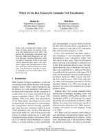
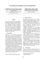
three divalent or six monovalent metals [1,4,6] (Fig. 1).
These residues are either bridging cysteines, which can
bind two divalent ions or they are terminal cysteines
that bind only one divalent metal [3,4,11].
When metal ions bind to apothionein, the polypep-
tide chain will rapidly fold resulting in the formation
of the two native, three-dimensional metal thiolate
clusters residing in each domain [3,5]. In the a-domain,
the only known MT secondary structure can be found
(a short a-helix present in case the protein is fully loa-
ded with divalent (not monovalent) metals) [5,6].
The antigenic part (epitope) of the MT-I + II pro-
teins is formed by a lysine-rich region, residues 20–25,
together with the seven N-terminal residues 1–7, which
after protein folding are seen in close proximity in the
three-dimensional structure [1,4,6].
The most studied human MT genes are found on
chromosome 16, which features very high levels of seg-
mentally duplicated sequence among the human auto-
somes and abundant genetic polymorphisms, which are
also existing in the MT-I + II genes [1,5].
In the chromosome 16 q13 region, MT genes are
tightly linked, and as a minimum they consist of 11
MT-I genes (MT-I-A, -B, -E, -F, -G, -H, -I, -J, -K, -L,
and -X) encoding functional or nonfunctional RNA,
and one gene for the other MT isoforms (the MT-2 A
Fig. 1. Schematic drawing of the mammalian MT-II protein showing the two metal-thiolate clusters (C-terminal a-domain and N-terminal
b-domain) including the 20 cysteine residues (blue squares) and their sulfur atoms (S), which bind to divalent or monovalent cations (in this
case Zn). The domains are linked by a short peptide containing amino acid residues 30–32 in mammalian MT-II (LINK). In the b-domain, three
divalent or six monovalent metal ions are coordinated, while in the a-domain four divalent or six monovalent cations can be bound.
Both bridging and terminal cysteines are present in mammalian MT-2. The bridging cysteines bind to two separate, divalent cations, while
the terminal cysteines chelate one divalent metal. If monovalent metals are bound, all cysteines can chelate two cations.
Metallothioneins are neuroprotective factors M. Penkowa
1858 FEBS Journal 273 (2006) 1857–1870 ª 2006 The Author Journal compilation ª 2006 FEBS
gene, MT-3 gene, and MT-4 gene) [1,3,4]. However, a
gene called MT-like 5 (MTL-5) has been described in
the q13 region of chromosome 11, and it encodes a
testis-specific MT-like protein named tesmin [3,4,6,7,12].
Compared with the human genes, the mouse MT
genes are less complex, as they only have one func-
tional gene for each major MT isoform (one gene
encoding MT-1, MT-2, MT-3 and MT-4) and these
are all located on chromosome 8. As in humans, the
mouse genome also contains an MTL-5 gene, which is
located on chromosome 19B [1–3,7,12].
However, this review will focus only on mammalian
MT-I + II isoforms, while all the other MT isoforms
and related MT-like structures (genes or their prod-
ucts) will not receive further attention. The major top-
ics reviewed here are the in vivo roles of mammalian
MT-I + II in immunoregulation, neuroprotection and
cerebral regeneration, a field receiving growing scienti-
fic interest.
Cerebral MT-I + II expression
Brain MT-I + II mRNA and proteins are present in
low amounts in physiological conditions and are
expressed during embryonic development and in
neonatals, and with increasing postnatal age MT-I + II
immunoreactivity increases and becomes continually
more widespread in the CNS [8,9]. In the brain,
astrocytes are the main source of MT-I + II, although
other cell types, such as choroid plexus epithelia,
endothelium and meningeal cells, may also show
MT-I + II [1,10]. In neurons the data on MT-I + II
expression have been inconsistent, and MT-I + II posi-
tive neurons have only been intermittently described
[9,13], although MT-I + II were demonstrated to exert
direct protective effects upon neurons, as shown in
primary neuronal cultures [14,15]. However, it is in
general agreed that the levels of MT-I + II are
several-fold higher in astrocytes relative to neurons.
Thus far, all the brain disorders studied in animals
and humans have shown that MT-I + II mRNA and
proteins are acutely and highly increased in reactive
astroglia as part of the acute inflammation and host
defense response [16–20]. To some extent, MT-I + II
are also increased in the vascular endothelium, choroid
plexus, ependyma, activated microglia ⁄ macrophages,
and meninges, while neuronal and oligodendroglial
MT-I + II immunoreactivity have not been consis-
tently reported [21–23].
MT-I + II mRNA increases are seen within 24 h
after an insult to the brain followed by many fold
increases in their protein levels as seen typically after
1–3 days postinjury [20–24].
Increased MT-I + II expression is seen in various
types of CNS pathology models such as in traumatic,
excitotoxic, and ischemic ⁄ hypoxic injury, multiple
sclerosis including its animal model experimental auto-
immune encephalomyelitis (EAE), amyotrophic lateral
sclerosis (ALS), Alzheimer’s disease (AD), Parkinson’s
disease (PD), Pick’s disease, pellagra encephalopathy,
immobilization stress, and peripheral nerve injury
[1,19,21,25–29].
Cellular MT-I + II distribution
MT-I + II have been considered as strictly intracellu-
lar proteins [4,29], but in recent years, mounting data
indicated that MT-I + II are distributed both intra-
and extracellularly [20,30–32].
Inside cells, MT-I + II are distributed in cytoplasm
and subcellular organelles like lysosomes and mito-
chondria. Depending on the cell cycle phase, differenti-
ation or in case of toxicity, MT-I + II are rapidly
translocated to the nucleus, as seen during early
S-phase and in oxidative stress [4,33–35]. Due to their
small size, MT-I + II can diffuse through nuclear pore
complexes, although the nuclear trafficking is relying
on specific cytosolic partner proteins and the appear-
ance of nuclear binding proteins, which in the presence
of ROS enhance the nuclear localization of MT-I + II
[29,36,37]. Also, perinuclear localization of MT
mRNA may contribute to the nuclear import of
MT-I + II proteins, as well as some structural altera-
tions in the proteins per se (such as lack of post-trans-
lational acetylation of lysine and cysteine) are
anticipated to regulate the nuclear trafficking [29,36].
Once in the nucleus, MT-I + II are selectively and
actively retained by nuclear factors, which are likely to
make use of saturable and energy-dependent binding
mechanisms, in that elimination of the ATP pool
hampers the nuclear translocation and ⁄ or retention of
MT-I + II [37]. However, the precise intracellular
MT-I + II trafficking system has yet to be clarified.
In addition, cells have been demonstrated to actively
secrete MT-I + II in vitro, although there is no known
signal peptide for cellular export [35,38].
In vivo, MT-I overexpressing transgenic mice display
significant MT-I + II immunoreactivity in the brain
extracellular space [20]. In the brain, the astrocytes, not
the neurons, are the major source of MT-I + II, even
though these proteins primarily protect the neurons
[9,31,39]. Hence, it is considered that astroglia may
secrete MT-I + II to the extracellular space in order for
them to protect the surrounding neurons [30]. This is
supported by studies of primary cell cultures, which
showed that extracellular MT-I + II exert direct effects
M. Penkowa Metallothioneins are neuroprotective factors
FEBS Journal 273 (2006) 1857–1870 ª 2006 The Author Journal compilation ª 2006 FEBS 1859
upon neurons, as MT-administration enhanced the sur-
vival, differentiation and postinjury recovery of cortical,
hippocampal, and dopaminergic neurons [14,15].
The experimental data from in vitro and in vivo stud-
ies that are reviewed here have shown consistently that
intra- and extracellular MT-I + II promote analogous
functions [14,28,29,31,35,40–41], which specifies that
MT-I + II have roles both in and outside cells.
Regulation of MT-I + II
MT-I + II are regulated in a coordinate manner [2]
and are rapidly increased by various pathological con-
ditions [1,20]. However, physiological and lifestyle-rela-
ted parameters like nutritional condition and physical
activity have also been reported to regulate MT-I + II
mRNA and proteins [7,11].
Administration of essential or toxic metals like Zn,
Cu, Cd, Hg increase MT-I + II biosynthesis by indu-
cing their transcription, for which several cis-acting
DNA elements, metal response elements (MREs) in
the promoter region are binding sites for trans-acting
transcription factors [3,7,43,44]. The MT-I + II gene
transcription is initiated when metals occupy the
MRE-binding transcription factor-1 (MTF-1), which is
a multiple Zn finger protein and the only known medi-
ator of the metal responsiveness of MT-I + II [3,44].
Reactive oxygen species (ROS) and oxidative stress
also increase expression of MT-I + II, which are
highly efficient free radical scavengers in the brain
[1,45]. ROS increase the MT-I + II transcriptional
response, as shown by exposure to free radicals like
superoxide anions and hydroxyl radicals, which rapidly
increase MT-I mRNA levels in a dose-dependent man-
ner [43,46]. The mechanism involves an antioxidant
response element (ARE) in the promoter region, ARE-
binding transcription factors, as well as the MTF-1,
transcription factors of the basic zipper type (Fos and
Fra-1), NF-E2-related factor 2, and the upstream stim-
ulatory factor family (USF, a basic helix–loop–helix–
leucine zipper protein), although it is likely that other
and yet unidentified proteins are involved [7,46].
Thereby, metals and ROS activate MT-I + II gene
transcription by different signaling pathways, response
elements and transcription factors.
In addition, MT-I + II are also increased by gluco-
corticoid hormones like corticosterone and dexametha-
sone, which signal through glucocorticoid response
elements (GREs) present in the gene regulatory region,
and also catecholamines (norepinephrine, isoprotere-
nol) activate MT-1 + II gene transcription [1,7,47].
During CNS inflammation, major MT-I + II regu-
latory factors are proinflammatory cytokines and espe-
cially interleukin (IL)-6 [1]. Accordingly, IL-6, IL-3,
tumor necrosis factor (TNF)-a, macrophage-colony
stimulating factor (M-CSF), and interferons increase
brain MT-I + II expression in a cytokine-specific
manner as demonstrated by using transgenic mice with
cytokine overexpression [48–51] or cytokine deficiency
[29,52–55].
Although the activation of MT-I + II gene tran-
scription is by far the best described regulatory mech-
anism, repression of MT gene activity has also been
reported [4,7]. Hence, during Zn deficiency, MTF-1
may form a complex with a Zn-responsive inhibitor,
named MT transcription inhibitor, which prevents
MTF-1 from interacting with the MREs, and thereby
MT-I + II gene transcription could be negatively con-
trolled due to the levels of trace metals [4,7,43]. In
human cells, MT-IIA gene activation is inhibited by
Zn finger protein PZ120, which interacts with the
MT-IIA transcription start site and inhibits gene
expression [56]. Also, transcription factors Fos and
Fra-1 can inhibit MT-I + II biosynthesis by inter-
action with ARE [7].
However, MT-I + II biosynthesis is also affected by
post-transcriptional events, since their protein levels do
not necessarily reflect the levels of mRNA expression
[4,57]. Hence, Cu treatment of adult rats reduced renal
MT-I + II mRNA levels while at the same time, the
renal MT-I + II protein expression was significantly
increased [58], which suggests that post-transcriptional
regulation occurs and this may likely affect either the
translation and ⁄ or the protein degradation. In fact,
MT-I + II are to some degree regulated by means of
intracellular protein degradation, which takes place in
both lysosomal and nonlysosomal compartments
[4,59]. In general, intracellular MT-I + II proteins
occur as either metal-containing proteins (MTs) or as
apothioneins, and their depletion and ⁄ or restitution
may depend on the bound metals, subcellular localiza-
tion, and the tissue examined. Hence, turnover rates of
cytosolic apothioneins versus lysosomal metal-bound
MT-I + II proteins are quite different, in that lyso-
somal MT-I + II proteolysis occurs more readily than
in the cytosol, although bound metals stabilize
MT-I + II proteins and prevent their lysosomal pro-
teolysis [59,60]. In the cytoplasm, the 26S proteasome
complex degrades apothionein, which due to the
lack of metals has a shorter half-life than MTs
[4,11]. The type of metal complex may also in itself
affect the MT-I + II degradation, as the half-life of
Cd-containing proteins is close to 3 days, while
Zn-binding reduces half-life to 18–20 h. Also, animal
age and the chemical pretreatment may determine the
half-life of MT-I + II, as well as MT-I in some cases
Metallothioneins are neuroprotective factors M. Penkowa
1860 FEBS Journal 273 (2006) 1857–1870 ª 2006 The Author Journal compilation ª 2006 FEBS
has reduced half-life relative to MT-2 [61]. Thus, it is
evident that other factors than the metals per se can
regulate MT-I + II turnover [4,11,61].
The CNS roles of endogenous MT-I + II
Recently, rising interest in MT-I + II neuroprotective
functions and therapeutic potential has been evident.
During the genomic era it became possible to modify
the MT-I + II genes in cultured cells and in animal
embryos leading to the generation of MT-I + II
knockout (MT-KO) mice [62] and transgenic MT-I
overexpressing (TgMT) mice [63]. These genotypes
have provided important answers concerning the roles
of MT-I + II in the disordered CNS, although at first
the data from MT-KO mice were rather disappointing,
since these mice developed normally, appeared viable
and fertile without any phenotypic changes [62]. Con-
sequently, MT-I + II were considered as dispensable
factors and ⁄ or proteins that may have abundant com-
pensatory backup systems.
Years later, this concept was substantially contradic-
ted, as it was demonstrated that neuronal survival and
brain tissue repair are compromised when MT-I + II
are absent. It became clear that during brain disorders,
MT-KO mice show significantly enhanced brain tissue
destruction, neuronal cell death, and clinical symptoms,
when compared with those of wild-type controls [49,64–
66]. Accordingly, even if the MT-I + II proteins may be
dispensable during healthy, physiological conditions,
they are unquestionably essential for coping with brain
damage [1,30,39]. The major histopathological changes
seen in brains of MT-KO mice are enhanced inflamma-
tory responses including increased recruitment of macro-
phages, lymphocytes and their CD34 + hematogenous
progenitor cells and enhanced secretion of proinflamma-
tory factors like IL-1, IL-3, IL-6, IL-12, TNF-a,
lymphotoxin-a (LTa), macrophage activator factor
(Mac-1), intercellular adhesion molecule (ICAM-1) and
acute phase response gene EB22 [31,49,65–69].
These studies also gave insight into the MT-I + II
in vivo antioxidant functions in the brain, as
MT-I + II deficiency resulted in amplified ROS
formation and oxidative stress including highly
increased lipid peroxidation, protein nitrosylation and
DNA oxidation, when compared with those of WT
controls [19,21,67,70]. In addition, MT-KO mice dis-
play significantly increased neurodegeneration and
apoptotic cell death relative to WT controls as shown
during traumatic brain injury, kainic acid-induced epi-
leptic seizures, 6-aminonicotinamide (6-AN)-induced
pellagra encephalopathy, ischemia, cytokine-induced
meningoencephalitis, peripheral nerve injury, PD, EAE
and ALS [23,27,40,62,65,67–71]. During these brain
disorders, the MT-KO mice also developed worse
clinical symptoms and showed significantly poorer
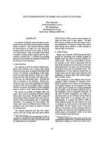
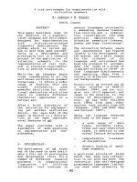
neurological outcome relative to WT controls (Fig. 2).
In contrast to brain disordered MT-KO mice, the
TgMT mice showed significantly less neuropathological
damage, while their tissue repair and neurological out-
come were improved relative to WT control mice
[13,20,28,31,40,55,72,73].
Thus, TgMT mice subjected to diverse brain disorders
display reduced inflammatory responses of macrophages
and lymphocytes including significantly decreased
levels of proinflammatory cytokines, matrix metallo-
proteinases (MMPs), and ROS. Also, the amounts of
delayed brain tissue damage consisting of oxidative
stress, neurodegeneration and apoptotic cell death were
radically reduced in TgMT mice relative to wild-type
controls [13,20,28,31,55,72]. To this end, comparisons of
the MT-I + II containing cells in the brain with the cell
populations suffering from oxidative stress and apopto-
tic death showed clearly that damaged and ⁄ or dying
cells are devoid of MT-I + II expression, which are
confined to surviving cells, and this likely reflects the
cytoprotection conferred by MT-I + II [19,72].
In addition, MT-I overexpression after brain injury
stimulates the astroglial responses including the expres-
sion of anti-inflammatory cytokines, growth factors,
neurotrophins and their receptors, such as IL-10, fibro-
blast growth factor (FGF), FGF-receptor (FGF-R),
transforming growth factor (TGF)-b, TGF- b-receptor
(TGF-b-R), vascular endothelial growth factor
(VEGF), nerve growth factor (NGF), neurotrophin
(NT)-3–5, brain-derived neurotrophic factor (BDNF),
glial-derived neurotrophic factor (GDNF) [13,20,28,31,
72,73]. Concomitantly, MT-I overexpression improves
brain tissue repair including neuronal regrowth and
vascular remodeling by angiogenesis, as well as the
TgMT mice show improved clinical outcome, when
compared with those of the wild-type control mice
[13,15,28,31,72].
To study brain restoration and tissue repair, a suit-
able model is the traumatic, focal brain injury to the
cortex, which results in a cortical necrotic cavity with-
out viable cells that gradually will be replaced with
glial scar tissue, vascular network and extracellular
matrix [10,24,54,74].
These processes are significantly enhanced by
MT-I + II, which are essential for the CNS wound
repair to occur [30,31,72,64]. Hence, in the injured
MT-KO mice the lesion cavity persists after 3 months,
by which severe inflammation is ongoing; while in wild-
type controls, the necrotic cavity is usually replaced with
a scar after 30 days [65,66], while in MT-Tg mice this
M. Penkowa Metallothioneins are neuroprotective factors
FEBS Journal 273 (2006) 1857–1870 ª 2006 The Author Journal compilation ª 2006 FEBS 1861
scar tissue is established before day 20 postinjury
[31,72].
Moreover, following brain pathology MT-I + II are
essential for the recruitment of neuroglial precursor
cells [19,22,75] and their migration towards the site of
injury. Hence, the increased tissue repair promoted by
MT-I + II after injury is likely mediated in part by
regeneration, where newly formed cells repopulate the
tissue and in part by regrowth and sprouting of survi-
ving cells.
The roles of exogenous MT-I + II
Shortly after the first data emerged from genetically
modified MT-KO and TgMT mice subjected to neuro-
pathology, new studies were conducted focusing on the
potential therapeutic use of MT-I + II proteins. For
this, adult rodents were injected intraperitoneally with
exogenous MT-I and ⁄ or MT-II (MT-I ⁄ II) proteins
during healthy conditions and neuropathological dis-
orders like brain injury, pellagra encephalopathy and
EAE [9,22,28,30,31,75]. The used MT-I ⁄ II proteins
contained metals, which were mainly Zn (the Zn con-
tent was approximately 7%) and small amounts of Cd
(the Cd content was < 0.5%). Therefore, these metals
were included in the control treatment regimen.
The intraperitoneal administration of exogenous
MT-I ⁄ II modulates immunoregulation and improves
neuroprotection and CNS recovery in vivo during brain
pathology, reflecting that MT-I + II have extracellular
roles. This was far from anticipated at the time of the
first publication (2000), as MT-I + II had been con-
sidered as strictly intracellular proteins [4].
At first, exogenous MT-I and ⁄ or MT-II proteins
were injected intraperitoneal in rats with EAE that
were evaluated clinically and histopathologically. In a
dose- and time-dependent manner, the MT-I ⁄ II treat-
ment reduced the severity of neurological symptoms
and the mortality relative to placebo control groups.
MT-I ⁄ II treatment in EAE reduced significantly the
activation and recruitment of macrophages and T
lymphocytes including levels of IL-1b, IL-6, IL-12,
Inhibition
Stimulation
MT-I+II
Oxidative stress
Microgliosis
Macrophages
&
T cells
Cytokine
release
Oxidative stress
Tissue Loss
Neurological
symptoms
Apoptosis
BBB
disruption
Cerebral ECs
Neurons
Neurodegeneration
Fig. 2. Schematic drawing of the main anti-
inflammatory, antioxidant and anti-apoptotic
actions of MT-I + II leading to neuroregen-
eration, angiogenesis and repair. MT-I + II
modulate an array of vital cellular functions
that involve cytoprotection, angiogenesis,
DNA repair and the maintenance of tissue
homeostasis. During pathology, MT-I + II
inhibit inflammation and cytokines and pro-
tect against oxidative stress, degeneration,
and apoptosis.
Metallothioneins are neuroprotective factors M. Penkowa
1862 FEBS Journal 273 (2006) 1857–1870 ª 2006 The Author Journal compilation ª 2006 FEBS
TNF-a and ROS, which was seen in brain, spleen, and
bone marrow [9,42]. The EAE lesions (plaques) with
demyelination, apoptotic cell death, axonal degener-
ation and transection were radically reduced by
MT-I ⁄ II administration relative to control treatment
[22]. Concomitantly, MT-I ⁄ II-treated animals dis-
played improved remyelination, regeneration and clin-
ical recovery from EAE relative to the placebo groups.
This therapeutic effect was due to MT-I + II-activa-
tion of oligodendroglial progenitors ⁄ stem cells and
enhanced expression of growth and trophic factors
(FGF, TGF-b, NT-3–5 and NGF), which were signifi-
cantly enhanced by MT-I + II in EAE and even more
during the recovery phases, when compared with those
of the placebo controls [22,76].
In later studies, exogenous MT-I ⁄ II proteins were
administered during experimental models of traumatic
brain injury (freeze lesion with dry ice) and pellagra
encephalopathy (induced by administration of 6-AN).
The acute (primary) injuries (the trauma- or 6-AN-
induced necrosis) were comparable in the treatment
groups, but in the following days, some pronounced
differences in the responses to pathology appeared.
Thus, animals receiving MT-I ⁄ II-treatment showed sig-
nificantly less oxidative stress, neurodegeneration and
apoptotic cell death (delayed damage) in the days ⁄
weeks following the primary injuries [28–31,75]. In
these studies, the MT-I ⁄ II treatment also enhanced
repair responses including expression of growth ⁄
trophic factors, astrogliosis, angiogenesis, neuronal
regrowth [75], and particularly after the traumatic
brain injury, it was evident that MT-I ⁄ II enhance
reorganization of the necrotic lesion cavity [31]. The
metal bound state of MT was preferred because the
metalloform is likely to be the more physiological
relevant form of the protein, and also because it is
significantly less susceptible to degradation than
apothionein. However, none of the effects of the
MT-I ⁄ II treatment were seen after administration of
the metals per se, but the latter may still be important
as MT-I ⁄ +II adopt their tertiary structure upon
chelation of metal ions [1,3,4]. However, the mole-
cular mechanisms by which the MT-I ⁄ II treatment
promoted neuroprotection and repair remain to be
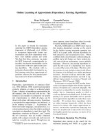
fully clarified (Fig. 3).
The MT-I + II molecular mechanisms
To clarify the specific functions of the MT-I + II pro-
teins, many different approaches and techniques have
been applied throughout thousands of studies. Although
they described the MT-I + II structure, chemical
characteristics, regulation, expression, distribution,
degradation and the consequences of reducing or
increasing MT-I + II in cells; they have not yet
clarified the precise signaling and mechanisms by
which MT-I + II exert immunoregulatory and neuro-
protective actions.
However, many possibilities are likely, since
MT-I + II are indeed multipurpose proteins involved
in a broad range of functions, which include, but are
not restricted to metal ion homeostasis, scavenging of
ROS, redox status, immune defense responses, pro-
tein–protein and protein–nucleotide interactions, regu-
lation of Zn fingers and Zn-containing transcription
factors, mitochondrial respiration, thermogenesis, body
energy metabolism, angiogenesis, cell cycle progression,
and cell survival and differentiation [1,6,29,30,33,
39,77,78]. Some of these MT-I + II actions may have
therapeutic relevance in a range of acute and chronic
neurological disorders, in which inflammation and
oxidative stress are central in the pathophysiology
[79–84]. Accordingly, MT-I + II may signal through
diverse molecular pathways.
The immunomodulatory actions of MT-I + II
reduce proinflammatory mediators including cytokines,
MMPs, and adhesion molecules [20,32,72].
The reduction of brain IL-1, IL-6, IL-12 and TNF-a
could be a central mechanism in the MT-I + II anti-
inflammatory effects, since these cytokines are major
immune activators that increase leukocyte activation,
transendothelial migration, and chemoattraction, thereby
leading to neuroinflammatory infiltrates [79–81].
Hence, genetic deficiency or overexpression of these
cytokines or their receptors will diminish or enhance
the brain inflammatory leukocytes [74,81,84,85]. Thus,
IL-6 knockout mice (genetic IL-6-deficient mice)
(IL-6KO) mice are resistent to EAE sensibilization,
while IL-6 overexpressors show spontaneous chronic
Fig. 3. Summary of the major biological functions of MT-I + II.
M. Penkowa Metallothioneins are neuroprotective factors
FEBS Journal 273 (2006) 1857–1870 ª 2006 The Author Journal compilation ª 2006 FEBS 1863
neuroinflammation and degeneration [73,86], which
reflects that IL-6 is activating hematopoiesis, acute
phase responses, and inflammation. IL-1 and IL-12 are
also central pro-inflammatory cytokines that are cru-
cial in the development of Th1 cells and initiation of
autoimmune attacks, demyelination and neurodegener-
ative diseases and neuronal cell death by apoptosis
and necrosis [49,80,82,84,85]. As the pro-inflammatory
cytokines mediate significant neurotoxicity and chronic
pathology, the MT-I + II-inhibition of their mRNA
and protein biosynthesis [66] will likely contribute to
improved neuroprotection.
It was recently shown that MT-I + II share certain
structural and functional similarities with beta- and
delta-chemokines CCL-17 and CX3CL-1 in vitro [87],
whereby MT-I + II may regulate leukocyte chemo-
taxis, although this has yet to be confirmed in vivo.
Other cell culture studies showed that MT-I + II may
inhibit monocytic activation and invasion including
secretion of cytokines [88–90]. Moreover, MT-I + II
inhibit macrophage-induced T cell proliferation and
the activation of cytotoxic T cells and antigen-specific
B cells [91–94].
To this end, MT-I + II may also reduce inflamma-
tion by interfering directly with cell–cell interactions as
MT-I + II were demonstrated to bind specifically to
the membranes of macrophages, T and B cells, which
thereby are inactivated [91–95].
These MT-I + II anti-inflammatory effects can
also be seen in humans, as patients with autoimmune
and allergic diseases show depletion of systemical
MT-I + II and occurrence of anti-MT-I + II auto-
IgGs against MT-I + II, an alteration that is most
pronounced during clinical exacerbations [96,97]. How-
ever, as in animals, the human MT-I + II levels can
be fully replenished by various agents, among which
steroids like glucocorticoid and cortisone can be used;
and interestingly, steroids in general increase
MT-I + II levels right before the patients show signifi-
cant clinical improvements [97,98]. Also in MS
patients, the MT-I + II expression levels are highest
during the recovery and remission of disease [76].
In fact, the molecular mechanism of steroid-medi-
ated immuno suppression could be a steroid-caused
MT-I + II augmentation, given that glucocorticoid-
treated patients show significant MT-II increases in
their peripheral leukocytes shortly before the therapeu-
tic effect of steroid commenced [98]. In support of this,
dexamethasone-induced MT-II can be used as an indi-
cator of glucocorticoid sensitivity [99]. This correlation
between steroids and MT-I + II also exists in the
brain, where MT-I + II mRNA and proteins are
enhanced significantly by glucocorticoids [100].
In case MT-I + II are central mechanisms of steroid
therapeutic effects, then MT-I + II might be used as a
more specific anti-inflammatory agent likely having less
side-effects than steroids.
As proinflammatory cytokine profiles are associated
with development of human type-2 diabetes, which
also affects the brain, we recently examined MT-I + II
in such patients. Interestingly, systemical MT-I + II
expression and function are depleted in type-2 diabet-
ics relative to healthy subjects [101]. Hence, both con-
stitutive and stress-related MT-I + II were deficient in
the patients versus the healthy control subjects, which
suggests that an absence of MT-I + II may have a
key role in the pathogenesis of type-2 diabetes [101].
Indeed, in a following study of experimental diabetes,
it was shown that diabetic MT-I + II depletion can be
fully restored by medication, and such MT-I + II
replenishment is associated with disease remission
[102].
In addition, the MT-I + II inhibition of MMPs,
which are Zn-dependent endopeptidases produced by
inflammatory cells, may also contribute to amelior-
ation of a number of human autoimmune diseases,
where MMPs are involved in pathophysiological events
like diapedesis of infiltrating cells, tissue degradation
and blood–brain barrier breakdown [103,104].
Furthermore, MT-I + II stimulate astroglial res-
ponses including expression of anti-inflammatory
signals, growth ⁄ trophic factors [72,66,68]. Although
astrogliotic scarring traditionally has been considered
as inhibitors of neuroregeneration, mounting and con-
vincing data have now shown that reactive astrocytes
provide essential neuroprotection and recovery. Hence,
astrocytes endow neurons with antioxidants, energy
substrates, anti-inflammatory and trophic ⁄ growth fac-
tors; and they improve neurogenesis and neurological
outcome [70,105,106].
Hence, ablation of astroglia during brain pathology
leads to massive increases in neurodegeneration, de-
myelination, infiltration by leucocytes, and cell
death [106]. Thus, astroglial responses activated by
MT-I + II may contribute to increased neuron survi-
val, regeneration and CNS recovery. Also, MT-I + II
increase expression of IL-10, FGF, TGF-b, VEGF,
NGF, NT-3–5, BDNF, GDNF and their receptors;
and this could in itself mediate neuroprotection as well
as contribute to the MT-I + II-mediated repair, angi-
ogenesis and vascular remodeling [19,20,22,30,32,39].
Together, these actions of MT-I + II can contribute
to overall improvements in CNS cell survival and
recovery [1,27,28,31,72,64–66,76].
Taken as a whole, these effects upon cerebral inflam-
mation suggest that MT-I + II could be causing a
Metallothioneins are neuroprotective factors M. Penkowa
1864 FEBS Journal 273 (2006) 1857–1870 ª 2006 The Author Journal compilation ª 2006 FEBS
general shift in the balance between pro- and anti-
inflammatory molecules.
The actual mechanisms through which MT-I + II
inhibit neurodegeneration and cell death remain to be
fully described, although a range of anti-apoptotic
effects have been shown in animals and humans. First,
the anti-inflammatory effects as well as the antioxidant
properties of MT-I + II could each contribute to
decreased neurodegeneration and cell loss [79–83],
although it is unlikely that these MT-I + II effects are
the only responsible mechanisms.
Hence, when the MT-I + II anti-inflammatory
actions are counterbalanced, as done by using double
transgenic mice overexpressing both MT-I and pro-
inflammatory cytokine IL-6, it was evident that
MT-I + II still reduce neurodegeneration and cell
death significantly [32,72,73]. However, the MT-I + II
antioxidant effects likely contribute to neuroprotection,
as the cerebral ROS formation and oxidative stress
are inversely related to the MT-I + II levels but
not to the expression of other antioxidants such
as Cu ⁄ Zn-super oxide dismutase (Cu ⁄ Zn-SOD),
Mn-SOD, and catalase [32,40,65,72]. During mitochon-
dria-specific oxidative stress, MT-I + II are indispens-
able and have key roles in the mitochondrial
protection, which did not relate to other antioxidants
like glutathione peroxidase, catalase, Mn-SOD, and
Cu ⁄ Zn-SOD [45].
MT-I + II may also prevent neuronal damage by
having critical roles in metal ion homeostasis. Partic-
ularly the Zn regulation by MT-I + II may have
major importance, since Zn is central for a broad
range of functions. However, tight control of the Zn
levels is necessary as an overload or deficiency of this
metal leads to severe neurotoxicity [1,107]. Also,
MT-I + II transfer Zn directly to mitochondrial fac-
tors, Zn-finger proteins and transcription factors,
which also are essential for several signaling pathways
and cell fate [4,34,78]. Along with Zn, various metal
ions with a neurotoxic potential are bound and
released by MT-I + II, which can thereby influence a
range of cellular metabolites and pathways in the
brain. A disrupted metal ion homeostasis causes oxida-
tive stress, degeneration and neuronal cell death, and
accordingly, dysregulation of metals has been associ-
ated with many pathologies including stroke, epilepsy,
PD, AD, and traumatic brain injury [6,10,21,44,107].
Hence, the MT-I + II regulation of metal ion availab-
ility and levels in the CNS is most likely to contribute
to the MT-I + II protective functions. Besides having
roles in metal ion regulation, MT-I + II proteins also
obtain their tertiary structure and enhanced molecular
stability from their chelation of metals [4,6,11].
To this end, it is important that the different
MT-I ⁄ II treatments injected into animals were all fully
loaded Zn
7
–MT complexes, as the metal ensures pro-
tein stability, folding and longer half-life [3,4].
However, MT-I + II interact and modulate many
intracellular messengers that are directly or indirectly
regulating the apoptotic cascade, and therefore
MT-I + II may affect additional pathways during
their responses to damage and promotion of tissue
repair. The nucleotides ATP and GTP [5,34,108] bind
to MT-I + II proteins, whereby both structural and
functional changes are seen in the proteins [108]. Also,
the MT-I + II and ATP levels inside cells are interre-
lated, which in itself could affect cell loss or survival,
since ATP depletion is part of the apoptotic cascade
[40]. The MT-I + II and ATP connection may also be
implicated in other actions, such as the MT-I + II-
caused stabilization and rejuvenation of the ageing
mitochondrial genome [40] and MT-I + II-regulation
of energy balance and metabolism [77,78]. To this end,
MT-I + II can donate Zn directly to mitochondrial
aconitase (m-aconitase) by means of direct protein–
protein interaction [109].
In addition, MT-I + II regulate the levels, activity
and cellular localization of the transcription factor
NFjB [10,70,95], which is involved in cell fate during
neuropathology. Besides, MT-I + II induce a range of
common proto-oncogenes (like bcl-2 and c-myc) whilst
pro-apoptotic proteins (like p53 and caspase-3) are
inhibited [11,20,32,33,41,74].
The roles of MT-I + II in cell fate and the
MT-I + II connection to other factors involved in cell
cycle regulation have led to many studies of
MT-I + II roles in cancer. It is not surprising that
MT-I + II may prevent tumor cell death by protecting
against pro-apoptotic treatment regimes [11,33]. How-
ever, when the cancer is located in ectodermal tissues
(such as colon, bladder and skin), a positive correla-
tion exists between increased MT-I + II levels and an
improved prognosis [11].
Final comments
This review summarizes the current knowledge and
advances in the understanding of MT-I + II roles in
immunomodulation and neuroprotection. The findings
indicate that MT-I + II inhibit efficiently proinflam-
matory cytokines, ROS, MMPs and pro-apoptotic sig-
nals, which all may cause a broad range of brain
disorders. As shown by many independent groups, the
MT-I + II levels are inversely related to the degree of
brain damage observed after traumatic injury, EAE,
epileptic seizures, ischemia, and neurodegenerative
M. Penkowa Metallothioneins are neuroprotective factors
FEBS Journal 273 (2006) 1857–1870 ª 2006 The Author Journal compilation ª 2006 FEBS 1865
diseases like PD, ALS and pellagra [9,15,20,22,27,30–
32,40,69–71]. Consequently, MT-I + II might provide
new drug targets against neurological disorders, especi-
ally those containing autoimmunity, neurodegeneration
and neuron loss. As MT-I + II compounds are in gen-
eral well tolerated, they may be used in the future as
therapeutic and ⁄ or preventive medications.
Acknowledgements
These studies were supported by IMK Almene Fond,
Vera og Carl Michaelsens Legat, The Lundbeck Foun-
dation, The Danish Medical Research Council, The
Danish Medical Association Research Fund, Toyota
Fonden, Frænkels Mindefond, Scleroseforeningen,
Kathrine og Vigo Skovgaards Fond, Fonden til
Lægevidenskabens Fremme, Dir. Leo Nielsens Legat,
Th. Maigaard’s Eftf. Fru Lily Benthine Lunds Fond.
Thanks are given to Adam Bohr and Kristian Kolind
for excellent procedural assistance.
References
1 Hidalgo J, Aschner M, Zatta P & Vasak M (2001)
Roles of the metallothionein family of proteins in the
central nervous system. Brain Res Bull 55, 133–145.
2 Searle PF, Davison BL, Stuart GW, Wilkie TM, Nor-
stedt G & Palmiter RD (1984) Regulation, linkage, and
sequence of mouse metallothionein I and II genes. Mol
Cell Biol 4 , 1221–1230.
3 Ghoshal K & Jacob ST (2001) Regulation of metal-
lothionein gene expression. Prog Nucleic Acid Res Mol
Biol 66, 357–384.
4 Klaassen CD, Liu J & Choudhuri S (1999) Metallo-
thionein: an intracellular protein to protect against
cadmium toxicity. Annu Rev Pharmacol Toxicol 39,
267–294.
5 Vallee BL (1979) Metallothionein: historical review and
perspectives. Experientia Suppl. 34, 19–39.
6 Vasak M (2005) Advances in metallothionein structure
and functions. J Trace Elem Med Biol 19, 13–17.
7 Haq F, Mahoney M & Koropatnick J (2003) Signaling
events for metallothionein induction. Mutat Res 533,
211–226.
8 Penkowa M, Nielsen H, Hidalgo J, Bernth N & Moos T
(1999) Distribution of metallothionein I + II and
vesicular zinc in the developing central nervous system:
correlative study in the rat. J Comp Neurol 412, 303–
318.
9 Penkowa M & Hidalgo J (2001) Metallothionein treat-
ment reduces proinflammatory cytokines IL-6 and
TNF-alpha and apoptotic cell death during experimen-
tal autoimmune encephalomyelitis (EAE). Exp Neurol
170, 1–14.
10 Penkowa M, Giralt M, Thomsen PS, Carrasco J &
Hidalgo J (2001) Zinc or copper deficiency-induced
impaired inflammatory response to brain trauma may
be caused by the concomitant metallothionein changes.
J Neurotrauma 18, 447–463.
11 Miles AT, Hawksworth GM, Beattie JH & Rodilla V
(2000) Induction, regulation, degradation, and biologi-
cal significance of mammalian metallothioneins. Crit
Rev Biochem Mol Biol 35, 35–70.
12 Quaife CJ, Findley SD, Erickson JC, Froelick GJ,
Kelly EJ, Zambrowicz BP & Palmiter RD (1994)
Induction of a new metallothionein isoform (MT-IV)
occurs during differentiation of stratified squamous
epithelia. Biochemistry 33, 7250–7259.
13 van Lookeren CM, Thibodeaux H, van Bruggen N,
Cairns B, Gerlai R, Palmer JT, Williams SP & Lowe
DG (1999) Evidence for a protective role of metal-
lothionein-1 in focal cerebral ischemia. Proc Natl Acad
Sci USA 96, 12870–12875.
14 Kohler LB, Berezin V, Bock E & Penkowa M (2003)
The role of metallothionein II in neuronal differentia-
tion. and Survival Brain Res 992, 128–136.
15 Chung RS, Vickers JC, Chuah MI & West AK (2003)
Metallothionein-IIA promotes initial neurite elongation
and postinjury reactive neurite growth and facilitates
healing after focal cortical brain injury. J Neurosci 23,
3336–3342.
16 Chung RS, Adlard PA, Dittmann J, Vickers JC, Chuah
MI & West AK (2004) Neuron-glia communication:
metallothionein expression is specifically up-regulated
by astrocytes in response to neuronal injury. J Neuro-
chem 88, 454–461.
17 Espejo C, Penkowa M, Demestre M, Montalban X &
Martinez-Caceres EM (2005) Time-course expression of
CNS inflammatory, neurodegenerative tissue repair
markers and metallothioneins during experimental
autoimmune encephalomyelitis. Neuroscience 132,
1135–1149.
18 Penkowa M, Camats J, Hadberg H, Quintana A, Rojas
S, Giralt M, Molinero A, Campbell IL & Hidalgo J
(2003) Astrocyte-targeted expression of interleukin-6
protects the central nervous system during neuroglial
degeneration induced by 6-aminonicotinamide. J Neu-
rosci Res 73, 481–496.
19 Penkowa M, Espejo C, Martinez-Caceres EM, Montal-
ban X & Hidalgo J (2003) Increased demyelination and
axonal damage in metallothionein I+II-deficient mice
during experimental autoimmune encephalomyelitis.
Cell Mol Life Sci 60, 185–197.
20 Penkowa M, Florit S, Giralt M, Quintana A, Molinero
A, Carrasco J & Hidalgo J (2005) Metallothionein
reduces central nervous system inflammation, neurode-
generation, and cell death following kainic acid-
induced epileptic seizures. J Neurosci Res 79, 522–534.
Metallothioneins are neuroprotective factors M. Penkowa
1866 FEBS Journal 273 (2006) 1857–1870 ª 2006 The Author Journal compilation ª 2006 FEBS
21 Hidalgo J, Penkowa M, Giralt M, Carrasco J & Moli-
nero A (2002) Metallothionein expression and oxidative
stress in the brain. Methods Enzymol 348, 238–249.
22 Penkowa M & Hidalgo J (2003) Treatment with metal-
lothionein prevents demyelination and axonal damage
and increases oligodendrocyte precursors and tissue
repair during experimental autoimmune encephalomye-
litis. J Neurosci Res 72, 574–586.
23 Stankovic RK (2005) Atrophy of large myelinated
axons in metallothionein-I, II knockout mice. Cell Mol
Neurobiol 25, 943–953.
24 Poulsen CB, Penkowa M, Borup R, Nielsen FC, Cac-
eres M, Quintana A, Molinero A, Carrasco J, Giralt
M & Hidalgo J (2005) Brain response to traumatic
brain injury in wild-type and interleukin-6 knockout
mice: a microarray analysis. J Neurochem 92, 417–432.
25 Ceballos D, Lago N, Verdu E, Penkowa M, Carrasco
J, Navarro X, Palmiter RD & Hidalgo J (2003) Role
of metallothioneins in peripheral nerve function. and
Regeneration Cell Mol Life Sci 60, 1209–1216.
26 Gong YH & Elliott JL (2000) Metallothionein
expression is altered in a transgenic murine model of
familial amyotrophic lateral sclerosis. Exp Neurol
162, 27–36.
27 Nagano S, Satoh M, Sumi H, Fujimura H, Tohyama
C, Yanagihara T & Sakoda S (2001) Reduction of
metallothioneins promotes the disease expression of
familial amyotrophic lateral sclerosis mice in a dose-
dependent manner. Eur J Neurosci 13, 1363–1370.
28 Penkowa M, Giralt M, Camats J & Hidalgo J (2002)
Metallothionein 1+2 protect the CNS during neuro-
glial degeneration induced by 6-aminonicotinamide.
J Comp Neurol 444, 174–189.
29 Ogra Y & Suzuki KT (2000) Nuclear trafficking of
metallothionein: possible mechanisms and current
knowledge. Cell Mol Biol 46, 357–365.
30 Chung RS & West AK (2004) A role for extracellular
metallothioneins in CNS injury and repair. Neuro-
science 123, 595–599.
31 Giralt M, Penkowa M, Lago N, Molinero A & Hidalgo
J (2002) Metallothionein-1+2 protect the CNS after a
focal brain injury. Exp Neurol 173, 114–128.
32 Penkowa M, Quintana A, Carrasco J, Giralt M,
Molinero A & Hidalgo J (2004) Metallothionein
prevents neurodegeneration and central nervous system
cell death after treatment with gliotoxin 6-aminonicotina-
mide. J Neurosci Res 77, 35–53.
33 Cherian MG & Apostolova MD (2000) Nuclear locali-
zation of metallothionein during cell proliferation and
differentiation. Cell Mol Biol 46, 347–356.
34 Maret W (2002) Optical methods for measuring zinc
binding and release, zinc coordination environments
in zinc finger proteins, and redox sensitivity and
activity of zinc-bound thiols. Methods Enzymol 348,
230–237.
35 Trayhurn P, Duncan JS, Wood AM & Beattie JH
(2000) Regulation of metallothionein gene expression
and secretion in rat adipocytes differentiated from pre-
adipocytes in primary culture. Horm Metab Res 32,
542–547.
36 Takahashi Y, Ogra Y & Suzuki KT (2005) Nuclear
trafficking of metallothionein requires oxidation of a
cytosolic partner. J Cell Physiol 202, 563–569.
37 Woo ES, Dellapiazza D, Wang AS & Lazo JS (2000)
Energy-dependent nuclear binding dictates metallothio-
nein localization. J Cell Physiol 182, 69–76.
38 Trayhurn P, Duncan JS, Wood AM & Beattie JH
(2000) Metallothionein gene expression and secretion in
white adipose tissue. Am J Physiol Regul Integr Comp
Physiol 279, R2329–R2335.
39 West AK, Chuah MI, Vickers JC & Chung RS (2004)
Protective role of metallothioneins in the injured mam-
malian brain. Rev Neurosci 15, 157–166.
40 Ebadi M, Brown-Borg H, El Refaey H, Singh BB,
Garrett S, Shavali S & Sharma SK (2005) Metallothio-
nein-mediated neuroprotection in genetically engineered
mouse models of Parkinson’s disease. Brain Res Mol
Brain Res 134, 67–75.
41 Ebadi M, Sharma SK, Ghafourifar P, Brown-Borg H
& El Refaey H (2005) Peroxynitrite in the pathogenesis
of Parkinson’s disease and the neuroprotective role of.
Metallothioneins Meth Enzymol 396, 276–298.
42 Penkowa M & Hidalgo J (2000) Metallothionein I+II
expression and their role in experimental autoimmune
encephalomyelitis. Glia 32, 247–263.
43 Klassen RB, Crenshaw K, Kozyraki R, Verroust PJ, Tio
L, Atrian S, Allen PL & Hammond TG (2004) Megalin
mediates renal uptake of heavy metal metallothionein
complexes. Am J Physiol Renal Physiol 287, F393–F403.
44 Andrews GK (2000) Regulation of metallothionein
gene expression by oxidative stress and metal ions.
Biochem Pharmacol 59, 95–104.
45 Kondoh M, Inoue Y, Atagi S, Futakawa N, Higashi-
moto M & Sato M (2001) Specific induction of
metallothionein synthesis by mitochondrial oxidative
stress. Life Sci 69, 2137–2146.
46 Andrews GK & Geiser J (1999) Expression of the
mouse metallothionein-I and -II genes provides a
reproductive advantage during maternal dietary zinc
deficiency. J Nutr 129, 1643–1648.
47 Beattie JH, Wood AM, Trayhurn P, Jasani B, Vincent
A, McCormack G & West AK (2000) Metallothionein
is expressed in adipocytes of brown fat and is induced
by catecholamines and zinc. Am J Physiol Regul Integr
Comp Physiol 278, R1082–R1089.
48 Carrasco J, Hernandez J, Gonzalez B, Campbell IL &
Hidalgo J (1998) Localization of metallothionein-I and
-III expression in the CNS of transgenic mice with
astrocyte-targeted expression of interleukin 6. Exp
Neurol 153, 184–194.
M. Penkowa Metallothioneins are neuroprotective factors
FEBS Journal 273 (2006) 1857–1870 ª 2006 The Author Journal compilation ª 2006 FEBS 1867
49 Carrasco J, Giralt M, Penkowa M, Stalder AK, Camp-
bell IL & Hidalgo J (2000) Metallothioneins are upre-
gulated in symptomatic mice with astrocyte-targeted
expression of tumor necrosis factor-alpha. Exp Neurol
163, 46–54.
50 Giralt M, Carrasco J, Penkowa M, Morcillo MA,
Santamaria J, Campbell IL & Hidalgo J (2001)
Astrocyte-targeted expression of interleukin-3 and
interferon-alpha causes region-specific changes in
metallothionein expression in the brain. Exp Neurol
168, 334–346.
51 Hernandez J, Molinero A, Campbell IL & Hidalgo J
(1997) Transgenic expression of interleukin 6 in the
central nervous system regulates brain metallothionein-
I and -III expression in mice. Brain Res Mol Brain Res
48, 125–131.
52 Carrasco J, Hernandez J, Bluethmann H & Hidalgo J
(1998) Interleukin-6 and tumor necrosis factor-alpha
type 1 receptor deficient mice reveal a role of IL-6 and
TNF-alpha on brain metallothionein-I and -III regula-
tion. Brain Res Mol Brain Res 57, 221–234.
53 Penkowa M, Molinero A, Carrasco J & Hidalgo J
(2001) Interleukin-6 deficiency reduces the brain inflam-
matory response and increases oxidative stress and neu-
rodegeneration after kainic acid-induced seizures.
Neuroscience 102, 805–818.
54 Penkowa M, Moos T, Carrasco J, Hadberg H, Molin-
ero A, Bluethmann H & Hidalgo J (1999) Strongly
compromised inflammatory response to brain injury in
interleukin-6-deficient mice. Glia 25, 343–357.
55 Penkowa M, Poulsen C, Carrasco J & Hidalgo J (2002)
M-CSF deficiency leads to reduced metallothioneins I
and II expression and increased tissue damage in the
brain stem after 6-aminonicotinamide treatment. Exp
Neurol 176, 308–321.
56 Tang CM, Westling J & Seto E (1999) trans repression
of the human metallothionein IIA gene promoter by
PZ120, a novel 120-kilodalton zinc finger protein. Mol
Cell Biol 19, 680–689.
57 Vasconcelos MH, Tam SC, Beattie JH & Hesketh JE
(1996) Evidence for differences in the post-transcrip-
tional regulation of rat metallothionein isoforms.
Biochem J 315, 665–671.
58 Vasconcelos MH, Tam SC, Hesketh JE, Reid M &
Beattie JH (2002) Metal- and tissue-dependent relation-
ship between metallothionein mRNA and protein.
Toxicol Appl Pharmacol 182, 91–97.
59 Steinebach OM & Wolterbeek BT (1992) Metallothio-
nein biodegradation in rat hepatoma cells: a compart-
mental analysis aided
35
S-radiotracer study. Biochim
Biophys Acta 1116, 155–165.
60 Hahn SH, Yoo OJ & Gahl WA (2001) Effect of metal
ions on the stability of metallothionein in the degrada-
tion by cellular fractions in vitro. Exp Mol Med 33,
32–36.
61 Kershaw WC & Klaassen CD (1992) Degradation and
metal composition of hepatic isometallothioneins in
rats. Toxicol Appl Pharmacol 112, 24–31.
62 Michalska AE & Choo KH (1993) Targeting and
germ-line transmission of a null mutation at the
metallothionein I and II loci in mouse. Proc Natl Acad
Sci USA 90, 8088–8092.
63 Masters BA, Kelly EJ, Quaife CJ, Brinster RL & Pal-
miter RD (1994) Targeted disruption of metallothio-
nein I and II genes increases sensitivity to cadmium.
Proc Natl Acad Sci USA 91, 584–588.
64 Natale JE, Knight JB, Cheng Y, Rome JE & Gallo V
(2004) Metallothionein I and II mitigate age-dependent
secondary brain injury. J Neurosci Res 78, 303–314.
65 Penkowa M, Carrasco J, Giralt M, Moos T & Hidalgo
J (1999) CNS wound healing is severely depressed in
metallothionein I- and II-deficient mice. J Neurosci 19,
2535–2545.
66 Penkowa M, Carrasco J, Giralt M, Molinero A,
Hernandez J, Campbell IL & Hidalgo J (2000) Altered
central nervous system cytokine-growth factor expres-
sion profiles and angiogenesis in metallothionein-I+II
deficient mice. J Cereb Blood Flow Metab 20,
1174–1189.
67 Giralt M, Penkowa M, Hernandez J, Molinero A,
Carrasco J, Lago N, Camats J, Campbell IL &
Hidalgo J (2002) Metallothionein-1+2 deficiency
increases brain pathology in transgenic mice with astro-
cyte-targeted expression of interleukin 6. Neurobiol Dis
9, 319–338.
68 Penkowa M, Giralt M, Moos T, Thomsen PS, Hernan-
dez J & Hidalgo J (1999) Impaired inflammatory
response to glial cell death in genetically metallothio-
nein-I- and -II-deficient mice. Exp Neurol 156, 149–
164.
69 Penkowa M, Espejo C, Martinez-Caceres EM, Poulsen
CB, Montalban X & Hidalgo J (2001) Altered inflam-
matory response and increased neurodegeneration in
metallothionein I+II deficient mice during experimen-
tal autoimmune encephalomyelitis. J Neuroimmunol
119, 248–260.
70 Carrasco J, Penkowa M, Hadberg H, Molinero A &
Hidalgo J (2000) Enhanced seizures and hippocampal
neurodegeneration following kainic acid-induced sei-
zures in metallothionein-I + II-deficient mice. Eur J
Neurosci 12, 2311–2322.
71 Trendelenburg G, Prass K, Priller J, Kapinya K, Polley
A, Muselmann C, Ruscher K, Kannbley U, Schmitt
AO, Castell S et al. (2002) Serial analysis of gene
expression identifies metallothionein-II as major neuro-
protective gene in mouse focal cerebral ischemia. J
Neurosci 22, 5879–5888.
72 Penkowa M, Camats J, Giralt M, Molinero A,
Hernandez J, Carrasco J, Campbell IL & Hidalgo J
(2003) Metallothionein-I overexpression alters brain
Metallothioneins are neuroprotective factors M. Penkowa
1868 FEBS Journal 273 (2006) 1857–1870 ª 2006 The Author Journal compilation ª 2006 FEBS
inflammation and stimulates brain repair in transgenic
mice with astrocyte-targeted interleukin-6 expression.
Glia 42, 287–306.
73 Molinero A, Penkowa M, Hernandez J, Camats J, Gir-
alt M, Lago N, Carrasco J, Campbell IL & Hidalgo J
(2003) Metallothionein-I overexpression decreases brain
pathology in transgenic mice with astrocyte-targeted
expression of interleukin-6. J Neuropathol Exp Neurol
62, 315–328.
74 Quintana A, Giralt M, Rojas S, Penkowa M, Campbell
IL, Hidalgo J & Molinero A (2005) Differential role of
tumor necrosis factor receptors in mouse brain inflam-
matory responses in cryolesion brain injury. J Neurosci
Res 82, 701–716.
75 Penkowa M, Tio L, Mecedes G, Quintana A, Molinero
A, Atrian S, Vasak M & Hidalgo J (2006) Specificity
and divergence in the neurobiological effects of differ-
ent Metallothioneins after brain injury. J Neurosci Res,
doi: 10.1002/jnr.20790.
76 Espejo C & Martinez-Caceres EM (2005) The role of
methallothioneins in experimental autoimmune ence-
phalomyelitis and multiple sclerosis. Ann NY Acad Sci
1051, 88–96.
77 Beattie JH, Wood AM, Newman AM, Bremner I,
Choo KH, Michalska AE, Duncan JS & Trayhurn P
(1998) Obesity and hyperleptinemia in metallothionein
(-I and -II) null mice. Proc Natl Acad Sci USA 95,
358–363.
78 Ye B, Maret W & Vallee BL (2001) Zinc metallothionein
imported into liver mitochondria modulates respiration.
Proc Natl Acad Sci USA 98, 2317–2322.
79 Allan SM & Rothwell NJ (2003) Inflammation in cen-
tral nervous system injury. Philos Trans R Soc Lond B
Biol Sci 358, 1669–1677.
80 Mhatre M, Floyd RA & Hensley K (2004) Oxidative
stress and neuroinflammation in Alzheimer’s disease
and amyotrophic lateral sclerosis: common links and
potential therapeutic targets. J Alzheimers Dis 6, 147–
157.
81 Safieh-Garabedian B, Haddad JJ & Saade NE (2004)
Cytokines in the central nervous system: targets for
therapeutic intervention. Curr Drug Targets CNS
Neurol Disord 3, 271–280.
82 Safieh-Garabedian B, Mouneimne GM, El Jouni W,
Khattar M & Talhouk R (2004) The effect of endo-
toxin on functional parameters of mammary CID-9
cells. Reproduction 127, 397–406.
83 Villoslada P & Genain CP (2004) Role of nerve growth
factor and other trophic factors in brain inflammation.
Prog Brain Res 146, 403–414.
84 Campbell IL (2001) Cytokine-mediated inflammation
and signaling in the intact central nervous system. Prog
Brain Res 132, 481–498.
85 Wang J, Asensio VC & Campbell IL (2002) Cytokines
and chemokines as mediators of protection and injury
in the central nervous system assessed in transgenic
mice. Curr Top Microbiol Immunol 265, 23–48.
86 Ishihara K & Hirano T (2002) IL-6 in autoimmune dis-
ease and chronic inflammatory proliferative disease.
Cytokine Growth Factor Rev 13, 357–368.
87 Yin X, Knecht DA & Lynes MA (2005) Metallothionein
mediates leukocyte chemotaxis. BMC Immunol 6, 21.
88 Koropatnick J & Zalups RK (1997) Effect of non-toxic
mercury, zinc or cadmium pretreatment on the capacity
of human monocytes to undergo lipopolysaccharide-
induced activation. Br J Pharmacol 120, 797–806.
89 Leibbrandt ME & Koropatnick J (1994) Activation of
human monocytes with lipopolysaccharide induces
metallothionein expression and is diminished by zinc.
Toxicol Appl Pharmacol 124, 72–81.
90 Leibbrandt ME, Khokha R & Koropatnick J (1994)
Antisense down-regulation of metallothionein in a
human monocytic cell line alters adherence, invasion,
and the respiratory burst. Cell Growth Differ 5, 17–25.
91 Canpolat E & Lynes MA (2001) In vivo manipulation
of endogenous metallothionein with a monoclonal anti-
body enhances a T-dependent humoral immune
response. Toxicol Sci 62, 61–70.
92 Lynes MA, Borghesi LA, Youn J & Olson EA (1993)
Immunomodulatory activities of extracellular metal-
lothionein I: metallothionein effects on antibody pro-
duction. Toxicology 85, 161–177.
93 Youn J & Lynes MA (1999) Metallothionein-induced
suppression of cytotoxic T lymphocyte function: an
important immunoregulatory control. Toxicol Sci 52,
199–208.
94 Youn J, Borghesi LA, Olson EA & Lynes MA (1995)
Immunomodulatory activities of extracellular metal-
lothionein II: effects on macrophage functions. J Toxi-
col Environ Health 45, 397–413.
95 Crowthers KC, Kline V, Giardina C & Lynes MA
(2000) Augmented humoral immune function in metal-
lothionein-null mice. Toxicol Appl Pharmacol 166, 161–
172.
96 Jin GB, Nakayama H, Shmyhlo M, Inoue S, Kondo
M, Ikezawa Z, Ouchi Y & Cyong JC (2003) High posi-
tive frequency of antibodies to metallothionein and
heat shock protein 70 in sera of patients with metal
allergy. Clin Exp Immunol 131, 275–279.
97 Miesel R & Zuber M (1993) Copper-dependent antioxi-
dase defenses in inflammatory and autoimmune rheu-
matic diseases. Inflammation 17, 283–294.
98 Knutsson U, Stierna P, Marcus C, Carlstedt-Duke J,
Carlstrom K & Bronnegard M (1995) Effects of intra-
nasal glucocorticoids on endogenous glucocorticoid
peripheral and central function. J Endocrinol 144 , 301–
310.
99 Bronnegard M, Werner S & Gustafsson JA (1991)
Regulation of glucocorticoid receptor expression in
cultured fibroblasts from a patient with familial
M. Penkowa Metallothioneins are neuroprotective factors
FEBS Journal 273 (2006) 1857–1870 ª 2006 The Author Journal compilation ª 2006 FEBS 1869
glucocorticoid resistance. J Steroid Biochem Mol Biol
39, 693–701.
100 Hidalgo J, Belloso E, Hernandez J, Gasull T & Moli-
nero A (1997) Role of glucocorticoids on rat brain
metallothionein-I and -III response to stress. Stress 1,
231–240.
101 Scheede-Bergdahl C, Penkowa M, Hidalgo J, Olsen
DB, Schjerling P, Prats C, Boushel R & Dela F (2005)
Metallothionein-mediated antioxidant defense system
and its response to exercise training are impaired in
human type 2 diabetes. Diabetes 54, 3089–3094.
102 Ayaz M, Celik HA, Aydin HH & Turan B (2005)
Sodium selenite protects against diabetes-induced
alterations in the antioxidant defense system of
the liver. Diabetes Metab Res Rev [Epub ahead of
print].
103 Avolio C, Giuliani F, Liuzzi GM, Ruggieri M, Paoli-
celli D, Riccio P, Livrea P & Trojano M (2003) Adhe-
sion molecules and matrix metalloproteinases in
multiple sclerosis: effects induced by interferon-beta.
Brain Res Bull 61, 357–364.
104 Gasche Y, Soccal PM, Kanemitsu M & Copin JC
(2006) Matrix metalloproteinases and diseases of the
central nervous system with a special emphasis on
ischemic brain. Front Biosci 11, 1289–1301.
105 Sofroniew MV (2005) Reactive astrocytes in neural
repair and protection. Neuroscientist 11, 400–407.
106 Faulkner JR, Herrmann JE, Woo MJ, Tansey KE,
Doan NB & Sofroniew MV (2004) Reactive astrocytes
protect tissue and preserve function after spinal cord
injury. J Neurosci 24, 2143–2155.
107 Frederickson CJ, Maret W & Cuajungco MP (2004)
Zinc and excitotoxic brain injury: a new model. Neuro-
scientist 10, 18–25.
108 Maret W, Heffron G, Hill HA, Djuricic D, Jiang LJ &
Vallee BL (2002) The ATP ⁄ metallothionein interaction:
NMR and STM. Biochem 41, 1689–1694.
109 Feng W, Cai J, Pierce WM, Franklin RB, Maret W,
Benz FW & Kang YJ (2005) Metallothionein transfers
zinc to mitochondrial aconitase through a direct inter-
action in mouse hearts. Biochem Biophys Res Commun
332, 853–858.
Metallothioneins are neuroprotective factors M. Penkowa
1870 FEBS Journal 273 (2006) 1857–1870 ª 2006 The Author Journal compilation ª 2006 FEBS