Báo cáo khóa học: Active site residues and mechanism of UDP-glucose dehydrogenase ppt
Bạn đang xem bản rút gọn của tài liệu. Xem và tải ngay bản đầy đủ của tài liệu tại đây (278.87 KB, 9 trang )
Active site residues and mechanism of UDP-glucose dehydrogenase
Xue Ge
1
, Lisa C. Penney
1
, Ivo van de Rijn
2
and Martin E. Tanner
1
1
Department of Chemistry, University of British Columbia, Vancouver, Canada;
2
Wake Forest University School of Medicine,
Winston-Salem, NC, USA
UDP-glucose dehydrogenase catalyzes the NAD
+
-depend-
ent twofold oxidation of UDP-glucose to give UDP-glucu-
ronic acid. A sequestered aldehyde intermediate is produced
in the first oxidation step and a covalently bound thioester is
produced in the second oxidation step. This work demon-
strates that the Streptococcus pyogenes enzyme incorporates
a single solvent-derived oxygen atom during catalysis and
probably does not generate an imine intermediate. The
reaction of UDP-[6¢¢,6¢¢-di-
2
H]-
D
-glucose is not accompan-
ied by a primary kinetic isotope effect, indicating that
hydride transfer is not rate determining in this reaction.
Studies with a mutant of the key active site nucleophile,
Cys260Ala, show that it is capable of both reducing the
aldehyde intermediate, and oxidizing the hydrated form of
the aldehyde intermediate but is incapable of oxidizing UDP-
glucose to UDP-glucuronic acid. In the latter case, a ternary
Cys260Ala/aldehyde intermediate/NADH complex is pre-
sumably formed, but it does not proceed to product as both
release and hydration of the bound aldehyde occur slowly. A
washout experiment demonstrates that the NADH in this
ternary complex is not exchangeable with external NADH,
indicating that dissociation only occurs after the addition of a
nucleophile to the aldehyde carbonyl. Studies on Thr118Ala
show that the value of k
cat
is reduced 160-fold by this
mutation, and that the reaction of UDP-D-[6¢¢,6¢¢-di-
2
H]-
glucose is now accompanied by a primary kinetic isotope
effect. This indicates that the barriers for the hydride transfer
steps have been selectively increased and supports a mech-
anism in which an ordered water molecule (H-bonded to
Thr118) serves as the catalytic base in these steps.
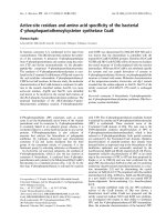
UDP-glucose dehydrogenase catalyzes an NAD
+
-depend-
ent twofold oxidation of UDP-glucose to generate UDP-
glucuronic acid (Fig. 1) [1]. In mammals, UDP-glucuronic
acid is used in the biosynthesis of hyaluronan and various
glycosaminoglycans such as heparin sulfate and chondroitin
sulfate [2]. In addition, it is used in the liver where the act of
glucuronidation targets molecules for excretion [3]. UDP-
glucuronic acid also serves as a precursor to UDP-xylose
which provides a major component of the cell wall polysac-
charides in plants [4]. In many strains of pathogenic bacteria,
such as group A streptoccoci and Streptococcus pneumoniae
type 3, UDP-glucuronic acid is used in the construction of
the antiphagocytic capsular polysaccharide [5,6]. This cap-
sule protects the bacteria from the immune system of the
host and thus serves as a major virulence factor.
UDP-glucose dehydrogenase is of mechanistic interest
because it belongs to a family of sugar nucleotide-modifying
enzymes that catalyze a net four-electron oxidation and
thus effectively serve as both alcohol dehydrogenases and
aldehyde dehydrogenases [7]. Other members of this
family include UDP-ManNAc dehydrogenase [8] and
GDP-mannose dehydrogenase [9,10]. Extensive studies on
both the bovine and S. pyogenes UDP-glucose dehydro-
genases have led to the mechanistic pathway outlined in
Fig. 1. The enzyme is thought to follow a Bi-Uni-Uni-Bi
ping-pong mechanism in which UDP-glucose is bound first
and UDP-glucuronic acid is released last [11,12]. The first
oxidation involves the transfer of the C-6¢ pro-R hydride
of UDP-glucose to the si face (B face) of NAD
+
to form
NADH and an aldehyde intermediate [13,14]. This inter-
mediate is bound tightly to the enzyme and is not accessible
to external aldehyde-trapping reagents [15,16]. Nevertheless,
it has been demonstrated that synthetic samples of the
aldehyde intermediate will serve as a kinetically competent
substrate for the second step of the reaction [17]. It is quite
likely that the bound form of the aldehyde exists largely as
a covalent hemithioacetal adduct as this species has been
implicated as an intermediate in the second step of catalysis,
although an imine linkage via an active site lysine has also
been proposed [18]. The second oxidation step involves the
addition of a cysteine thiol to the aldehyde to generate the
thiohemiacetal intermediate with subsequent hydride trans-
fer to the second NAD
+
molecule [19,20]. The resulting
thioester is hydrolyzed in a final step to generate the
product, UDP-glucuronic acid.
Strong evidence in support of this covalent catalysis
mechanism was obtained when a Cys260Ser mutant of the
S. pyogenes enzyme was incubated with UDP-glucose and
NAD
+
[20]. The mutant enzyme was essentially inactive;
however, mass spectral analysis indicated that a covalent
adduct had accumulated in which UDP-glucuronic acid was
attached to Ser260 via an ester linkage. The mutant enzyme
was therefore capable of catalyzing both oxidation steps of
the reaction but was incapable of hydrolyzing the unnatural
ester bond at any significant rate. Interestingly, when the
Correspondence to M. E. Tanner, Department of Chemistry,
University of British Columbia, 2036 Main Mall, Vancouver,
British Columbia, Canada V6T 1Z1.
Tel.: +1 604 822 9453, Fax: +1 604 822 2847,
E-mail:
Abbreviations: GAPDH, glyceraldehyde phosphate dehydrogenase.
Enzyme: UDP-glucose dehydrogenase (EC 1.1.1.22).
(Received 29 August 2003, revised 9 October 2003,
accepted 14 October 2003)
Eur. J. Biochem. 271, 14–22 (2004) Ó FEBS 2003 doi:10.1046/j.1432-1033.2003.03876.x
Cys260Ala mutant was examined, the enzyme was essen-
tially inactive towards UDP-glucose oxidation but readily
oxidized the intermediate aldehyde at a rate within an order
of magnitude of that seen with the wild-type enzyme.
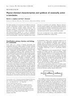
Apparently, the normal reaction proceeds via covalent
catalysis; however, with the alanine mutant the second
oxidation step can proceed directly from the hydrated
aldehyde (Fig. 2).
In subsequent work, X-ray structures were solved of the
native and Cys260Ser dehydrogenases from S. pyogenes
in complex with UDP-xylose/NAD
+
and UDP-glucuronic
acid/NAD(H), respectively [21]. In each case, Cys/Ser260
was positioned appropriately to participate in covalent
catalysis, as expected. Other active site residues that could
potentially play roles in the hydride transfer and/or hydro-
lysis steps were also identified. These include Thr118,
Glu141, Glu145, Lys204, Asn208, and Asp264. All but
Glu145 are strictly conserved among all family members.
Inspection of the hydrogen bonding networks at the site that
would be occupied by the C-6¢ hydroxyl of UDP-glucose in
the Michaelis complex led to the proposal of two scenarios
for the hydride transfer steps (presumably both hydride
transfer steps will employ the same catalytic residues). In
the first scenario, Lys204 acts as the catalytic base that
deprotonates the C-6¢ hydroxyl, whereas Asn208 and an
ordered water molecule serve as hydrogen bond donors to
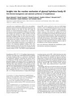
the hydroxyl oxygen (Fig. 3A). In the second scenario, the
ordered water molecule serves as the catalytic base with the
assistance of Asp264 as the ultimate proton acceptor
(Fig. 3B). In this case, Asn208 and Lys204 (presumably in
the ammonium form) serve as hydrogen bond donors to the
hydroxyl oxygen, and Thr118 and a ribose hydroxyl of
NAD
+
form hydrogen bonds with the ordered water. The
identity of the catalytic residues involved in hydrolysis of
the thioester intermediate are harder to predict from these
structures; however, Glu141 and Glu145 are potential
candidates.
In this work, the possibility of imine formation involving
Lys204 is examined with the use of H
2
18
O, and the nature
of the rate determining steps of catalysis is probed using
deuterated UDP-glucose. Studies on the Cys260Ala
mutant and other active site mutants are described which
help to further delineate the roles these residues play in
catalysis.
Experimental procedures
General procedures
UDP-glucose dehydrogenase from S. pyogenes was
expressed in Escherichia coli using the plasmid pGAC147
as described elsewhere [5]. All proteins were purified as
described previously [12] and analyzed by ESI-MS to ensure
they had the expected molecular masses. All enzymatic
assays for dehydrogenase activity used the method des-
cribed previously [12], unless otherwise indicated. The
aldehyde intermediate, uridine diphospho-a-
D
-gluco-
hexodialdose, was synthesized as described previously [17],
and stock concentrations were calculated from measure-
ments of A
262
using a value of e
262
¼ 8700
M
)1
for the
uridine chromophore. H
2
18
O (95% enriched) was from
Cambridge Isotope Laboratories. [6,6-di-
2
H]-
D
-Glucose
(98% enriched) was from Aldrich. Commercial enzymes
and sugar nucleotides were from Sigma or Boehringer
Mannheim Biochemicals, unless stated otherwise.
Site-directed mutagenesis and mutant protein
expression
The mutagenesis protocol to produce the active site mutants
followed an adaptation of enzymatic PCR [22] using
plasmid pGAC147 [5] which contains a copy of hasBas
template. The primers for the construction of the mutant
plasmids were as follows: T118A, 5¢-CGCGGTACCAAC
TCTTATCATCAAATCAGCAATTC-3¢ and 5¢-ATAG
GTACCATGGCTATTAACGCTTAGTACC-3¢; E141Q,
5¢-CGCGGTACCTTTATATGACAACTTATATC-3¢
and 5¢-ATAGGTACCTTTAGATTCTCTTAAAAACTG
AGGGC-3¢; E145Q, 5¢-CGCGGTACCTTTATATGAC
AACTTATATC-3¢ and 5¢-ATAGGTACCTTTAGACTG
TCTTAAAAATTCAG-3¢;K204A,5¢-CGCGGTACCC
AATACTTATTTAGCG-3¢ and 5¢-ATAGGTACCAAA
TAGTGCTACTGCTTCAGC-3¢; N208A, 5¢-CGCGGTA
CCGTTAAGGGTAGC-3¢ and 5¢-ATAGGTACCTAAA
Fig. 1. The mechanism of the reaction cata-
lyzed by UDP-glucose dehydrogenase.
Ó FEBS 2003 Active site residues of UDP-glucose dehydrogenase (Eur. J. Biochem. 271)15
TAAGTAGCGGCAAATAG-3¢; D264N, 5¢-CGCGGTA
CCCAATTATTGGCAAATTAC-3¢ and 5¢-ATAGGTAC
CTTCGTGTTTTTAGGTAGACA-ATAAC-3¢.Nucleo-
tides designated in bold led to the desired mutations.
Cys260Ala was expressed using the plasmid pGAC400 as
described previously [20]. All mutations and constructs were
confirmed by sequencing the entire gene.
Expression experiments of the mutant constructs
were initiated by transforming each plasmid into E. coli
JM109(DE3) and colonies were grown overnight in TYPG
(3 mL). The TYPG media contained 8 g of tryptone, 8 g
of yeast extract, 2.5 g of NaCl, 1.25 g of K
2
HPO
4
and
2.5 g of glucose per 500 mL of distilled water. For
inductions, either the JM109(DE3) culture harboring
pGAC147 (3 mL) or the JM109(DE3) culture harboring
the desired mutant construct (0.5 mL) was inoculated into
TYPG (250 mL) and grown at 37 °C with vigorous
shaking until the cultures reached D
600
¼ 0.8–1.2. An
aliquot of the culture (10 mL) was removed just prior to
induction by isopropyl thio-b-
D
-galactoside (0.4 m
M
final
concentration) and the incubation was resumed. Following
induction aliquots of culture (10 mL) were removed at 1-h
intervals, rapidly chilled on ice, and a portion (1.5 mL)
was prepared for SDS/PAGE analysis. Bacteria were
sedimented at 13 000 g (10 s), the pellets were resuspended
in TE (150 lL; 10 m
M
Tris pH 7.5, 1 m
M
EDTA), and
finally the cells were solubilized by boiling for 5 min in the
presence of 6 · loading buffer (30 lL [23]). SDS/PAGE
was performed by the method of Laemmli [24] using 10%
gels and loading 20 lL lysate per lane to confirm the
overexpression of the mutant constructs.
Solvent isotope incorporation study
A solution of sodium phosphate buffer (50 m
M
, 500 lL),
pH 8.0, containing 50% H
2
18
O (v/v), UDP-glucose
(50 m
M
), NAD
+
(10 m
M
), FMN (10 m
M
), dithiothreitol
(2 m
M
), and UDP-glucose dehydrogenase (0.35 mg) was
incubated at 37 °C for 20 h. At periodic intervals the
reaction was gently mixed and exposed to atmospheric
oxygen. After the incubation, EDTA (16 m
M
) was added
and the mixture was lyophilized to dryness. The residue was
redissolved in D
2
Oand
13
C NMR spectra were recorded. A
control sample was run containing 100% H
2
16
O.
For further purification, the samples were applied to a
column of Bio-Gel P-2 resin (40 mL) and eluted with
water. Fractions containing the UDP-glucuronic acid
product were lyophilized and submitted for MALDI MS
analysis.
Kinetic isotope effect studies
UDP-[6¢¢,6¢¢-di-
2
H]-
D
-glucose was prepared using a slight
modification of a procedure previously described for the
preparation of tritiated UDP-glucose [25]. A solution
of Tris/HCl buffer (70 m
M
, 40 mL) pH 7.8, containing
[6,6-di-
2
H]-
D
-glucose (8 mg, 44 lmol, 1.1 m
M
), ATP
(73 mg, 132 lmol), UTP (64 mg, 132 lmol), glucose
1,6-diphosphate (0.165 lmol), MgSO
4
(36 mg, 146 lmol),
hexokinase (66 U), phosphoglucomutase (109 U), UDP-
glucose pyrophosphorylase (12.5 U), and inorganic pyro-
phosphatase (33 U) was incubated at 30 °Cfor20h.The
reaction mixture was applied to a column of DE-52 anion
exchange resin (65 mL) and eluted with a linear gradient of
0–400 m
M
triethylammonium bicarbonate (800 mL total).
Fractions containing UDP-glucose (as assayed by UDP-
glucose dehydrogenase and NAD
+
) were pooled, lyophi-
lized, redissolved in water, and lyophilized a second time.
The product was redissolved in water and passed through a
column of Amberlite IR-120 (plus) resin (10 mL, Na
+
form, eluted with water). The resulting solution was applied
to a column of Bio-Gel P-2 resin (2.5 · 45 cm) and eluted
with water. UV active fractions were pooled and lyophilized
to dryness to give the disodium salt of UDP-[6¢¢,6¢¢-di-
2
H]-
D
-glucose as a white solid (29 lmol, 66% yield assayed
enzymatically, see below).
1
H NMR and mass spectra were
consistent with the assignment of the product as UDP-
[6¢¢,6¢¢-di-
2
H]-
D
-glucose, and indicated the extent of deuter-
ium incorporation to be > 95%; LSI(–) MS (thioglycerol)
m/z 567 (M(di-
2
H)–H
+
, 100%).
The rates of the dehydrogenase reaction were determined
under saturating conditions for both substrates using a
previously described kinetic assay [12]. The values of [UDP-
glucose] (deuterated or undeuterated) and [NAD
+
]were
as follows: Wild-type enzyme (0.5 m
M
,1.2m
M
), T118A
(0.5 m
M
,4m
M
), E141Q (0.5 m
M
,2m
M
), and E145Q
(0.8 m
M
,2m
M
). The error reported for the kinetic isotope
effects are the SD of the data points from the average
determined ratios (five independent measurements).
The concentrations of the stock UDP-glucose solu-
tions (both deuterated and undeuterated) were determined
Fig. 3. Two scenarios outlining the residues involved in catalyzing the
hydride transfer steps. (A) Lys204 serving as the key acid/base residue.
(B) An ordered water molecule and Asp264 serving as the key acid/
base residue. X ¼ H for the first oxidation step, X ¼ SR (from
Cys260) for the second oxidation step.
Fig. 2. The proposed mechanism for the oxidation of the hydrated
aldehyde intermediate by the Cys260Ala mutant.
16 X. Ge et al. (Eur. J. Biochem. 271) Ó FEBS 2003
enzymatically by running the UDP-glucose dehydrogenase
reaction to completion. The substrate concentrations were
calculated from the changes in A
340
assuming a stoichio-
metry of two equivalents of NADH produced for every
molecule of UDP-glucose consumed, and an extinction
coefficient for NADH of e ¼ 6220
M
)1
.
Cys260Ala studies
Initial velocity kinetics for the reduction of the aldehyde
intermediate. Assays were performed at 30 °CinTrien/
HCl buffer (50 m
M
, 0.50 mL total volume) pH 8.7, con-
taining NADH (0.15 m
M
) and dithiothreitol (2 m
M
)with
varying amounts of aldehyde. Initial velocities were meas-
ured during the first 60 s after initiation with the Cys260Ala
mutant and calculated from the decrease in A
340
using an
extinction coefficient for NADH of e ¼ 6220
M
)1
.
Full time course analysis of incubations with the aldehyde
intermediate and NADH. Thealdehydeintermediate
(75 l
M
with wild-type enzyme, 92 l
M
with Cys260Ala)
was incubated at 30 °C with NADH (0.15 m
M
) and either
wild-type dehydrogenase (0.22 l
M
)ortheCys260Ala
mutant (0.22 l
M
) in Trien/HCl buffer (50 m
M
,0.50mL
total volume), pH 8.7, containing dithiothreitol (2 m
M
).
The changes in NADH concentrations were monitored
using A
340
assuming an extinction coefficient for NADH of
e ¼ 6220
M
)1
. To confirm the identity of the products at the
end-point of these reactions, aliquots were analyzed by ion-
paired reversed phase HPLC as described previously and
compared with authentic standards [14].
Deuterium washout experiment. A solution of Trien/HCl
buffer (50 m
M
, 2 mL total volume) pH 8.7, containing
UDP-
D
-[6¢,6¢-di-
2
H]-glucose (2 m
M
), NADH (20 m
M
),
NAD
+
(4 m
M
), dithiothreitol (2 m
M
), and the Cys260Ala
mutant (0.1 mg) was incubated at 30 °C for 23 h. The
resulting solution was applied to a column of DE-52 anion
exchange resin (60 mL) and eluted with a linear gradient of
0–300 m
M
ammonium bicarbonate (800 mL total). Frac-
tions containing UDP-glucose (as assayed by UDP-glucose
dehydrogenase and NAD
+
) were pooled and lyophilized.
The resulting solids were dissolved in water and passed
through a column of Amberlite IR-120 (plus) resin (10 mL,
Na
+
form, eluted with water). The eluent was lyophilized to
dryness, redissolved in water, applied to a column of Bio-
Gel P-2 resin (2.5 · 45cm),andelutedwithwater.UV
active fractions were pooled and lyophilized to dryness. The
UDP-
D
-[6¢¢,6¢¢-di-
2
H]-glucose obtained in this fashion was
analyzed for deuterium content by
1
H NMR spectroscopy
and LSI-MS, and showed identical spectral characteristics
to the original labeled material.
Results
Solvent
18
O-isotope incorporation study
The fact that the aldehyde intermediate in the UDP-glucose
dehydrogenase reaction is never released into solution and is
inaccessible to external trapping reagents led to the sugges-
tion that this species is covalently bound to an active site
lysine residue via an imine linkage [18]. This suggestion was
supported by experiments on the bovine liver enzyme in
which the active site cysteine thiol had been chemically
modified to a thiocyanide moiety. When this inactivated
form of the enzyme was incubated with UDP-glucose and
NAD
+
(or with the aldehyde intermediate alone) and then
treatedwithNaBH
4
, a covalent enzyme-substrate adduct
was generated. The properties of the adduct were consistent
with that expected for a reduction product of a Schiff’s base
formed between the aldehyde intermediate and an active site
lysine, and the authors concluded that an imine was formed
on the normal reaction pathway. The suggestion was
somewhat at odds with earlier
18
O-isotopic labeling studies
showing that the reaction proceeds with the incorporation
of only one solvent-derived oxygen atom into the carboxy-
late product [26]. The proposal of an imine intermediate
could only be consistent with this observation if the original
C6¢¢ oxygen atom was sequestered in the active site during
the second oxidation step and then re-delivered during
hydrolysis of the thioester.
More recent work has focused on the bacterial enzyme
from S. pyogenes, and the X-ray crystal structure has shown
that Lys204 is in the active site and is in close proximity
to C6¢ of UDP-glucose [21]. In order to re-examine the
possibility of Schiff base formation with this enzyme, the
reaction was carried out in H
2
18
O water and the UDP-
glucuronic acid produced was examined for
18
O-isotope
content. Samples of UDP-glucose were incubated with
substoichiometric amounts of NAD
+
in the presence of
FMN and oxygen [27,28]. The FMN/O
2
acts as an NAD
+
regenerating system and facilitates purification of the UDP-
glucuronic acid from the minor dinucleotide contaminants.
Reactions were carried out in both H
2
16
Oand50%
H
2
16
O/50% H
2
18
O, and the resulting UDP-glucuronic acid
was analyzed by MALDI TOF MS. In the latter case,
signals corresponding to both unlabeled (m/z 648.3,
M-2H
+
+3Na
+
) and singly
18
O-labeled product (m/z
650.3) were observed in approximately equal amounts,
confirming that the reaction was accompanied by incor-
poration of a single solvent-derived oxygen atom. In order
to determine the position of the incorporated
18
O-atom,
both samples were analyzed by
13
C NMR spectroscopy. In
the control sample, a single carboxylate signal is observed at
176.671 p.p.m., whereas, in the 50% H
2
16
O/50% H
2
18
O
sample two signals of approximately equal intensity were
observed at 176.673 and 176.647 p.p.m. (Fig. 4). The
0.026 p.p.m. separation of the signals in the latter sample
is consistent with the magnitude expected for an
18
O-isotope
induced shift in a mono-labeled carboxylate, and confirms
that the label was introduced into the carboxylate of
UDP-glucuronic acid [28].
Deuterium kinetic isotope effect study
In order to determine if either hydride transfer step in
the reaction catalyzed by UDP-glucose dehydrogenase
was rate determining, samples of 6¢¢,6¢¢-dideuterated-
UDP-glucose were prepared and analyzed for the pres-
ence of a primary kinetic isotope effect on catalysis.
Since both C6¢¢ positions were labeled, a single experi-
ment could be used to simultaneously probe both
hydride transfer steps. UDP-[6¢¢,6¢¢-di-
2
H]-
D
-glucose was
prepared enzymatically from [6,6-di-
2
H]-
D
-glucose using
Ó FEBS 2003 Active site residues of UDP-glucose dehydrogenase (Eur. J. Biochem. 271)17
hexokinase, phosphoglucomutase, UDP-glucose pyro-
phosphorylase, and inorganic pyrophosphatase. The
resulting UDP-[6¢¢,6¢¢-di-
2
H]-
D
-glucose was found to be
> 95% enriched with two deuterium labels when ana-
lyzed by MS. The rates of the UDP-glucose dehydro-
genase reaction for both the labeled and unlabeled
substrates were determined under saturating conditions
and the value of k
H
/k
D
was found to be 1.1 ± 0.1. This
demonstrates that there is no primary kinetic isotope
effect on the reaction of this substrate and therefore the
hydride transfer steps are not rate determining in this
reaction.
Site-directed mutagenesis
In an effort to further understand the roles of the active site
residues in catalysis several mutant proteins were targeted
for study. Plasmids encoding Thr118Ala, Glu141Gln,
Glu145Gln, Lys204Ala, Asn208Ala, Cys260Ala, and
Asp264Asn were generated and shown to result in high
levels of protein expression. Unfortunately, with several of
the key mutants (K204A, N208A, and D264N), all of the
expressed protein was produced in insoluble inclusion
bodies and attempts to solubilize these proteins were
unsuccessful. With Cys260Ala, much of the protein was
present in inclusion bodies, however, enough remained in
solution to allow for the purification and study of this
mutant. Thr118Ala, Glu141Gln, and Glu145Gln were
soluble and could be isolated in good yield.
Studies on Cys260Ala
Cys260 provides the key active site thiol involved in covalent
catalysis with this enzyme and thus mutants of this residue
warrant further investigation. In previous studies, the
Cys260Ala mutant was reported to be essentially inactive
towards the oxidation of UDP-glucose (< 0.01% activity
of wild-type enzyme) [20]. However, the mutant was quite
capable of catalyzing the oxidation of the aldehyde inter-
mediate at rates within an order of magnitude of that of
thewild-typeenzyme(k
cat
¼ 0.19 s
)1
, K
M
¼ 0.26 m
M
vs.
k
cat
¼ 1.2 s
)1
, K
M
¼ 0.014 m
M
, respectively). Since this
mutant is presumably incapable of participating in covalent
catalysis, it would appear that it simply binds the hydrated
form of the aldehyde from solution and oxidizes it directly
to UDP-glucuronic acid (Fig. 2). The question remains,
however, as to why UDP-glucose is not a substrate for the
Cys260Ala mutant. One possibility is that the Cys260 thiol
plays a key role in the first oxidation step of the reaction, in
addition to its role in covalent catalysis during the second
oxidation step. An alternate possibility is that Cys260 is not
required for the first oxidation step and the aldehyde
intermediate is readily formed by the mutant enzyme;
however, it is tightly bound and there is no mechanism by
which it can be hydrated and proceed forward to the second
oxidation step.
In order to address the previous question, the ability of
the Cys260Ala mutant to catalyze the reverse of the first step
in catalysis, namely reduction of the aldehyde intermediate,
was examined. The aldehyde intermediate was generated via
chemical synthesis and was found to be an excellent
substrate for reduction by NADH using the Cys260Ala
mutant. The values of k
cat
and K
M
for the aldehyde were
determined (at 0.15 m
M
NADH) to be 1.9 ± 0.1 s
)1
and
58 ± 7 l
M
, respectively (it was not possible to measure
these values with the wild-type enzyme due to a dismutation
process, vide infra). It should be noted that the value of K
M
essentially represents an Ôapparent K
M
Õ since the majority of
the aldehyde in solution exists as a hydrate [17] and the
reduction process requires that the enzyme bind the
unhydrated form of the aldehyde. The observation that
Cys260Ala catalyzes a reasonably rapid reduction of the
aldehyde clearly demonstrates that Cys260 does not play a
key role in catalysis of the first oxidation step. It also means
that Cys260Ala must be capable of catalyzing the reverse
reaction, namely the oxidation of UDP-glucose to give the
free aldehyde. The Haldane equation dictates that the
extremely slow rate of this oxidation process must ulti-
mately be attributed to an unfavorable equilibrium constant
reflecting the higher energies of the free aldehyde and
NADH.
These observations are further supported by monitoring
the full time course of the reaction of the aldehyde
intermediate with excess NADH (Fig. 5). When the wild-
type enzyme is the catalyst, an initial decrease in A
340
is
observed due to the reduction of the aldehyde that generates
NAD
+
and UDP-glucose. As NAD
+
accumulates, how-
ever, it is possible for the enzyme to oxidize either the
aldehyde intermediate or UDP-glucose to UDP-glucuronic
acid and regenerate NADH. Ultimately, the A
340
value
returns to its initial position, indicating there is no net
consumption of NADH. This phenomenon can be under-
stood by considering a dismutation process in which the
aldehyde disproportionates between the alcohol and the acid
without consuming NADH (Fig. 6). The thermodynamic
stability of UDP-glucuronic acid ultimately drives the
Fig. 4.
13
C NMR spectra of the carboxyl group of UDP-glucuronic acid
(A) generated in 50% H
18
2
O/50% H
16
2
O and (B) generated in
100% H
16
2
O.
18 X. Ge et al. (Eur. J. Biochem. 271) Ó FEBS 2003
dismutation to completion. With the Cys260Ala mutant, a
rapid decrease in A
340
is observed due to reduction of the
aldehyde intermediate and the consumption of NADH
(Fig. 5). In this case, however, the NADH is not regenerated
by the dismutation process and the value of A
340
does not
return to its original position. This occurs because the UDP-
glucose that is generated by reduction of the aldehyde
cannot be oxidized by this mutant and is therefore kinetically
trapped in that form. By calculating the amount of NAD
+
consumed, it was found that 80% of the aldehyde was
converted to UDP-glucose in this fashion. The remaining
20% of the aldehyde did undergo dismutation since a
fraction of it was oxidized directly to the acid by the enzyme
and the accumulating NAD
+
.Inbothexperiments,HPLC
analysis with authentic standards of the UDP-sugars was
used to confirm the expected product distributions. This
kinetic behavior is entirely consistent with the curious
observation that Cys260Ala can both oxidize the aldehyde
to the acid and reduce it to the alcohol, but that it cannot
oxidize the alcohol to the acid at any appreciable rate.
Since the Cys260Ala mutant is capable of catalyzing the
first hydride transfer step, the ternary complex of bound
NADH and aldehyde intermediate must be formed upon
incubation of the alcohol and NAD
+
. In order to proceed
with the second oxidation step, however, a nucleophile must
add to the aldehyde, and in the absence of the active site
thiol a water molecule must assume this role. If neither
release, nor hydration, of the bound aldehyde can proceed
at a reasonable rate, no turnover would be observed and the
only fate of the bound species would be to return to starting
materials via the reverse reaction. In order to probe further
the nature of this ternary complex an experiment was
devised to determine whether the bound NADH could
exchange into bulk solution at any measurable rate. A
sample of UDP-
D
-[6¢¢,6¢¢-di-
2
H]-glucose was incubated with
the Cys260Ala mutant in the presence of 20 m
M
NADH
and 4 m
M
NAD
+
. An initial oxidation event should
produce a ternary complex of the monodeuterated aldehyde
and NAD
2
H. If the bound NAD
2
H could exchange with
unlabeled NADH, then back reaction would lead to the
formation of monodeuterated UDP-glucose, and a net
ÔwashoutÕ of the pro-R deuterium should be observed. Even
upon extensive incubations, however, no significant loss of
deuterium label could be detected in the recovered UDP-
D
-
[6¢¢,6¢¢-di-
2
H]-glucose as analyzed by both MS and
1
HNMR
spectroscopy. This indicates that the NADH is not released
into solution from the mutant ternary complex and suggests
that in the wild-type reaction, the first-formed NADH is not
released until the Cys260 thiolate adds to bound aldehyde
and generates the thiohemiacetal intermediate.
Studies on Thr118Ala, Glu141Gln, and Glu145Gln
The three remaining mutants that could be obtained in a
soluble form were analyzed for their ability to catalyze the
UDP-glucose dehydrogenase reaction (Table 1). E141Q
and E145Q showed similar behavior in that the values for
Fig. 6. The dismutation of the aldehyde intermediate during an incuba-
tion with NADH and wild-type UDP-glucose dehydrogenase. Boxed
structures represent species present at the completion of the reaction.
Fig. 5. Kinetic trace following the full time-course of the incubation of
the aldehyde intermediate and NADH with either wild-type UDP-
glucose dehydrogenase (solid line) or the Cys260Ala mutant (dashed
line).
Table 1. Kinetic constants and kinetic isotope effects for the reactions
catalyzed by the wild-type and mutant UDP-glucose dehydrogenases.
Enzyme
UDP-Glc
K
M
(l
M
)
NAD
+
K
M
(l
M
) k
cat
(s
)1
) k
H
/k
D
a
Wild-type 20 ± 4 65 ± 6 1.8 ± 0.1 1.1 ± 0.1
T118A 59 ± 9 400 ± 100 0.011 ± 0.003 1.9 ± 0.1
E141Q 60 ± 9 135 ± 7 0.14 ± 0.02 1.4 ± 0.1
E145Q 125 ± 24 187 ± 20 0.20 ± 0.04 1.4 ± 0.1
a
Rate of UDP-glucose oxidation vs. the rate of UDP-[6¢¢,6¢¢-
di-
2
H]glucose oxidation measured under saturating conditions (see
Experimental procedures).
Ó FEBS 2003 Active site residues of UDP-glucose dehydrogenase (Eur. J. Biochem. 271)19
k
cat
were 10-fold lower than those obtained with the wild-
type dehydrogenase, and the values for K
M
were two- to six-
fold higher. The modest changes in the catalytic constants
suggest these active site residues do not play key roles in
either catalysis or binding. In the case of Thr118Ala, the
value of k
cat
dropped by 160-fold, whereas the K
M
values
increased slightly. This indicates that Thr118 is reasonably
important for catalysis, although it is probably not serving
as a key acid/base catalyst or nucleophile. In order to probe
the nature of the steps that were affected by the mutations,
the mutants were examined for the possible presence of a
primary kinetic isotope effect on the oxdiation of UDP-
[6¢¢,6¢¢-di-
2
H]-
D
-glucose. In the case of both E141Q and
E145Q, the k
cat
isotope effects were found to be 1.4 ± 0.1.
In the case of Thr118Ala, however, k
H
/k
D
was found to be
1.9 ± 0.1, consistent with the presence of a primary kinetic
isotope effect. This indicates that with the Thr118Ala
mutant, one or both of the hydride transfer steps has
become the rate-limiting step of catalysis.
Discussion
The isotope incorporation studies described in this manu-
script confirm that the S. pyogenes UDP-glucose dehydro-
genase reaction proceeds with the incorporation of a single
solvent derived oxygen atom into the product carboxylate.
This is consistent with previous results obtained using the
bovine liver enzyme [26]. This observation does not disprove
the existence of an imine intermediate during catalysis, since
the original C6¢¢ oxygen atom from UDP-glucose could
conceivably be sequestered within the active site and
redelivered into the product carboxylate during the final
hydrolysis step. Nevertheless, the absence of any di-labeled
product, combined with a lack of any chemical rationale as
to a how imine formation could facilitate catalysis, argues
against the formation of such an intermediate. Instead, it is
likely that once the aldehyde is formed, the thiol of Cys260
readily adds to generate the covalently bound thiohemi-
acetal intermediate (Fig. 1). In the previous studies that
used the thiocyanide-modified bovine enzyme and NaBH
4
trapping, the isolated adduct was probably formed as a
result of the unnatural modification to the active site thiol
that prevents the aldehyde from proceeding forward in
catalysis [18]. Thus, the imine formation observed with the
modified enzyme probably does not reflect a step that
occurs in the normal reaction pathway.
The absence of any primary kinetic isotope effect during
the oxidation of UDP-
D
-[6¢¢,6¢¢-di-
2
H]-glucose indicates that
neither of the hydride transfer steps are rate determining in
the reaction of the wild-type enzyme. Instead, another
chemical step such as the hydrolysis of the thioester
intermediate may be rate determining. This was clearly the
case with the Cys260Ser mutant in which the ester
intermediate accumulated and could be isolated [20]. Kinetic
studies have also led to the suggestion that an irreversible
thioesterhydrolysisstepisratedetermininginthecaseofthe
beef liver enzyme [11].
Studies with the Cys260Ala mutant showed that this
mutant is still capable of oxidizing the aldehyde intermedi-
ate at a reasonable rate [20]. It is likely that the mutant is
binding the hydrated aldehyde from solution and oxidizing
it directly to the acid without using covalent catalysis
(Fig. 2). Similar observations have been made with the
phosphorylating glyceraldehyde 3-phosphate dehydro-
genase (GAPDH) from E. coli [29]. This enzyme normally
oxidizes an aldehyde using a thiol-based covalent catalysis
strategy similar to the second step of the UDP-glucose
dehydrogenase reaction. The resulting thioester intermedi-
ate is attacked by phosphate to generate the acyl phosphate
product, 1,3-diphosphoglycerate. When the active site
cysteine was converted to an alanine, however, the resulting
mutant catalyzed the formation of 3-phosphoglycerate as
the sole product. It was suggested that the mutant had
oxidized the hydrated form of the aldehyde directly to the
acid, and was thereby converted into a nonphosphorylating
GAPDH. Other enzymes, such as histidinol dehydrogenase
[30,31] and alcohol dehydrogenase [32,33], are also known
to be able to oxidize hydrated aldehydes directly to acids
without using covalent catalysis.
The observation that the Cys260Ala mutant of UDP-
glucose dehydrogenase can efficiently catalyze the reverse of
the first oxidation step, namely reduction of the aldehyde
intermediate, indicates that the Cys260 is not required for
this step. Instead, the inability of this mutant to catalyze the
overall oxidation of the alcohol to the acid must be due to
the reasonably high energy of the aldehyde intermediate. In
the reaction of the wild-type enzyme, this intermediate is
stabilized by binding interactions with active site residues
and is readily converted to the thiohemiacetal by attack of
Cys260. In the case of Cys260Ala, however, the only way
for catalysis to proceed is for the intermediate to be released
into solution and then rebound as the hydrate. Apparently
release of the aldehyde intermediate is very slow, and
hydration of the bound aldehyde does not readily occur,
hence the overall rate of catalysis is also very slow. This
phenomenon is readily apparent when the full time course
of the aldehyde reduction is observed. With the wild-type
enzyme, an initial reduction of the aldehyde is observed,
followed by oxidation of the aldehyde/alcohol as NAD
+
accumulates. The net result is a dismutation in which the
aldehyde is converted into equimolar amounts of alcohol
and acid, with no net consumption of NADH. Similar
dismutation processes have been observed with alcohol
dehydrogenases that show aldehyde dehydrogenase activit-
ies [32,33]. In the case of Cys260Ala, however, the initial
reduction phase generates a great deal of UDP-glucose that
is kinetically trapped and cannot be reoxidized at any
appreciable rate. Only 20% of the aldehyde underwent
dismutation in this case. The final experiment with
Cys260Ala was a test for NADH exchange in the ternary
dehydrogenase/aldehyde/NADH complex. The absence of
deuterium washout from UDP-[6¢¢,6¢¢-di-
2
H]-
D
-glucose dur-
ing an incubation with the mutant, NAD
+
, and excess
NADH shows that the NAD
2
H formed in the initial
oxidation step does not exchange with free NADH. Its only
fate, therefore, is a back reaction to produce the initial
starting materials. A similar ternary complex is also formed
in the case of the wild-type reaction; however, the thiol
group of Cys260 can readily add to the aldehyde carbonyl
group. The first formed NADH may then exchange with
NAD
+
and the second oxidation step may proceed. The
results obtained with the mutant enzyme are consistent with
the idea that cofactor exchange only takes place after the
nucleophilic thiol adds to the carbonyl. This helps to explain
20 X. Ge et al. (Eur. J. Biochem. 271) Ó FEBS 2003
how the enzyme efficiently sequesters the aldehyde inter-
mediate during catalysis.
Our attempts at investigating the roles of other key active
site residues via mutagenesis studies were hampered by an
inability to isolate several of the mutants (Lys204Ala,
Asn208Ala, and Asp264Asn) in a soluble form. Of the three
mutants that were amenable to purification (Thr118Ala,
Glu141Gln, and Glu145Gln), only Thr118Ala showed a
substantial reduction in catalytic efficiency. This mutant
also showed a primary kinetic isotope effect upon the
oxidation of UDP-[6¢¢,6¢¢-di-
2
H]-
D
-glucose, indicating that a
hydride transfer step was rate limiting for catalysis. Con-
sidering the two scenarios for residues involved in promo-
ting the hydride transfer steps [21], these observations are
most consistent with the one in which the ordered water
molecule serves as the catalytic base with the assistance of
Asp264 as the ultimate proton acceptor (Fig. 3B). The
water molecule is within hydrogen bonding distance of
Thr118 and removing such an interaction would certainly
perturb its environment. If the water molecule does play a
key role in the hydride transfer steps, then mutations to
Thr118 may increase the barrier to these steps and render
them rate determining. Further investigations will be
necessary to fully outline the roles of the active site residues
in both the hydride transfer and thioester hydrolysis steps of
this interesting enzymatic transformation.
Acknowledgements
This research was supported by NSERC (operating grant to M.E.T)
and the NIH (Public Health Service Grant AI37320 to I.v.d.R).
References
1. Oppenheimer, N.J. & Handlon, A.L. (1992) Mechanism of NAD-
dependent enzymes. Enzymes 20, 453–504.
2. Rode
´
n, L. (1980) In The Biochemistry of Glycoproteins and Pro-
teoglycans (Lennarz, W.J. & , eds), pp. 267–371. Plenum Pub-
lishing Co, New York, NY.
3. Dutton, G.J. (1980) Glucuronidation of Drugs and Other Com-
pounds. CRC Press Inc., Boca Raton, FL.
4. Dalessandro, G. & Northcote, D.H. (1977) Changes in enzymic
activities of nucleoside diphosphate sugar interconversions during
differentiation of cambium to xylem in sycamore and poplar.
Biochem. J. 162, 267–279.
5. Dougherty, B.A. & van de Rijn, I. (1993) Molecular character-
ization of hasB from an operon required for hyaluronic
acid synthesis in group A streptococci. J. Biol. Chem. 268, 7118–
7124.
6. Arrecubieta, C., Lo
´
pez, R. & Garcı
´
a, E. (1994) Molecular char-
acterization of cap3A, a gene from the operon required for the
synthesis of the capsule of Streptococcus pneumoniae type 3:
Sequencing of mutations responsible for the unencapsulated
phenotype and localization of the capsular cluster on the pneu-
mococcal chromosome. J. Bacteriol. 176, 6375–6383.
7. Feingold, D.S. & Franzen, J.S. (1981) Pyridine nucleotide-linked
four electron- transfer dehydrogenases. Trends Biochem. Sci. 6,
103–105.
8. Kawamura, T., Ishimoto, N. & Ito, E. (1979) Enzymatic synthesis
of uridine diphosphate N-acetyl-
D
-mannosaminuronic acid.
J. Biol. Chem. 254, 8457–8465.
9. Snook, C.F., Tipton, P.A. & Beamer, L.J. (2003) Crystal structure
of GDP-mannose dehydrogenase: a key enzyme of alginate bio-
synthesis in P. aeruginosa. Biochemistry 42, 4658–4668.
10. Naught,L.E.,Gilbert,S.,Imhoff,R.,Snook,C.,Beamer,L.&
Tipton, P. (2002) Allosterism and cooperativity in Pseudomonas
aeruginosa GDP-mannose dehydrogenase. Biochemistry 41, 9637–
9645.
11. Ordman, A.B. & Kirkwood, S. (1977) UDP-glucose dehydro-
genase. Kinetics and their mechanistic implications. Biochim.
Biophys. Acta 481, 25–32.
12.Campbell,R.E.,Sala,R.F.,vandeRijn,I.&Tanner,M.E.
(1997) Properties and kinetic analysis of UDP-glucose dehydro-
genase from group A streptococci. J. Biol. Chem. 272, 3416–
3422.
13. Ridley, W.P. & Kirkwood, S. (1973) The stereospecificity of
hydrogen abstraction by uridine diphosphoglucose dehydro-
genase. Biochem. Biophys. Res. Commun. 54, 955–960.
14. Campbell, R.E. & Tanner, M.E. (1999) UDP-Glucose analogues
as inhibitors and mechanistic probes of UDP-glucose dehydro-
genase. J. Org. Chem. 64, 9487–9492.
15. Strominger, J.L., Maxwell, E.S., Axelrod, J. & Kalckar, H.M.
(1957) Enzymatic formation of uridine diphosphoglucuronic acid.
J. Biol. Chem. 224, 79–90.
16. Nelsestuen, G.L. & Kirkwood, S. (1971) The mechanism of action
of uridine diphosphoglucose dehydrogenase. J. Biol. Chem. 246,
3828–3834.
17. Campbell, R.E. & Tanner, M.E. (1997) Uridine diphospho-a-
D
-gluco-hexodialdose: Synthesis and kinetic competence in the
reaction catalyzed by UDP-glucose dehydrogenase. Angew. Chem.
Int., English edition 36, 1520–1522.
18. Ordman, A.B. & Kirkwood, S. (1977) Mechanism of action of
uridine diphosphoglucose dehydrogenase. J. Biol. Chem. 252,
1320–1326.
19. Ridley, W.P., Houchins, J.P. & Kirkwood, S. (1975) Mechanism
of action of uridine diphosphoglucose dehydrogenase. J. Biol.
Chem. 250, 8761–8767.
20. Ge, X., Campbell, R.E., van de Rijn, I. & Tanner, M.E. (1998)
Covalent adduct formation with a mutated enzyme: Evidence for a
thioester intermediate in the reaction catalyzed by UDP-glucose
dehydrogenase. J. Am. Chem. Soc. 120, 6613–6614.
21. Campbell, R.E., Mosimann, S.C., van de Rijn, I., Tanner, M.E. &
Strynadka, N.C.J. (2000) The first structure of UDP-glucose
dehydrogenase reveals the catalytic residues necessary for the two-
fold oxidation. Biochemistry 39, 7012–7023.
22. Hughes, M.J. & A.D. (1996) Creation of deletion, insertion and
substitution mutations using a single pair of primers and PCR.
Biotechniques 20, 192–196.
23. Ausubel, F.M., Brent, R., Kingston, R.E., Moore, D.D., Seid-
man, J.G., Smith, J.A. & Struhl, K. (1987) Current Protocols in
Molecular Biology. Wiley, New York.
24. Laemmli, U.K. (1970) Cleavage of structural proteins during
the assembly of the head of bacteriophage T4. Nature 227, 680–
685.
25. Snetkova,E.V.,Akulov,G.P.,Gordeeva,L.S.&Kaminskii,Y.L.
(1987) Synthesis of UDP-[1-
3
H]glucose and UDP-[6-
3
H]glucose.
Khimiya Prirodnykh Soedinenii 1, 125–128.
26. Schiller, J.G., Bowser, A.M. & Feingold, D.S. (1972) Studies on
the mechanism of action of UDP-
D
-glucose dehydrogenase from
beef liver. Carbohydr. Res. 25, 403–410.
27. Jones, J.B. & Taylor, K.E. (1976) Nicotinamide coenzyme
regeneration. Flavin mononucleotide (riboflavin phosphate) as an
efficient, economical, and enzyme compatible recycling agent.
Can. J. Chem. 54, 2969–2973.
28. Grubmeyer, C.T. & Insinga, S. (1990) Histidinol dehydrogenase:
18
O isotope shift in
13
C NMR reveals the origin of histidine oxy-
gens. J. Am. Chem. Soc. 112, 5906–5908.
29. Corbier, C., Della Seta, F. & Branlant, G. (1992) A new chemical
mechanism catalyzed by a mutated aldehyde dehydrogenase.
Biochemistry 31, 12532–12535.
Ó FEBS 2003 Active site residues of UDP-glucose dehydrogenase (Eur. J. Biochem. 271)21
30. Nagai, A., Kheirolomoom, A. & Ohta, D. (1993) Site-directed
mutagenesis shows that the conserved cysteine residues of histi-
dinol dehydrogenase are not essential for catalysis. J. Biochem.
114, 856–861.
31. Teng, H., Segura, E. & Grubmeyer, C. (1993) Conserved cysteine
residues of histidinol dehydrogenase are not involved in catalysis.
Novel chemistry required for enzymatic aldehyde oxidation.
J. Biol. Chem. 268, 14182–14188.
32. Henehan, G.T.M., Chang, S.H. & Oppenheimer, N.J. (1995)
Aldehyde dehydrogenase activity of Drosophila melanogaster
alcohol dehydrogenase: Burst kinetics at high pH and aldehyde
dismutase activity at physiological pH. Biochemistry 34, 12294–
12301.
33. Henehan, G.T.M. & Oppenheimer, N.J. (1993) Horse liver alcohol
dehydrogenase-catalyzed oxidation of aldehydes: Dismutation
precedes net production of reduced nicotiamide adenine dinu-
cleotide. Biochemistry 32, 735–738.
22 X. Ge et al. (Eur. J. Biochem. 271) Ó FEBS 2003