Báo cáo khóa học: CD38 is expressed as noncovalently associated homodimers on the surface of murine B lymphocytes doc
Bạn đang xem bản rút gọn của tài liệu. Xem và tải ngay bản đầy đủ của tài liệu tại đây (262.67 KB, 10 trang )
CD38 is expressed as noncovalently associated homodimers
on the surface of murine B lymphocytes
Miguel E. Moreno-Garcı
´
a
1,2
, Santiago Partida-Sa
´
nchez
3
, Julie Primack
3
, Adriana Sumoza-Toledo
2
,
He
´
le
`
ne Muller-Steffner
4
, Francis Schuber
4
, Norman Oppenheimer
5
, Frances E. Lund
3
and
Leopoldo Santos-Argumedo
2
1
Departamentos de Biologı
´
a Celular and
2
Biomedicina Molecular, CINVESTAV-IPN, Mexico;
3
Trudeau Institute, Saranac Lake,
New York, USA;
4
Laboratoire de Chimie Bioorganique, UMR 7514 CNRS/ULP, Strasbourg-Illkirch, France;
5
Department of
Pharmaceutical Chemistry, UCSF, San Francisco, USA
CD38 is a transmembrane glycoprotein that functions as
an ectoenzyme and as a receptor. Based on the structural
similarity between CD38 and ADP-ribosyl cyclase from
Aplysia californica, it was hypothesized that CD38 is
expressed as a homodimer on the surface of cells. Indeed,
CD38 dimers have been reported, however, the structural
requirements for their stabilization on the plasma mem-
brane are unknown. We demonstrate that the majority of
CD38 is assembled as noncovalently associated homo-
dimers on the surface of B cells. Analysis of CD38
mutants, expressed in Ba/F3 cells, revealed that truncation
of the cytoplasmic region or mutation of a single amino
acid within the a1-helix of CD38 decreased the stability
of the CD38 homodimers when solubilized in detergent.
Cells expressing the unstable CD38 homodimers had
diminished expression of CD38 on the plasma membrane
and the half-lives of these CD38 mutant proteins on the
plasma membrane were significantly reduced. Together,
these results show that CD38 is expressed as noncova-
lently associated homodimers on the surface of murine
B cells and suggest that appropriate assembly of CD38
homodimers may play an important role in stabilizing
CD38 on the plasma membrane of B cells.
Keywords: B lymphocytes; CD38; homodimer stability;
NAD
+
glycohydrolase; protein structure.
CD38 is a type II transmembrane ectoenzyme expressed by
many cell types [1–3]. CD38 plays an important role in
calcium signaling as it catalyzes the production of several
calcium mobilizing metabolites including adenosine(5¢)-
diphospho(5)-b-
D
-ribose (ADP-Rib), cyclic adeno-
sine(5¢)diphospho(5)-b-
D
-ribose (cADP-Rib) and nicotinic
acid-adenine(5¢)diphosphate (NAADP
+
) [4,5]. In addition
to its role as an enzyme, CD38 can also serve as a receptor
on the plasma membrane of leukocytes and lymphocytes.
For example, incubation of B lymphocytes with agonistic
antibodies to CD38 induces calcium mobilization, protein
phosphorylation, proliferation, class switching, rescue from
cell death and induction of apoptosis [1,6–10]. In order to
understand the dual receptor and enzyme properties of
CD38, a number of structure/function studies have been
performed. These studies have been guided by analyses
of two CD38 homologues, the cytosolic Aplysia califor-
nica ADP-ribosyl cyclase [11,12] and the mammalian
GPI-anchored NAD
+
glycohydrolase, CD157 [13,14].
Crystallographic and X-ray diffraction analyses of these
two proteins indicated that both proteins form noncova-
lently associated homodimers [15,16]. Thus, it has been
proposed that CD38 is also likely to be expressed as a
homodimer on the plasma membrane and, in agreement
with this hypothesis, initial reports showed that high
molecular mass aggregates of CD38 are formed after
incubation of human erythrocytes with NAD
+
or 2-mercapto-
ethanol [17]. In addition, it was reported that CD38 formed
dimers and oligomers on the membrane of CD38 trans-
fected HeLa cells [18]. It is not clear, however, whether
CD38 is always present in a dimeric form on the surface
of cells as many groups have reported finding only the
monomeric form of CD38 [19–21]. Furthermore, it remains
to be determined whether CD38 dimers are formed via
covalent or noncovalent interactions between monomers.
For example, it has been suggested that two extracellular
cysteines in CD38 (Cys119 and Cys201 in human CD38 or
Cys123 and Cys205 in mouse CD38) could form interdi-
sulfide bonds between CD38 monomers [22]. In agreement
with this hypothesis, studies carried out with porcine heart,
rat lung and rat hepatocytes showed that under nonreduc-
ing conditions CD38 forms dimers, while under reducing
conditions CD38 is present in a monomeric form [23–25].
On the other hand, Umar et al. have shown that CD38
oligomers, expressed by retinoic acid stimulated HL60 cells,
are covalently stabilized by transglutaminase, suggesting an
alternate biochemical mechanism for the stabilization of
covalent CD38 oligomers [26]. As these previous results are
difficult to reconcile with one another, it is still unclear
Correspondence to L. Santos-Argumedo, Departamento de
Biomedicina Molecular, CINVESTAV-IPN, Av. IPN #2508 Col.
Zacatenco, cp 07360, Me
´
xico D.F., Me
´
xico.
Fax: + 52 55 5747 7134, Tel.: + 52 55 5061 3323,
E-mail:
Abbreviations:BS
3
, Bis(sulfosuccinimidyl)suberate; NP-40,
Nonidet-P-40; IEF, isoelectric focusing.
(Received 17 October 2003, revised 22 December 2003,
accepted 20 January 2004)
Eur. J. Biochem. 271, 1025–1034 (2004) Ó FEBS 2004 doi:10.1111/j.1432-1033.2004.04006.x
whether CD38 normally forms dimers, and if so, whether
stable CD38 dimer formation is dependent on covalent
bonds between monomers. In this report, we show that
CD38 primarily forms homodimers on the plasma mem-
brane of B lymphocytes. Furthermore, we demonstrate that
the stability of the CD38 homodimers is highly dependent on
the detergent used to solubilize the cells and is less dependent
on the formation of interdisulphide bonds between CD38
monomers, indicating that the CD38 homodimers in B cells
are likely to be stabilized via noncovalent monomer–
monomer interactions. Finally, using Ba/F3 cells stably
transfected with a number of different CD38 mutants, we
identified two domains of the CD38 protein that confer
stability to the homodimeric form of CD38. Functional
analysis revealed that the half-life of these unstable CD38
homodimers on the plasma membrane was significantly less
than wild-type CD38 resulting in reduced plasma membrane
expression. Thus, these results suggest that assembly of
CD38 homodimers may influence the stable expression of
CD38 on the plasma membrane of B cells.
Materials and methods
Mice, B cell purification and cell lines
Splenic B lymphocytes were purified from 6 to 8-week-old
BALB/c, C3H/HeJ, NMRI, C57BL/6 or C57BL/6-Cd38
–/
/
–
[27] mice with magnetic beads coupled with antibodies to
B220 (Miltenyi Biotech, Auburn, CA, USA). All research
mice at CINVESTAV and Trudeau Institute were eutha-
nized by CO
2
narcosis in accordance with the recommenda-
tions of the Panel on Euthanasia of the AVMA and in
compliance with the CINVESTAV and Trudeau Institute
IACUC guidelines. The IL-3 dependent murine pro-B cell
line, Ba/F3 (a generous gift from D. Campana, St. Jude
Children’s Hospital, Memphis, TN, USA) was cultured in
complete B cell media [21] supplemented with 10% (w/v)
WEHI-3 supernatant (containing IL-3).
Cell lysis, immunoprecipitation, Western blot and
isoelectric focusing
B cells were lysed with 10 m
M
Tris/HCl (pH 7.3), 2 m
M
Na
3
VO
4
,0.4m
M
EDTA, 10 m
M
NaF, 1 m
M
phenyl-
methanesulfonyl fluoride, 2 lgÆmL
)1
aprotinin and leupep-
tin and 1% of one of the following detergents: Nonidet-P-40
(v/v) (NP-40), Triton X-100 (v/v) (Sigma), Chaps (w/v)
(Polysciences Inc., Warrington, PA, USA), deoxy-BigChap
(w/v) (Pierce, Rockford, IL, USA), or digitonin (w/v) (Wako
pure chemicals Ltd, Japan). Cell lysates were incubated over-
nightat4°Cwith5lg of anti-mouse CD38 monoclonal
antibody (NIM-R5 [1]) or a nonspecific rat IgG2a (Zymed,
San Francisco, CA, USA) together with a 30 lLslurryof
protein G beads (Zymed). Complexes were boiled in
Laemmli buffer containing 2-mercaptoethanol or dithiotre-
itol (Sigma) at the concentration indicated in the text. To
analyze the samples under nonreducing conditions, the
samples were suspended in the Laemmli buffer in the absence
of reducing agents and then heated at 50 °Cfor3min.
Immunoprecipitated proteins (25 lL) were loaded into
10% polyacrylamide gels, electrophoresed and transferred
to nitrocellulose (Scleicher and Schuell, Dassel, Germany).
The membranes were blocked with 5% bovine serum
albumin (BSA) (Research Organics, Del Mar, CA, USA)
and incubated with rabbit polyclonal antibody against
CD38 [28] overnight at 4 °C followed by an anti-rabbit–
HRP (DAKO, Carpinteria, CA, USA) for 2 h at room
temperature. Proteins were developed using chemilumines-
cence (Amersham Pharmacia Biotech, Buckingamshire,
England).
The two-dimensional isoelectric focusing (IEF) analysis
of immunoprecipitated CD38 was performed as reported by
O’Farrel [29].
Preparation of CD38 mutant cDNA constructs
and Ba/F3 stable transfectants
Expression vectors containing thefull length coding region of
murine CD38 (CD38-WT-pME18S/neo) or the CD38
cytoplasmic region mutant (CD38-lATG-pME18S/neo)
have been previously described [19,30]. CD38-E150L and
CD38-G68E were generated by PCR using the CD38-WT
expression vector as a template and the primers below.
Restriction sites are underlined and the altered nucleotides
that correspond to the replacement amino acid codons are
indicated in lower case italics: CD38-E150L, primer 1:
5¢-(TACTT
GGATCCAGGGAAAGATGTTCACCCTG
ctGGACACCCTG)-3¢; CD38-E150L, primer 2: 5¢-(CC
C
TCTAGACCAGATCCTTCACGTATTAAGTCT
ACACG)-3¢; CD38-G68E, primer 1: 5¢-(GACATCTTC
CTCGagCGCTGCCTCATC)-3¢; CD38-G68E, primer 2:
5¢-(CCC
TCTAGACCAGATCCTTCACGTATTAAGTC
TACACG)-3¢; CD38-G68E, primer 3: 5¢-(GATGAGGC
AGCG
CTCGagGAAGATGTC)-3¢; CD38-G68E, primer
4: 5¢-(GGG
GAATTCATGGCTAACTATGAATTTAGC
CAG)-3¢.
The E150L PCR product was digested with BamHI/XbaI
and was used to replace the BamHI/XbaI fragment of
CD38-WT in pME18S/neo. The two PCR products for
G68E were digested with XhoI, XbaIandEcoRI and cloned
by three way ligation into the EcoRI and XbaIsitesof
pME18S/neo. The entire CD38 coding sequence was then
sequenced in both directions to ascertain that the appropri-
ate mutation was introduced and that no polymerase or
cloning errors had occurred.
Ba/F3 cells (5 · 10
6
) were electroporated as described
previously [21] and were cultured in Ba/F3 media containing
Geneticin (G418, Gibco-BRL, Grand Island, NY, USA)
at 500 lgÆmL
)1
. After 10 days, the surviving CD38
+
cells
were single cell cloned into a 96-well plate using a
FACSVantage-DIVA (Becton-Dickinson, San Jose, CA,
USA). At least 20 independent clones from each transfec-
tion were stained to determine CD38 expression levels and
at least five individual clones were picked to expand and
analyze experimentally.
Measurement of cyclase and glycohydrolase activity
in Ba/F3 transfectant cell homogenates
Transfected Ba/F3 cells were washed with NaCl/P
i
,
pelleted, snap frozen and stored at )70 °C. The mem-
brane fraction was obtained and resuspended in 1 mL
potassium phosphate buffer (50 m
M
,pH6.8)usinga
Dounce–Potter homogenizer (Wheaton Science Products,
1026 M. E. Moreno-Garcı
´
a et al.(Eur. J. Biochem. 271) Ó FEBS 2004
Milville, NJ, USA). Protein concentration was determined
with the BCA protein assay (Pierce). The catalytic activity
of CD38 in Ba/F3 cell homogenates was determined
by HPLC using the radiolabeled substrates [carbo-
nyl-
14
C]NAD and [adenosine-U-
14
C]NAD
+
as described
previously [30]. To normalize the enzyme activity of the
cell homogenates from the various Ba/F3 transfectants,
the enzyme activity (V
max
) was multiplied by a correction
factor that compensated for the total protein per cell and
the amount of CD38 expressed per cell. This correction
factor was obtained by dividing the amount of protein per
cell (1.54 · 10
)7
mg) by the amount of CD38 expressed
on the membrane of each Ba/F3 cell (mean fluorescence
intensities) and is represented in arbitrary units of CD38
per mg of total protein. The protein concentration per cell
was determined by lysing a known number of Ba/F3 cells
and determining protein concentration by Bradford ana-
lysis. This was repeated multiple times and the number
represents the average amount of protein (in mgs) per cell.
Crosslinking with BS
3
B cells were washed, resuspended in 7 mL NaCl/P
i
,and
560 lLofa25m
M
solution of bis(sulfosuccinimidyl)suber-
ate (BS
3
, Pierce, Rockford, IL, USA) in 5 m
M
sodium citrate
buffer was added dropwise to the cell suspensions giving a
final concentration of 2 m
M
BS
3
. Cells were incubated for 1 h
at 4 °C with gentle shaking. The reaction was stopped with
140 lL1
M
Tris/HCl (pH 7.5). Cell suspensions were
washed with NaCl/P
i
and prepared for lysis.
FACS analysis
To measure CD38 expression on Ba/F3 cells, 5 · 10
5
cells
were stained with anti-CD38 Ig (NIM-R5-FITC, dilution
1 : 500) (Southern Biotech, Birmingham, AL, USA) for
30 min at 4 °C. The cells were analyzed by cytometry using
a FACSCalibur (Becton-Dickinson, San Jose, CA, USA).
Surface biotinylation of proteins
To analyze the stability of CD38 on the plasma membrane of
the different Ba/F3 mutants, labeling of the surface proteins
with the membrane impermeable reagent sulfo-NSH-LC-
biotin (Pierce) was performed as described [31] and following
the manufacturer instructions. Briefly, cultured Ba/F3 cells
(1 · 10
7
) or splenic B cells (2 · 10
8
)werewashedtwotimes
with sterile NaCl/P
i
and resuspended in 3 mL of NaCl/P
i
containing 0.5 mgÆmL
)1
of sulfo-NHS-LC-biotin. The cells
were incubated for 30 min at room temperature or on ice,
followed by three washings with NaCl/P
i
. The Ba/F3 clones
were then resuspended in complete Ba/F3 media and splenic
B cells were resuspended in supplemented RPMI media
containing 100 UÆmL
)1
of IL-4. The cells were cultured at
37 °C, and 2 · 10
6
Ba/F3 cells or 5 · 10
7
splenic B cells were
harvested at 0, 2, 10, 20 and 30 h. The cells were lysed with
0.5–1 mL of lysis buffer containing 1% (v/v) NP-40, and
CD38 was immunoprecipitated as described above. Immu-
noprecipitated CD38 was analyzed by Western blot using
streptavidin-HRP (Sigma), and then the membrane was
stripped and reanalyzed with rabbit anti-CD38 Ig and
finally anti-rabbit Ig–HRP.
Results
CD38 forms homodimers in murine splenic B cells
To address whether murine CD38 is expressed as a
homodimer in splenic B cells, we purified these cells from
CD38 expressing and CD38 deficient (CD38 KO) mouse
strains and lysed them in buffer containing 1% (v/v) NP-40.
CD38 was immunoprecipitated with an anti-mouse CD38
monoclonal antibody (NIM-R5), electrophoresed under
reducing or nonreducing conditions and then analyzed by
Western blot using a polyclonal rabbit anti-mouse CD38 Ig
(Fig. 1A). Under nonreducing conditions, no CD38 reactive
proteins were detected in the immunoprecipitates from
CD38 KO cells (Fig. 1A, lane 1). In contrast, two distinct
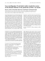
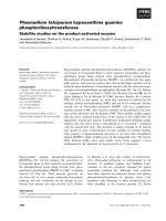
Fig. 1. CD38 forms 95 kDa homodimers in B lymphocytes. (A)
B lymphocytes (5 · 10
7
) were isolated from the indicated mouse
strains, including CD38 deficient mice (CD38-KO [27]). The cells were
lysed with 1% (v/v) NP-40 and CD38 was immunoprecipitated with
monoclonal antibody to CD38, NIM-R5. The samples were prepared
either in the absence (lanes 1–5) or presence of 5% (v/v) 2-mercapto-
ethanol (lanes 6–10) and CD38 was detected by Western blot as des-
cribed in Materials and methods. The relative molecular mass markers
are indicated on the left of each figure. The nonspecific IgH band
present in all of the reduced samples (including the CD38-KO sample)
is indicated with an asterisk. (B) Immunoprecipitated CD38 from
BALB/c B cell lysates was boiled in the presence of 2-mercaptoethanol
(lanes 1–3) or dithiothreitol (lanes 4–6) at the concentrations shown
in the figure. (C) CD38 was immunoprecipitated from splenic B cell
lysates, resolved by 2D isoelectric focusing (IEF) and detected by
Western blot. IEF spots of monomeric and dimeric forms of CD38 are
indicated by arrows. b-ME, 2-mercaptoethanol; DTT, dithiothreitol.
Ó FEBS 2004 CD38 homodimers are noncovalently stabilized in B cells (Eur. J. Biochem. 271) 1027
molecular mass forms of CD38 were observed in the
immunoprecipitates from CD38-expressing cells; a 42 and a
95 kDa protein (p42 and p95) (Fig. 1A, lanes 2–5). The
42 kDa protein is the expected size of glycosylated mono-
meric CD38 [19] while the 95 kDa protein is the approxi-
mate size of a CD38 dimer. When the samples were boiled
and reduced in 2-mercaptoethanol, we observed nonspecific
bands of 68 kDa (corresponding to the immunoglobulin
heavy chain present in B lymphocytes) and 200 kDa
(data not shown) in all immunoprecipitates, including the
sample from the CD38 KO mice (Fig. 1A, lane 6). In
addition to observing the nonspecific bands, we still detected
the p42 and p95 forms of CD38 in the CD38-expressing cells
(Fig. 1A, lanes 7–10). This indicates that p95 was partially,
although not fully, resistant to reduction by 2-mercapto-
ethanol. Interestingly, even addition of higher concentra-
tions of 2-mercaptoethanol or another reducing agent,
dithiotreitol, did not completely ablate the p95 form of
CD38 (Fig. 1B).
To determine the structural composition of the p95 form
of CD38, we first ruled out the possibility that the p95 form
was composed of a CD38 monomer associated with the
immunoglobulin heavy chain from the precipitating anti-
CD38 Ig (data not shown). Next, we showed that the p95
form of CD38 was easily detected when iodoacetamide was
included in all of the buffers in order to block any reactive
free cysteines (data not shown). This ruled out the possibility
that p95 was formed during the lysis and immunopreci-
pitation process. Finally, we compared p42 and p95 for their
pattern of isoelectric points by IEF and 2D polyacrylamide
gel electrophoresis. For p42 we observed two dominant
isoelectric points of 7.7 and 7.2 and two minor points at 7.4
and 7.1 (Fig. 1C). Analysis of p95 revealed isoelectric points
of 7.7, 7.2 and 7.1 (Fig. 1C). These points were located at
similar positions to the corresponding points in the p42
monomeric form. We did not detect a protein spot at 7.4 in
p95; however, this protein species only represented a minor
form even in the CD38 p42 monomer. Taken together, the
data indicate that the p95 form of CD38 appears to
represent a homodimeric form of CD38 as it is recognized
by both monoclonal and polyclonal antibodies against
CD38, and has essentially identical IEF points as the p42
monomer form of CD38.
CD38 homodimers are expressed on the surface
of splenic B cells and are destabilized when solubilized
with type B surfactants (steroid-based detergents)
To determine whether CD38 is normally expressed in the
homodimeric form on the plasma membrane of B cells, we
purified splenic B cells from normal and CD38 KO mice
and treated them for 1 h with a nonpermeable chemical
crosslinker, BS
3
, in order to stabilize the CD38 homodimers
during the lysis and immunoprecipitation steps. As expec-
ted, no CD38 protein was detected in the CD38 deficient
cells (Fig. 2A, lanes 2 and 5). Similarly to the previous
results, the majority of CD38 protein was of monomeric size
(p42) in the cells that were not treated with BS
3
(Fig. 2A,
lanes 1 and 4). In contrast, in cells that had been treated
with crosslinker, CD38 was found predominantly in the
p95 homodimeric form (lanes 3 and 6). As the ratio of
homodimers to monomers was approximately five-fold
increased when the crosslinker was used (Fig. 2A, compare
lanes 1 and 3 or lanes 4 and 6), these results indicate that
a large proportion of the total CD38 is expressed in a
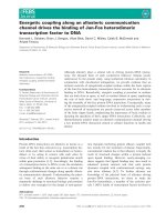
Fig. 2. CD38 is found as homodimers on the surface of splenic
B lymphocytes and the stability of the dimers depends on the detergent
used to solubilize the cells. (A) Purified B cells were incubated with the
nonpermeable crosslinker BS
3
for 1 h at 4 °C(lanes2,3,5and6)or
left untreated (lanes 1 and 4). Crosslinked cells were lysed and CD38
was immunoprecipitated from cells expressing CD38 (CD38-WT,
lanes 1, 3, 4 and 6) or lacking CD38 (CD38-KO, lanes 2 and 5).
Immunoprecipitated proteins were treated with (lanes 4–6) or without
5% (v/v) 2-mercaptoethanol (lanes 1–3) and CD38 was detected by
Western blot. (B) Splenic B cells were solubilized with 1% (v/v) NP-40
(lanes 1, 2, 5 and 6) or Chaps (lanes 3, 4, 7 and 8) and CD38 was
immunoprecipitated. The samples were heated in the presence (lanes
5–8) or absence (lanes 1–4) of 5% (v/v) 2-mercaptoethanol and CD38
was detected by Western blot. (C) B cells were solubilized with 1%
(v/v) NP-40 (lane 1), Triton X-100 (lanes 2 and 3), digitonin (lanes 4
and 5), Chaps (lanes 6 and 7) or deoxy-BigChap (lanes 8 and 9).
Lysates were immunoprecipitated with antibody to CD38 (NIM-R5)
and CD38 was detected by Western blot. The nonspecific IgH band
present in all samples, including samples immunoprecipitated with an
isotype control antibody (IgG2a), is indicated with an asterisk. b-ME,
2-mercaptoethanol.
1028 M. E. Moreno-Garcı
´
a et al.(Eur. J. Biochem. 271) Ó FEBS 2004
homodimeric form on the surface of live B lymphocytes and
suggest that most of the CD38 dimers must fall apart when
the cells are solubilized in detergent. Crosslinkers like
disuccinimidyl suberate, that have the same reactivity and
spacer arm length as BS
3
(11.4 A
˚
) also stabilized the CD38
homodimers. However, crosslinkers such as 3,3¢-dithio-
bis(sulfosuccinimidyl propionate), sulfo-disulfosuccinimidyl
tartarate and sulfo-bis[2-(sulfosuccinimidooxycarbonyl-
oxy)ethyl]sulfone, that have the same reactivity as BS
3
but
have different spacer arm lengths (12, 6.4 and 13 A
˚
,
respectively), were unable to stabilize the homodimers (data
not shown). These results suggest that the stabilization
of CD38 homodimers by crosslinkers depends strongly
on the conformation and orientation between the CD38
monomers.
It has been reported that NP-40 and Triton X-100
stabilize noncovalent hetero- or homo-dimerization of
proteins, while detergents like Chaps and octylglucoside
disrupt these interactions [32,33]. Up to now, in all our
experiments, the cells were solubilized in NP-40, a deter-
gent that might help to stabilize or protect the CD38
dimers from dissociating during the solubilization process.
In sharp contrast, when the B cells were solubilized with
Chaps we found significantly less CD38 homodimers,
whether under reducing (Fig. 2B, lanes 5–8) or nonreduc-
ing (Fig. 2B, lanes 1–4) conditions. This demonstrates that
the detergent used to solubilize the cells influenced the
amount of CD38 homodimers that could be immuno-
precipitated.
To analyze whether the stabilization of CD38 dimers
was a property of the family of detergents utilized, we used
several different detergents to solubilize the cells. As
shown in Fig. 2C, CD38 dimers were precipitated when
the cells were solubilized with NP-40 or Triton X-100;
detergents that belong to the polyoxyethylene family
(Fig. 2C, lanes 1–3). In contrast, when the cells were
solubilized with Chaps, digitonin or deoxy-BigChap,
members of the steroid-based detergent family, only
CD38 monomers were detected (Fig. 2C, lanes 4–9).
These results suggest that CD38 homodimer stability is
dependent on noncovalent interactions between CD38
monomers.
Structural requirements for CD38 homodimerization
To investigate the structural requirements for dimer
stabilization, we determined whether different CD38
mutants were capable of forming homodimers when
transfected into Ba/F3 cells. Ba/F3 cells, stably transfected
with full length wild-type CD38 (CD38-WT) or with
different CD38 mutants, were solubilized in NP-40 lysis
buffer and CD38 was immunoprecipitated, run on SDS/
PAGE under nonreducing conditions, and detected by
Western blot. A summary of the results, presented in
Table 1, indicates that CD38 homodimers were present in
the lysates of most of the transfectants expressing CD38
mutants, including, CD38-E150L, a CD38 active site
mutant (Table 2, [34]) and CD38-C123K, a mutant that
is unable to form the postulated interdisulphide bond
between two CD38 monomers [22]. These data indicate
that CD38 homodimers can be formed even when the
active site is altered and the putative interdisulphide
bridge formed betweeen CD38 monomers is disrupted.
Interestingly, however, CD38 dimers were absent in
lysates from two of the other mutant Ba/F3 transfectants.
Table 1. Expression of CD38 homodimers in different CD38 mutants
expressed in Ba/F3 pro-B cells. Each of the mutant CD38 cDNAs
listed, was stably expressed in Ba/F3 cells (Materials and methods) or
A20 cells as described previously [30]. The cells were solubilized in 1%
(v/v) NP-40. CD38 was immunoprecipitated, run on SDS/PAGE gels
under nonreducing conditions and then analyzed for the presence (Y)
or absence (N) of the p42 monomer and p95 homodimer by Western
blot. WT, wild-type.
CD38 mutant p42 p95
WT Y Y
lATG Y N
C123K Y Y
E150L Y Y
E150Q Y Y
D151V Y Y
E150QD151N Y Y
G68E Y N
Table 2. NAD
+
glycohydrolase activity of membrane homogenates from Ba/F3 transfectants. Each of the mutant CD38 cDNAs listed, was stably
expressed in Ba/F3 cells and the enzyme activity (V
max
) of the membrane homogenates was determined as described previously [30]. The V
max
was
adjusted to reflect differences in CD38 expression levels between the various mutants and is reported as nmol of product formed per minute per
arbitrary unit of CD38. Briefly, the total amount of protein per Ba/F3 cell was determined by the Bradford method. The average amount of CD38
expressed on the membrane of each Ba/F3 cell was determined by FACS and is reported as mean fluorescence intensity (Fig. 3C shows values of
each of the clones). The relative enzyme activity of each of the mutants is given in parentheses. It was determined by setting the V
max
adjusted
activity of CD38-WT to 100% and then calculating the percentage activity of each of the mutants relative to CD38-WT. MFI, mean fluorescence
intensity; WT, wild-type.
Mutant
V
max
(nmolÆmin
)1
Æmg
)1
protein)
Protein per cell
(mg protein per cell · 10
)7
)
CD38 per cell
arbitrary units CD38 per cell
(1/MFI · 10
)4
)
V
max
adjusted
(nmolÆmin
)1
per
arbitrary units of
CD38 · 10
)10
)
WT 843.2 1.54 9.0 1180.0 (100%)
E150L 5.54 1.54 11.4 9.73 (0.8%)
G68E 56.9 1.54 35.8 310 (26%)
lATG 284.1 1.54 30.8 1350 (114%)
Ó FEBS 2004 CD38 homodimers are noncovalently stabilized in B cells (Eur. J. Biochem. 271) 1029
In one of the mutants (CD38-lATG), the 22 amino acid
cytoplasmic region of CD38 was replaced with a 4 amino
acid tail (Met-Lys-Val-Lys), and in the second mutant
(CD38-G68E), the glycine at position 68 was replaced by
the polar residue glutamate. The G68 residue is within the
a1-helix that has been previously postulated to be a dimer
interface site in the Aplysia enzyme [16]. As shown in
Fig. 3A (lanes 2 and 5), CD38 homodimers were preci-
pitated from Ba/F3 transfectants expressing CD38-WT or
expressing a mutant form of CD38 in which a single
residueintheactivesitewasmutated(CD38-E150L).
However, no CD38 homodimers were detected in
immunoprecipitations from transfectants expressing the
CD38-G68E or CD38-lATG mutant proteins (Fig. 3A,
lanes 3 and 4). This result suggests that the cytoplasmic
region and first a-helix interface region of CD38 are
important for dimer stability.
To determine whether these two regions were necessary
for CD38 dimer stabilization on the plasma membrane of
the Ba/F3 cells, the transfectants expressing CD38-lATG
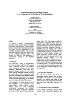
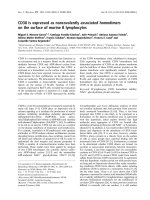
Fig. 3. The stability and membrane expression of CD38 homodimers is dependent on at least two separate domains of CD38. (A) Ba/F3 cells
transfected with control vector, CD38-WT, CD38-G68E, CD38-lATG or CD38-E150L were lysed with 1% (v/v) NP-40 and CD38 was imu-
noprecipitated, run on SDS/PAGE under nonreducing conditions and detected by Western blot. (B) The Ba/F3 transfectants listed above were
treated (as described in Fig. 2) with the crosslinker BS
3
(lanes 2, 4, 6, 8 and 10) or left untreated (lanes 1, 3, 5, 7 and 9). CD38 was detected by
Western blot as in Fig. 2A. (C) Ba/F3 mutants were analyzed for expression of CD38 on the plasma membrane by FACS using the antibody, NIM-
R5, conjugated to FITC. Dead cells were excluded by propidium iodide incorporation. Light line histograms, nontransfected Ba/F3 cells; dark line
histograms, Ba/F3 transfectants. The mean fluorescence intensity of the cells from each of the transfectants is listed above the histogram. (D) Post-
nuclear supernatants were prepared from Ba/F3 clones lysed with 1% (v/v) NP-40, the protein concentration was determined and equivalent
amounts of protein were run on SDS/PAGE gels under reducing conditions. CD38 and actin expression were analyzed by Western blot using rabbit
polyconal anti-CD38 Ig and mouse monoclonal antibody to actin, followed by HRP-labeled anti-rabbit IgG and anti-mouse IgG, respectively.
(E) Comparison of plasma membrane and total CD38 expression levels in the Ba/F3 clones. To determine the relative plasma membrane expression
levels of CD38 between the different Ba/F3 clones the mean fluorescence intensity for each of the clones was determined (C) and the relative levels
were normalized to that of the CD38-WT transfectant which was set at 100%. To quantitate the total CD38 expression levels for the various Ba/F3
transfectant clones, densitometric analysis of CD38 and actin Western blots (D) were performed using
SIGMAGEL
.
LNK
. The CD38 levels for each
clone were first normalized to actin by dividing the densitometric value of CD38 by the densitometric value of actin. Then the relative total CD38
expression levels for each clone were normalized to CD38-WT which was set at 100%.
1030 M. E. Moreno-Garcı
´
a et al.(Eur. J. Biochem. 271) Ó FEBS 2004
or CD38-G68E were crosslinked with BS
3
, solubilized in
NP-40 lysis buffer, and CD38 was detected by immuno-
precipitation and Western blot (Fig. 3B). As we have
previously observed, homodimers of CD38 were absent in
the immunoprecipitates from the noncrosslinked CD38-
lATG and CD38-G68E transfectants (lanes 5 and 9).
However, when the crosslinker was added, CD38 homo-
dimers could be visualized (lanes 6 and 10). Indeed, similar
ratios of homodimers to monomers were observed in the
crosslinked CD38-G68E and CD38-lATG mutants com-
pared to crosslinked CD38-WT and CD38-E150L (compare
lanes 4, 6, 8 and 10). Therefore, the cytoplasmic region and
a1-helix domains are not critical for CD38 dimer stabiliza-
tion in B cells, however, the two domains must contribute to
the overall stability of CD38 homodimers because the
mutated dimers fell apart even under ÔpermissiveÕ solubili-
zation and nonreducing conditions.
CD38-G68E and CD38-lATG are less efficiently
expressed and have a reduced half-life
on the plasma membrane
The previous results indicated that CD38-lATG and
CD38-G68E are not obligatory for dimer stabilization
but do contribute to the overall stability of the dimers,
particularly upon detergent solubilization. It has been
reported that inappropriate folding of proteins or inappro-
priate assembly of multimeric protein complexes can
influence the surface and overall expression of these proteins
in cells and can also alter the half-life of the misfolded or
disorganized protein complexes [35,36]. Given that the
stability of CD38-lATG and CD38-G68E homodimers is
reduced when the proteins are solubilized in permissive
detergents, immunoblotting and FACS experiments were
performed in order to analyze the total and surface levels of
CD38 in all the Ba/F3 transfectants (Fig. 3C–E). These
experiments revealed that the total amount of CD38 (as
assessed by immunoblotting), as well as the amount of
CD38 expressed on the plasma membrane (as assessed by
FACS) was similar in the CD38-WT and CD38-E150L
transfected Ba/F3 cells (Fig. 3C,D). In contrast, CD38
expression levels (both total and plasma membrane levels) in
transfectants expressing CD38-G68E and CD38-lATG
were significantly decreased relative to CD38-WT
(Fig. 3C,D). Similar results were obtained upon analysis
of multiple independent Ba/F3 clones expressing CD38-
lATG or CD38-G68E (data not shown). Upon densito-
metric analysis of the immunoblots it became clear that the
amount of CD38 expressed on the plasma membrane and
the total amount of CD38 expressed by the various
transfectants correlated very well with one another
(Fig. 3E), strongly suggesting that the reduced cell surface
expression of CD38 by the CD38-lATG and CD38-G68E
transfectants was not due simply to inefficient transport of
the protein to the plasma membrane. In addition, confocal
microscopic analysis of CD38 expression in Ba/F3 trans-
fectants revealed that neither CD38 nor any of the CD38
mutant proteins were present in intracellular compartments
(data not shown). Therefore, we next considered the
possibility that the reduced plasma membrane expression
of the CD38-lATG and CD38-G68E mutant proteins
could be due to a faster turnover rate for these mutant
molecules on the plasma membrane [35]. To analyze this
possibility, we performed pulse-chase experiments using
normal B cells and the Ba/F3 transfectants. To first
determine whether the surface half-life of CD38 in Ba/F3
cells is comparable to that of splenic B cells, we biotinylated
the surface of splenic B cells and Ba/F3 clones (CD38-WT
and lATG) and compared the plasma membrane half-life
of CD38. In these experiments the biotinylation was carried
out for 30 min on ice in order to avoid any potential
internalization of biotin or biotinylated proteins. The cells
were then washed to remove the reactive biotin and cultured
for up to 30 h. The amount of cell surface biotin-labeled
CD38 at various timepoints was determined by immuno-
precipitating with anti-CD38 and immunoblotting with
SA-HRP to detect the cell surface biotinylated-CD38 and
anti-CD38 to detect the total CD38 pool. The plasma
membrane expression of CD38 on CD38-WT Ba/F3
transfectant cells and on normal B cells was quite stable
over time with a half-life of approximately 28 h on both cell
types (Fig. 4A,B), indicating that the turnover rate of CD38
in both cell types is similar and comparable. In contrast, the
surface half-life of CD38-lATGwaslessthanhalfthat
observed for CD38 expressed by normal B cells or Ba/F3
transfectants, suggesting that this mutant protein is less
stably expressed on the plasma membrane (Fig. 4A,B). To
confirm these results, we repeated the biotinylation experi-
ment using Ba/F3 transfectants expressing other CD38
mutant proteins. The plasma membrane expression of both
CD38-WT and CD38-E150L was quite stable over time
with a half-life of approximately 28 h (Fig. 5A,B). In
striking contrast, the half-lives of plasma membrane bound
CD38-G68E and CD38-lATG were less than half that
observed with CD38-WT (Fig. 5A,B). Thus, both of the
CD38 mutant proteins that form unstable homodimers also
have significantly reduced stability on the plasma mem-
brane, suggesting that appropriate assembly or stabilization
of CD38 into homodimers may be required for its extended
expression on the plasma membrane.
Enzyme activity is not dependent on the presence
of stable homodimers
As the mutations in the cytoplasmic region and the a1-helix
of CD38 affected dimer stability upon solubilization,
plasma membrane expression levels and surface half-life, it
was also possible that these mutations would affect the
enzyme activity of the proteins. To test the enzyme activity
of the CD38 mutants, membrane homogenates from the
various Ba/F3 transfectants were prepared and NAD
+
glycohydrolase activity in the membranes was measured by
HPLC. As shown in Table 2, the enzyme activity of the
active site mutant, CD38-E150L was greatly decreased
compared to CD38-WT. Interestingly, the glycohydrolase
activities of CD38-G68E and CD38-lATG were also less
than CD38-WT. As the membrane expression levels of
CD38-lATG and CD38-G68E were reduced compared to
CD38-WT (Fig. 3C,D), we performed a calculation to
adjust the enzyme activity (V
max
adjusted) to reflect the
amount of total protein and CD38 protein expressed on
a per cell basis. Upon adjusting the enzyme activity to
compensate for the membrane CD38 expression levels, we
found that the enzyme activity of CD38-lATG was at least
Ó FEBS 2004 CD38 homodimers are noncovalently stabilized in B cells (Eur. J. Biochem. 271) 1031
as high as CD38-WT (Table 2). These data indicate that the
catalytic activity of CD38 is not dependent on the formation
of stable CD38 homodimers. Interestingly, however, even
when expression levels of CD38-G68E were accounted for,
the NAD
+
glycohydrolase activity of CD38-G68E was only
27% of CD38-WT. This result shows that a single point
mutation in the first a-helix of CD38, a residue that is far
removed from the active site of CD38, can significantly
influence CD38 enzyme activity.
Discussion
In this work we show that homodimers of CD38 are
expressed on the surface of B lymphocytes. Although CD38
dimers that are sensitive to reducing agents have been
previously reported [22–25], we found that the stability of
CD38 dimers expressed in B cells correlated better with the
type of detergent used to solubilize the cells (Fig. 2) than the
presence or absence of reducing agents (Fig. 1). Previous
reports have shown that heterodimerization of proteins such
as Bax with Bcl-2 or Bax with Bcl-X
L
are dependent on the
detergent used to solubilize the cells [32,33]. Thus, NP-40
and Triton X-100, detergents that form large micelles,
stabilized the hydrophobic interactions between Bax and its
partners, while Chaps and octyl glucoside, detergents that
form small micelles, could not accommodate the hetero-
dimers. Interestingly, we found the same pattern with CD38
homodimers in that they were stabilized in the polyoxy-
Fig. 5. The mutants expressing unstable CD38 homodimers present a
reduced CD38 half-life on the plasma membrane. (A) Ba/F3 transfectant
cells expressing CD38-WT or each of the different mutants were sur-
face labeled with sulfo-NHS-LS-biotin for 30 min at room tempera-
ture. The cells were washed and then cultured at 37 °Cforan
additional 30 h. 2 · 10
6
cells were harvested at 0, 2, 10, 20 and 30 h
after biotin labeling. Cell viability, as measured by trypan blue exclu-
sion, was over 95% at each time point. Immunoprecipitation and
Western-blotting was performed as described in Materials and
methods and Fig. 4. (B) Densitometric analyses using the program
SIGMAGEL.LNK
were performed to compare the relative amounts of
biotin-labeled CD38 in each Ba/F3 clone. Densitometry was per-
formed as described in Fig. 4.
Fig. 4. The half-life of CD38 is the same in splenic B cells and CD38-
WT Ba/F3 transfectants. Purified splenic B cells, CD38-WT and
CD38-lATG Ba/F3 transfectants were labeled with sulfo-NHS-LS-
biotin for 30 min on ice. The cells were washed and then cultured at
37 °C for an additional 30 h. 2 · 10
6
Ba/F3 cells or 5 · 10
7
splenic
B cells were harvested at 0, 10, 20 and 30 h after biotin labeling. Cell
viability, as measured by trypan blue exclusion, was over 95% for the
Ba/F3 transfectants at each time point and was 97, 90, 87 and 75% for
splenic B cells at 0, 10, 20 and 30 h after biotinylation, respectively.
(A) At each timepoint, the cells were lysed in 1% (v/v) NP-40, CD38
was immunoprecipitated with anti-CD38 Ig, and the immunoprecipi-
tated protein was analyzed by SDS/PAGE and Western blotting. The
amount of plasma membrane associated (biotinylated-CD38) and
total CD38 was determined by immunoprecipitation, SDS/PAGE and
Western blotting. Plasma membrane biotinylated-CD38 was detected
with streptavidin-HRP (left). Total immunoprecipitated CD38 was
detected using the polyclonal rabbit antibody to CD38 (right).
(B) Densitometric analyses using the program
SIGMAGEL
.
LNK
were
performed to compare the relative amounts of biotin-labeled CD38
(membrane CD38) in each Ba/F3 clone. To determine the relative
amount of biotin-labeled CD38 present in each clone, the densito-
metric value of biotin-CD38 was divided by the densitometric value of
total CD38. The ratio of cell surface CD38 to total CD38 at time 0
was set at 100% and all other time points were compared relative to
this.
1032 M. E. Moreno-Garcı
´
a et al.(Eur. J. Biochem. 271) Ó FEBS 2004
ethylene detergents (i.e. NP-40 and Triton X-100) and were
destabilized with detergents such as digitonin, Chaps
and deoxy-BigChap (Fig. 2). Importantly, these differences
could not be attributed to differences in the ability of the
various detergents to solubilize CD38 (data not shown).
Furthermore, when we used the crosslinker, BS
3
,the
majority of CD38 was ÔcapturedÕ in the homodimer form
indicating that CD38 must be dimerized via noncovalent
interactions that were partially disrupted when the cells were
solubilized in detergent. However, it is also clear that
conformation and folding of the individual CD38 mono-
mers is strongly influenced by the five known intradisul-
phide bonds present in each monomer and their reduction is
also expected to greatly influence the stability of the
noncovalently associated CD38 homodimers.
As CD38 dimers appear to be stabilized via noncovalent
interactions between monomers, a reasonable assumption is
that mutations within the potential interface domains might
alter the formation or stability of CD38 homodimers. When
cells expressing two different mutant forms of CD38, a
cytoplasmic region mutant and an a1-helix mutant, were
solubilized under nonreducing conditions in a permissive
detergent such as NP-40, we were unable to detect the
presence of CD38 homodimers (Fig. 3A), suggesting that
these two domains play an important role in homodimer
stability. Because homodimers of CD38-G68E and CD38-
lATG were observed when the cells were crosslinked with
BS
3
(Fig. 3B), these data suggest that these domains are not
obligate for the stabilization of the dimers, but rather must
contribute to the overall stability of the dimers. Crystallo-
graphic analysis of the Aplysia cyclase indicates four
putative oligomerization sites including the a1, a4anda10
helices and residues between 242 and 248 [16]. Thus, we
propose that multiple contact points contribute in an
additive or synergistic manner to CD38 homodimer stabil-
ization. Although the mutants described here are not
sufficient, in themselves, to control CD38 homodimer
stabilization, the results clearly demonstrate a role for the
a1-helix and the cytoplasmic region in stabilizing the
solubilized homodimers.
The mutations in the cytoplasmic tail and a1-helix of
CD38 not only affected the stability of the CD38 homo-
dimer upon solubilization, but also significantly diminished
the expression of the CD38 homodimer on the plasma
membrane. Indeed, we were never able to isolate CD38-
G68E expressing transfectants expressing levels of CD38
comparable to CD38-WT, despite screening more than 25
individual clones (data not shown). As a whole, these data
suggest that CD38-G68E and CD38-lATG mutants were
either inefficiently assembled and transported to the mem-
brane or were less stable on the surface. Our pulse-chase
experiments (Figs 4 and 5) clearly showed that the plasma
membrane half-life of these CD38 mutants was significantly
less than CD38-WT, suggesting that extended plasma
membrane expression of CD38 may depend on the presence
of stable CD38 homodimers. Although further experiments
will be needed to prove this hypothesis, similar results were
obtained analyzing mutants of the dipeptidylpeptidase IV
CD26 [36]. The close correlation between surface expression
of CD38 and total expression of CD38 suggests that the
mutant forms of CD38 are not preferentially retained in
the intracellular compartments (Fig. 3C–E). Furthermore,
intracellular expression analysis of CD38 on Ba/F3 trans-
fectants and normal B cells using confocal microscopy or
subcellular fractionation and immunobloting revealed that
neither CD38 nor any of the CD38 mutant proteins
analyzed in this study were detected in intracellular mem-
branes (data not shown).
CD38 homodimers have been proposed to play a number
of different functional roles including formation of a
transmembrane pore, allowing for transport of cADPR
into the cytosol [37]. However, it is clear that stable
homodimers are not obligate for enzyme activity as the
unstable CD38 homodimer mutant, CD38-lATG, had
perfectly normal enzyme activity when the activity was
adjusted to reflect CD38 expression levels on the membrane
(Table 2). In agreement with this, we also observed
NADase activity from the p42 monomeric form of CD38
(data not shown), suggesting that CD38 monomers are
enzymatically active. This is in agreement with the results of
Bruzzone et al. [18]. Interestingly, although mutations in the
active site do not decrease the formation or stability of
CD38 homodimers (Table 2), the cells expressing the
unstable CD38 homodimer, CD38-G68E, had significantly
decreased CD38 dependent enzyme activity (Table 2).
Thus, while CD38 enzyme activity is not critically
dependent on the presence of stable CD38 homodimers, it
is clear that mutating a single residue in the a1-helix
interface can decrease both homodimer stability and
enzyme activity. In conclusion, we have shown that CD38
is normally expressed as a noncovalently associated
homodimer on the plasma membrane of B cells. Mutations
that affect the stability of the CD38 homodimer do not
necessarily alter CD38-dependent enzyme activity; however,
these mutations do result in reduced plasma membrane
stability and decreased expression of CD38 on the plasma
membrane.
Acknowledgements
The authors would like to thank Troy Randall for discussions and
critical reading of this manuscript. The authors also thank Dr Jose
´
Manuel Herna
´
ndez-Herna
´
ndez for technical advice and Q. F. B.
He
´
ctor Romero Ramı
´
rez for technical assistance. M. E. M G., A. S T.
and L. S A. are supported by CONACyT Me
´
xico grants, # 28093N,
33497N and 40218Q. J. P., S. P-S., and F. E. L. are supported by NIH
grant AI-43629 and the Trudeau Institute.
References
1. Santos-Argumedo, L., Teixeira, C., Preece, G., Kirkham, P.A. &
Parkhouse, R.M.E. (1993) A B lymphocyte surface molecule
mediating activation and protection from apoptosis via calcium
channels. J. Immunol. 15, 3119–3130.
2. Mehta, K., Shahid, U. & Malavasi, F. (1996) Human CD38, a
cell-surface protein with multiple function. FASEB J. 10, 1408–
1417.
3. Lund, F.E., Cockayne, D.A., Randall, T.D., Solvason, N.,
Schuber, F. & Howard, M.C. (1998) CD38: a new paradigm in
lymphocyte activation and signal transduction. Immunol. Rev.
161, 79–93.
4. Howard,M.C.,Grimaldi,J.C.,Bazan,J.F.,Lund,F.E.,Santos-
Argumedo, L., Parkhouse, R.M.E., Walseth, T.F. & Lee, H.C.
(1993) Formation and hydrolysis of cyclic ADP-ribose catalyzed
by lymphocyte antigen CD38. Science 262, 1056–1059.
Ó FEBS 2004 CD38 homodimers are noncovalently stabilized in B cells (Eur. J. Biochem. 271) 1033
5. Aarhus, R., Graeff, R.M., Dickey, D.M., Walseth, T.F. & Lee,
H.C. (1995) ADP-ribosyl cyclase and CD38 catalyze the synthesis
of a calcium-mobilizing metabolite from NADP
+
. J. Biol. Chem.
270, 30327–30333.
6. Kirkham, P.A., Santos-Argumedo, L., Harnett, M.M. & Park-
house, R.M.E. (1994) Murine B-cell activation via CD38 and
protein tyrosine phosphorylation. Immunology 83, 513–516.
7. Kitanaka, A., Suzuki, T., Ito, C., Nishigaki, H., Coustain-Smith,
E., Tanaka, T., Kubota, Y. & Campana, D. (1999) CD38-medi-
ated signaling events in murine pro-B cells expressing human
CD38 with or without its cytoplasmic domain. J. Immunol. 162,
1952–1958.
8. Kumagai, M., Coustan-Smith, E., Murray, D.J., Silvennoinen, O.,
Murti, K.G., Evans, W.E., Malavasi, F. & Campana, D. (1995)
Ligation of CD38 suppresses human B lymphopoiesis. J. Exp.
Med. 181, 1101–1110.
9. Zubiar, M., Guirado, M., Terhorst, C., Malavasi, F. & Sancho, J.
(1999) The CD3-cde transducing module mediates CD38-induced
protein-tyrosine kinase and mitogen-activated protein kinase
activation in Jurkat T cells. J. Biol. Chem. 274, 20633–20642.
10.Yasue,T.,Baba,M.,Mori,S.,Mizoguchi,C.,Uehara,S.&
Takatsu, K. (1999) IgG1 production by sIgD+ splenic B cells and
peritoneal B-1 cells in response to IL-5 and CD38 ligation. Int.
Immunol. 11, 915–923.
11. Hellmich, M.R. & Strumwasser, F. (1991) Purification and char-
acterization of a molluscan egg-specific NADase, a second-
messenger enzyme. Cell Regul. 2, 193–202.
12. Lee, H.C. & Aarhus, R. (1991) ADP-ribosyl cyclase: an enzyme
that cyclizes NAD
+
into a calcium-mobilizing metabolite. Cell
Regul. 2, 203–209.
13. Dong, C., Wang, J., Neame, P. & Cooper, M.D. (1994) The
murine BP-3 gene encodes a relative of the CD38/NAD
+
glyco-
hydrolase family. Int. Immunol. 6, 1353–1360.
14. Itoh, M., Ishihara, K., Tomizawa, H., Tanaka, H., Kobune, Y.,
Ishikawa, J., Kaisho, T. & Hirano, T. (1994) Molecular cloning of
murine BST-1 having homology with CD38 and Aplysia ADP-
ribosyl cyclase. Biochem. Biophys. Res. Commun. 203, 1309–1317.
15. Yamamoto-Katayama, S., Ariyoshi, M., Ishihara, K., Hirano, T.,
Jingami, H. & Morikawa, K. (2002) Crystallographic studies on
human BST-1/CD157 with ADP-ribosyl cyclase and NAD gly-
cohydrolase activities. J. Mol. Biol. 316, 711–723.
16. Prasad, G.S., McRee, D.E., Stura, E.A., Levitt, D.G., Lee, H.C. &
Stout, C.D. (1996) Crystal structure of Aplysia ADP ribosyl
cyclase, a homologue of the bifunctional ectozyme CD38. Nat.
Struct. Biol. 3, 957–964.
17. Franco, L., Zocchi, E., Calder, L., Guida, L., Benatti, U. &
De Flora, A. (1994) Self-aggregation of the transmembrane
glycoprotein CD38 purified from human erythrocytes. Biochem.
Biophys. Res. Commun. 202, 1710–1715.
18. Bruzzone, S., Guida, L., Franco, L., Zocchi, E., Corte, G. & De
Flora, A. (1998) Dimeric and tetrameric forms of catalytically
active transmembrane CD38 in transfected HeLa cells. FEBS Lett.
433, 275–278.
19. Harada, N., Santos-Argumedo, L., Chang, R., Grimaldi, J.C.,
Lund, F.E., Brannan, C.I., Copeland, N.G., Jenkins, N.A., Heath,
A.W. & Parkhouse, R.M. (1993) Expression cloning of a cDNA
encoding a novel murine B cell activation marker. Homology to
human CD38. J. Immunol. 151, 3111–3118.
20. Jackson, D.G. & Bell, J.I. (1990) Isolation of a cDNA encoding
the human CD38 (T10) molecule, a cell surface glycoprotein with
an unusual discontinuous pattern of expression during lympho-
cyte differentiation. J. Immunol. 144, 2811–2815.
21. Lund,F.E.,Yu,N.,Kim,K.M.,Reth,M.&Howard,M.C.
(1996) Signaling through CD38 augments B cell antigen
receptor (BCR) responses and is dependent on BCR expression.
J. Immunol. 157, 1455–1467.
22. Han, M.K., Kim, S.J., Park, Y.R., Shin, Y.M., Park, H.J., Park,
K.J., Park, K.H., Kim, H.K., Jang, S.I., An, N.H. & Kim, U.H.
(2002) Antidiabetic effect of a prodrug of cysteine,
L
-2-
oxothiazolidine-4-carboxylic acid, through CD38 dimerization
and internalization. J. Biol. Chem. 277, 5315–5321.
23. Chidambaram, N., Wong, E.T. & Chang, C.F. (1998) Differential
oligomerization of membrane-bound CD38/ADP-ribosyl cyclase
in porcine heart microsomes. Biochem. Mol. Biol. Int. 44, 1225–
1233.
24. Khoo, K.M. & Chang, C.F. (1998) Purification and character-
ization of CD38/ADP-ribosyl cyclase from rat lung. Biochem.
Mol. Biol. Int. 44, 841–850.
25. Khoo, K.M. & Chang, C.F. (2000) Localization of plasma
membrane CD38 is domain specific in rat hepatocyte. Arch. Bio-
chem. Biophys. 373, 35–43.
26. Umar, S., Malavasi, F. & Mehta, K. (1996) Post-translational
modification of CD38 protein into a high molecular weight form
alters its catalytic properties. J. Biol. Chem. 271, 15922–15927.
27. Cockayne, D.A., Muchamuel, T., Grimaldi, J.C., Muller-Steffner,
H., Randall, T.D., Lund, F.E., Murray, R., Schuber, F. &
Howard, M.C. (1998) Mice deficient for the ecto-nicotinamide
adenine dinucleotide glycohydrolase CD38 exhibit altered
humoral immune responses. Blood 92, 1324–1333.
28. Lund, F.E., Solvason, N.W., Cooke, M.P., Health, A.W.,
Grimaldi, J.C., Parkhouse, R.M., Goodnow, C.C. & Howard,
M.C. (1995) Signaling through murine CD38 is impaired in anti-
gen receptor-unresponsive B cells. Eur. J. Immunol. 25, 1338–1345.
29. O’Farrel, P.H. (1975) High resolution Two-dimensional electro-
phoresis of proteins. J. Biol. Chem. 250, 4007–4021.
30. Lund, F.E. & Muller-Steffner, H.M., Yu, N., Stout, C.D., Schu-
ber, F. & Howard, M.C. (1999) CD38 signaling in B lymphocytes
is controlled by its ectodomain but occurs independently of
enzymatically generated ADP-ribose or cyclic ADP-ribose.
J. Immunol. 162, 2693–2702.
31. Cavet, M.E., Akhter, S., Murtazina, R., Sanchez de Medina, F.,
Tse, C.M. & Donowitz, M. (2001) Half-lives of plasma membrane
Na
+
/H
+
exchangers NHE1-3: plasma membrane NHE2 has a
rapid rate of degradation. Am. J. Physiol. Cell Physiol. 281,
C2039–C2048.
32. Hsu, Y.T. & Youle, R.J. (1997) Nonionic detergents induce
dimerization among members of the Bcl-2 family. J. Biol. Chem.
272, 13829–13834.
33. Hsu, Y.T. & Youle, R.J. (1998) Bax in murine thymus is a soluble
monomeric protein that displays differential detergent-induced
conformations. J. Biol. Chem. 273, 10777–10783.
34. Munshi,C.,Aarhus,R.,Graeff,R.,Walseth,T.F.,Levitt,D.&
Lee, H.C. (2000) Identification of the enzymatic active site of
CD38 by site-directed mutagenesis. J. Biol. Chem. 275, 21566–
21571.
35. Haardt, M., Benharouga, M., Lechardeur, D., Kartner, N. &
Lukacs, G.L. (1999) C-terminal truncations destabilize the cystic
fibrosis transmembrane conductance regulator without impairing
its biogenesis. A novel class of mutation. J. Biol. Chem. 274,
21873–21877.
36. Fan, H., Meng, W., Kilian, C., Grams, S. & Reutter, W. (1997)
Domain-specific N-glycosylation of the membrane glycoprotein
dipeptidase IV (CD26) influences its subcellular trafficking,
biological stability, enzyme activity and protein folding. Eur.
J. Biochem. 246, 243–251.
37. Franco,L.,Guida,L.,Bruzzone,S.,Zocchi,E.,Usai,C.&De
Flora, A. (1998) The transmembrane glycoprotein CD38 is a
catalytically active transporter responsible for generation and
influx of the second messenger cyclic ADP-ribose across mem-
branes. FASEB J. 12, 1507–1520.
1034 M. E. Moreno-Garcı
´
a et al.(Eur. J. Biochem. 271) Ó FEBS 2004