Báo cáo khoa học: Characterization of a digestive carboxypeptidase from the insect pest corn earworm (Helicoverpa armigera) with novel specificity towards C-terminal glutamate residues pptx
Bạn đang xem bản rút gọn của tài liệu. Xem và tải ngay bản đầy đủ của tài liệu tại đây (329.7 KB, 12 trang )
Characterization of a digestive carboxypeptidase from the insect
pest corn earworm (
Helicoverpa armigera
) with novel specificity
towards C-terminal glutamate residues
David P. Bown and John A. Gatehouse
School of Biological and Biomedical Sciences, University of Durham, UK
Carboxypeptidases were purified from guts of larvae of corn
earworm (Helicoverpa armigera), a lepidopteran crop pest,
by affinity chromatography on immobilized potato carb-
oxypeptidase inhibitor, and characterized by N-terminal
sequencing. A larval gut cDNA library was screened using
probes based on these protein sequences. cDNA HaCA42
encoded a carboxypeptidase with sequence similarity to
enzymesofclanMC[Barrett,A.J.,Rawlings,N.D.&
Woessner, J. F. (1998) Handbook of Proteolytic Enzymes.
Academic Press, London.], but with a novel predicted spe-
cificity towards C-terminal acidic residues. This carboxyp-
eptidase was expressed as a recombinant proprotein in the
yeast Pichia pastoris. The expressed protein could be acti-
vated by treatment with bovine trypsin; degradation of
bound pro-region, rather than cleavage of pro-region from
mature protein, was the rate-limiting step in activation.
Activated HaCA42 carboxypeptidase hydrolysed a synthetic
substrate for glutamate carboxypeptidases (FAEE, C-ter-
minal Glu), but did not hydrolyse substrates for carboxy-
peptidase A or B (FAPP or FAAK, C-terminal Phe or Lys)
or methotrexate, cleaved by clan MH glutamate carboxy-
peptidases. The enzyme was highly specific for C-terminal
glutamate in peptide substrates, with slow hydrolysis of
C-terminal aspartate also observed. Glutamate carboxyp-
eptidase activity was present in larval gut extract from
H. armigera. The HaCA42 protein is the first glutamate-
specific metallocarboxypeptidase from clan MC to be iden-
tified and characterized. The genome of Drosophila mel-
anogaster contains genes encoding enzymes with similar
sequences and predicted specificity, and a cDNA encoding a
similar enzyme has been isolated from gut tissue in tsetse fly.
We suggest that digestive carboxypeptidases with sequence
similarity to the classical mammalian enzymes, but with
specificity towards C-terminal glutamate, are widely distri-
buted in insects.
Keywords: clan MC metalloproteinase; digestive proteinase;
glutamate carboxypeptidase; insect herbivore; proteinase
activation.
Carboxypeptidases are exopeptidases that remove a single
amino acid residue from the C-terminus of a protein or
peptide substrate. They play an important role in protein
digestion in the guts of higher animals, acting to liberate free
amino acids from the peptides produced by endopeptidase
action, thus completing the digestive process and generating
molecules that can be absorbed by the gut, via amino acid
transporters. Mammals contain three genes encoding
digestive carboxypeptidases, designated carboxypeptidases
A1, A2 and B [1]. All three proteins are zinc-containing
metallopeptidases of clan MC [2].
The specificity of the digestive carboxypeptidases in
mammals has been extensively investigated. These enzymes
show a specificity directed towards the C-terminal amino
acid residue in their substrates. Carboxypeptidases A1 and
A2 prefer neutral amino acids, with A1 favouring smaller
amino acid side chains, whereas A2 favours bulkier side
chains [3]. Carboxypeptidase B is highly specific for basic
C-terminal residues (with arginine favoured over lysine [4]).
Specificity is primarily determined by interaction of the side
chain of the C-terminal amino acid of the substrate with a
binding pocket on the enzyme, with amino acid 255
(human carboxypeptidase A1 numbering) at the bottom
[5]. The side chain of this residue interacts with the
substrate side chain; in carboxypeptidase B amino acid 255
is negatively charged aspartic acid to interact with positively
charged basic side chains, whereas in carboxypeptidase A1
and A2 it is isoleucine, to form a hydrophobic interaction
with neutral side chains. Amino acids Tyr248 (hydrogen
bond to P1 amino group), Arg71 (hydrogen bond to P2
carbonyl oxygen), Asn144, Arg145 (bind C-terminal carb-
oxylate group of substrate) and Tyr198 are also important
for substrate binding [2,6].
The presence of digestive carboxypeptidases in insects
was established by Ward [7] who partially purified an
enzyme with carboxypeptidase A activity from gut
Correspondence to J. A. Gatehouse, School of Biological and
Biomedical Sciences, University of Durham, South Road, Durham,
DH1 3LE, UK. Fax: + 44 191 334 1201, Tel.: + 44 191 334 1264,
E-mail:
Abbreviations: FAAK, furylacryloyl-Ala-Lys; FAEE,
furylacryloyl-Glu-Glu; FAPP, furylacryloyl-Phe-Phe; PCI, potato
carboxypeptidase inhibitor; SKTI, soya bean Kunitz trypsin inhibitor,
ACE, angiotension 1 converting enzyme.
Enzymes: glutamate carboxypeptidases (EC 3.4.17).
Note: A website is available at
(Received 4 February 2004, revised 19 March 2004,
accepted 24 March 2004)
Eur. J. Biochem. 271, 2000–2011 (2004) Ó FEBS 2004 doi:10.1111/j.1432-1033.2004.04113.x
extracts of the webbing clothes moth, Tineola bisselliella.
Soluble carboxypeptidase activity was subsequently found
in gut extracts from larvae of both coleopteran
(mealworm; Tenebrio molitor [8]); and lepidopteran
(armyworm; Spodoptera frugiperda [9]); insect species. In
addition to carboxypeptidase A activity, carboxypeptidase
B activity has been detected in corn earworm (Heli-
coverpa armigera) although at much lower levels than
carboxypeptidase A activity [10]. The molecular charac-
terization of these enzymes has been carried out through
the identification of cDNA clones encoding them. The
first putative insect digestive carboxypeptidase to be
cloned was from black fly (Simulium vittatum [11]),
followed by enzymes from corn earworm (H. armigera
[12]), mosquito (Anopheles gambiae [13]; Aedes aegypti
[14]), tsetse fly (Glossinia morsitans [15]), and bertha
armyworm (Mamestra configurata [16]). On the basis of
sequence similarity, the genes from black fly, mosquito
and corn earworm were predicted to encode proteins with
similar specificity to carboxypeptidase A, whereas the
cDNA from tsetse fly was predicted to encode a protein
showing similar specificity to carboxypeptidase B. These
predicted enzyme activities have not been directly demon-
strated except in the case of a carboxypeptidase A-like
enzyme from corn earworm, which has been expressed as a
recombinant protein in insect cells using a baculovirus-based
expression system, and has been shown to hydrolyse a
synthetic substrate for carboxypeptidase A, but not a
synthetic substrate for carboxypeptidase B [10]. Although
other insect carboxypeptidases have not been expressed as
recombinant enzymes, expression of putative digestive
carboxypeptidases has been shown to be strongly gut-
specific, and upregulated by feeding [11,13,14,17]. Insects
also contain other types of metallopeptidases, such as
angiotensin I-converting enzyme (clan MA) [18], but these
have not been shown to be involved in digestion.
Mammalian digestive carboxypeptidases are synthesized
as inactive proenzymes (after cotranslational removal of
signal peptides) which contain a long N-terminal pro-
region [19]. Activation of the carboxypeptidase results
from cleavage of the peptide bond between the pro-region
and the mature enzyme, catalysed by an activating
proteinase (trypsin in mammals). The insect digestive
carboxypeptidases also show evidence of the presence of
pro-regions, on the basis of sequence similarity, but the
activation process has only been directly demonstrated
with the carboxypeptidase A-like enzyme from Helicoverpa
armigera [20].
The present paper describes the identification of further
digestive carboxypeptidases in corn earworm, which are
predicted to show differing specificities towards C-terminal
amino acid residues. One of these enzymes is shown to
have a novel specificity towards C-terminal glutamate
residues.
Experimental procedures
Materials
Cultures of H. armigera were obtained from Syngenta plc
(Jealott’s Hill Research Station, Bracknell, Berks, UK) and
were maintained at 25 °C,witha16hdaylengthina
licenced facility (DEFRA PHL 179/4428). Larvae were
routinely reared on the standard artificial diet described by
Bown et al.[12].
Purification and characterization of carboxypeptidases
from
H. armigera
larval gut extract
Gut extract was prepared from fourth instar lavae of
H. armigera as described previously [10]. Extract from 65
larvae (13.25 mL) was diluted with an equal volume of
2· loading buffer (1 · ¼ 50 m
M
Tris/HCl, 100 m
M
NaCl
pH 7.5), centrifuged at 10 000 g for 10 min, and filtered
through a GF/C glass fibre disc (Whatman Biochemicals)
followed by a 0.47 lm cellulose acetate membrane. The
extract was applied to a column of immobilized potato
carboxypeptidase inhibitor (PCI) which had been prepared
by coupling 2 mg PCI (gift from F. X. Aviles, Institut de
Biolotechnologia i de Biomedicina, Universitat Autonoma
de Barcelona, Spain) to a 1 mL Hi-Trap NHS-activated
Sepharose column as described by the manufacturer
(Amersham-Pharmacia Biotech). The column was washed
successively with 6 mL portions of: loading buffer; 0.25
M
NaCl; 2
M
glycine/HCl pH 2.0; 0.25
M
NaCl; 0.1
M
glycine/
NaOH, pH 12.0; 0.25
M
NaCl; and 6
M
guanidine hydro-
chloride in loading buffer. Pooled fractions were acetone
precipitated and analysed by SDS/PAGE. N-terminal
sequencing was carried out on proteins blotted onto poly
(vinylidene difluoride) membrane after SDS/PAGE by
standard Edman degradation procedures using an Applied
Biosystems Model 477A Protein Sequencer.
Isolation and characterization of cDNAs encoding
H. armigera
carboxypeptidase
The construction of a cDNA library in the phage k vector
Lambda Uni-ZAP XR (Stratagene) from RNA extracted
from gut tissue of H. armigera larvae has been described
previously [12]. The library was screened (as described by
Sambrook & Russell [21]) using PCR products as probes for
carboxypeptidases. The probes were generated by PCR
amplification of the library with specific primers encoding
carboxypeptidase N-terminal sequences: Band C Fig. 1A,
5¢-ATIACITGGGA(C/T)ACITA(C/T)TA(C/T)(A/C)G-3¢;
band D Fig. 1A, 5¢-TT(C/T)GA(C/T)CA(A/G)ATITA
(C/T)CA(C/T)C-3¢; and a generic vector primer (T7 primer),
5¢-GTAATACGACTCACTATAGGGCG-3¢. PCR prod-
ucts were cloned in pCR2.1 using the TOPO cloning method
(Invitrogen) and checked by DNA sequencing. Clones
identified in the primary screen of the library were plaque-
purified, excised into pBluescript SK+, and characterized
by DNA sequencing as described previously [12]. 5¢ RACE
was carried out using a BD SMART RACE cDNA
Amplification Kit, using the manufacturer’s protocol
( and the gene-
specific primer: 5¢-CCTCGTCAATGGAGTACTCGTAG
CCATCAG-3¢. The amplified product was cloned in pCR2.1
as described for DNA sequencing. DNA sequences were
determined by standard dideoxynucleotide sequencing pro-
tocols as adapted for ABI automated DNA sequencers,
carried out by the DNA Sequencing Service, School of
Biological and Biomedical Sciences, University of Durham.
Both DNA strands were fully sequenced on overlapping
Ó FEBS 2004 Glutamate-specific insect carboxypeptidase (Eur. J. Biochem. 271) 2001
fragments. Sequences were assembled using
SEQUENCHER
software (Genecodes; ) running
on Apple MacOS computers. Sequence analysis was carried
out using
BLAST
searches to identify sequence similarities
( and
SIGNALP
[22]
to identify signal peptides ( />SignalP-2.0/). Multiple sequence comparison and phylo-
genetic tree analysis was carried out using the Clustal method
in the
MEGALIGN
program (DNAStar LaserGene software;
www.dnastar.com).
Preparation of expression construct for recombinant
HaCA42 procarboxypeptidase
A complete ORF for the predicted HaCA42 procarboxy-
peptidase (i.e., without the signal peptide) was produced by
PCR using primers designed to match the first 21 and last 21
bases of the coding sequence of the proprotein, which had
extra bases added to include PstI(N-terminal)andSalI
(C-terminal) restriction sites: Forward, 5¢-CGCGCTGCA
GGTCATGAGAAATATGAAGGA-3¢;Reverse,5¢-GC
GCGTCGACTGAATAGTTTTGCAAGACGTACTG-3¢.
They were designed to allow the amplified sequences
encoding the proproteins to be ligated into the pGAPZa
(Invitrogen Life Technologies) to form a continuous reading
frame from the a-factor secretion signal of the vector,
through the procarboxypeptidase sequence, and into the
6 · His-tag and stop codon of the vector. The PCR
products were first cloned into the pCR2.1 vector using a
TOPO cloning system (Invitrogen). After confirming the
identity of the intermediate clone, the coding sequence
fragment was excised by restriction with PstIandSalI, and
ligated to pGAPZa which had been restricted with the same
enzymes. Vector constructs were transformed into chemi-
cally competent TOP10F¢ cells (Invitrogen) and maintained
on medium containing zeocin (Invitrogen) at a final
concentration of 50 lgÆmL
)1
. All expression constructs
were sequenced through the ligation sites and inserted
sequence to ensure that no errors had been introduced into
the expressed polypeptides by the PCR process, and that the
construct had been correctly assembled.
Expression and purification of recombinant
HaCA42 procarboxypeptidase
Competent Pichia pastoris cells (protease-deficient strain
SMD1168H) were prepared using the Pichia EasyComp
Transformation Kit (Invitrogen) following the manufac-
turer’s protocol. Cells were transformed using linearized
DNA (restricted with BlnI) from the expression construct.
Transformed yeast cells were selected by plating on YPDS
agar medium (10 gÆL
)1
yeast extract, 20 gÆL
)1
peptone,
20 gÆL
)1
dextrose, 1
M
sorbitol, 20 gÆL
)1
agar) containing
zeocin at a final concentration of 100 lgÆmL
)1
. Selected
colonies were screened for the presence of the expression
construct by colony PCR. Selected PCR-positive colonies
were screened for expression of the recombinant protein by
immuno dot-blot analysis [23] of culture supernatant from
small-scale (10 mL) cultures grown for 72 h at 30 °Cin
YPD/zeocin medium (10 gÆL
)1
yeast extract, 20 gÆL
)1
peptone, 20 gÆL
)1
dextrose, 100 lgÆmL
)1
zeocin). Recom-
binant protein was detected with anti-His(C-term) primary
antibodies (Invitrogen) followed by horseradish peroxidase-
linked goat antimouse secondary Ig (Bio-Rad). Bound
peroxidase activity was visualized with a chemiluminesent
ECL detection system (Amersham Biosciences).
The highest-expressing clone was grown in large-scale
culture in a 2.5 L laboratory fermenter (BioFlo 3000, New
Brunswick Scientific Co. Inc.; www.nbsc.com) using the
method described in Rogelj et al. [24], but omitting the
methanol induction step. After pelleting the yeast cells by
centrifugation at 8000 g for 30 min at 4 °C, NaCl was
added to the resulting culture supernatant to a final
concentration of 2
M
. Recombinant protein was purified
from this solution by hydrophobic interaction chromato-
graphy on a column of phenyl-Sepharose (1 cm i.d., 25 mL
volume), equilibrated in and washed with 2
M
NaCl. The
column was eluted with water, and the eluted peak of
protein was pooled. The pooled fractions were adjusted to
20 m
M
Tris/HCl pH 7.8, 0.5
M
NaCl (buffer A) and 5 m
M
imidazole by adding concentrated buffer solutions. The
recombinant protein was finally purified by nickel affinity
chromatography on a Ni/nitriolitriacetic acid agarose
(Qiagen) column (1 cm i.d., 5 mL volume). The column
was washed with Buffer A plus 5 m
M
imidazole. The
recombinant 6 · His-tagged protein bound comparatively
weakly, and eluted with both Buffer A plus 20 m
M
imidazole and Buffer A plus 300 m
M
imidazole. These
fractions were pooled and the protein was precipitated with
ammonium sulphate to 90% saturation. The precipitated
protein was resuspended in a minimum volume of buffer
and desalted by gel filtration. Glycerol was added to the
excluded peak and this material was stored frozen in
aliquots at )20 °C. The frozen aliquots were thawed before
use in all subsequent assays; no loss of activity occurred on
storage under these conditions.
Activation of HaCA42 carboxypeptidase with trypsin
The HaCA42 procarboxypeptidase was activated by treat-
ment with bovine trypsin in 50 m
M
Tris/HCl pH 8 at 37 °C.
Both the molar ratio of trypsin/procarboxypeptidase and
the time of incubation were varied. Samples were removed
and diluted 1 : 5 into ice-cold sodium borate buffer pH 8.5.
Samples of diluted enzyme were assayed for activity against
furylacryoyl-Glu-Glu (FAEE, see below) as a substrate, and
the remaining protein was precipitated by acetone. The
protein pellet was redissolved in SDS sample buffer and
analysed by SDS/PAGE.
Carboxypeptidase assays and expression
of HaCA42 mRNA
Carboxypeptidase assays using the synthetic substrates
furylacryloyl-Phe-Phe (FAPP), furylacryloyl-Ala-Lys
(FAAK) and FAEE and Northern blotting of RNA from
larval gut tissue, were carried out as described previously
[10].
Peptide digestion by HaCA42 carboxypeptidase
Recombinant HaCA42 procarboxypeptidase was activated
by treatment with bovine trypsin as described above. The
activated enzyme was diluted into buffer containing 1 m
M
2002 D. P. Bown and J. A. Gatehouse (Eur. J. Biochem. 271) Ó FEBS 2004
benzamidine to inhibit trypsin, and the mixture was treated
with phenyl methylsulphonyl fluoride or aminoethyl-ben-
zene sulphonyl fluoride (1 m
M
) to inactivate the serine
proteinase. Diluted carboxypeptidase was incubated with
peptide substrates at a concentration of 1–2 l
M
in 10 m
M
Tris/HCl pH 7.5 for varying times up to 120 min at 30 °C,
routinely at an enzyme/substrate molar ratio of 1 : 200.
Other ratios were used as required. Reactions were sampled
and quenched by adding dithiothreitol to 20 m
M
,and
spotted onto Ciphergen H4 protein chips (www.ciphergen.
com). Peptides were analysed by surface enhanced laser
desorption/ionization MS, using a Ciphergen instrument, as
described in the manufacturer’s literature. Mass ion sizes
were estimated by calibrating the instrument with size
standards covering the range analysed.
Results
Purification of carboxypeptidase enzymes
from
H. armigera
larval gut
In order to characterize the total complement of digestive
carboxypeptidases in larval corn earworm, a H. armigera
larval gut extract was subjected to affinity chromatography
using immobilized PCI as a ligand. The gut extract was
applied to the column under nondenaturing conditions at
neutral pH, and the column was washed extensively prior
to elution under successively more denaturing conditions.
Eluted protein fractions were pooled, concentrated and
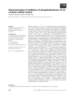
analysed by SDS/PAGE (Fig. 1A). No protein bands were
visible in the fraction eluted using buffer at pH 2 (data not
shown). Subsequent elution of the column with buffer at
pH 12 gave a fraction containing a number of discrete
polypeptides, with major bands at 25, 50 and
55 kDa. Finally, the column was eluted under highly
denaturing conditions, using buffer containing 6
M
guani-
dine hydrochloride; the eluted fraction contained three
polypeptides, a major band at 35 kDa, and a closely
spaced doublet of bands at 30 kDa.
Proteins were identified by N-terminal sequencing of
polypeptide bands blotted from gel electrophoretic separa-
tions (Table 1). None of the major bands eluted at pH 12
contained N-terminal sequences similar to carboxypepti-
dases present in the databases. The two proteins migrating
at 50 and 55 kDa (bands A and B; Fig. 1A) were
identified from their N-terminal sequences as similar to
a-amylase (accession no. AAA17751) from silkworm
(Bombyx mori). The 25 kDa polypeptide band (band F;
Fig. 1A) gave an N-terminal sequence which corresponded
to that predicted by a cDNA previously isolated from the
H. armigera larval gut library [12]. This cDNA, SR21
(accession No. Y12274) encodes a protein with sequence
similarity to serine proteases, but which appears to lack
members of the catalytic triad required for enzyme activity.
Binding of these proteins to PCI may be a result of specific
interactions between the inhibitor and the proteins them-
selves, although this would not be expected on the basis of
their functional properties and sequences, or may suggest
that they are present in a tightly bound complex with
carboxypeptidase(s) in vivo.
In contrast to the bands eluted at pH 12, the polypeptides
eluted by 6
M
guanidine hydrochloride had N-terminal
sequences with similarity to, or identity with carboxypep-
tidases. The band estimated as 35 kDa (band C; Fig. 1A)
gave an N-terminal sequence which had 41% identity over
29 amino acid residues to the N-terminal region of a crayfish
carboxypeptidase (P04069). The less strongly stained bands
estimated as 30 kDa (bands D and E; Fig. 1A) contained
two different N-terminal sequences. The lower molecular
mass band of the doublet (band E; Fig. 1) gave an
N-terminal sequence identical over 32 amino acid residues
to the N-terminal region of carboxypeptidase A from
H. armigera larval gut (sequence predicted by cDNAs
AJ005176–8). The higher molecular mass band of the
doublet (band D; Fig. 1A) gave an N-terminal sequence of
20 amino acid residues which was similar to (47% identity),
rather than identical with the N-terminal region of
H. armigera carboxypeptidase A.
Identification of cDNAs encoding carboxypeptidases
in
H. armigera
larval gut library
The characterization of three similar cDNAs encoding
carboxypeptidase A-like digestive proteases as a result of
screening a cDNA library prepared from RNA extracted
from gut tissue of corn earworm larvae has been described
previously [10]. In order to isolate cDNA clones encoding
other digestive carboxypeptidases, degenerate oligonucleo-
tide primers were designed using the N-terminal sequence
data obtained from gut polypeptides, as described above.
Using these specific primers, and primers directed against
vector sequences, PCR was carried out on the larval gut
cDNA library. Both N-terminal primers in combination
with a generic 3¢ primer gave products of 1.0 kb. PCR
products were individually excised from gel, purifed, and
cloned. At least three independent clones for each product
were characterized by a preliminary DNA sequencing run.
Fig. 1. Purification of native and recombinant carboxypeptidases. (A)
Affinity chromatography of gut extract from larval H. armigera on
immobilized PCI. Fractions eluted under conditions as shown were
analysed by SDS/PAGE. Bands A–F refer to polypeptides subjected to
N-terminal sequence analysis (Table 1). (B) Purification of recombin-
ant HaCA42 carboxypeptidase from culture medium after expression
in P. pastoris. The purified protein was analysed by SDS/PAGE.
Ó FEBS 2004 Glutamate-specific insect carboxypeptidase (Eur. J. Biochem. 271) 2003
The PCR reactions using the two separate carboxypepti-
dase-specific primers each gave essentially a single product
with similarity to carboxypeptidases (although minor het-
erogeneity, potentially resulting from amplification errors,
was present). These sequences were then used as probes to
screen the cDNA library. cDNAs detected by each of the
two PCR products were isolated and sequenced.
Characterization of
H. armigera
carboxypeptidase-
encoding cDNAs
cDNAs encoding the 35 kDa H. armigera carboxypepti-
dase (band C, Fig. 1A) are exemplified by a clone designa-
ted HaCA42. This cDNA (accession no. AJ626862) was
fully sequenced on both strands; it is truncated at the 5¢ end,
and starts at nucleotide 8 of the coding sequence. The
sequence at the 5¢ end of the mRNA was completed
by 5¢ RACE, from which two independent clones gave
identical sequences at the same starting point for the
mRNA. A poly(A) tail is present. The corresponding
mRNA thus contains an 11 base 5¢ untranslated region
(UTR), a coding sequence of 1275 bases (including stop
codon), and an 89 base 3¢ UTR excluding the poly(A)
sequence. A cDNA clone with 98% identity with HaCA42
at the nucleotide level over the coding sequence and 99%
identity with HaCA42 in the deduced amino acid sequence
was also sequenced, and represents a second member of the
subfamily of carboxypeptidase genes exemplified by
HaCA42. The deduced amino acid sequence of HaCA42
(Fig. 2) predicts that this is a secreted protein, the first 18
residues constituting a typical signal peptide (SignalP
prediction, vs. 2.0). The predicted proprotein is therefore
is 406 amino acids in length, with a predicted MW of
46.0 kDa. When this sequence was used to query the
protein sequence databases, the closest similarity (38–40%
identity, based on identity of corresponding amino acid
residues) was to the carboxypeptidase A sequences encoded
by the cDNAs previously isolated from H. armigera
Fig. 2. Predicted protein sequence from cDNA HaCA42. The predicted signal peptide is indicated; propeptide and mature protein are designated
from N-terminal sequence determined for carboxypeptidase purified from H. armigera larval gut extract (shaded). Sequence features of clan MC
carboxypeptidases are denoted as follows (numbering from human carboxypeptidase A sequence): *, catalytically active residues (Arg127, Glu270);
d, zinc ligand residues (His69, Glu72, His196); b, substrate binding residues (Arg71, Asn144, Arg145, Tyr198, Tyr248); s,S1¢ site residue (Arg255).
Potential N-glycosylation sequences are boxed.
Table 1. N-terminal sequences of polypeptides eluted from affinity column containing immobilized PCI at pH 12 and with 6
M
guanidine hydrochloride.
Partial amino acid sequences predicted by specified cDNAs (accession numbers in brackets) are given in italic type.
Band
(kDa)
N-terminal sequence determined/sequence predicted
from cDNA (partial) Identification
PCI
F (25)
SSSPARXEDYPSTVQLETGI Ha cDNA SR21
AYSSSSPA RIEDYPSTVQLETGIGRV Similar to serine protease (Y12274)
B (50)
YKNPYYAPGR(S)VNVN Bombyx mori
ALAY KNPHYAS GR T TMVHLFE a-amylase (AAA17751)
A (55)
YLNPXY Bombyx mori
ALAYKNPHYASGRTTMVHLFE a-amylase (AAA17751)
6
M
guanidine hydrochloride
E (30–)
LSFDKIHSYEEVDAYLQELAKEFPNVVTVV Ha cDNA CM1
RSRLSFDKIHSYEEVDAYLQEL AKEFP NVVTVVEGG carboxypeptidase (AJ005176)
D (30+)
LD(F/S)LPFDQIYTYHQVDTFLA Ha cDNA CB6
ASRLD S LPFDQIYTYHQVDTFLDMLA carboxypeptidase
C (35)
SITWDTYYRHDEINDYLDELAEQNSD(L/I)XTV Ha cDNA CA42
SGKSITWDTYYR HDEINDYLDELAEQNSD L VTVINA carboxypeptidase
2004 D. P. Bown and J. A. Gatehouse (Eur. J. Biochem. 271) Ó FEBS 2004
(accession numbers AJ005176–8). Similar levels of similarity
were found to sequences of ORFs found in the genomes
of Drosophila (34–37% identity, NM139861-3), Anopheles
(42% identity, AAAB01008960) and Caenorhabditis elegans
(40% identity, NM074283). A carboxypeptidase B enzyme
from crayfish also lies within the group of sequences
showing the high levels of similarity to HaCA42 (37%
identity, P04069).
The N-terminal sequence determined for the 35 kDa
carboxypeptidase from H. armigera larval guts (band C;
SITWDTY…; Table 1) is located 98 amino acids from the
predicted N-terminus of the pro-region (Fig. 2). The amino
acid sequence predicted by the cDNA is identical to the
sequence determined (29 amino acid residues). Removal of
the pro-region results in a predicted protein of 308 amino
acids, MW 34.8 kDa, in close agreement with that deter-
mined by SDS/PAGE. Other features of the predicted
protein sequence are consistent with the conserved residues
in metallocarboxypeptidases of clan MC [2]. The mature
sequence contains amino acid residues His69, Glu72 and
His196 (numbering based on human carboxypeptidase A)
which ligate the catalytic zinc ion in metallocarboxypeptid-
ases; Arg127 and Glu270 also involved in catalysis; and
Arg71, Asn144, Arg145, Tyr198 and Tyr248, which parti-
cipate in substrate binding (Fig. 2). A distinguishing feature
of this predicted protein sequence is the amino acid residue
at position 255, which determines substrate specificity by
interacting with the side chain of the P
1
¢ residue. In the
protein predicted by HaCA42 this residue is arginine. There
are also two consensus N-glycosylation sites within the
amino acid sequence predicted by HaCA42, both of which
lie within the mature protein, with one near the C-terminus.
The cDNAs encoding the polypeptide present in the
upper band of the 30 kDa doublet of H. armigera carb-
oxypeptidases (band D, Fig. 1A) are exemplified by a clone
designated HaCB6 (accession no. AJ626863). This cDNA
also encodes a clan MC metallocarboxypeptidase enzyme,
and will be described elsewhere.
Expression and purification of recombinant
procarboxypeptidase HaCA42
A construct to allow the protein encoded by HaCA42 to
be expressed in the yeast P. pastoris was assembled by
amplifying the coding sequence of the cDNA by PCR.
Primers were designed to allow the PCR product to be
inserted into the Pichia expression vector pGAPZaBwith
the N-terminus of the proprotein in-frame and adjacent to
the cleavage point of the yeast a-mating factor secretion
signal encoded by the vector. In addition, a (His)
6
-tag
encoded by the vector was added to the C-terminus of the
protein before the stop codon. The construct was verified by
DNA sequencing after assembly, and linearized plasmid
DNA was used to transform competent P. pastoris.After
selection for transformation on zeocin plates, colonies were
screened by PCR for the presence of the HaCA42 sequence.
Positive colonies were individually grown in small-scale
cultures, and samples of culture medium were assayed for
expression of His-tagged protein by immunodot blot. The
transformant that showed the highest level of expression
was chosen for protein production, and was grown up under
optimized conditions in a 2 L laboratory fermentor.
The recombinant protein (referred to subsequently as
HaCA42 procarboxypeptidase or carboxypeptidase) was
purified from culture medium by hydrophobic interaction
chromatography followed by affinity chromatography on
immobilized nickel ions. The purified protein ran as a single
band when analysed by SDS/PAGE, with an estimated
MW of 50 kDa (Fig. 1B). The yield of purified protein
was 5mgÆL
)1
of fermenter culture.
Activation of recombinant procarboxypeptidase HaCA42
The recombinant HaCA42 procarboxypeptidase enzyme
had no detectable activity when assayed against synthetic-
furylacryloyl (FA)–peptide substrates for carboxypeptidases.
Three substrates were assayed: FAPP, with a C-terminal
phenylalanine residue (hydrolysed by carboxypeptidase A);
FAAK, with a C-terminal lysine residue (hydrolysed by
carboxypeptidase B) and FAEE, with a C-terminal gluta-
mate residue (hydrolysed by glutamate carboxypeptidase).
However, after the protein was treated with substoichio-
metric amounts of bovine trypsin (procarboxypeptidase/
trypsin molar ratio > 5 : 1) carboxypeptidase activity
against FAEE could be detected. Trypsin gave no activity
against any of these substrates in the absence of the
recombinant carboxypeptidase. Because the activation of
mammalian digestive procarboxypeptidases in vivo is
known to be caused by cleavage of the propeptide by
trypsin [19], the results suggested that a similar activation
process was necessary for the HaCA42 procarboxypepti-
dase, and that endogenous yeast proteases in the protease-
deficient Pichia strain used were not sufficient to cause
activation.
To further investigate the activation process, recombinant
HaCA42 procarboxypeptidase was incubated with trypsin
(9.4 : 1 molar ratio) at 37 °C. At various timepoints samples
were withdrawn and trypsin activity was quenched; the
carboxypeptidase activity against FAEE was then assayed,
and the polypeptides present were analysed by SDS/PAGE.
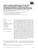
Results are shown in Fig. 3A. A control sample of HaCA42
procarboxypeptidase (track C) contained no detectable
polypeptides of < 48 kDa, but even after a nominal zero
incubation time (track 0), corresponding to sampling the
mixture of procarboxypeptidase and trypsin immediately
after addition of trypsin, polypeptides of 36 kDa and
13 kDa are present in the sample. Neither of these
polypeptides was present in trypsin when this enzyme was
analysed by SDS/PAGE (data not shown). By analogy with
the activation of mammalian carboxypeptidases, these
polypeptides correspond to the active HaCa42 carboxy-
peptidase (36 kDa polypeptide) and the pro-region (13 kDa
polypeptide). After 5 min the majority, and by 20 min all, of
the original 50 kDa protein had been digested, with an
increase in staining of the bands of 36 kDa and
13 kDa. On further incubation with trypsin the 13 kDa
polypeptide is itself digested by trypsin, and decreases in
amount until it is no longer detectable after 80 min of
digestion, but the amount of 36 kDa polypeptide remains
constant up to 2 h digestion under these conditions. When
the carboxypeptidase activity (FAEE substrate) of the
mixture was assayed, there was a qualitative correlation
between the appearance of the putative 36 kDa activated
carboxypeptidase polypeptide, and the level of activity
Ó FEBS 2004 Glutamate-specific insect carboxypeptidase (Eur. J. Biochem. 271) 2005
detected. Thus, the control procarboxypeptidase sample
had no detectable activity, but the zero time sample
contained detectable activity (5% of maximum activity)
which increased with time (Fig. 3B). However, when
quantitative estimates of activity were compared to results
of the gel analysis, it was apparent that cleavage alone was
not sufficient for activation. After 20 min digestion by
trypsin, all of the procarboxypeptidase band at 50 kDa
had been cleaved to 36 kDa and 13 kDa bands, but the
carboxypeptidase activity was only 55% of the maximum
activity (Fig. 3A,B). The carboxypeptidase activity only
reaches a maximum after 60–80 min incubation with
trypsin, and further incubation with trypsin to 120 min does
not affect the level of activity against this substrate.
Attainment of maximum carboxypeptidase activity in this
assay corresponds to the disappearance of the 13 kDa
pro-region polypeptide (Fig. 3A,B); once this polypeptide
has been completely digested by trypsin, the carboxypepti-
dase activity is maximal.
Characterization of recombinant HaCA42
carboxypeptidase activity
The pH optimum for hydrolysis of FAEE by the activated
HaCA42 carboxypeptidase was determined over the range
2.2–10.5 using a variety of buffer systems. There was a
marked optimum activity at pH 8.5 in borate buffer with
activity declining to 50% of maximum at pH 7.5 and 10.0
(data not shown). Various diagnostic inhibitors were used to
characterize the activity of the recombinant enzyme. No
inhibition (< 10% reduction in activity compared to
enzyme preincubated without inhibitor) was observed after
preincubation with: the cysteine protease inhibitor E-64
(10
)5
M
final concentration); the aspartic protease inhibitor
pepstatin (10
)5
M
); the serine protease inhibitors phenyl
methylsulphonyl fluoride, (2 · 10
)5
M
) and soybean kunitz
trypsin inhibitor (5 · 10
)7
M
); chymostatin (10
)5
M
)an
inhibitor of chymotrypsin; and benzamidine (10
)2
M
)an
inhibitor of trypsin.
The metalloprotease inhibitors phenanthroline (5 ·
10
)3
M
) and EDTA (10
)2
M
) both had marked effects on
activity (82% inhibition and 96% inhibition, respectively)
as did the protein carboxypeptidase inhibitor PCI (94%
inhibition at 2.5 · 10
)6
M
). Interestingly, preincubation
with zinc, used by many authors in activating carboxy-
peptidase, has a deleterious effect on activity; 10
)5
M
ZnCl
2
inhibits activity by 68% and 10
)6
M
ZnCl
2
inhibits activity
by 21%. The reducing agent dithiothreitol also inhibits
activity of the recombinant HaCA42 carboxypeptidase at
concentrations above 10
)5
M
, resulting in 45% inhibition at
10
)4
M
and 86% inhibition at 10
)3
M
.
The kinetic parameters for hydrolysis of FAEE by the
recombinant HaCA42 carboxypeptidase were determined
by a standard Michaelis–Menten analysis using varying
substrate concentrations. K
m
was estimated as 6 · 10
)5
M
(mean of three determinations), and V
max
was estimated as
7.3 · 10
)7
moles FAEE hydrolysedÆs
)1
Æmg protein
)1
. Assu-
ming a MW of 36 kDa for the active recombinant enzyme,
and that all the proenzyme has been activated and remains
active in the assay, these figures give values of 26 s
)1
for
i
at
and 4.3 · 10
5
s
)1
Æ
M
)1
for k
cat
/K
m
.
Substrate specificity of recombinant HaCA42
carboxypeptidase
The activated recombinant HaCA42 carboxypeptidase
hydrolysed FAEE (Glu C-terminal residue), but gave no
detectable hydrolysis of synthetic substrates for carboxy-
peptidase A (FAPP, Phe C-terminal residue) or carboxy-
peptidase B (FAAK, Lys C-terminal residue) even when
used in large amount for extended digestion periods. The
specificity of the activated enzyme was investigated in more
detail by incubation with a selection of peptides of known
sequence in the presence of trypsin inhibitors to prevent
digestion by the activating enzyme. The presence or absence
of digestion was assayed by MS over a mass range which
included the peptide substrates. Results are presented in
Table 2, which defines the peptide sequences and their
abbreviations.
When hydrolysing peptide substrates at enzyme/substrate
ratios of 1 : 200, the HaCA42 carboxypeptidase had a
similar specificity to that observed when synthetic dipeptide
Fig. 3. Activation of HaCA42 carboxypeptidase by trypsin. (A) SDS/
PAGE of cleavage of procarboxypeptidase, sampled after stated times
of digestion with bovine trypsin (9.4 : 1 molar ratio procarboxypept
idase/trypsin). Pro-, procarboxypeptidase; Mature, mature carboxy-
peptidase; Activn. peptide, pro-region. The faint band at 25 kDa is
from the trypsin used for activation. (B) Carboxypeptidase activity
(digestion of FAEE substrate) after stated times of digestion with
bovine trypsin.
2006 D. P. Bown and J. A. Gatehouse (Eur. J. Biochem. 271) Ó FEBS 2004
substrates were used. Angiotensin 1, a peptide with a
C-terminal neutral, hydrophobic amino acid (Leu) was
not hydrolysed, like the synthetic substrate FAPP (Phe
C-terminal residue). Similarly, fibrinopeptide B, with a
C-terminal basic residue (Arg), like FAAK (Lys C-terminal
residue), was not hydrolysed. The neutral hydrophilic
C-terminal serine of angiotensin (1–14), and the C-terminal
proline of ACTH 1–24 were also not hydrolysed. On the
other hand, a peptide with a C-terminal glutamate residue
(b-endorphin amino acids 61–91) was readily cleaved by the
HaCA42 carboxypeptidase, like the synthetic substrate
FAEE (C-terminal Glu residue). The specificity was further
explored by using peptide substrates with the C-terminal
side-chain amide residues, asparagine (PDI substrate) and
glutamine (Cys-CD36). Neither peptide was hydrolysed
by the HaCA42 carboxypeptidase, suggesting that the
C-terminal amino acid must carry a negative charge on the
side chain. Finally, an angiotensin 1 converting enzyme
(ACE) inhibitor peptide with a C-terminal aspartate residue
was assayed; this was hydrolysed by the carboxypeptidase,
but very slowly. Under conditions sufficient to completely
cleave the b-endorphin substrate, < 5% of the ACE-
inhibitor peptide was cleaved, as estimated by the
appearance of a new peptide with lower molecular mass
(data not shown).
The specificity of the HaCA42 carboxypeptidase was also
investigated by carrying out a time-course experiment for
digestion of the b-endorphin substrate. Results are presented
in Fig. 4. At an enzyme/peptide ratio of 1 : 5000, appear-
ance of a peptide product of correct mass for cleavage of the
C-terminal glutamate from the b-endorphin peptide was
observed after 1 min. The amount of this product relative to
the undigested peptide increased with time, until digestion
was essentially complete after 90 min. After removal of the
C-terminal glutamate, the next residue is a glycine, but there
was no evidence for removal of this residue from the initial
product of HaCA42 carboxypeptidase digestion in the
timescale of this experiment (up to 120 min), or in experi-
ments where HaCA42 carboxypeptidase was present at
ratios up to 1 : 200 with respect to substrate.
The HaCA42 carboxypeptidase was also assayed for its
ability to hydrolyse the folate analogue methotrexate, which
contains a glutamate residue linked via an amide bond to
pteroic acid. No activity against this substrate could be
detected in a spectrophotometric assay in the presence of
excess enzyme.
Glutamate carboxypeptidase activity in
H. armigera
larvae
Activity towards synthetic substrates for carboxypeptidase
A and B (FAPP and FAAK) has previously been charac-
terized in gut extracts from H. armigera larvae [10].
However, crude extracts of H. armigera larval gut contents
showed little detectable activity towards the glutamate
carboxypeptidase substrate FAEE, although carboxypep-
tidase A activity, and low levels of carboxypeptidase B
activity could be detected in the same material. To confirm
that the digestive carboxypeptidases in this insect did
include enzymes with activity towards substrates with
C-terminal glutamate residues, two approaches were taken.
When insects were induced to regurgitate gut contents, and
the regurgitant was collected and analysed, carboxypepti-
dase activity towards the FAEE substrate could be readily
detected. The activity was shown to be present in bulk gut
contents by partial purification of total gut content proteins
by ammonium sulphate precipitation. The redissolved
ammonium sulphate pellet was assayed for carboxypepti-
dase activity, and hydrolysis of both FAPP (carboxypepti-
dase A activity) and FAEE (glutamate carboxypeptidase
activity) were detected, although more activity towards the
former substrate was present. Quantitative analysis gave
values of 4.3 · 10
)8
moles FAPP hydrolysedÆmin
)1
Ægut
equivalent
)1
and 7.3 · 10
)9
moles FAEE hydro-
lysedÆmin
)1
Ægut equivalent
)1
under the conditions of this
assay, suggesting that approximately six times as much
carboxypeptidase A activity as glutamate carboxypeptidase
activity is present in bulk gut contents.
A Northern blot of RNA extracted from gut tissue of
larval H. armigera was probed with the HaCA42 cDNA. A
single band of estimated size 1.45 kb was observed after
autoradiography (Fig. 5), consistent with the estimated size
of the mRNA, and its assumed abundance in gut tissue.
Discussion
Carboxypeptidases specific for glutamate have been char-
acterized from a number of bacterial species; they are
referred to as carboxypeptidase G (various subtypes), or
more correctly, glutamate carboxypeptidases (NC-IUBMB
preferred [25]), and have been given the EC classification
3.4.17.11. These enzymes are able to cleave C-terminal
glutamate residues in peptides, and also the glutamate
Table 2. Digestion of peptide substrates by activated recombinant HaCA42 carboxypeptidase. Digestion was detected by MS after varying times of
digestion up to 2 h. 0, no digestion detectable; ±, slight digestion detectable; +++, digestion readily detectable. ACTH, adrenocorticotropic
hormone.
Peptide C-terminal amino acid Sequence (M
r
) Digestion
[Glu1]-Fibrinopeptide B Arginine
EGVNDNEEGFFSAR 0
PDI substrate Asparagine
NRCSQGSCWN 0
ACE inhibitor Aspartate
PTHIKYGD +)
b-endorphin (aa 61–91) Glutamate YGGFMTSEKSQTPLVTLFKNAIIKNAYKKGE +++
Cys-CD36 (aa 139–155) Glutamine CNLAVAAASHIYQNQFVQ 0
Angiotensin 1 Leucine
DRVYIHPFHL 0
ACTH (1–24) Proline
SYSMEHFRWGKPVGKKRRPVKVYP 0
Angiotensinogen 1–14 (rat) Serine
DRVYIHPFHLLYYS 0 (unstable)
Ó FEBS 2004 Glutamate-specific insect carboxypeptidase (Eur. J. Biochem. 271) 2007
residue linked via its a-amino group to pteroic acid in folic
acid and folate analogues, such as the drug methotrexate
(4-amino-N
10
-methylpteroylglutamate). A distinct enzyme,
known as glutamate carboxypeptidase II (EC 3.4.17.21),
which is active towards acidic dipeptides with C-terminal
glutamate, and folate analogues, is present in mammalian
nervous tissue and prostate [26]. These enzymes all belong to
clan MH of metalloproteinases, and have little sequence
similarity or structural similarity to clan MC carboxy-
peptidases. The enzyme described in the present paper is
different from these previously described glutamate
carboxypeptidases in belonging to the clan MC of metallo-
carboxypeptidases. It is also more specific than the clan
MH glutamate carboxypeptidases, as it has no detectable
activity towards glutamate residues linked to folic acid. No
other carboxypeptidase in clan MC has a similar specificity
to the HaCA42 enzyme, and to date the only eukaryotic
digestive carboxypeptidase activity demonstrated has been
of the -A or -B type [2,27]. The HaCA42 enzyme is therefore
the first example of a new peptidase. The best nomenclature
for this enzyme would be carboxypeptidase C (which would
emphasize its similarity to carboxypeptidases A and B), but
this name is already used for a subclass of serine carboxy-
peptidases, although not for any specific enzyme in this
class. ÔGlutamate carboxypeptidase MCÕ is a possible
alternative.
It seems unlikely that this type of carboxypeptidase is
unique to H. armigera, and it would be reasonable to expect
similar enzymes to be present in other lepidopteran
Fig. 4. Time-course for digestion of b-endorphin peptide substrate by
activated HaCA42 carboxypeptidase. Traces show mass spectra from
peptide sampled after varying times of digestion. Mass ion at m/e
3465.0 corresponds to uncleaved peptide; mass ion at 3335.9 corres-
ponds to removal of a glutamate residue from the C-terminus; this
product is then stable to further C-terminal exopeptidase action. The
small peak at m/e 3150.7 visible after extended digestion results from
cleavage between lysine residues in the peptide C-terminal sequence
(…YKKGE) caused by residual trypsin activity from the activating
enzyme.
Fig. 5. Expression of HaCA42 in gut mRNA. RNA extracted from
midgut tissue of H. armigera larvae fed on control diet (C) and diet
supplemented with SKTI (S) was separated by formaldehyde/agarose
gel electrophoresis and blotted onto nitrocellulose. The blot was pro-
bed with the HaCA42 cDNA (coding sequence and 3¢ UTR) and
washed to a final stringency of 0.1 · NaCl/Cit, 0.1% SDS at 50 °C.
The size of the hybridizing band was estimated from markers run on
separate tracks of the same gel, which were excised and stained.
2008 D. P. Bown and J. A. Gatehouse (Eur. J. Biochem. 271) Ó FEBS 2004
herbivores, and possibly in a wider range of arthropods. The
Drosophila melanogaster (fruit fly) genome contains 19 genes
encoding proteins with sequence similarity to the HaCA42
carboxypeptidase (
BLAST
comparison, E < 10
)30
), plus two
genes encoding proteins with a low level of similarity
(CG4122, 4678; E ¼ 7 · 10
)6
,2· 10
)6
, respectively). A
phylogenetic tree based on sequence comparison between
HaCA42 and similar proteins predicted by the Drosophila
genome is shown in Fig. 6A. The HaCA42 carboxypepti-
dase maps within the phylogenetic tree of similar Drosophila
predicted proteins. Although not all the Drosophila genes
encode active carboxypeptidase enzymes, the majority
contain the residues necessary for activity, and have
sufficient similarity over the region corresponding to
residues 248–270 in human carboxypeptidase A to allow
the equivalent residue to amino acid 255, the specificity
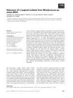
determining residue, to be identified. Three genes, CG4408,
CG12374 and CG14820, predict proteins with lysine
residues at position 255 (Fig. 5B), where a positively
charged basic side chain should give these proteins a similar
specificity to HaCA42. All these proteins are predicted to
have metallocarboxypeptidase activity; the CG12374
product is annotated in FlyBase as having carboxy-
peptidase A activity, but this assignment is based only
on overall sequence similarity and, we suggest, is
probably incorrect.
In contrast with the situation in Drosophila,theAnopheles
gambiae (mosquito) genome does not contain genes enco-
ding carboxypeptidases with similar predicted specificity to
HaCA42. There are 22 genes predicting proteins with
sequence similarity to HaCA42 (E < 10
)29
), but none of
the genes predicting active enzymes have a basic residue at
positions equivalent to Ile255 in human carboxypeptidase
A, all being carboxypeptidase A- or B-like in predicted
specificity. In support of a wider distribution of glutamate-
specific carboxypeptidases beyond H. armigera,examina-
tion of the global sequence databases suggests that a further
enzyme similar in sequence to HaCA42, and with a similar
predicted cleavage specificity, is present in one other insect
species. An incomplete cDNA from tsetse fly (Glossinia
morsitans morsitans), designated GmZcp (accession number
AAK07479; amino acid sequence given is not complete),
Fig. 6. Sequence comparisons for carboxypeptidases. (A) Phylogenetic tree for predicted carboxypeptidases of clan MC, family M14, from
D. melanogaster (designated by CG-gene identifier) compared to HaCA42 carboxypeptidase (shaded branch). The S
1
¢ site residue (AA255) and the
predicted carboxypeptidase activity (A-like, B-like or glutamate-) based on this residue are as indicated. Sequence similarity over the region
corresponding to amino acids 248–270 (human carboxypeptidase A numbering) is not present for the predicted products of CG32379 and CG8945;
CG15679 lacks both E270 and Y248, and CG3097 and CG8564 lack Y248. These genes are predicted to encode proteins inactive as carboxy-
peptidases. (B) Sequence alignment over the region including amino acids 246–272 (human carboxypeptidase A numbering) for human carb-
oxypeptidase A, and enzymes of clan MC, family M14 predicted to show glutamate carboxypeptidase activity. H. armigera (shaded) and
D. melanogaster genes are designated as above; GmZcp cDNA, protein predicted by G. morsitans (tsetse fly) gut cDNA clone.
Ó FEBS 2004 Glutamate-specific insect carboxypeptidase (Eur. J. Biochem. 271) 2009
encodes a carboxypeptidase which contains a lysine residue
at position 255 (Fig. 6B), like the Drosophila sequences,
predicting similar specificity to HaCA42. In this case the
enzyme has been incorrectly predicted to have carboxy-
peptidase B-like specificity [15]. The tsetse fly sequence is
predicted to encode a digestive enzyme, as it was cloned
from gut tissue, and the corresponding mRNA increases in
level in response to feeding, but the role(s) of the Drosophila
genes are not known. It seems likely that further enzymes
with this predicted specificity will be found in other insects,
and possibly in a wider range of eukaryotes, as more
sequence data become available.
Expression of the HaCA42 carboxypeptidase as a
recombinant protein in P. pastoris has allowed its functional
properties to be fully characterized. A similar approach has
been taken for a second carboxypeptidase from this species,
the digestive carboxypeptidase A-like enzyme described
by Bown et al. [10]. A study using recombinant enzyme
produced in P. pastoris showed that the insect enzyme had a
broader substrate specificity than human carboxypeptidase
A, showing some hydrolytic activity towards both aliphatic
and basic C-terminal residues as well as more hydrophobic
residues [20]. In that case also, activation of the proenzyme
produced in Pichia by bovine trypsin was necessary for
activity.
The activation of human carboxypeptidase A by trypsin
in the intestine involves both the cleavage of a susceptible
peptide bond between pro-region and mature polypeptide,
and the subsequent degradation of the pro-region peptide
by trypsin and other digestive enzymes. The pro-segment
behaves as an inhibitor of the carboxypeptidase prior to
activation, and can be shown to act as a potent inhibitor
(K
i
in the n
M
range) when produced separately and added
to activated enzyme [28]. Structural studies have shown
thatthepro-segmentbindstotheactivesiteregionofthe
enzyme, rendering the active centre inaccessible to protein
and peptide substrates [19]. The activation process is thus
limited by degradation of the bound pro-region, because the
carboxypeptidase will be inhibited as long as the activation
domain of the pro-region is kept in place. Structural studies
on recombinant carboxypeptidase A-like enzyme from
H. armigera have shown the presence of an inhibitory
pro-region in the enzyme prior to activation with bovine
trypsin [29], and suggest that the activation process for this
enzyme in vivo is similar to that established for mammalian
enzymes, with insect trypsin-like enzymes being responsible
for the cleavage and degradation of the pro-region.
Subsequent studies suggested that the lysine-specific endo-
protease LysC was a more effective activator of the enzyme
in vitro [20], although enzymes of this type have not been
detected in the insect host, whereas trypsin-like enzymes are
abundant [12], and lepidopteran trypsins have been shown
to hydrolyse more efficiently at Lys than at Arg residues
[30]. The activation process for the HaCA42 carboxypep-
tidase corresponds well to this general model; there is a lysine
reside immediately prior to the mature peptide N-terminus
(Fig. 2), so that peptide bond between propeptide and
mature protein can be cleaved by Helicoverpa trypsin. There
are a further five basic residues in the preceding 11 amino
acids of the propeptide, giving this region a high probability
for cleavage by trypsin. Cleavage of the recombinant
protein by trypsin in vitro takes place at or near the
N-terminus of the mature peptide produced in vivo,asthe
estimated molecular masses of pro- and mature polypep-
tides in vitro correspond to cleavage having taken place in
this region.
The presence of hydrolytic activity towards FAEE in
H. armigera larval gut extract and regurgitant shows that
activated glutamate carboxypeptidase is present in vivo,a
conclusion confirmed by the isolation of a polypeptide with
the predicted N-terminal sequence of the mature protein by
affinity chromatography on immobilized potato carboxy-
peptidase inhibitor. The H. armigera carboxypeptidases
bound very tightly to this inhibitor, with complete denatur-
ationin6
M
guanidine hydrochloride necessary for elution.
This inhibitor forms stable complexes with a wide range of
carboxypeptidases, with dissociation constants in the nano-
molar range, but without binding being dependent on the
cleavage specificity of the enzyme (the C-terminal residue of
the inhibitor is glycine). The purified polypeptides are thus
likely to represent the range of digestive carboxypeptidases
in the gut extract. An unexpected result was that the most
abundant carboxypeptidase to be eluted from the PCI
affinity column was not the carboxypeptidase A-like
enzyme previously characterized [10], but (apparently) the
glutamate carboxypeptidase encoded by HaCA42, whereas
gut carboxypeptidase activity towards synthetic substrates
shows more A-like activity than other types. The kinetic
parameters for the recombinant HaCA42 carboxypeptidase
are similar to those found for the carboxypeptidase A-like
enzyme from this insect [10,20] and therefore the difference
in FAPP-specific and FAEE-specific carboxypeptidase
activities in vivo cannot be explained by differences in
specific activities of the respective enzymes. Possibly the
majority of the glutamate carboxypeptidase enzyme remains
associated with its inhibitory pro-peptide in vivo in gut
contents. Further studies will be required to fully charac-
terize the complement of digestive carboxypeptidases in this
insect.
Acknowledgements
The authors thank John Gilroy for coaxing some excellent protein
sequence data out of an ageing instrument, and Prof. F. X. Aviles for
the generous gift of recombinant PCI. This work was funded in part
by EC Programme FAIR6-CT98-4239 and by the McKnight
Foundation.
References
1. Clauser, E., Gardell, S.J., Craik, C.S., Macdonald, R.J. & Rutter,
W.J. (1988) Structural characterization of the rat carboxy-
peptidase A1 and carboxypeptidase B Genes – comparative ana-
lysis of the rat carboxypeptidase gene family. J. Biol. Chem. 263,
17837–17845.
2. Barrett, A.J., Rawlings, N.D. & Woessner, J.F. (1998) Handbook
of Proteolytic Enzymes. Academic Press, London.
3. Gardell, S.J., Craik, C.S., Clauser, E., Goldsmith, E.J., Stewart,
C.B., Graf, M. & Rutter, W.J. (1988) A novel rat carboxy-
peptidase, Cpa2 – characterization, molecular cloning, and
evolutionary implications on substrate specificity in the
carboxypeptidase gene family. J. Biol. Chem. 263, 17828–17836.
4. Tan, A.K. & Eaton, D.L. (1995) Activation and characterization
of procarboxypeptidase B from human plasma. Biochemistry 34,
5811–5816.
2010 D. P. Bown and J. A. Gatehouse (Eur. J. Biochem. 271) Ó FEBS 2004
5. Titani, K., Ericsson, L.H., Walsh, K.A. & Neurath, H. (1975)
Amino acid sequence of bovine carboxypeptidase B. Proc. Natl.
Acad. Sci. USA 72, 1666–1670.
6. Aviles, F.X., Vendrell, J., Guasch, A., Coll, M. & Huber, R. (1993)
Advances in metallo-procarboxypeptidases – emerging details on
the inhibition mechanism and on the activation process. Eur. J.
Biochem. 211, 381–389.
7. Ward, C.W. (1976) Properties of the major carboxypeptidase
in the larvae of the webbing clothes moth Tineolla bisselliella.
Biochim. Biophys. Acta 429, 564–572.
8. Ferreira, C., Bellinello, G.L., Ribeiro, A.F. & Terra, W.R. (1990)
Digestive enzymes associated with the glycocalyx, microvillar
membranes and secretory vesicles from midgut cells of Tenebrio
molitor larvae. Insect Biochem. 20, 839–847.
9.Ferreira,C.,Capella,A.N.,Sitnik,R.&Terra,W.R.(1994)
Digestive enzymes in midgut cells, endoperitrophic and ectoperi-
trophic contents, and peritrophic membranes of Spodoptera
frugiperda (Lepidoptera) larvae. Arch. Insect Biochem. Physiol. 26,
299–313.
10. Bown, D.P., Wilkinson, H.S. & Gatehouse, J.A. (1998) Midgut
carboxypeptidase from Helicoverpa armigera (Lepidoptera:
Noctuidae) larvae: enzyme characterisation, cDNA cloning and
expression. Insect Biochem. Mol. Biol. 28, 739–749.
11. Ramos, A., Mahowald, A. & Jacobs-Lorena, M. (1993) Gut-
specific genes from the black fly Simulium vittatum encoding
trypsin-like and carboxypeptidase-like proteins. Insect Mol. Biol.
1, 149–163.
12. Bown, D.P., Wilkinson, H.S. & Gatehouse, J.A. (1997) Differen-
tially regulated inhibitor-sensitive and insensitive protease genes
from the phytophagous insect pest, Helicoverpa armigera,are
members of complex multigene families. Insect Biochem. Mol.
Biol. 27, 625–638.
13. Edwards, M.J., Lemos, F.J., Donnelly-Doman, M. & Jacobs-
Lorena, M. (1997) Rapid induction by a blood meal of a car-
boxypeptidase gene in the gut of the mosquito Anopheles gambiae.
Insect Biochem. Mol. Biol. 27, 1063–1072.
14. Edwards, M.J., Moskalyk, L.A., Donelly-Doman, M., Vlaskova,
M., Noriega, F.G., Walker, V.K. & Jacobs-Lorena, M. (2000)
Characterization of a carboxypeptidase A gene from the mos-
quito, Aedes aegypti. Insect Mol. Biol. 9, 33–38.
15. Yan, J., Cheng, Q., Li, C.B. & Aksoy, S. (2002) Molecular char-
acterization of three gut genes from Glossina morsitans morsitans:
cathepsin B, zinc-metalloprotease and zinc-carboxypeptidase.
Insect Mol. Biol. 11, 57–65.
16. Hegedus, D., Baldwin, D., O’Grady, M., Braun, L., Gleddie, S.,
Sharpe, A., Lydiate, D. & Erlandson, M. (2003) Midgut proteases
from Mamestra configurata (Lepidoptera: Noctuidae) larvae:
characterization, cDNA cloning, and expressed sequence tag
analysis. Arch. Insect Biochem. Physiol. 53, 30–47.
17. Xiong, B. & Jacobslorena, M. (1995) Gut-specific transcriptional
regulatory elements of the carboxypeptidase gene are conserved
between black flies and Drosophila. Proc. Natl Acad. Sci. USA 92,
9313–9317.
18. Ekbote, U.V., Weaver, R.J. & Isaac, R.E. (2003) Angiotensin I-
converting enzyme (ACE) activity of the tomato moth, Lacanobia
oleracea: changes in levels of activity during development and after
copulation suggest roles during metamorphosis and reproduction.
Insect Biochem. Mol. Biol. 33, 989–998.
19. Vendrell, J., Querol, E. & Aviles, F.X. (2000) Metallocarboxy-
peptidases and their protein inhibitors: structure, function and
biomedical properties. Biochim. Biophys. Acta 1477, 284–298.
20. Bayes, A., Sonnenschein, A., Daura, X., Vendrell, J. & Aviles,
F.X. (2003) Procarboxypeptidase A from the insect pest Helico-
verpa armigera and its derived enzyme. Two forms with new
functional properties. Eur. J. Biochem. 270, 3026–3035.
21. Sambrook, J. & Russell, D.W. (2001) Molecular Cloning: a
Laboratory Manual, 3rd edn. Cold Spring Harbor Laboratory
Press, Cold Spring Harbor, NY.
22. Nielsen, H., Engelbrecht, J., Brunak, S. & von Heijne, G. (1997)
Identification of prokaryotic and eukaryotic signal peptides and
prediction of their cleavage sites. Protein Eng. 10, 1–6.
23. Gatehouse, A.M.R., Davison, G.M., Newell, C.A., Merryweather,
A., Hamilton, W.D.O., Burgess, E.P.J., Gilbert, R.J.C. & Gate-
house, J.A. (1997) Transgenic potato plants with enhanced
resistance to the tomato moth, Lacanobia oleracea:growthroom
trials. Mol. Breed 3, 49–63.
24. Rogelj, B., Strukelj, B., Bosch, D. & Jongsma, M.A. (2000)
Expression, purification, and characterization of equistatin in
Pichia pastoris. Protein Expr. Purific. 19, 329–334.
25. Sherwood, R.F. & Melton, R.G. (1998) Glutamate carboxy-
peptidase. In Handbook of Proteolytic Enzymes (Barrett, A.J.,
Rawlings, N.D. & Woessner, J.F., eds), pp. 1416–1420. Academic
Press, London.
26. Carter, R.E. & Coyle, J. (1998) Glutamate carboxypeptidase II. In
Handbook of Proteolytic Enzymes (Barrett, A.J., Rawlings, N.D.
& Woessner, J.F., eds), pp. 1434–1437. Academic Press, London.
27. Reznik, S.E. & Fricker, L.D. (2001) Carboxypeptidases from A to
Z: implications in embryonic development and Wnt binding. Cell.
Mol. Life Sci. 58, 1790–1804.
28. Sansegundo, B., Martinez, M.C., Vilanova, M., Cuchillo, C.M. &
Aviles, F.X. (1982) The severed activation segment of porcine
pancreatic procarboxypeptidase A is a powerful inhibitor of the
active enzyme – isolation and characterization of the activation
peptide. Biochim. Biophys. Acta 707, 74–80.
29. Estebanez-Perpina, E., Bayes, A., Vendrell, J., Jongsma, M.A.,
Bown, D.P., Gatehouse, J.A., Huber, R., Bode, W., Aviles, F.X. &
Reverter, D. (2001) Crystal structure of a novel mid-gut pro-
carboxypeptidase from the cotton pest Helicoverpa armigera.
J. Mol. Biol. 313, 629–638.
30. Lopes, A.R., Juliano, M.A., Juliano, L. & Terra, W.R. (2004)
Coevolution of insect trypsins and inhibitors. Arch. Insect Bio-
chem. Physiol. 55, 140–152.
Ó FEBS 2004 Glutamate-specific insect carboxypeptidase (Eur. J. Biochem. 271) 2011