Báo cáo khoa học: Genomic organization, tissue distribution and deletion mutation of human pyridoxine 5¢-phosphate oxidase pptx
Bạn đang xem bản rút gọn của tài liệu. Xem và tải ngay bản đầy đủ của tài liệu tại đây (280.27 KB, 10 trang )
Genomic organization, tissue distribution and deletion mutation
of human pyridoxine 5¢-phosphate oxidase
Jeong Han Kang
1
, Mi-Lim Hong
1
, Dae Won Kim
2
, Jinseu Park
2
, Tae-Cheon Kang
3
, Moo Ho Won
3
,
Nam-In Baek
4
, Byung Jo Moon
1
, Soo Young Choi
2
and Oh-Shin Kwon
1
1
Department of Biochemistry, College of Natural Sciences, Kyungpook National University, Taegu, Korea;
2
Department of Genetic
Engineering, Division of Life Sciences, and
3
Department of Anatomy, College of Medicine, Hallym University, Chunchon, Korea;
4
Graduate School of Biotechnology & Plant Metabolism Research Center, Kyunghee University, Suwon, Korea
We used a combined computer and biochemical approach
to characterize human pyridoxine 5¢-phosphate oxidase
(PNPO). The human PNPO gene is composed of seven
exons and six introns, and spans approximately 8 kb. All
exon/intron junctions contain the gt/ag consensus splicing
site. The absence of TATA-like sequences, the presence of
Sp1-binding sites and more importantly, the presence of
CpG islands in the regulatory region of the PNPO gene are
characteristic features of housekeeping genes. Northern blot
analyses showed two species of poly(A)
+
RNA of 2.4 and
3.4 kb at identical intensity, whereas Western blot analysis
showed that no protein isoform exists in any of the tissues
examined. PCR-based analysis led to the idea that two
messages are transcribed from a single copy gene, and that
the size difference is due to differential usage of the poly-
adenylation signal. The major sites of PNPO expression are
liver, skeletal muscle and kidneys while a very weak signal
was detected in lung. The mRNA master dot-blot for mul-
tiple human tissues provided a complete map of the tissue
distribution not only for PNPO but also for pyridoxal kinase
and pyridoxal phosphatase. The data indicate that mRNA
expression of all three enzymes essential for vitamin B
6
metabolism is ubiquitous but is highly regulated at the level
of transcription in a tissue-specific manner. In addition,
human brain PNPO cDNA was expressed in Escherichia
coli, and the roles of both the N- and C-terminal regions
were studied by creating sequential truncation mutants. Our
results showed that deletion of the N-terminal 56 residues
affects neither the binding of coenzyme nor catalytic activity.
Keywords: deletion mutation; genomic organization; PNP
oxidase; polyadenylation; tissue distribution.
Pyridoxal 5¢-phosphate (PLP), the metabolically active form
of vitamin B
6
, is a required coenzyme for numerous
enzymes involved in amino acid metabolism [1]. The
functions of PLP include coenzymatic participation in
reactions leading to the formation of several neurotrans-
mitters [2]. Moreover, it appears that PLP modulates
steroid–receptor interactions and is involved in the regula-
tion of immune function [3]. The enzymes that are
conventionally involved in vitamin B
6
metabolism are an
ATP-dependent pyridoxal kinase (PDXK; EC 2.7.1.35)
[4,5], FMN-dependent pyridoxine 5¢-phosphate oxidase
(PNPO, EC 1.4.3.5) [6,7] and pyridoxal phosphatase
(PDXP, EC 3.1.3) [8,9].
PNPO catalyzes the conversion of pyridoxine 5¢-phos-
phate (PNP) and pyridoxamine-5¢-phosphate (PMP) to
PLP, with O
2
as an electron acceptor. Kinetic studies
published by Choi et al. [10], have established that the
oxidase can function via either a binary or ternary complex
mechanism, depending upon the nature of the substrate.
The enzyme isolated from mammalian tissues is a dimer
composed of two identical subunits each of 30 kDa.
FMN acts as a coenzyme and is absolutely required for
catalytic activity [11]. Extensive studies with the Escherichia
coli enzyme revealed that there are two molecules of FMN
per dimer and not one FMN as reported previously [12].
The enzyme was first obtained in pure from rabbit liver
and several of its properties were characterized [13]. It has
also been studied in preparations from pig brain [14], sheep
brain [15], yeast [16], and bacteria [17–19]. Interestingly,
Ngo et al. [7] reported that no PNPO activity was detected
in liver and neurally derived tumour cells, which suggested
that tumour tissue uses a different pathway for the synthesis
of PLP than that used by normal tissues. Thus the absence
of oxidase activity and its relationship to other metabolic
processes occurring in abnormal cells remains to be
explained. The characterization of the cDNA encoding
PNPO opens new avenues of research designed to under-
Correspondence to O S. Kwon, Department of Biochemistry,
Kyungpook National University, Taegu, 702-701, Korea.
Fax: + 82 53 943 2762, Tel.: + 82 53 950 6356,
E-mail: and S.Y. Choi, Department of Genetic
Engineering, Division of Life Sciences, Hallym University, Chunchon,
200-702, Korea. Fax: + 82 33 241 1463, Tel.: + 82 33 248 2112,
E-mail:
Abbreviations: PLP, pyridoxal 5¢-phosphate; PNPO, pyridoxine
5¢-phosphate oxidase; PNP, pyridoxine 5¢-phosphate; PMP, pyridox-
amine 5¢-phosphate; PDXK, pyridoxal kinase; PDXP, pyridoxal
phosphatase; EST, expressed sequence tag; EBI, European
Bioinformatics Institute.
Enzymes: ATP-dependent pyridoxal kinase (EC 2.7.1.35); FMN-
dependent pyridoxine 5¢-phosphate oxidase (PNPO, EC 1.4.3.5);
pyridoxal phosphatase (EC 3.1.3).
(Received 23 February 2004, revised 16 April 2004,
accepted 20 April 2004)
Eur. J. Biochem. 271, 2452–2461 (2004) Ó FEBS 2004 doi:10.1111/j.1432-1033.2004.04175.x
stand the structure and regulatory mechanisms of this
enzyme. A high degree of sequence homology exists between
PNPO from different sources suggesting that all members of
this enzyme group share a common three-dimensional fold
and catalytic mechanism. Recently, the E. coli [20–22] and
human enzymes [23] have been cloned and crystallized.
In contrast with the abundant data on the mechanism of
catalysis very little is known about the genomic structure
and expression of PNPO. Here we present a characteriza-
tion of the genomic organization, the structure of the
mRNA isoforms produced by alternative polyadenylation,
and the tissue distribution of the transcript. To our
knowledge this study describes the first detailed investiga-
tion of the transcription of human PNPO. In addition, the
minimum size necessary for enzymic function was deter-
mined by deletion mutagenesis.
Materials and methods
Materials
A Marathon-Ready
TM
cDNA library from human brain, a
multiple tissue Northern blot (MTN
TM
Blot) and a dot blot
array (MTE
TM
Array) containing poly(A)
+
RNAs from
human tissues were purchased from Clontech. pET-28a(+)
expression vector from Novagen, and restriction endonuc-
leases and other cloning reagents were from New England
Biolabs Inc. or Promega. Double-stranded DNA probes
were radiolabeled with [a-
32
P]dCTP (3000 CiÆmmol
)1
)using
a commercial random priming kit (both from Amersham
Pharmacia Biotech). Human tissue specimens for Western
blot analysis were obtained from The Medical Center,
Hallym University, Chunchon, South Korea, and approved
by the Institutional Review Board.
Cloning and deletion mutagenesis
NCBI
BLAST
searches revealed an expressed sequence tag
(EST) clone (GenBank
TM
accession number AK001397)
encoding a full-length ORF for human PNPO. This clone
was used to design PCR primers for the cloning of human
brain PNPO gene. We used a PCR amplification using wild-
type PNPO specific primers (Table 1) and Marathon Ready
cDNA library (human whole brain, Clontech) as a template.
PCR was carried out in GeneAmp PCR system 2400
(PerkinElmer Life Sciences) for 30 cycles of denaturation
94 °C for 1 min, annealing 55 °C for 1 min and extension
72 °C for 2 min. The PCR product was cloned into the
pGEM-T vector (Promega) and sequenced (GenBank
TM
/
EBI accession number AF468030).
To facilitate expression vector construction, a BamHI
recognition site was introduced at both ends of the ORF by
PCRwithprimersshowninTable1.ThePCRmixturewas
analysed on a 0.8% agarose gel, and the product band was
extracted from the gel, purified, and ligated into the pGEM
vector. Then a BamHI digested fragment was subcloned
into pET28a expression vector (pET28a/PNPOx) and used
to transform BL21(DE3) competent cells.
For the construction of deletion mutants, convenient
restriction sites and PCR-based strategies were used
(Table 1). Each PNPO deletion mutant was subcloned into
pET28a. These constructs encode the following residues
of human PNPO: D1–56, residues 57–262; D1–72, residues
73–262; D238–262, residues 1–237. The structures of these
plasmids were verified by restriction and sequence analysis
to ensure that the reading frame was maintained.
In silico
analysis
The full-length ORF sequence of PNPO (GenBank
TM
/EBI
accession number AF468030) was used to query human
genome sequences using
BLASTN
in order to elucidate the
genomic structure. To identify putative transcription factors
binding sites in the promoter regions, an analysis of the
5¢-upstream sequence of the PNPO gene was performed
in silico by using the
MATINSPECTOR PROFESSIONAL
program
in genomatix suite () [24] and
TFSEARCH
software ( />TFSEARCH.html). The CpG island as defined by Gardiner-
Garden and Frommer [25] was analysed using CpG plot/
CpG report [26] of the European Molecular Biology Open
Software Suite (EMBOSS). The program
CPGPLOT
was used
to plot all CpG rich areas.
Northern analysis
A Northern filter containing eight human tissue-specific
poly(A)
+
RNAs and a dot blot array containing human
poly(A)
+
RNAs from various adult tissues, foetal tissues,
and cancer cell lines were prehybridized at 65 °Cfor1h
in ExpressHyb
TM
Hybridization solution (Clontech). The
filters were then hybridized at 65 °Cfor16hwith
32
P-labelled specific cDNA probes containing either the
complete ORF or the 3¢-UTR of PNPO as required. The
3¢-UTR of 1 kb had been cloned using the PNPO-specific
primers (sense, 5¢-TACACAGGGTGGTCCACAAGC
Table 1. PCR primers used in the expression constructions for wild-type and deletion mutants. PNPO deletion mutants were constructed using PCR
amplication of the relevant portions of PNPO cDNA followed by restriction digestion and subsequent subcloning into pGEM and pET28a vector.
Primer Primer sequence Restriction enzyme
Wild Forward 5¢-TAAGGATCCCCCATGACGTGC-3¢ BamHI
Reverse 5¢-CAGGATCCAGAGTTAAGGTGCAAG-3¢ BamHI
D1–56 Forward 5¢-CCGAATTCGACCCAGTGAAACAGTTT-3¢ EcoRI
Reverse 5¢-GGAAGCTTAGTTAAGGTGCAAGTCTCTC-3¢ HindIII
D1–72
a
Forward 5¢-CGGATCCGAGGAGGCTGTTCAGTGT BamHI
D238–262
a
Reverse 5¢-AGGATCCCTAGGGTAGGCCCCGCCG-3¢ BamHI
a
The reverse and forward primer of wild-type were used for constructions of D1–72 and D238–262, respectively.
Ó FEBS 2004 Human pyridoxine 5¢-phosphate oxidase (Eur. J. Biochem. 271) 2453
CAGG-3¢;antisense,5¢-GGGGCGGTAACGGCTGG
ACAGAGAA-3¢). To obtain the full-length ORF, we
performed PCR amplifications using the specific primers for
human PDXP [9] and human PDXK (sense, 5¢-CAG
GCCCCATATGGAGGAGGAGTGCCGG-3¢;antisense,
5¢-GGGGATCCTCACAGCACCGTGGC-3¢) [27]. After
washing as recommended by the manufacturer, blots were
exposed to X-ray films at )70 °C with an intensifying screen
for the appropriate time period. Blots were reprobed with a
human b-actin as a loading control. For scanning densi-
tometry, the blot was scanned and BioLab Image software
was used to quantify the signals.
Western analysis
The proteins separated by SDS/PAGE were electropho-
retically transferred to nitrocellulose membrane, and the
membrane was rinsed briefly in distilled water and then
air-dried. The blot was blocked with Blotto (Bio-Rad,
Richmond, VA, USA) for 1 h at 37 °C. After rinsing with
TBS, the blots were incubated for 1 h with a mAb against
sheep PNPO [28], then washed three times in TBS
containing Tween 20 at 5 min intervals. The membrane
was incubated for 1 h at 37 °C with horseradish peroxidase-
conjugated, goat antimouse IgG antibodies, and diluted
1 : 5000 in TBS containing 0.05% (v/v) Tween-20. Finally,
the bound conjugate was identified by incubating the
membrane in a substrate buffer [0.5 mgÆmL
)1
4-chloro-1-
naphtol in 1 : 5 (v/v) methanol/TBS and 0.015 H
2
O
2
]for
5 min at room temperature.
Expression in
E. coli
and purification of recombinant
human PNPO
The PNPO cDNA was cloned between the BamHI of
pET28a expression vector (Novagen Inc.) after PCR
amplification. Transformants of E. coli BL21(DE3) har-
bouring pET28a/PNPO were cultured at 37 °CinLuria–
Bertani medium with 50 lgÆmL
)1
kanamycin. When that
culture had grown to an A
600
of 0.5, isopropyl thio-b-
D
-galactoside was added to a final concentration of 1 m
M
.
After inducing the expression of the PNPO protein for 3 h
at 37 °C, cells were harvested by centrifugation (10 000 g at
4 °C for 10 min), and the pellet was suspended in lysis
buffer (20 m
M
Tris/HCl pH 7.4, 1 m
M
EDTA, 200 m
M
NaCl, 10 m
M
2-mercaptoethanol, 0.5 m
M
phenyl-
methylsulfonyl fluoride). The cell suspension was sonicated,
and the lysate was cleared by centrifugation at 12 000 g and
4 °C for 20 min. The supernatant was then poured into the
column loaded with nickel-nitrilotriacetic acid agarose
(Qiagen), washed with Tris buffer containing 40 m
M
imidazole, and protein was eluted with 200 m
M
imidazole.
The purity of the eluted protein was evaluated by SDS/
PAGE on 12% acrylamide and visualized using Coomassie
blue staining.
Enzyme assay
The spectrophotometric method was used in the assay of
PNPO activity. The rate of the formation of PLP was
measured by following the increase in absorbance at 410 nm
in 0.1
M
Tris/HCl pH 8.4 containing 0.1 m
M
PNP. At this
wavelength, the Schiff base formed between Tris and PLP
has an extinction coefficient of 5900
M
)1
Æcm
)1
. One unit of
specific activity is defined as the amount of protein that
catalyses the formation of 1 lmol PLPÆmin
)1
at 25 °C. The
value of K
m
and k
cat
were determined from double
reciprocal plots of initial velocity and substrate concentra-
tion. The concentration of enzyme was determined by the
Bradford method.
Results and discussion
Genomic organization of human PNPO
Using PNPO cDNA as a query sequence, a
BLAST
analysis
(available through the NCBI web site) mapped the PNPO
gene to human chromosome 17q21.32. The gene spans over
7743 bp, and the coding region of the gene was divided into
seven discrete exons as shown in Fig. 1A. All exon/intron
boundaries were found to contain the canonical 5¢ donor GT
and 3¢ acceptor AG sequences (Table 2). The ORF encodes
a 261 amino acid protein with a molecular mass of 30 kDa.
A computer calculation reveals that the isoelectric point for
the protein is 6.61.
SCANPROSITE
software analysis by
EXPASY
showed that the deduced human protein has the following
putative post-translational modification sites: a sulfation
site, nine phosphorylation sites, three N-myristoylation sites
and an RGD cell attachment sequence. The genomic
sequences were examined for the presence of CpG islands
using the CpG plot program from the European Bioinfor-
matics Institute (EBI). The human PNPO gene contains
CpG islands with a CG
obs
/CG
exp
ratioinexcessof0.6anda
G + C content of 62% spanning two regions from )377 to
)158 and from )137 to +136 of the start codon. Such a
CpG island is indicative of the presence of a promoter region
and indicates a widespread expression. Analysis of the
5¢-flanking human PNPO gene sequence using
PROMOTOR-
INSPECTOR
software (Genomatix Software GmbH, Munich,
Germany) resulted in no apparent core promoter region.
The
MATINSPECTOR
program in Genomatix, however,
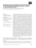
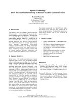
Fig. 1. Genomic organization of the PNPO. Schematic diagram of the
exon/intron organization of the human (A) and mouse (B) PNPO
gene. Exons are designated by closed boxes, and introns by bold lines.
The ORF is marked black, and grey boxes denote the 5¢-and3¢-UTR
sequences. The locations of CpG islands are indexed relative to the
start codon, and indicated by the open boxes with numbers.
2454 J. H. Kang et al. (Eur. J. Biochem. 271) Ó FEBS 2004
revealed that ) similar to the mouse ) the proximal
5¢-flanking region lacked a TATA-box but contained two
Sp1 sites (data not shown). The absence of a TATA-box is
indeed a noticeable feature of many housekeeping genes [29].
The mouse gene encodes a protein of 261 amino acids of
m 30 114 Da, and it is located on chromosome 11 which has
a very similar genomic organization to that of humans
(Fig. 1B). The longest cDNA contains 1991 bp consisting
of a 786 bp ORF, a 118 bp 5¢-untranslated region and a
1087 bp 3¢-noncoding region. As in humans, the mouse
PNPO gene is encoded by seven exons and the intron/exon
junctions also follow the GT/AG rule. The 3¢-end of the
sequence contains a poly(A) stretch, preceded by a putative
polyadenylation signal AATAAA. The mouse PNPO gene
has CpG islands extending from position )511 to 276 and
from )82 to +227 with a CG content of 61%. The deduced
protein with a predicted pI of 8.35 has a putative sulfate
site, eight phosphorylation sites, two N-myristoylation sites
and one RGD cell attachment sequence. Human and mouse
PNPO share 90% identity at the amino acid level.
Table 2. The intron/exon junctions of the human PNPO gene. The nucleotide sequences at exon (uppercase letters) and intron (lowercase letters)
junction are shown. Exon and intron sizes are indicated in bp.
Exon (bp) 5¢-splice donor Intron (bp) 3¢-Splicing acceptor Exon
I (243)
CGAGAG/gtgccg 1 (1492) tcctag/GCATTT II
II (125)
CACCAG/gtgggc 2 (1185) tcctag/AGATGG III
III (100)
GAGCTG/gtgggt 3 (843) ttctag/GACTCT IV
IV (54)
CGTCAG/gtgagt 4 (248) gagcag/GTGCGT V
V (129)
CGGGAG/gtgagt 5 (333) ggacag/TATCTG VI
VI (71) ATCCTG/gtgagt 6 (220) ttatag/GGGTGG VII
VIIa (1662)
AGATTA
VIIb (2700) ATTGAT
Consensus G/gtg ag/
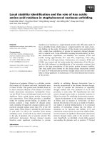
Fig. 2. Splicing pattern of the PNPO mRNA isoforms. (A) Northern blot analysis of the expression of the PNPO gene in human tissues. Two
micrograms of poly(A)
+
RNA prepared from the tissues indicated were analysed by Northern hybridization. The blots in the upper panel were
hybridized with
32
P-labelled probes corresponding to the coding region (left) and the 3¢-UTR of human PNPO cDNA (right). The membrane was
stripped and reprobed with a b-actin cDNA probe (bottom). The approximate sizes of the isoforms are indicated. (B) The scheme of two mRNA
species is given. Exons are indicated by open boxes, and coding regions and UTR used for probes are delineated by black and grey box, respectively.
The putative polyadenylation signal is indicated.
Ó FEBS 2004 Human pyridoxine 5¢-phosphate oxidase (Eur. J. Biochem. 271) 2455
Northern blot analysis of human PNPO
To determine the size of human PNPO mRNA transcripts,
Northern blot analyses were performed with the full-length
PNPO cDNA. As shown in Fig. 2, the PNPO mRNAs are
expressed in all human tissue examined, but their relative
abundance varies markedly. Of note, two transcripts of 2.4
and 3.4 kb were detectable with almost identical intensity
in all tissues examined (Fig. 2A, left). Although performed
under very stringent conditions, all blots revealed the
presence of double bands.
BLAST
analysis suggests that both signals arise from the
PNPO locus as there were no data to indicate the existence
of a highly related gene that cross-hybridizes with the PNPO
probe. There are several possible mechanisms by which
multiple transcripts could be generated from the same gene:
(1) use of alternative polyadenylation sites; (2) use of
alternate transcription start sites; and (3) differential splicing
of pre-mRNA. In the Western blot analysis as shown in
Fig. 3, no protein with a molecular mass higher than
30 kDa could be detected with mAbs against sheep PNPO.
This line of evidence may rule out the existence of an
alternative splicing product.
To further elucidate the presence of isoform message, this
filter was reprobed with the DNA probes specific for the
3¢-UTR between the two potential poly(A) signals. The
results showed that only the 3.4 kb band was detected
(Fig. 2A, right), which supports the hypothesis that the two
mRNA species are generated by alternate usage of poly-
adenylation sequences. The putative schematic structure of
the mRNA isoforms is shown in Fig. 2B.
Two putative polyadenylation signals ) one an ATT
AAA motif 1472 bp downstream of the termination codon
and the other an AATAAA motif 27 bp upstream of the
end of the gene ) were found within the genomic primary
sequence. It is known that the most common polyadeny-
lation signal is AATAAA, and that ATTAAA is 80% as
efficient as the terminal sequence [30]. Thus, both polyade-
nylation sites of PNPO worked, implying some read-
through of the first site by an unknown mechanism. A
search of the human EST database with the human PNPO
sequence also supported this hypothesis. Alternate usage of
polyadenylation signals is frequently seen in testis tissue.
However, in mouse, such putative isoforms resulting from
the alternative usage of polyadenylation could not be found
in EST sequences. Human cells, unlike cells of other
mammalian species, generate more than one PNPO tran-
script, resulting from the preferential poly(A) site selection.
This feature strongly suggests the possibility of evolutionary
changes of the 3¢-UTR, which is characterized by more
degrees of freedom than the 5¢-UTR and the ORF [31,32].
Tissue distribution of PNPO, PDXK and PDXP
As shown in Fig. 2, Northern blot analysis indicated that
the mRNA level of PNPO is highest in liver. Skeletal muscle
and kidney contained considerable amounts of the tran-
script while lower levels were detected in lungs. In addition,
a human multiple tissue expression array (MTE
TM
)was
analysed by hybridization with mRNAs from various
human tissue. As shown in Fig. 4, we provide a complete
set of the tissue distribution of PNPO mRNA in humans.
Although the level of mRNA expression in the brain is low
compared to that in other organs such as the liver, a
densitometric analysis of the dot blot array showed a similar
basal expression of PNPO in the entire brain subregion. The
transcripts of foetal PNPO are relatively low compared with
those of adults. Notably, the widespread distribution of
PNPO in human tissue is consistent with its essential role in
cellular metabolism.
Another interesting aspect of our work is the finding that
three key PLP metabolic enzymes, PNPO, PDXK and
PDXP have remarkably different expression profiles. The
Fig. 3. Western blot analysis of human PNPO. SDS/PAGE (A) and
immunoblot with mAb (B) for human tissue and cell homogenates.
LaneM,Molecularmassstandards;lane1,brain;lane2,liver;lane3,
lung; lane 4, prostate; lane 5, human breast cancer (MCF-7); lane 6,
human uterine carcinoma (HL3T1); lane 7, stomach tissue.
2456 J. H. Kang et al. (Eur. J. Biochem. 271) Ó FEBS 2004
mRNA expression levels in selected tissue for each enzyme
are shown in Table 3. Consistent with their ubiquitous role
in vitamin B
6
metabolism, all three transcripts have been
detected in a wide variety of tissue. Analysis of the array
revealed that human PDXK was expressed in essentially all
organs with the highest levels observed in descending order
testes, kidneys and placenta. A relatively high level of
PDXK transcript was expressed in foetal organs. In
contrast, human PDXP mRNAs appear to be strikingly
abundant in the brain indicating a more specific role [9].
These results imply that the three enzymes are differentially
expressed and regulated in a tissue specific manner.
The regulation of PLP could be controlled by several
factors. The synthesis of PLP requires the joint action of
PDXK and PNPO, and the PLP availability is dependent
on the degree of protein binding of the synthesized
coenzyme and transport of the precursors [33,34], and
phosphatase action [35]. PNPO does play a kinetic role in
regulating in vivo PLP formation [2,36], whereas PDXK
plays an additional trapping role whereby pyridoxal is
diffusible across the cell membrane [33]. Tissue with high
oxidase activities, however, produce PLP not only for
internal consumption, but also for an external supply to
other tissue with low oxidase activities. Thus, the complete
metabolic network for PLP homeostasis remains to be
investigated.
Functional organization by deletion mutagenesis
To investigate enzymatic properties, cDNA-encoded
human PNPO was expressed in E. coli as a fusion protein
with a His tag. The size of the recombinant protein, as well
Fig. 4. Multiple tissue analysis of human PNPO mRNA expression. Tissue-specific expression of the PNPO mRNA was analysed with poly(A)
+
RNA dot-blot. The human multiple tissue expression (MTE
TM
) array was hybridized with a
32
P-labelled PNPO-specific cDNA probe. Tissue
sources for the RNA are indicated below the blot.
Ó FEBS 2004 Human pyridoxine 5¢-phosphate oxidase (Eur. J. Biochem. 271) 2457
as the purity, was determined by SDS/PAGE. As shown in
Fig. 5A, the fusion protein of a wild-type PNPO showed an
apparent molecular mass of 34 kDa, in good agreement
with the theoretical size (33.5 kDa). Recombinant PNPO
was catalytically active. Steady-state kinetic analyses
were carried out on the recombinant enzyme. The apparent
K
m
of 2.1 l
M
and 6.2 l
M
were obtained for the substrate
PNP and PMP, respectively, from Lineweaver–Burk
(double-reciprocal) plots (Table 4).
In order to delineate the region of human PNPO that is
essential for catalysis, we expressed the sequential trunca-
tion mutants in E. coli and determined the effect of each
deletion on activity. In this work, the role of both the N- and
C-terminal regions of human PNPO were studied by the
truncation mutants: D1–56, D1–72 and D238–262 (Fig. 5B).
V
max
values of 0.10 and 0.05 lmolÆmin
)1
Æmg
)1
for the
recombinant wild-type enzyme were obtained for PNP and
PMP, respectively, whereas the deletion of the noncon-
served 56-amino acid at N-terminal domain (D1–56) caused
about a twofold increase in catalytic activity (Table 4). The
K
m
value of the mutant, however, is about threefold higher
Table 3. Comparison of mRNA expression levels of vitamin B
6
regula-
ting enzymes. A dot blot array containing human poly(A)
+
RNAs
from various tissues were hybridized with probes as described in Fig. 4.
Expression levels of selected tissues for PNPO, PDXK, and PDXP are
compared. Values are given relative to the highest expressing tissue for
each enzyme that was arbitrarily set to 100.
PNPO PDXK PDXP
Whole brain 23.8 29.0 83.6
Cerebral cortex 25.9 27.0 100.0
Frontal lobe 12.4 14.3 81.8
Parietal lobe 13.6 32.2 82.4
Occipital lobe 14.7 25.3 89.1
Temporal lobe 12.6 24.4 84.6
Paracentral gyrus of cerebral cortex 9.5 18.5 73.2
Pons 6.2 10.3 47.9
Cerebellum, left 13.3 21.9 90.5
Cerebellum, right 25.0 31.1 91.1
Corpus callosum 18.4 19.8 37.7
Amygdala 13.0 22.6 94.2
Caudate nucleus 19.0 28.4 74.2
Hippocampus 12.3 24.6 81.8
Medulla oblongate 5.0 15.8 41.7
Putamen 2.0 17.9 54.2
Accumbens nucleus 5.2 19.4 70.8
Thalamus 10.8 23.1 85.2
Spinal cord 1.6 3.7 10.8
Heart 3.7 9.1 18.7
Aorta 1.2 4.4 0.1
Atrium, left 14.0 14.7 19.6
Atrium, right 11.6 14.7 25.0
Ventricle, left 4.1 16.7 17.2
Ventricle, right 20.5 21.7 21.7
Interventricular septum 16.2 34.8 39.9
Apex of the heart 1.8 18.2 34.4
Oesophagus 7.5 13.4 2.8
Stomach 17.2 43.5 26.1
Duodenum 9.3 27.2 18.9
Jejunum 13.3 43.1 26.1
Ileum 5.8 23.0 21.2
Ilocecum 6.1 60.6 35.9
Appendix 0.7 20.5 21.2
Colon, ascending 1.2 5.7 21.9
Colon, transverse 7.9 8.0 21.6
Colon, desending 2.8 9.4 6.5
Rectum 2.3 16.4 15.8
Kidney 85.6 58.0 31.4
Skeletal muscle 34.1 15.4 20.7
Spleen 13.5 32.0 8.8
Thymus 6.4 29.9 18.6
Peripheral blood leukocyte 0.4 26.3 13.3
Lymph node 4.6 51.2 17.8
Bone morrow 11.2 42.0 22.8
Trachea 4.8 23.0 5.8
Lung 4.6 24.8 7.6
Placenta 30.2 61.4 8.1
Bladder 6.8 12.4 9.2
Uterus 2.9 19.4 9.5
Prostate 16.5 37.0 18.2
Testis 8.4 100.0 49.3
Ovary 4.3 20.1 28.9
Liver 100.0 56.0 61.8
Table 3. (Continued).
PNPO PDXK PDXP
Pancreas 2.5 59.0 33.3
Adrenal gland 11.5 34.4 32.6
Thyroid gland 20.3 18.5 12.3
Salivary gland 10.0 45.0 39.2
Leukaemia, HL-60 2.3 3.4 17.4
HeLa S3 3.5 20.2 14.8
Leukaemia, K-562 2.5 10.5 20.5
Leukaemia, MOLT-4 8.1 1.5 19.3
Burkitt, lympoma, Raji 6.5 8.0 27.5
Burkitt, lympoma, Daudi 1.1 1.4 25.6
Colorectal adenocacrinoma, SW480 5.1 9.4 8.1
Lung carcinoma, A549 0.7 1.8 4.3
Foetal brain 1.9 12.5 34.7
Foetal heart 3.9 22.5 8.4
Foetal kidney 9.7 54.1 5.6
Foetal liver 30.2 29.3 15.9
Foetal spleen 1.9 41.7 8.2
Foetal thymus 6.6 41.7 14.1
Foetal lung 8.5 24.1 16.0
Table 4. Kinetic parameters of wild-type and N-terminal deletion
mutant. PNPO activities of wild-type and deletion mutant were
measured in 0.1
M
Tris/HCl at pH 8.4. Data shown are the average of
three determinations ± SD.
Enzyme Compound
K
m
or K
i
a
(l
M
)
V
max
(lmolÆmin
)1
Æmg
)1
)
k
cat
/K
m
(
M
)1
ÆS
)1
)
Wild-type PNP 2.1±0.2 0.10±0.06 5.2 · 10
4
PMP 6.2±0.3 0.05±0.01 8.2 · 10
3
PLP 3.8
D1–56 PNP 6.2±0.2 0.21±0.02 3.1 · 10
4
PMP 20.8±0.4 0.08±0.01 3.6 · 10
3
PLP 23.0
a
Inhibition constant for product PLP determined with the sub-
strate PNP.
2458 J. H. Kang et al. (Eur. J. Biochem. 271) Ó FEBS 2004
than that of the full-length PNPO. Thus, the value for the
specificity constant (k
cat
/K
m
) is compensated. PLP is a
competitive inhibitor, and the K
i
values for the wild-type
enzyme and D1–56 were 3.8 and 23 l
M
, respectively. Since
the mechanism of PNPO is not yet fully understood, we
cannot explain the changes in kinetic parameters. The
N-terminal segment, however, would remain flexible and
disordered in a solution, and it would form a lid over the
active site [23]. This may play at least a partial role in
binding and catalytic activity.
Further truncation (D1–72) resulted in completely abol-
ished enzymatic activity, indicating that the first highly
conserved helix segment (residues 57–72) is required for
activity. Previous studies showed that the peptide fragment
of approximately two-thirds of the molecular mass yielded
by a limited chymotryptic cleavage of sheep PNPO
endowed with full catalytic activity [36]. This discrepancy
may be due to a sequence difference between species or a
disturbance in the folding process during expression caused
by a missing structural unit. The presence of the first helical
sequence might be solely structural, as it does not have a
direct interaction with either PLP or FMN [23]. In addition,
a deletion of 25 residues at the C terminus (D238–262)
resulted in essentially inactive enzymes, indicating that this
region is required for function.
Conclusions
In this report, we have described the genomic organization
of PNPO, tissue distribution and deletion mutagenesis.
(1) The human PNPO gene is composed of seven exons
and six introns spanning 7.7 kb of the genomic DNA.
The 5¢-flanking region has the characteristic features of
housekeeping genes. Due to alternate usage of polyadeny-
lation sites, two species of mRNA existed in all examined
tissue. Nevertheless, no protein isoforms were detected.
Fig. 5. Deletion analysis of recombinant PNPO. (A) Expression and purification of recombinant human PNPO. SDS/PAGE analysis (12%
acrylamide)ofcrudecellextractsofE. coli BL21(DE3) containing the expression vector without and with the coding sequences for the wild-type or
mutants. Lane M, Low molecular mass standards (Bio-Rad); lane 1, crude extracts from cultured cells harbouring pET28a; lane 2, cells containing
pET28a/PNPO in the presence of 1 m
M
isopropyl thio-b-
D
-galactoside; lane 3, purified recombinant PNPO from Ni
2+
resin; lanes 4–6, purified
deletion mutants: D1–56, D1–72 and D238–262, respectively. (B) Left, schematic structure of wild-type PNPO and the N- and C-terminal deletion
mutants used in this study. Numbers refer to the amino acid position along the primary sequence of PNPO. Right, the effect of N- and C-terminal
deletion on PNPO activity was expressed as a percentage of enzymatic activity in wild-type enzyme. Solid black and crosshatched bars are for
substrate PNP and PMP, respectively. The results shown are the means ± SD from triplicate assays.
Ó FEBS 2004 Human pyridoxine 5¢-phosphate oxidase (Eur. J. Biochem. 271) 2459
(2) The widespread distribution of PNP oxidase mRNA
in human tissue agrees with its essential function in
vitamin B
6
homeostasis. Three key enzymes for vitamin B
6
metabolism ) PNPO, PDXK and PDXP ) have remark-
ably different expression profiles. (3) The catalytic core
of PNPO was determined by sequential deletion mutants.
The deletion of the N-terminal 56 residues did not affect
binding of coenzyme, or catalytic activity, whereas deletion
of the C-terminal region resulted in an inactive enzyme.
The results obtained here will contribute directly to future
studies aimed at a better understanding of the catalytic
mechanism of PNPO and vitamin B
6
metabolism. In
particular, the tissue-specific effects on mRNA stability
and the regulatory mechanism governing the PNPO gene
expression require further investigation.
Acknowledgements
This work was supported by Grant R01-2002-000-00008-0 from Basic
Research Program of the Korea Science & 21st Century Brain Frontier
Research Grant (M103KV010019–03K2201-01910) from the Ministry
of Science and Technology, Korea.
References
1. Snell, E.E. (1990) Vitamin B6 and decarboxylation of histidine.
Ann. NY Acad. Sci. 585, 1–12.
2. McCormick, D.B. & Merrill, A.H. (1980) Pyridoxamine (pyri-
doxine) 5¢-phosphate oxidase. In Vitamin B
6
Metabolism and Role
in Growth (Tryfiates, G.P., ed.), pp. 1–26. Food and Nutrition
Press, Westport, CT.
3. Robson, L.C. & Schwartz, M.R. (1975) Vitamine B
6
deficiency
and the lymphoid system I. Effects on cellular immunity and in
vitro incorporation of
3
H-uridine by small lymphocytes. Cell.
Immunol. 16, 135–144.
4. McCormick, D.B., Gregory, M.E. & Snell, E.E. (1961) Pyridoxal
phosphokinase I: assay, distribution, purification and properties.
J. Biol. Chem. 236, 2076–2084.
5. Hanna, M.C., Turner, A.J. & Kirkness, E.F. (1997) Human
pyridoxal kinase. cDNA cloning, expression, and modulation by
ligands of the benzodiazepine receptor. J. Biol. Chem. 272, 10756–
10760.
6. Kwok, F. & Churchich, J.E. (1992) Pyrdoxine-5¢-P oxidase. In
Chemistry and Biochemistry of Flavoenzymes (Muller, F., ed.),
Vol. 3, pp. 1–20. CRC Press, London.
7. Ngo,E.O.,LePage,G.R.,Thanassi,J.W.,Meisler,N.&Netter,
L.M. (1998) Absence of PNP oxidase (PNPO) activity in neo-
plastic cells: isolation, characterization, and expression of PNPO
cDNA. Biochemistry 37, 7741–7748.
8. Fonda, M.L. (1992) Purification and characterization of vitamine
B
6
-phospate phosphatase from human erythrocytes. J. Biol.
Chem. 267, 15978–15983.
9. Jang,Y.M.,Kim,D.W.,Kang,T.C.,Won,M.H.,Baek,N.I.,
Moon, B.J., Choi, S.Y. & Kwon, O.S. (2003) Human Pyridoxal
Phosphatase: Molecular cloning, functional expression and tissue
distribution. J. Biol. Chem. 278, 50040–50046.
10. Choi, J.D., Bowers-Komro, D.M., Davis, M.D., Edmondson,
D.E. & McCormick, D.B. (1983) Kinetic properties of pyridox-
amie (pyridoxine) 5¢-phosphate oxidase from rabbit liver. J. Biol.
Chem. 258, 840–845.
11. Wada, H. & Snell, E.E. (1961) The enzymatic oxidation of pyri-
doxine and pyridoxamine-phosphates. J. Biol. Chem. 236, 2089–
2095.
12. DiSalvo,M.,Yang,E.,Zhao,G.,Winkler,M.E.&Schirch,V.
(1998) Expression, purification and characterization of recom-
binant Escherichia coli pyridoxine 5¢-phosphate oxidase. Protein
Express. Purif. 13, 349–356.
13. Kazarinoff, M.N. & McCormick, D.B. (1975) Rabbit liver pyri-
doxamine (pyridoxine) 5¢-phosphate oxidase. J. Biol. Chem. 250,
3436–3442.
14. Churchich, J.E. (1984) Brain pyridoxine-5-phosphate oxidase: a
dimeric enzyme containing one FMN site. Eur. J. Biochem. 138,
327–332.
15. Choi, S.Y., Churchich, J.E., Zaiden, E. & Kwok, F. (1987) Brain
pyridoxine-5¢-phosphate oxidase: modulation of its catalytic
activity by reaction with pyridoxal 5¢-phosphate and analogs.
J. Biol. Chem. 262, 12013–12017.
16. Tsuge, H., Itoh, K., Akatsuka, F., Okada, T. & Ohashi, K. (1987)
Inactivation of pyridoxamine-5¢-P oxidase by aliphatic primary
amines. Biochem. Int. 6, 743–749.
17. Notheis, C., Drewke, C. & Leistner, E. (1995) Purification and
characterization of the pyridoxol-5¢-phosphate: oxygen oxido-
reductase (deaminating) from Escherichia coli. Biochim. Biophys.
Acta 1247, 265–271.
18. Zhao, G. & Winkler, M. (1995) Kinetic limitation and cellular
amount of pyridoxine (pyridoxamine) 5¢-phosphate oxidase of
Escherichia coli K-12. J. Bacterol. 177, 883–891.
19. Di Salvo, M.L., Safo, M.K., Musayev, F.N., Bossa, F. & Schirch, V.
(2003) Structure and mechanism of Escherichia coli pyridoxine
5¢-phosphate oxidase. Biochim. Biophys. Acta 1647, 76–82.
20.Safo,M.K.,Mathews,I.,Musayev,F.N.,DiSalvo,M.L.,
Thiel, D.J., Abraham, D.J. & Schirch, V. (2000) X-ray structure of
Escherichia coli pyridoxine 5¢-phosphate oxidase complexed with
FMN at 1.8 A
˚
resolution. Structure 8, 751–762.
21. Safo, M.K., Musayev, F.N., De Salvo, M.L. & Schirch, V. (2001)
X-ray structure of Escherichia coli pyridoxine 5¢-phosphate oxi-
dase complexed with pyridoxal 5¢-phosphate at 2.0A
˚
resolution.
J. Mol. Biol. 310, 817–826.
22. Di Salvo, M.L., Ko, T.P., Musayev, F.N., Raboni, S., Schirch, V.
& Safo, M.K. (2002) Active site structure and stereospecificity of
Escherichia coli pyridoxine-5¢-phosphate oxidase. J. Mol. Biol.
315, 385–397.
23. Musayev, F.N., Di Salvo, M.L., Ko, T.P., Schirch, V. & Safo, M.K.
(2003) Structure and properties of recombinant human pyridoxine
5¢-phosphate oxidase. Protein Sci. 12, 1455–1463.
24. Quandt, K., Frech, K., Karas, H., Wingender, E. & Werner, T.
(1995) MatInd and MatInspector: new fast and versatile tools
from detection of consensus matches in nucleotide sequence data.
Nucleic Acids Res. 23, 4878–4884.
25. Gardiner-Garden, M. & Frommer, M. (1987) CpG islands in
vertebrate genomes. J. Mol. Biol. 196, 261–282.
26. Rice, P., Longden, I. & Bleasby, A. (2000) EMBOSS: the Euro-
pean Molecular Biology Open Software Suite. Trends Genet. 16,
276–277.
27. Lee, H S., Moon, B.J., Choi, S.Y. & Kwon, O.S. (2000) Human
pyridoxal kinase: Overexpression and properties of the recom-
binant enzyme. Mol. Cells 10, 452–459.
28. Bahn, J.H., Kwon, O.S., Joo, H.M., Jang, S.H., Park, J., Hwang,
I.K.,Kang,T.C.,Won,M.H.,Kwon,H.Y.,Kwok,F.,Kim,H.B.,
Cho, S.W. & Choi, S.Y. (2002) Immunohistochemical studies of
brain pyridoxine-5¢-phosphate oxidase. Brain Res. 925, 159–168.
29. Weis, L. & Lindenberg, D. (1992) Transcription by RNA poly-
merase II: initiator-directed formation of transcription-competent
complexes. FASEB J. 6, 3300–3309.
30. Wickens, M. (1990) How the messenger got its tail: addition of
poly (A) in the nucleus. Trends Biochem. Sci. 15, 277–281.
31. Grzybowska, E.A., Wilczynska, A. & Siedlecki, J.A. (2001) Reg-
ulatory functions of 3¢ UTRs. Biochem. Biophys. Res. Commun.
288, 291–295.
32. Qu, X., Qi, Y. & Qi, B. (2002) Generation of multiple mRNA
transcripts from the novel human apoptosis-inducing gene hap
2460 J. H. Kang et al. (Eur. J. Biochem. 271) Ó FEBS 2004
by alternative polyadenylation utilization and the translational
activation function of 3¢ untranslated region. Arch. Biochem.
Biophys. 400, 233–244.
33. Snell, E.E. & Haskell, B.E. (1971) The metabolism of vitamine B
6
.
In Metabolism of Vitamins and Trace Elements (Florkin,M.&
Stotz, E.H., eds), Vol. 21, pp. 47–71. Elsevier Scientific Publishing
Co, Amsterdam.
34. Anderson, B.B., Newmark, P.A. & Rawlins, M. (1974) Plasma
binding of vitamine B6 compound. Nature 250, 502–504.
35. Lumeng, L. & Li, T.K. (1975) Characterization of the pyridoxal
5¢-phosphate and pyridoxamine 5¢-phosphate hydrolase activity in
rat liver. Identity with alkaline phosphatase. J. Biol. Chem. 250,
8126–8131.
36. Kwon, O.S., Kwok, F. & Churchich, J.E. (1991) Catalytic
and regulatory properties of native and chymotrypsin-treated
pyridoxine-5-phosphate oxidase. J. Biol. Chem. 266, 22136–
22140.
Ó FEBS 2004 Human pyridoxine 5¢-phosphate oxidase (Eur. J. Biochem. 271) 2461