Báo cáo khoa học: The HNF1b transcription factor has several domains involved in nephrogenesis and partially rescues Pax8/lim1-induced kidney malformations docx
Bạn đang xem bản rút gọn của tài liệu. Xem và tải ngay bản đầy đủ của tài liệu tại đây (681.86 KB, 14 trang )
The HNF1b transcription factor has several domains involved
in nephrogenesis and partially rescues Pax8/lim1-induced
kidney malformations
Guizhi Wu, Silvia Bohn and Gerhart U. Ryffel
Institut fu
¨
r Zellbiologie, Universita
¨
tsklinikum Essen, Germany
The tissue-specific transcription factors HNF1a and HNF 1b
are closely related homeodomain proteins conserved in
vertebrate evolution. Heterozygous mutations in human
HNF1b
1
but not in HNF1a genes are associated with kidney
malformations. Overexpression of HNF1b in Xenopu s
embryos leads to defective pronephros development, while
HNF1a has n o e ffect. W e have defin ed th e regions respon-
sible for this functional difference between HNF1b and
HNF1a in transfected HeLa cells as well as in injected
Xenopus embryos. Using domain swapping experiments, we
located a nuclear localization signal in the POU
H
domain of
HNF1b, and showed that the POU
S
and POU
H
domains of
HNF1b mediate a high transactivation potential in trans-
fected cells. In injected Xenopus embryos three HNF1b
domains are involved in nephrogenesis. These include the
dimerization domain, the 26 amino acid segment specific for
splice variant A as well as the POU
H
domain. As HNF1b
together with Pax8 and lim1 constitute the earliest regulators
in the pronephric anlage, it is possible that they cooperate
during early nephrogenesis. We have shown here that
HNF1b can overcome the enlargement and the induction of
an ectopic pronephros mediated by overexpression of Pax8
and lim1. However, the phenotype induced by Pax8 and lim1
overexpression and characterized by cyst-like structures and
thickening of the p ronephric tubules was not altered by
HNF1b overexpression. Taken together, HNF1b acts ant-
agonistically to Pax8 and lim1 in only some processes during
nephrogenesis, and a simple antagonistic relationship does
not completely describe the f unctions of thes e genes. We
conclude that HNF1b has some distinct morphogenetic
properties during nephrogenesis.
Keywords: HNF1 b; lim1; nephrogenesis; Pax8; pronephros.
The tissue-specific transcription factors, HNF1a (TCF1)
and HNF1b (vHNF1, TCF2), are two unique homeo-
domain proteins [1]. The POU homeodomains (POU
H
)are
divergent from other homeodomain proteins in that they
contain an extra 21 amino acid ( aa) loop between helices 2
and 3 [2,3]. Both transcription factors are encoded in distinct
genes on separate chromosomes, a nd are highly conserved
in vertebrates with homologues in fish [4,5], frog [6,7]
and m ammals, including humans [8–10]. The evolutionary
conservation is also seen in the exon/intron patterning
which remains essentially the same between Xenopus and
mammals [11]. B oth HNF1 p roteins contain a highly
conserved N -terminal dimerization domain, a b ipartite
DNA binding region and a more divergent C-terminal
transactivation domain (Fig. 1) . Based on the crystal
structure of the dimer, the dimerization domain has been
identified as an intertwined four-helix bundle that allows the
formation o f homo- or heterodimers of the HNF1 proteins
[12,13]. The DNA binding domain is composed of a POU
specific domain (POU
S
) and the divergent POU homeo-
domain (POU
H
). Recent three-dimensional structural ana-
lysis of the HNF1a protein indicates that the POU
S
domain
interacts with the 21 aa loop of the POU
H
domaintocreate
a stable in terface between the two DNA binding domains.
This feature distinguishes HNF1a from other, more flexible,
POU
H
factors [14]. As the primary structures of HNF1a
and HNF1b are very similar within the DNA binding
region, it is reasonable to assume that this structure i s also
present in the HNF1b protein. Depending on the splice
variant, there is a 26 aa insertion between the POU
S
and
POU
H
domain in the HNF1b protein. This variant is found
in mammalian and also Xenopus HNF1b proteins (Fig. 1),
but never in the HNF1a proteins. In contrast to these rather
conserved domains, the C-terminal transactivation domain
is the most divergent protein area when the HNF1a and
HNF1b proteins are compared.
It is not resolved whether the differences between the
HNF1a and HNF1b proteins that are highly conserved
throughout vertebrate evolution reflect distinct functions.
Consistent with distinct functional roles, the temporal and
spatial expression patterns of HNF1a and HNF1b differ
significantly. During murine embryogenesis, HNF1a is
expressed in the yolk sac endoderm at day 8.5 of gestation as
well as in the developing liver, kidney, intestine, pancreas
and stomach [15–17]. In contrast, HNF1b is expressed
earlier in the primitive and visceral endoderm. Starting at
day 4.5 of gestation, th e anterior p art of the neural tube as
Correspondence to G. U. Ryffel, Institut fu
¨
r Zellbiologie (Tumor-
forschung), Universita
¨
tsklinikum Essen, D-45122 Essen, Germany.
Fax: +49 201723 5905, Tel.: +49 201723 3110,
E-mail: gerhart.ryff
Abbreviations: NLS, nuclear localization signal; POU
S
,POUspecific
domain; POU
H
, POU homeodomain.
(Received 2 June 2004, revised 22 July 2004, accepted 29 July 2004)
Eur. J. Biochem. 271, 3715–3728 (2004) Ó FEBS 2004 doi:10.1111/j.1432-1033.2004.04312.x
well as the developing kidney, liver, gut and pancreas
express HNF1b [18]. Additionally, HNF1b is also expressed
in the primordia for the genitalia and the lung. HNF1b
expression persists in these organs in the adult, whereas
HNF1a is never active in t hese tissues [19,20].
The embryonic expression pattern o f the HNF1 proteins
is evolutionarily conserved in vertebrates. The expression of
HNF1b occurs prior to HNF1 a in Xenop us embryos
[21,22], and only HNF1b is expressed in the develo ping
brain in Xenopus [7]aswellasinzebrafish[5].Inagreement
with the differential embryonic expression patterns of the
two HNF1 proteins, inactivation of the corresponding
genes in the mouse has different effects. Homozygous
knock-out of the HNF1b gene led to early embryonic
lethality at day 7.5 of gestation with poorly organized
ectoderm and no discernible visceral endoderm [18,23]. In
contrast, HNF1a was not required for embryonic devel-
opment, but HNF1a-deficient mice died during postnatal
life due to hepatic, pancreatic and renal dysfunction
[24–28]. These results clearly establish different roles for
the t wo HNF1 genes. Whether differential properties of the
two transcription factors are the cause of these differences,
or rather the differential expression patterns, remains to be
seen. A functional equivalence of the HNF1a and HNF1b
protein has recently been shown in embryonic stem cells, as
the introduction of HNF1a restores the formation and
differentiation of a mature visceral endoderm in HNF1b-
deficient embryonic stem cells [29]. Further su pport for
functional differences can b e deduced from human dis-
eases. Biallelic inactivation of the HNF1 a gene has been
described as an early step in hepatocellular carcinoma [30].
However, HNF1b has not been associated with tumori-
genesis to date. Heterozygous mutations in both genes lead
to maturity onset diabetes of the young but HNF1b
mutations are additionally associated with severe nondia-
betic renal defects as well as genital malformations in
females [31–34]. In this c ontext, we showed the specific role
of HNF1b during development of the first form of
vertebrate kidney, the pronephros, using overexpression
experiments in Xenopus embryos. The expression of
HNF1b led specifically to a reduced formation of the
pronephros, whereas HNF1a had n o effect [35]. This
indicates that these two transcription factors have different
intrinsic biochemical properties. Most recently, the renal-
specific inactivation of the HNF1b gene in mice [36] and the
kidney-specific expression of mutated HNF1b [37] have
linked the HNF1b transcriptional network to genes causing
polycystic kidney d isease.
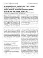
Fig. 1. The related human t ranscription factors, HNF1a and HNF1b. HNF1b and H NF1a are represented schematically (top). The domains are
indicated and numbers below the domains refer to the amino acid positions. Amino acid identity of the domains between HNF1a and HNF1b is
showninbold(homology)
16
. The 26 aa segment between the POU
S
and POU
H
domains of the human HNF1a and HNF1b proteins as well as of the
human and Xenopus HNF1b protein are aligne d (bottom) with missing amino acids indicated by ‘)’. The 26 aa segment deleted in the B s plice
variant of the HNF1b is indicated (green). Identical amino acids between b and a or human b and Xenopus b sequences are shown and
17
conserved
amino acid changes are indicated by +.
3716 G. Wu et al. (Eur. J. Biochem. 271) Ó FEBS 2004
In vertebrates, three d istinct types of kidneys (pro-
nephros, mesonephros and metanephros) are formed
progressively during development [38]. Similar regulators
are expressed in all three kidneys, and thus, the molecular
processes by which the different kidn eys develop appear to
be closely related [39–41]. The pronephros is the simplest
vertebrate kidney, and consists of a single nephron with an
external glomus. It represents an attractive s ystem to study
molecular events during kidney development, as several
key regulators have been functionally identified by inject-
ing mRNA into Xenopus embryos [ 41,42]. Using the
Xenopus system, we have shown that overexpression of
human HNF1b in the developing frog embryo leads to
agenesis of the pronephric tubules and duct. The s ame
phenotype is seen for some human HNF1b mu ta nts
leading to defective renal development, whereas an
enlargement of the pronephros occurs with other mutants
[35,43]. An enlargement of the pronephros has also been
observed b y the overexpression of the transcription factors,
Pax8 and lim1, and this effect was additive [44]. Further-
more, the artificial expression of Pax8 and lim1 in the
Xenopus embryo induced ectopic pronephric structures, a
phenotype never seen in embryos overexpressing HNF1b.
Interestingly, HNF1b, Pax8 a nd lim1 are the earliest
known regulators in the pronephric anlage, implying that
they may cooperate during early events of nephrogenesis
[41,42].
In the present communication, we functionally mapped
the protein domains of HNF1b, s pecifically participating in
nephrogenesis via injection of chimeric HNF1a and HNF1b
proteins into Xenopus embryos. We also explored whether
Pax8- a nd lim1-mediated effects can be overcome by
simultaneous HNF1b overexpression.
Materials and methods
Plasmid constructions
The pCSGFP2, myc-Rc/CMVHNF1b and m yc -Rc/
CMVHNF1a expression vectors have been described
previously [35]. HNF1aaa and HNF1bbb were generated
by inserting an EcoRI- XbaI fragment encoding 1–321 aa
of the human HNF1a and 1–352 a a of the human
HNF1b, respectively. A BamHI site was introduced both
at G69 (a)andG79(b) without changing the amino acid
sequence. The Eco RI-BamHI and BamHI-XbaI frag-
ments were derived from PCR products made with the
following primers. HNF1aaa:
2
5¢-CG
GAATTCAATGG
TTTCTAAACTGAGCC-3¢ (forward) , 5¢-CGC
GGATCC
CCGAGTCTCCCCC-3¢ (reverse); 5¢-CGC
GGATCCGA
GGACGAGACGG-3¢ (forward), 5¢-GC
TCTAGATTA
GCGCACACCGTGGAC-3¢ (reverse); HNF1bbb: 5¢-CG
GAATTCAATGGTGTCCAAGCTCACGT-3¢ (forward),
5¢-CGC
GGATCCCTCGTCGCCGGACAA-3¢ (reverse);
5¢-CGC
GGATCCGAGGACGGCGACGA-3¢ (f orward),
5¢-GC
TCTAGATTAGCGCACTCCTGACAGC-3¢ (re-
verse). The restriction sites for cloning are underlined.
HNF1abb and HNF1baa were generated by exchanging
the EcoRI-BamHI fragments between HNF1bbb and
HNF1aaa. HNF1bbbD was generated by replacing the
BamHI-HincII fragment of the HNF1bbb expression vector
with the BamHI-HincII fragment of a PCR product
made with the forward primer, 5¢-CGC
GGATCCGA
GGACGGCGACGA-3¢, and the reverse primer, 5¢-GCT
CT
GTTGACTGAATTGTCGGAGGATCTCTCGT-3¢,
containing complementary sequ ences upstream and down-
stream to a segment encoding the 26 aa to be dele ted.
HNF1bD was generated by replacing the PvuI fragment
encoding 1–251 aa of HNF1 b with the c orresponding
fragment of HNF1bbbD.
HNF1aab, HNF1aabins26 and HNF1aaains26 con-
structs were generated using the Quickchange site-directed
Mutagenesis Kit (Stratagene) and a PCR fragment gener-
ated from the HNF1bbb sequence using the following
primers: HNF1aab: 5¢-GATGAGCTACCAACCAAGAA
GATGCGCCGCA-3¢ (forward), 5 ¢-GCCGCTCTAGATT
AGCGCACTC-3¢ (reverse); HNF1aabins26: 5¢-CGAGA
GGTGGCGCAGCAGTTCAACCAGACAGTCCAG-3¢
(forward), 5¢-GCCGCTCTAGATTAGCGCACTC-3¢ (re-
verse); HNF1aaains26: 5¢-CGAGAGGTGGCGCAGCA
GTTCAACCAGACAGTCCAG-3¢ (forwa rd), 5¢-CTCC
CTGCCCTGCATGGGTGAACTCTGGAAAGAGAA
AC-3¢ (reverse).
3
HNF1aabH and HNF1aabHS were generated by repla-
cing the BamHI-XbaI fragment of HNF1aab with the
BamHI-XbaI fragment of a PCR product generated using
the primers 5¢-CGC
GGATCCGAGGACGAGACGG-3¢
(forward) and 5¢-GC
TCTAGATTAGCTATAGGCGTCC
ATGG-3¢ (reverse) and 5¢-CGC
GGATCCGAGGACGAG
ACGG-3¢ (forward) and 5¢-GC
TCTAGATTATTGCCGG
AATGCCTCCT-3¢ (reverse), respectively. HNF1 bhomeo
was a mplified by PCR using the primers 5¢-CG
GAA
TTCAAAGAAGATGCGCCGCAAC-3¢ (forward) and
5¢-GC
TCTAGATTAGCTATAGGCGTCCATGG-3¢ (re-
verse). All amplified HNF1 f ragments were verified by
sequencing, digested with EcoRI and XbaI, then inserted
into the GFP-Rc/CMV and pCS2+MT [45] expression
vectors.
GFP-Rc/CMV was constructed by inserting th e HindIII-
EcoRI GFP PCR fragment produced using the
5¢-GGC
AAGCTTCTGGCCACCATGAGTAAAGGA-3¢
(forward) and 5¢-CG
GAATTCGTTTTGTATAGTTCAT
CCATGC-3¢ (reverse) primers to amplify a region of the
pCSGFP2 vector [46] into the Rc/CMV e xpression vector
(Invitrogen). The expression clone encoding Xenopus
HNF1b was kindly provided by R. Vignali, University of
Pisa, Italy
4
[47], and the plasmids encoding Xenopus lim1 and
Pax8 were kindly supplied by P. D. Vize, University of
Calgary, Canada [44].
Cell culture, transfection and luciferase assay
HeLa cells (our lab stock)
5
were cultured at 37 °Cin
Dulbecco’s modified Eagle’s medium supplemented with
penicillin (100 UÆmL
)1
), streptomycin (100 UÆmL
)1
)and
10% (v/v) heat-inactivated fetal bovine serum. The cells
were seeded at a density of 3 · 10
5
cells per 3.3 cm dish. The
transfection was performed 24 h a fter seeding using 1.3 lg
of reporter gene, 0.3 lg o f expression vector, and 6 lLof
lipofectamine (Invitrogen). The final DNA concentration
was equalized by the addition of Rc/CMV vector. The
transactivation activity was measured after 20 h using the
luciferase reporter assay system (Promega) and a Lumat LB
9501 luminometer (Berthold, Wilbad, Germany).
Ó FEBS 2004 HNF1b in nephrogenesis (Eur. J. Biochem. 271) 3717
Embryos, microinjection of synthetic mRNA and
immunohistochemistry
In vitro fertilization and culture of Xenopus laevis
6
embryos
were performed as described previously [48]. Adult Xenopus
laevis were obtained from Xenopus I, Inc. (Dexter, M I,
USA) and the animal experimentation guidelines were
followed (Regierungspra
¨
sidium Du
¨
sseldorf, Germany). The
developmental stages are taken from the ÔNormal Table of
Xenopus laevisÕ [49].
7
The expression vectors encoding
HNF1 chimeric proteins and the GFP encoding expression
vector (pCSGFP2) were linearized with NotIandPvuII,
respectively, then in vitro transcribed with SP6 RNA
polymerase [35]. A total of 250 pg of capped mRNA
encoding a chimeric protein together with 100 pg of capped
green fluorescent protein
8
(GFP) mRNA w ere injected into
one blastomere of two-cell stage embryos. After 2 days, the
injected side was scored under a stereofluorescence micro-
scope for the presence of GFP. At the swimming larval stage
(45), the animals were fixed in MEMFA [0.1
M
MOPS,
pH 7.4, 2 m
M
EGTA, 1 m
M
MgSO
4
, 3.7% (v/v) formal-
dehyde], subsequently dehydrated in methanol and stored at
)20 °C. For whole-mount immunostaining, the embryos
were rehydrated in NaCl/P
i
and blocked with NaCl/P
i
and
0.1% (v/ v) Triton X -100 (P BT)/20% (v /v) g oat s erum for
1 h at room temperature. Incubation with hybridoma
supernatant of the monoclonal antibodies, 3G8 and 4A6
(kindly provided by E. A. Jones, University of Warwick, UK
9
[50]), was performed overnight at 4 °C after a 1 : 2 dilution
in PBT/20% (v/v) goat serum. After washing five times with
PBT at 20–25 °C
10; 11
, incubation with a 1 : 1000 diluted cyanine
Cy3
10; 11
-conjugated rat a nti-(mouse) Ig (Jackson Immuno-
Research, West Grove
12
, PA, USA) was performed overnight.
Embryos were w ashed fi ve times with PBT at room
temperature, then analyzed by fluorescence microscopy.
Statistical analysis
The difference between the injected and the noninjected
sides was evaluated by measuring the w hole area using the
lateral view with t he widest diameter from the dorsal to the
ventral side of the immunostained pronephros. The area
included the pronephric tubules and the anterior part of the
pronephric duct. The measurements were made using the
computer program
KAPPA IMAGE BASE METEO
(opto-elec-
tronics GmbH, Gleichen, Germany), and the noninjected
side was used as a reference for each animal. No size
difference was set as 100. The values representing kidney
size obtained from each mutant were compared to values
obtained from GFP control-injected embryos. Significant
differences were scored using the Mann–Whitney test to
calculate P-values.
Results
The conserved 26 aa segment of HNF1b affects
the transactivation potential
We first explored whether the 26 aa segment specifically
deleted in t he splice variant HNF1b-B (Fig. 1 ) c ould interfere
with nephrogenesis. The splice variant B (HNF1bD) was
constructed by deleting the 26 aa segment as shown in
Fig. 2A. A second construct was created from a truncated
HNF1b protein ( HNF1bbb) that corresponds to the
human Y352insA HNF1b mutation, that we have shown
in previous experiments to be sufficient to induce agenesis of
the pronephros in Xenopus [43]. By deletion of the 26 aa
segment from HNF1bbb we constructed a truncated protein
lacking the 26 aa se gment (HNF1bbbD, Fig. 2 A). As a third
type we generated a HNF1a variant containing the 26 aa
segment from HNF1b that is normally not present in
HNF1a. As the full-length HNF1a protein has no effect on
renal development [35], we assumed t hat the truncated
HNF1a protein (HNF1aaa) lacking the transactivation
domain would not have an effect either. By adding the 26 aa
segment to t his truncated version of HNF1a we produced the
HNF1aaains26 construct (Fig. 2A).
The subcellular localization of these constructs was first
assayed in transfected HeLa cells. Previous experiments have
shown that HNF1a is localized primarily in the nucle us but
also to a certain extent in the cytoplasm [51]. Localization of
HNF1b, however, is exclusively nuclear [43]. To define the
subcellular d istribution o f these various proteins, w e
expressed GFP fusion proteins of these constructs in HeLa
cells. All HNF1b-derived constructs (HNF1b, HNF1bD,
HNF1bbb and HNF1bbbD) were localized exclusively in
the nucleus (Fig. 2 B). In contrast, the HNF1a-de rived
constructs (HNF1aaa and HNF1aaains26) were present in
both the nucleus and the cyto plasm (Fig. 2B), as ob served
previously for full-length HNF1 a [51].
Additionally, the transactivation potential of these HNF1
derivatives were investigated. Expression vectors encoding
these proteins were cotransfected into HeLa cells lacking
endogenous HNF1 proteins t ogether w ith a luciferase
reporter plasmid containing an HNF1 i nducible promoter.
Deletion of the 26 aa sequence present in HNF1b reduced
the transactivation potential 30% compared to the full-
length HNF1 b transcription factor (Fig. 2C). As observed
previously [43], the truncated HNF1b protein lacking the
transactivation domain retained substantial transactivation
potential (compare HNF1bbb with HNF1b in Fig. 2C).
Typically, HNF1bbb was less active at 10–30 ng expression
vector, but as active as the full-length protein when 150–
300 n g expression vector were transfected. The truncated
HNF1b construct missing the sequence encoding the 26 aa
segment (HNF1bbbD) transactivated similarly to H NF1bbb
(Fig. 2 C). In contrast, the truncated HNF1a protein
(HNF1aaa) had only a residual act ivity even when 300 ng
expression vector were transfected (Fig. 2C). This is consis-
tent with the i nitial description of the HNF1a transcription
factor and the definition of the C-terminal activation domain
of HNF1a [52,53]. The insertion of the b-specific 26 aa
segment into the truncated HNF1a construct (HNF1aaa)
abolished residual activity. This indicates that the 26 aa
segment plays some role in the transactivation potential.
The conserved 26 aa segment of HNF1b interferes with
pronephros development in
Xenopus laevis
The morphogenetic potential of the various HNF1 con-
structs were examined in the developing Xenopus embryo by
injecting mRNA encoding these proteins into one blasto-
mere of the two-cell stage embryo. As initial experiments
revealed that the GFP-HNF1 fusion proteins fluoresced too
3718 G. Wu et al. (Eur. J. Biochem. 271) Ó FEBS 2004
weakly for the identification of the injected side (data not
shown), GFP mRNA was coinjected with RNA for the
myc-tagged version of the constructs (Fig. 3A) as per-
formed previously [35]. Injected embryos were raised to free
swimming tadpoles (stage 45) and processed to v isualize the
pronephros using a mixture of monoclonal antibodies for
the pronephric tubules and duct [50]. Only embryos that
were otherwise phen otypically normal were scored f or
effects on pronephric development. Examples of dorsal
views of such larvae a re given in Fig. 3C. The pronephric
size was measured in the lateral view ( Fig. 3B) of a whole
series of larvae, and the quantification of these ph enotypic
changes together with the statistical analysis for significance
are summarized in Fig. 3D.
As found previously [35,43], full length HNF1b ledtoa
significant reduction of the size of the pronephros (Fig. 3D),
and this effect was even more p ronounced for the truncated
HNF1b protein (HNF1bbb, Fig. 3D). As expected, the
truncated HNF1a protein (HNF1aaa) did not interfere with
pronephros development (Fig. 3D). The HNF1b protein
lacking the 26 aa segment (HNF1bD) had no effect on
pronephric size, implying a crucial role of this 26 aa segment
in nephrogenesis (Fig. 3D). However, the truncated HNF1b
protein lacking this 26 aa segment (HNF1bbbD) led to a
reduction of the pronephric size (Fig. 3D), indicating
additional nephrogenic segments in this truncated protein.
The insertion of the 26 aa segment into the HNF1a protein
(HNF1aaains26) led to a reduction of pronephric size
(Fig. 3 D), illustrating that the nephrogenic potential of the
26 aa segment is transferable. A dramatic lethality at the
injected side was observed when the truncated HNF1b
construct lacking the 26 aa segment (HNF1bbbD) was
overexpressed (Fig. 3E,F). More than 90% of the injected
embryos died during gastrulation. Even when the amount of
HNF1bbbD mRNA was halved, 70% of the e mbryos still
died during gastrulation. The majority of the surviving
tadpoles were distorted (Fig. 3H–J) compared to control
animals (Fig. 3G).
13
Therefore, a relatively small number (36)
of healthy larvae were available for immunostaining and the
examination of the pronephros-specific effects. Neverthe-
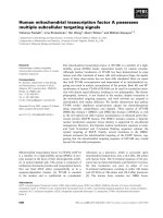
Fig. 2. Subcellular localization and transactivation potential of HNF1 constructs with deletion or insertion of the 26 aa segment. (A) The domains
encoded by each HNF1 construct are shown diagrammatically together with their designation. The black box indicates the 26 aa segment deleted in
HNF1b splice variant B. (B) Immunofluorescence of HeLa cells expressing GFP fusion proteins of the various constructs shown in A. Bar, 10 lm.
(C) Increasing amounts of GFP-HNF1b expression constructs (shown in A) were cotransfected with a HNF1-dependent luciferase reporter gene
into HeLa cells. T he fold-activation induced by each of t he HNF1 expression constructs is shown. Error b ars indicate standard de viation of the
mean of at least six replicates.
Ó FEBS 2004 HNF1b in nephrogenesis (Eur. J. Biochem. 271) 3719
less, this group was sufficient for significant analysis. This
abnormal development was not observed with any of the
other constructs.
To control the efficien cy of protein production, we tested
the amount of HNF1 proteins made by Western blots . As
exemplified in Fig. 3K, very similar levels were found in
the injected embryos. The t runcated HNF1a protein
(HNF1aaa) was as abundant as the truncated HNF1b
protein (HNF1bbb) demonstrating that both proteins are
equally expressed. Thus, the presence of HNF1aaa has in
contrast to HNF1bbb no effect on pronephric development.
Function of the dimerization domain of HNF1b
As overexpression of the t runcated HNF1b derivative
lacking the 26 aa segment (HNF1bbbD) also reduced the
pronephric size (Fig. 3D), we postulated that other seg-
ments present in this molecule may interfere with nephro-
genesis. To explore the function of the dimerization domain
of the HNF1b protein, we constructed chimeras of the
HNF1a and HNF1b proteins as shown in Fig. 4A. The
molecular and cellular properties of these chimeric con-
structs were assayed in transfected HeLa cells as well as in
developing Xenopus embryos.
The construct encoding the HNF1b-derived POU
S
and
POU
H
domains fused to t he HNF1a dimerization domain
(HNF1abb) was localized exclusively in the nucleus of
transfected HeLa c ells. In contrast, the construct encoding
the HNF1a-derived POU
S
and POU
H
domains fused to the
HNF1b dimerization domain (HNF1baa) was localized
both in the nucleus and the cytoplasm (Fig. 4B). These data
indicate that the POU
S
and POU
H
, but not the HNF1b-
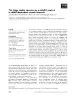
Fig. 3. Pronephric phenotype in Xenopus larvae after expression of
HNF1 proteins lacking or containing the HNF1b-s pecific 26 aa segment.
(A) Control ne urula expressing GFP o n the injected side . (B) Lateral
view of a larvae (stage 45) ex pressing full-length HNF1b protein. (C)
Dorsal view of larvae (stage 45) ex pressing the H NF1 protein desig-
nated. Whole-mount i mmun ostaining for the pronephric t ubul es and
duct using a Cy3-conjugated secondary antibody is shown as red
fluorescence. T he injecte d side is marked by an arrow. Bar, 3 00 lm.
(D) Statistical analysis of pronephric size in injected vs. noninjected
sides after expression of various HNF1 proteins. Boxes include 75% of
the values, and the vertical line represents the group median, and
whiskers represent the outer quartile. The P-value calculated using the
Mann–Whitney test and the animal number scored per group shown at
the far right. The reference indicates the GFP-injected control animals.
(E, F) Example of embryo exhibiting defects at gastrulation illumin-
ated at normal light (E) or under green fluorescence (F). N ote cell
death was only ob served in the injected ( GFP positive) region. Bar,
300 lm. (G) Stage 44 control embryo injected with GFP alone. Bar,
1 mm . (H–J) Developmenta l defects of different degrees were observed
in tadpoles expressing the truncated HNF1b protein lacking the 26 aa
segment (HNF1bbbD). Animals shown in panel H and I could not be
scored for pronephric morphology. (K) Western blot of protein
extracts derived from neurulae stage embryos injected with RNA
encoding HN F1aaa, HNF1bbb, HNF1aab or GFP
18
alone using th e
myc-tag specific antibody GE10 [35]. Each sample was an aliquot
representing one embryo o f a pool of 60 injected em bryos. At later
stages, the amount of HNF1 p rotein s was too low to be quantified.
3720 G. Wu et al. (Eur. J. Biochem. 271) Ó FEBS 2004
derived dimerization domain, determine t he exclusively
nuclear localization.
Transfection of the chimeric HNF1 constructs together
with an HNF1 dependent luciferase reporter plasmid was
used to measure the transactivation activity in HeLa cells.
Only the construct encoding the POU
S
and POU
H
of the
HNF1b protein (HNF1abb) resulted in transactivation of
the reporter gene s imilar to that mediated by the truncated
HNF1b construct (HNF1bbb, Fig. 4C). The presence of
the HNF1b-derived dimerization domain in the chimeric
protein ( HNF1baa) failed to increase the transactivation of
the reporter compared to the truncated HNF1a protein
(HNF1aaa, Fig. 4C).
The i nfluence of the chimeric constructs on kidney
development was tested in overexpression experiments in
Xenopus embryos. Injection of mRNA encoding chimeric
proteins with either the HNF1b-specific dimerization
domain (HNF1baa) or the b-specific DNA binding
domains (HNF1abb) of the HNF1b protein led to a
reduction in pronephric size (Fig. 4 D). This indicates that
the dimerization domain as well as the DNA binding
domain of HNF1b interfere with pronephric development.
HNF1aaa
HNF1baa
HNF1abb
HNF1bbb
1
1
1
1
351
321
80
71
321
70
81
351
HNF1abb
HNF1baa
A
0 50 100 150 200
relative pronephros size
B
fold induction
0
2
4
6
8
10
12
0 50 100 150 200 250 300
n
g
expression vector
HNF1aaa
HNF1bbb
HNF1abb
HNF1baa
C
localization
transactivation
-
N
N/C
N/C
N
-
11.1
4.4
4.4
11.2
D
phenotype
reference (95)
4.6 e-24 (226)
0.129 (147)
0.001 (114)
5.7 e-8 (151)
GFP
Dim
POU
S
POU
H
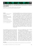
Fig. 4. Function of the dimerization domain of HNF1b. (A) The domains included in the HNF1 constructs are shown diagrammatically. HNF1b is
shown i n purple and HNF1a in blue. The black box indic ates the 26 aa segment deleted fro m the HNF1 b splice variant B. (B) Molecular and
cellular properties of HNF1 constructs were assayed in transfected cells as well as in developing embryos. On the left, N and N/C refer to nuclear
and nuclear plu s cytoplasmic l ocalizatio n, respectively. In t he middle, the fold induction of the HNF1-dependent luciferase r eporter after trans-
fection of the HNF1 constructs into HeLa cells is shown. On the right, statistical analysis of pronephric size in injected vs. noninjected sides after
expression of various HN F1 proteins. B oxes inc lude 75% o f the v alues, and t he vertical line represents the group median, and whiskers represent the
outer quartile. The P-value calculated using the Mann–Whitney test and the animal number scored per group shown at the far right. The reference
indicates the GFP-injected control animals. (C) Increasing amounts of HNF1 expression constructs were cotransfected together with a HNF1-
dependent luciferase reporter into HeLa cells. Mean of fold-activation of the reporter is represented by points, and error bars represent standard
deviation of at least six replicates. (D) Whole-mount immunostaining for pronephric tubules and duct in Xenopus larvae overexpressing the HNF1
protein indicated on one s ide. The in jected side is marked by a n arrow. Bar, 300 lm.
Ó FEBS 2004 HNF1b in nephrogenesis (Eur. J. Biochem. 271) 3721
However, the quantification shows a clear distinction in the
extent of the effect (Fig. 4 B), as the construct containing
only the dimerization domain of HNF1b (HNF1baa) was
considerably less efficient than the construct containing the
POU
S
and POU
H
domains of HNF1b (HNF1abb).
The homeodomain of HNF1b is essential for nuclear
localization and interferes with pronephric development
To explore the function of the HNF1b homeodomain
(POU
H
) in more detail, chimeric constructs were created
containing various parts of the HNF1b homeodomain
region. The chimeric gene c onstructs generated are shown
diagrammatically in Fig. 5A. Functional performance as
measured by subcellular localization, transactivation activ-
ity and effect on kidney development is summarized in
Fig. 5B. All chimeric constructs containing the HNF1b
homeodomain were found exclusively in t he nuclear com-
partment, implying t hat this domain contributes to nuclear
localization (Fig. 5B). With regard to the transactivation
potential, we observed that all chimeric constructs (Fig. 5C)
were less active than th e t runcated HNF1 b protein
HNF1aabins26
HNF1aab
HNF1aabH
HNF1aabHS
0 50 100 150 200
relative pronephros size
A
GFP
localization
tansactivition
-
N
N
N
N
-
-
3.2
5.5
4.3
2.0
-
HNF1aab
HNF1aabins26
1
1
351
351
176
183
196
229
HNF1aabH
1
319
196
229
HNF1aabHS
1
311
196
229
HNF1βHomeo
319
229
D
B
0 50 100 150 200 250 300
HNF1aab
HNF1aabH
HNF1aabins26
HNF1aabHS
n
g
expression vector
fold induction
C
phenotype
reference (95)
8.5e-9 (148)
2.7 e-14 (45)
2.3e-14 (42)
0.182 (85)
0.641 (81)
Dim
POU
S
POU
H
Fig. 5. The homeodomain of HNF1b is essential for nuclear localization and interferes with pronephric development. (A) The domains included in the
HNF1 constructs are shown diagrammatically. HNF1b isshowninpurpleandHNF1a in blue. The black box indicates the 26 aa segment of the
HNF1b splice variant B. (B) Molecular and cellular properties of HNF1 constructs were assayed in transfected cells as well as in developing
embryos.
19
See Fig 4 legend for details. (C) Increasing amounts of HNF1 expression constructs were cotransfected together with a HNF1-dependent
luciferase reporter into HeLa cells. Mean of fold-activation of the reporter is represented by points, and error bars represent standard deviation ofat
least six rep licates. (D) Whole-mount immu nostaining f or pronephric tubules and duct in Xenopus larvae overexpressing the HNF1 protein
indicated on one side. T he injected side is m arked by an arrow. Bar, 300 lm.
3722 G. Wu et al. (Eur. J. Biochem. 271) Ó FEBS 2004
(HNF1bbb) in transfection assays (Fig. 4C). While the
constructs containing the HNF1b homeodomain but lack-
ing the 26 aa segment (HNF1aab) gave approximately a
fivefold transactivation of the reporter plasmid, t he corres-
ponding construct containing the 26 a a segment (HNF1aab-
ins26) gave a threefold activation. Successive truncation at
the C-terminal end o f the ho meodomain in the constructs
lacking the 26 aa segment (HNF1aabH, HNF1aabHS) led
to a further decrease in the transactivation, but was still
twofold above base level (Fig. 5C).
To identify whether the homeodomain influences
kidney development in Xenopus embryos, mRNA from
the chimeric constructs were injected into one cell at the
two-cell s tage, a nd the pronephric size was measured
(Fig. 5B,D). Three chimeric constructs with the HNF1b-
specific homeodomain ( HNF1aab, HNF1aabins26 a nd
HNF1aabH) led to a reduction of the pronephric size with
the constructs lacking the 26 aa segment (HNF1aab and
HNF1aabH) being most effective (Fig. 5B). In contrast, the
construct lacking eight amino acid at the C-terminal p art of
the homedomain (HNF1aabHS) had no e ffect on proneph-
ric size indicating the critical C-terminal border. A construct
producing the homeodomain alone (HNF1bHomeo) had
no effect on pronephric size (Fig. 5B), indicating that the
HNF1 backbone is required to allow the protein fun ction
that interferes with kidney development. We observed that
both c himeric constructs lacking the 26 aa segment
(HNF1aab and HNF1aabH) had an adverse effect on
normal development as found for the truncated HNF1b
lacking the 26 aa segment (HNF1bbbD, Fig. 3E–J). In fact,
most surviving animals were distorted allowing only a
minority to be a nalyzed at stage 45. This adverse effect
on embryogenesis was absent in the construct with the
C-terminal truncation (HNF1aabHS) that h as also lost its
effect on nephrogenesis.
Partial rescue of Pax8/lim1-mediated pronephros
malformation by HNF1b injection
It has been reported that overexpression of the transcription
factors, Pax8 and lim1, in Xenopus embryos led to the
development of an abnormally large pronephros, and to the
formation of ectopic pronephric tissue [44]. As both these
transcription factors are expressed at t he neurula s tage
together with HNF1b in the pronephric anlage, we won-
dered whether simultaneous overexpression of HNF1b
could overcome the effects o f Pax8 a nd lim1. Overexpres-
sion of Pax8 or lim1 by themselves led only to marginal
effects, but synergized to have a pronounced effect [44]. We
coinjected RNA encoding Pax8 and lim1 into one blasto-
mere of the two-cell stage embryo together with GFP
mRNA as a tracer. Injected embryos w ere raised to the
swimming tadpo le stage, and processed to visualize pro-
nephric tubules and duct. Overexpression of Pax8 together
with lim1 led to an enlargement of the pronephros as
compared to embryos injected with GFP alone (Fig. 6A).
This size difference was shown to be significant using the
Mann–Whitney test (Fig. 6G). More importantly, ectopic
pronephric tubules a nd small c ysts close to the main
pronephric body were observed using immunostaining on
the injected side (Fig. 6B). Such structures were seen in 16%
of the injected embryos (Table 1), but never observed in
injections with mRNA encoding G FP o r a ny HNF1
derivative. Furthermore, 24% of the larvae coinjected with
Pax8 and lim1 displayed cyst-like structures or a thickening
of the tubules on the injected side (Table 1). Such abnor-
malities were seen in only 4% of larvae injected with the
truncated HNF1b protein (HNF1bbb). Our results are
similar to those using a different injection protocol reported
previously [44]. We coinjected mRNAs encoding Pax8 and
lim1 together with HNF1b and GFP as a tracer into one cell
of the two-cell stage Xenopus embryos. Immunostaining for
the pronephric tubules and duct at the tadpole stage showed
that these embryos had pronephric structures similar to
embryos injected with Pax8 and lim1 alone (Fig. 6C,D).
The pronephros appeared smaller in some larvae, but the
size difference was not significant when compared with
larvae injected with Pax8 and lim1 alone (Fig. 6G).
Furthermore, 17% of the samples were found to have
ectopic tubules (Table 1). Cyst-like structures or thickening
of the tubules were also found in 27% of the samples. These
data imply that the overexpression of Pax8 and lim1 is
dominant to the effect of HNF1b. It was not possible to
inject higher concentrations of HNF1b mRNA, b ut as the
truncated HNF1 b protein (HNF1bbb) was more a ctive i n
reducing the pronephric size (Fig. 3 C), this construct was
coinjected together with Pax8 and lim1. These larvae had
slightly smaller pronephroi in the injected side (Fig. 6F),
suggesting that HNF1bbb coinjection c ould overcome t he
effect mediated by Pax8 and lim1. More importantly, no
larvae had ectopic tubules (Table 1). H owever, 28% of the
samples were found to have cyst-like structures or thicken-
ing of t he tubules (Fig. 6E,F), similar to t he fraction
showing this phenotype in Pax8 and lim1 coinjected
embryos (Table 1). Therefore, cyst-like structures and
thicker tubules mediated by Pax8 and lim1 were not rescued
by HNF1bbb. Taken together these results indicate that the
Pax8- and lim1-induced phenotype has two separate
qualities. One is the enlargement and ectopic formation of
pronephros which could be antagonized by HNF1b and the
other is the induction of cyst-like structures which could not
be antagonized by HNF1b.
Discussion
The transcription factors, HNF1a and HNF1b,display
extensive structural s imilarities with indistinguishable DNA
sequence binding specificity [2]. Our data imply that they
have acquired distinct functions during evolution as
homologous domains of these two factors display disparate
properties. These include the subcellular localization, the
transactivation potential as well as th e ability to affect
nephrogenesis.
HNF1b has a nuclear localization sequence located
in the homeodomain
Analyzing the subcellular localization of various chimeric
HNF1 proteins, we observed an exclusively nuclear staining
in transfected HeLa cells in all constructs containing the
POU homeodomain (POU
H
) of the HNF1b protein. This
finding is consistent with our previous data showing nuclear
localization of all truncated HNF1b transcription factors
retaining the POU
H
domain [43]. The occurrence of a
Ó FEBS 2004 HNF1b in nephrogenesis (Eur. J. Biochem. 271) 3723
nuclear localization signal (NLS) in the homeodomain of
the HNF1b protein is supported by the presence in the
N-terminal region of the h omeodomain (amino acid
229–235, Fig. 7) of the amino acid sequence, KKMRRNR,
predicted to be a NLS (PredictNLS Online, http://
cubic.bioc.columbia.edu). The NLS of the HNF1b protein
and t he HNF1a protein (KKGRRNR) differ by only one
amino acid (M fi G). This change may hinder efficient
nuclear translocation of HNF1a in transfected HeLa cells,
and probably results in the nuclear as well as cytoplasmic
localization typical for HNF1a.
Differential transactivation potential of the HNF1a
and HNF1b protein
The C-terminal transactivation domains of HNF1a and
HNF1b are only weakly conserved (Fig. 1), and in most
transactivation a ssays HNF1a is approximately twofold
Fig. 6. Partial rescue of Pax8/lim1-inducedkidneymalformationbyHNF1b. (A–F) L ateral views of two representative larvae expressing the
proteins listed at the left on one side. Larvae are immunostained to visualize the pronephric tubules and duct. (A, B) Enlarged pronephroi in Pax8/
lim1 (125 pg mRNA each per embryo) c oinjected embryos. (C, D) Enlarged pronephroi in embryos coinjected with Pax8 (125 pg mRNA per
embryo), lim1 (125 pg mRNA per embryo), and HNF1b (250 pg mRNA per embryo). (E, F) Reduced pronephric size in embryos coinjected with
Pax8 (125 p g mRNA per embryo), lim1 (125 pg mRNA per embryo) and truncated HNF1b (HNF1bbb, 250 pg mRNA per embryo). Anterior is
to the left for the injected sides, and to the right for the noninjected sides, and dorsal is up. Thickened tubules (T) characterized by a wider diameter
and cyst-like structures or b ubbles (B) are indicated by a rrows. E ctopic pronephric tubules are indicated by arrow heads. Bar, 200 lm. (G)
Statistical analysis of pronephric size in injected vs. noninjected sides after expression of various HNF1 proteins. Boxes include 75% of the values,
and the vertical line represents the group median, and whiskers represent the outer quartile. The P-value calculated using the Mann–Whitney test
and the animal nu mber scored per g roup shown at the far r ight. The reference indicates the GFP-injected control animals.
3724 G. Wu et al. (Eur. J. Biochem. 271) Ó FEBS 2004
more potent t han HNF1b [2]. The truncated HNF1 b
protein (HNF1bbb) transactivated the reporter gene
strongly at saturating amounts, implying transactivational
properties outside of the classical transactivation domain.
This is distinct from HNF1a where the deletion of the
transactivation domain results in low activity even a t high
vector concentrations. This confirms initial reports that
transactivation a ctivity is confined to the C-terminal
region in HNF1a, leading to the designation as trans-
activation domain in both HNF1a and HNF1b proteins
[52,53].
The 26 a a segment located between the POU
S
and POU
H
domains of the HNF1b protein is highly e volutionarily
conserved, and is the most striking d ifference between the
HNF1a and HNF1 b proteins (Fig. 1). Our transactivation
assays showed that this b-specific segment plays distinct roles
dependent on the HNF1 protein background. In the full-
length HNF1b protein, it accentuated the transactivation
activity (Fig. 2), which is consistent with previous results
[29,54]. I n the t runcated HNF1b protein (HNF1bbb),
deletion of this segment made no difference on its trans-
activation potential. Finally, in truncated HNF1a protein
(HNF1aaa), the insertion of the 26 aa segment (HNF1aaa-
ins26) abolished the residual transactivation potential
(Fig. 2 ). These results imply that the 26 aa segment may
interact in a con text-dependent manner w ith other factors
and/or alters the conformation of the overall protein
structure.
We showed that the dimerization domain of the HNF1b
protein failed to increase the transactivation potential of the
truncated HNF1a protein. However, the replacement of
either the POU
S
and POU
H
domains or of the POU
H
domain alone with those from the HNF1b protein was
sufficient to increase transactivation activit y (Figs 4 and 5 ).
Even though both domains are highly conserved between
the two proteins there appears t o be functional differences.
As there is a progressive increase in the transactivation
potential with the length of the HNF1b protein derived
segment, we deduce that several features contribute to the
transactivation potential of the POU
S
and POU
H
domains
of HNF1b.
Domains of HNF1b involved in nephrogenesis
The simplicity
14
of the Xenopus system allowed us to
differentiate the properties of the HNF1a and HNF1b
proteins during kidney development. The analysis of the
molecularpropertiesoftheHNF1proteinsincellculturesis
too simplistic to evaluate functional properties in a devel-
oping organism. Our analysis of the morphogenetic poten-
tial of chimeric HNF1 proteins during k idney d evelopment
in Xenopus is most meaningful to this end. Although we
concentrated our analysis on HNF1 proteins of human
origin, it is unlikely that protein functions are species
specific. In fact, we have shown that the overexpression of
Xenopus HNF1b protein in Xenopus embryos also l ed to a
Fig. 7. The n ephrogenic effects of domains in the human HNF1b transcription factor and its m utants. Functional domains are i ndicated above t he
schematic representation of H NF1b, and numbers refer to the amino acid positions. The b lack box indicates the 26 aa segment deleted in the
HNF1b splice variant B. The three regions involved in nephrogenesis are marked by black lines beneath the HNF1b diagram. The NLS is marked
by a red line above the HNF1b diagram. Naturally occuring HNF1b mutations are shown below as line diagrams to indicate what regions of the
protein are missing. Wh ether these HN F1b mutants cause an enlargement or a reduction of pronephric size [43] i s indicated at the far r ight.
Table 1. Frequency of enlarged or ectopic pronephric tubules in mRNA-injected embryos. Enlarged, enlarged relative pronephros size of injected side/
uninjected side > 120%. Normal, normal relative to pronephros size of injected side/uninjected side between 80–120%. Smaller, smaller relative to
pronephros size of injected side/uninjected side < 80% .
Embryos
Pronephric tubules (%)
Cyst-like structures
or thickening (%) NEnlarged Normal Smaller Ectopic
Pax8 + lim1 49 34 1 16 24 83
Pax8 + lim1 + HNF1b 40 38 5 17 27 77
Pax8 + lim1 + HNF1bbb 20 38 42 0 28 111
HNF1bbb 5 17 78 0 4 226
Ó FEBS 2004 HNF1b in nephrogenesis (Eur. J. Biochem. 271) 3725
reduction in pronephric size (data not shown), supporting
the conserved function of HNF1b from Xenopus to humans.
As summarized in Fig. 7, we identified three domains of the
HNF1b protein that interfere with pronephric d evelopment
when swapped into the HNF1a protein. These include the
dimerization domain, the 26 a a segment and the homeo-
domain. It is noteworthy that the dimerization domain of
the HNF1b protein interferes with pronephros formation,
despite that swapping of this region had no effect on the
transactivation potential in transient transfection assays
(Fig. 4C). This indicates that the nephrogenic effect is
distinct from the simple ability to transactive a reporter
gene. This emphasizes t he importance of a complex
transcription factor b ackground present in an appropriate
developmental context. We also showed that the 26 aa
HNF1b-specific segment plays an important role in pro-
nephric deve lopment. This is most interesting, as this 26 aa
segment is the characteristic feature of the splice variant A.
Whereas the full-length splice variant A of HNF1b led to
agenesis of the pronephros in Xenopus embryos [35,43], the
splice variant B ( HNF1bD) lacking the 26 aa segment did
not interfere with pronephric development (Fig. 3D). As the
ratio of splice variant A : B alters during kidney develop-
ment [15], our data support that this differential splicing
pattern plays a key role in nephrogenesis. The functional
difference between the A and B splice v ariants in nephro-
genesis contrasts to their role during early embryogenesis,
where either variant can compensate f or the loss of the
native HNF1b gene during the differentiation of visceral
endoderm from embryonic stem cells [29].
Although the in sertion of t he 26 aa segment into
truncated HNF1a protein (HNF1aaa) generated a HNF1
protein (HNF1aaains26) with nephrogenic properties, dele-
tion of this segment from the truncated HNF1b protein
(HNF1bbb) did not affect its ability to reduce t he
pronephric size (HNF1bbbD in Fig. 3D). This indicates
that other regions o f HNF1b protein contribute to the
nephrogenic properties of HNF1b. In fact, chimeric
proteins both containing the homeodomain o f HNF1b,
but lacking the 26 aa segment (HNF1aab a nd
HNF1aabH), led to agenesis of the pronephros (Fig. 5B),
thus, demonstrating the i mportance o f the HNF1b home-
odomain in kidney development. We were able to restrict
the region of the homeodomain responsible for this effect to
the POU
H
domain from 229 to 319 (Fig. 5B). However, the
homeodomain alone was unable to reduce proneph ric size,
indicating that the homeodomain of HNF1b functions only
in the co ntext of the HNF1 backbone. Deletion of the C-
terminal amino acids of the HNF1b homeodomain (311–
319) abolished its potential to interfere with pronephric
formation. The corresponding eight amino acid in the
HNF1a protein are not required for DNA binding [14]. As
two amino acids w ithin this e ight amino acid region (Q311
and A317) are different in the HNF1a and HNF1b protein,
it is possible that one or both of these two amino acids play
a functional role of the HNF1b POU
H
domain during
nephrogenesis. Alternatively, the entire POU
H
homeo-
domain of HNF1b may be necessary for proper function.
Expression of all truncated HNF1 proteins lacking the
26 aa segment but containing the HNF1b homeodomain
(HNF1bbbD, HNF1aabH or HNF1aab) had an adverse
effect on the survival of the embryos and resulted in a high
proportion of defects starting at gastrulation. It is not clear
why the expression of these HNF1 proteins caused these
early developmental problems. A possible explanation is,
that HNF1b has several functions in early embryogenesis
distinct from nephrogenesis. Knock-out experiments in the
mouse established that HNF1b is required for yolk sac
differentiation [18,23], and overexpression in Xenopus of a
dominant negative form of HNF1b interferes with meso-
derm induction [47]. Furthermore, HNF1b mRNA injection
into zebrafish showed it to be involved in the specification of
the rhombomeres identity in the hindbrain [55]. It is possible
that some of our constructs may have disturbed similar
early developmental processes outside of the pronephric
anlage in the frog.
In a recent report, we have found that the introduction of
human HNF1b mutant genes
15
into Xenopus embryos leads
to either a r eduction or an enlargement of the pronephros
[43]. T hese observed phenotypes co uld not be correlated
directly to the structure of the mutated HNF1b protein
(summarized in Fig. 7). All truncated HNF1b proteins
retaining t he DNA binding domain (e.g. Y352insA) as well
as a HNF1b mutant with an in-frame internal deletion in
the POU
S
domain (R137–K161) that destroys DNA
binding resulted in a r eduction in pronephric size. In
contrast, all truncated HNF1b proteins with impaired DNA
binding (e.g. A263insGG and E101X) resulted in an
enlargement of the pronephros. In this report we have
identified three regions having a n ephrogenic potential. We
deduce that these three regions must be present in a HNF1b
mutant for a reduction in pronephric size, otherwise an
enlargement occurs.
Partial cooperation of Pax8, lim1 and HNF1b
in nephrogenesis
There are at least two other early expressed transcription
factors involved in kidney development in vertebrates. In
the Xenopus embryo, both Pax8 and lim1 are expressed
initially in the pronephric anlage at the time when HNF1b is
expressed [41]. Both these transcription factors are func-
tionally important, as overexpression of either protein led to
an enlarged pronep hros with ectopic pronephric structures
[44]. This effect was additive when both transcription factors
were coexpressed, and the effect of Pax8 could be mimicked
by Pax2 [44], w hose expression starts s hortly after Pax8 in
the pronephric anlage [56]. The importance of lim1 [57] and
Pax2 [58] in mammalian d evelopment was shown in k nock-
out mice that had severe defects in organogenesis including
agenesis of the kidney. The nephrogenic role of Pax8 has
only been identified in a Pax2-deficient background. Mice
lacking Pax8 additionally are unable to form any nephric
structure due to a block in the mesenchymal-epithelial
transition [59].
In an effort to evaluate whether HNF1b cooperates with
Pax8 and lim1 during kidney development, we coinjected all
three transcription factors into Xenopus embryos. We
confirmed that overexpression of Pax8 together with lim1
results i n a n enlargement o f the pronephros an d t he
development of ectopic pronep hric tubules [44]. As HNF1b
overexpression inhibits kidney formation and Pax8/lim1
overexpression is nephrogenic, it is possible that a simple
antagonism exists between these factors during kidney
3726 G. Wu et al. (Eur. J. Biochem. 271) Ó FEBS 2004
development. We show here that HNF1b can only partially
rescue Pax8/lim1-induced nephrogenesis. T he truncated
HNF1b protein rescued the Pax8/lim1-induced enlargement
and ectopic tubule formation. However, Pax8/lim1-induced
thickening of tubules and cyst-like structure formation
remained essentially unchanged. These r esults suggest that
HNF1b activity can overcome part of the nephrogenic
potential of Pax8 and lim1. Most importantly, the data also
reveal that Pa x8/lim1 and HNF1 b are not simple antago-
nists during nephrogenesis, but that Pax8/lim1 also h ave
distinct morphogenetic properties.
Acknowledgements
WearemostgratefultoR.VignaliandP.D.VizefortheXenopus
HNF1b and lim1, Pax2/8 cDNAs, respectively, and Elizabeth A. Jones
for antibodies 3G8 and 4A6. We thank Kathy Astrahantseff, Christoph
Waldner and Karin D udziak for critical reading of the manuscript.
This work was supported by the Deutsche Forschungsgemeinschaft
(Ry5/7–2).
References
1. Gehring, W.J., A ffolter, M . & Burglin, T. (1994) Homeodomain
proteins. Annu. Rev. Biochem. 63, 487–526.
2. Cereghini, S. (1996) Liver-enriched transcription factors and
hepatocyte differentiation. FASEB J. 10, 267–282.
3. Pontoglio, M. (2000) Hepatocyte nuclear factor 1, a transcription
factor at the crossroads of glucose homeostasis. J. Am. Soc.
Nephrol. 11 (Suppl. 16), S140–S143.
4. Deryckere, F., Byrnes, L., Wagner, A., McMorrow, T. & Gannon,
F. (1995) Salmon HNF1: cDNA sequenc e, evolution, tissue spe-
cificity and binding to the salmon serum albumin promoter.
J. Mol. Biol. 247, 1–10.
5. Sun, Z. & Hopkins, N. (2001) vhnf1, the MODY5 and familial
GCKD-associated gene, regulates regional specification of the
zebrafish gut, pron ep hros, and hindbrain. Genes Dev. 15, 3217–
3229.
6. Bartkowski, S., Zapp, D., Web er, H., Eb erle, G., Zoidl, C., Senkel,
S., Klein-Hitpass, L. & Ryffel, G.U. (1993) Developmental reg-
ulation and tissue distribution of the liver transcription factor
LFB1 (HNF1) in Xenopus laevis. Mol. Cell Biol. 13, 421–431.
7. Demartis, A., Maffei, M., Vignali,R.,Barsacchi,G.&DeSimone,
V. (1994) Cloning and developmental expression of LFB3/HNF1
beta transcription factor in Xenopus laevis. Mech. Dev. 47, 19–28.
8. Frain, M., Swart, G., Monaci, P., Nicosia, A., Stampfli, S., Frank,
R. & Cortese, R. (1989) The liver-specific transcription factor
LF-B1 contains a highly diverged homeobox DNA binding
domain. Cell 59, 145–157.
9. Bach,I.,Mattei,M.G.,Cereghini,S.&Yaniv,M.(1991)Two
members of an HNF1 homeoprotein family are expressed in
human liver. N ucleic Acids. R es. 19, 3553–3559.
10. Baumhueter, S., Mendel, D.B., Conley, P.B., Kuo, C.J., Turk, C.,
Graves, M.K., Edwards, C.A., Courtois, G. & Crabtree, G.R.
(1990) HNF-1 shares three sequence motifs with the POU domain
proteins and is identical to LF-B1 and AP F. Genes D ev. 4,
372–379.
11. Zapp, D., Bartkowski, S., Zoidl, C., Klein-Hitpass, L. & R yffel,
G.U. (1993) Genomic structure of the Xenopus laevis liver tran-
scription factor LFB1. Gene 134, 251–256.
12. Rose, R.B., Endrizzi, J.A., Cronk, J.D., Holton, J. & Alber, T.
(2000) High-resolution structure of the HNF-1alpha dimerization
domain. Biochemistry 39 , 15062–15070.
13. Narayana, N., Hua, Q . & Weiss, M.A. (2001) The dimerization
domain of HNF-1alpha: Structure and plasticity of an intertwined
four-helix bundle with application to diabetes m ellitus. J. Mol.
Biol. 310, 635–658.
14. Chi,Y.I.,Frantz,J.D.,Oh,B.C.,Hansen,L.,Dhe-Paganon,S.&
Shoelson, S.E. (2002) Diabetes mutations delineate an atypical
POU domain in HNF-1alpha. Mol. Cell 10, 1129–1137.
15. Cereghini, S., Ott, M.O., Power, S. & Maury, M. (1992) Expres-
sion patterns of vHNF1 and HNF1 homeopr oteins in early
postimplantation embryos s uggest distinct and seque ntial devel-
opmental roles. Development 116, 783–797.
16. Ott, M.O., Rey-Campos, J., Cereghini, S. & Yaniv, M. (1991)
vHNF1 is expressed in epithelial cells of distinct embryonic origin
during development and precedes HNF1 expression. Mech. Dev.
36, 47–58.
17. Lazzaro, D., De Simone, V., De Magistris, L., Lehtonen, E. &
Cortese, R. (1992) LFB1 and LFB3 h omeoproteins are sequen-
tially expressed durin g kidney development. Development 114,
469–479.
18. Barbacci, E., Reber, M., Ott, M., Breillat, C., Huetz, F . & Cere-
ghini, S. (1999) Variant hepatocyte nuclear factor 1 is required for
visceral endoderm specification. Development 126 , 4795–4805.
19. Coffinier, C., Barra, J., Babinet, C. & Yaniv, M. (1999) Expression
of the vHNF 1/HNF1b eta homeoprotein gene during m ouse
organogenesis. M ech. Dev. 89, 211–213.
20. Reber, M. & Cereghini, S . (2001) Variant hepatocyte nuclear
factor 1 expression in the mouse genital tract. Mech. Dev. 100,
75–78.
21. Pogge, V., Strandmann , E., Nastos, A., Holewa, B ., Senkel, S.,
Weber, H. & Ryffel, G.U. (1997) Patterning the expression of a
tissue-specific transcription factor in embryogenesis: HNF1 alpha
gene activation during Xenopus developm ent. Mech. Dev. 64,
7–17.
22. Weber, H., Strandmann, E.P., Holewa, B., Bartkowski, S., Zapp,
D., Zoidl, C. & Ryffel, G.U. (1996) Regulation and function of the
tissue-specific transcription factor HNF1 alpha (LFB1) during
Xenopus development. Int. J. Dev. Biol. 40, 297–304.
23. Coffinier, C., The
´
pot, D., Babinet, C., Yaniv, M. & Barra, J.
(1999) Essential role for the homeoprotein vHNF1/HNF1b in
visceral endoderm differentiation. Developmen t 126, 4785–4794.
24. Pontoglio, M., Barra, J., Hadchouel,M.,Doyen,A.,Kress,C.,
Bach, J.P., Babinet, C. & Yaniv, M. (1996) Hepatocyte nuclear
factor 1 inactivation resu lts in hepatic d ysfunction , phenyl-
ketonuria, and renal Fanconi syndrome. Cell 84 , 575–585.
25. Pontoglio, M., Sreenan, S., Roe, M., Pugh, W., Ostrega, D.,
Doyen,A.,Pick,A.J.,Baldwin,A.,Velho,G.,Froguel,P.,Levi-
setti, M., Bonner-Weir, S., Bell, G.I.,Yaniv,M.&Polonsky,K.S.
(1998) Defective insulin secretioninhepatocytenuclearfactor
1alpha- deficient mice. J. Clin. Invest. 101, 2215–2222.
26. Pontoglio, M., Prie, D., Cheret, C., Doyen, A., Leroy, C., Frogu el,
P.,Velho,G.,Yaniv,M.&Friedlander,G.(2001)HNF1alpha
controls renal glucose reabsorption in mouse and man. EMBO
Reports 1, 359–365.
27. Shih, D.Q., Screenan, S., Munoz, K.N., Philipson, L.,
Pontoglio, M., Yan iv, M., Polonsky, K.S. & Stoffel, M. (2001)
Loss of HNF-1alpha function in mice leads to abnormal expres-
sion of genes involved i n pancreatic islet development and meta-
bolism. Diabetes 50, 2472–2480.
28. Lee, Y.H., Sauer, B. & Gonzalez, F.J. (1998) Laron dwarfism and
non-insulin-depende nt diabetes mellitus i n the Hnf-1alpha
knockout mouse. Mol. Cell Biol. 18, 3059–3068.
29. Haumaitre, C., Reber, M. & Cereghini, S. (2003) Functions of
HNF1 family members in differentiation of the visceral endoderm
cell lineage. J. Biol. Chem. 278, 40933–40942.
30. Bluteau, O., Jeannot, E., Bioulac-Sage, P., Marques, J.M.,
Blanc, J.F., Bui, H., Beaudoin, J.C., Franco, D., Balabaud, C.,
Laurent-Puig, P. & Zucman-Rossi, J. (2002) Bi-allelic inactivation
of TCF1 in hepatic adenomas. Nat. Genet. 32, 312–315.
Ó FEBS 2004 HNF1b in nephrogenesis (Eur. J. Biochem. 271) 3727
31. Ryffel, G.U. (2001) Mutations in the human genes encoding the
transcription factors of the hepatocyte nuclear factor (HNF) 1 and
HNF4 families. functional and pathological consequences. J. Mol.
Endocrinol. 27 , 11–29.
32. Bingham, C., Ellard, S., Cole, T.R., J ones, K.E., Allen, L .I.,
Goodship, J.A., Goodship, T .H., Bakalinova-Pugh, D., Russell,
G.I., Woolf, A.S., Nicholls, A.J. & Hattersley, A.T. (2002) Solitary
functioning kidney and diverse g enital tract malformations asso-
ciated with hepatocyte nuclear factor-1beta mutations. Kidney Int.
61, 1243–1251.
33. Bingham, C., Ellard, S., Van’t Hoff, W.G., Simmonds, H.A.,
Marinaki, A.M., Badman, M.K., Winocour, P.H., Stride, A.,
Lockwood, C.R., Nicholls, A.J., Owen, K.R., Spyer, G., Pearson,
E.R. & Hattersley, A.T. (2003) Atypical familial juvenile hyper-
uricemic nephropathy associated with a hepatocyte nuclear factor-
1beta gene mutation. Kidney Int. 63, 1 645–1651.
34. Woolf, A.S., Wynyard, P.J.D., Hermanns, M.M. & Welham,
S.J.M. (2003) Maldevelopm ent of t he human k idney and l ower
urinary tract: An Overview. In The Kidne y: fro m No rmal D evel-
opment to Congenital Disease (Vize,P.D.,Woolf,A.S.&Bard,
J.B.L., eds), pp. 377–393. Academic Press, Amsterdam.
35. Wild, W., Pogge, V., Strandmann, E., Nastos, A., Senkel, S.,
Lingott-Frieg, A., Bulman, M., Bingham, C., Ellard, S., H atters-
ley, A.T. & Ryffel, G .U. (2000) T he mutated human gene
encoding hepa tocyte nuc lear factor 1 beta inhibits kidney forma-
tion in developing Xenopus embryos. Proc. Natl Acad. Sci. USA
97, 4695–4700.
36. Gresh, L., Fischer, E., Reimann, A., Tanguy, M., Garbay, S.,
Shao, X., Hiesberger, T., Fiette, L., Igarashi, P., Yaniv, M. &
Pontoglio, M. (2004) A transcriptional network in polycystic
kidney disease. EMBO J.
37. Hiesberger, T ., Bai, Y., Shao, X., McNally, B.T., Sinclair, A.M.,
Tian, X., Somlo, S. & Igarashi, P. (2004) Mutation of hepatocyte
nuclear factor-1beta inhibits Pkhd1 gene expression and produces
renal cysts in mice. J. Clin. Invest. 113, 814–825.
38. Vize, P.D., Carroll, T.J. & W allingford, J.B. (2003) Introduction:
Embryonic Kidneys and o ther n ephrogenic mode ls. In The Kid-
ney. From Normal Development to Congenital Disease (Vize, P.D.,
Woolf, A.S. & Bard, J.B.L., eds), pp. 1–6. Academic Press,
Amsterdam.
39. Jones, E.A. (2003) Molecular control of pronephric development:
An overview. In The Kidney. From Normal Development t o C on-
genital Disease (Vize,P.D.,Woolf,A.S.&Bard,J.B.L.,eds),
pp. 93–118. Academic Press, Amsterdam.
40. Carroll, T.J. & McMahon, A.P. (2003) Overview: The Molecular
Basis of Kidney Develop ment. In Th e Kidne y: from N orma l
Development to Congenital Disease (Vize, P.D., Woolf, A.S. &
Bard, J.B.L., eds), pp. 343–376. Academic Press, Amsterdam.
41. Ryffel, G.U. (2003) What can a frog tell us about human kidney
development. Nephron Exp. N ephrol. 94, e35–e43.
42. Bra
¨
ndli, A.W. (1999) Towards a molecular anatomy of the Xen-
opus pronephric kidney. Int. J. Dev. Biol. 43, 381–395.
43.Bohn,S.,Thomas,H.,Turan,G.,Ellard,S.,Bingham,C.,
Hattersley, A.T. & Ryffel, G.U. (2003) Distinct molecular and
morphogenetic properties of mutations in th e human H NF1beta
gene th at lead to defective kidney development. J. Am. Soc.
Nephrol. 14, 2033–2041.
44. Carroll, T.J. & Vize, P.D. (1999) Synergism between Pax-8 and
lim-1 in embryonic kidney development. Dev. Biol. 214, 46–59.
45. Rupp, R.A., Snider, L. & Weintraub, H. (1994) Xenopus embryos
regulate the nuclear localization of XMyoD. Genes Dev. 8, 1311–
1323.
46. Ryffel, G.U. & Lingott, A. (2000) Distinct promoter elements
mediate endodermal and mesodermal expression of the
HNF1alpha promoter in transgenic Xenopus. Mech. Dev. 90,
65–75.
47. Vignali, R., Poggi, L., Madeddu, F. & Barsacchi, G. (2000)
HNF1beta is required for mesoderm induction in the Xenopus
embryo. Development 127, 1455–1465.
48. Peng, H.B. (1991) Xenopus laevis: Practical uses in cell and
molecular biology. Solutions and protocols. Methods Cell Biol. 36,
657–662.
49. Nieuwkoop, P.D. & Faber, J. (1975) Normal Table of Xenopus
Laevis (Daudin). Elsevier/North-Holland Publishing Co, Amster-
dam, the Netherlands.
50. Vize, P.D., Jones, E.A. & Pfister, R. (1995) Development of the
Xenopus pronephric system. Dev. Biol. 171, 531–540.
51. Thomas,H.,Badenberg,B.,Bulman,M.,Lemm,I.,Lausen,J.,
Kind, L., Roosen, S., Ellard, S., Hattersley, A.T. & Ryffel, G.U.
(2002) Evidence for haploinsufficiency of the human HNF1alpha
gene revealed by functional characterization of MODY3-asso-
ciated mutations. Biol. Chem. 383 , 1691–1700.
52. Nicosia, A., Monaci, P ., To mei, L ., De F rancesco, R., Nuzzo,
M.,Stunnenberg,H.&Cortese,R.(1990)Amyosin-like
dimerization helix and an extra-large homeodomain are essential
elements of the tripartite DNA binding structure of LFB1. Cell 61,
1225–1236.
53. Sourdive, D.J., Chouard, T. & Y aniv, M. (1993) The HNF1
C-terminal domain contributes to transcriptional activity
and modulates nuclear localisation. C. R. Acad. Sci. III. 316,
385–394.
54. Ringeisen, F., Rey-Campos, J. & Yaniv, M. (1993) The trans-
activation potential of variant hepatocyte nuclear factor 1
is modified by alternative splicing. J. Biol. Chem. 268, 25706–
25711.
55. Wiellette, E.L. & Sive, H. (2003) vhnf1 and Fgf signals synergize to
specify rhombomere identity in the zebrafish hindbrain. Develop-
ment 130, 3821–3829.
56. Heller, N. & Bra
¨
ndli, A.W. (1999) Xenopus Pax-2/ 5/8 ortho logu es.
novel insights into Pax gene evolution and identification of Pax-8
as the earliest marker for otic and pronephric cell lineages. Dev.
Genet. 24, 208–219.
57. Shawlot, W. & Behringer, R.R. (1995) Requirement for Lim1 in
head-organizer function. Nature 374, 425–430.
58. Torres,M.,Gomez-Pardo,E.,Dressler,G.R.&Gruss,P.(1995)
Pax-2 controls m ultiple steps of urogenital development. Devel-
opment 121, 4057–4065.
59. Bouchard, M ., So uabni, A., Mandler, M., N eubuser, A. & Bus-
slinger, M. (2002) Nephric lineage specification by Pax2 and Pax8.
Genes Dev. 16, 2958–2970.
3728 G. Wu et al. (Eur. J. Biochem. 271) Ó FEBS 2004