Báo cáo khoa học: Aptamers toEscherichia colicore RNA polymerase that sense its interaction with rifampicin, r-subunit and GreB ppt
Bạn đang xem bản rút gọn của tài liệu. Xem và tải ngay bản đầy đủ của tài liệu tại đây (368.4 KB, 11 trang )
Aptamers to
Escherichia coli
core RNA polymerase that sense its
interaction with rifampicin, r-subunit and GreB
Andrey Kulbachinskiy
1,2
, Andrey Feklistov
2,3
, Igor Krasheninnikov
3
, Alex Goldfarb
1
and Vadim Nikiforov
1,2
1
Public Health Research Institute, Newark, New Jersey, USA;
2
Institute of Molecular Genetics, Moscow, Russia;
3
Department of Molecular Biology, Moscow State University, Moscow, Russia
Bacterial RNA polymerase (RNAP) is the central enzyme of
gene expression that is responsible for the synthesis of all
types of cellular RNAs. The process of transcription is
accompanied by complex structural rearrangements of
RNAP. Despite the recent progress in structural studies
of RNAP, detailed mechan isms o f c onformational c han ges
of RNAP that occur at different stages of transcription
remain unknown. The goal of this work was to obtain novel
ligands to RNAP which w ould target d ifferent epitopes o f
the enzyme and serve a s specific probes to study the mech-
anism of transcription and conformational flexibility of
RNAP. Using in vitro selection m ethods, we obtained 13
classes of ssDNA aptamers against Escherichia coli core
RNAP. The minimal nucleic acid scaffold (an oligonucleo-
tide construct imitating DNA and RNA in elongation
complex), rifampicin and the r
70
-subunit inhibited binding
of the aptamers t o RNAP core but did not affect the d isso-
ciation r ate o f p reformed RNAP–aptamer complexes. We
argue t hat these ligands sterically block access of the
aptamers to their binding sites within the main RNAP
channel. In contrast, transcript cleavage factor GreB
increased the rate of dissociation of preformed RNAP–
aptamer complexes. This suggested that GreB that binds
RNAP outside the main channel actively disrupts R NAP–
aptamer complexes by inducing conformational changes in
the channel. We propose t hat the aptamers obtained in this
work will be useful for s tudying the interactions of RNAP
with various ligands and regulatory factors and for investi-
gating the conformational flexibility of the enz yme.
Keywords: aptamers; conformational changes; elongation
complex; Gr eB; R NA polymerase.
DNA-directed RNA polymerase ( RNAP, E C 2.7.7.6) is a
complex molecular machine undergoing multiple intra-
molecular rearrangements in the process of R NA synthe sis
[1–3]. D uring the transcription cycle, R NAP makes specific
and nonspecific contacts with double and single stranded
(ss) DNA, the RNA/DNA hybrid and nascent RNA.
Recent advances in structural studies of bacterial and yeast
RNAPs [ 4–8] made it possible to create three-dimensional
models of the promoter and elongation c omplexes and to
propose the roles for various RNAP domains in interactions
with DNA and RNA [6,8–11].
The m ost striking s tructural feature of RNAP is a deep
cleft ( the main channel) formed by the t wo largest R NAP
subunits (b and b¢ in the bacterial enzyme) that runs along
the full length of the molecule [4,12]. In the elongation
complex, the main channel a ccommodates t he RNA/DNA
hybrid, duplex DNA downstream from the hybrid and
RNA behind the hybrid. The 8-bp-long DNA/RNA hybrid
is lodged between t he catalytic Mg
2+
ion a nd a s tructural
element of b¢ called the rudder (Fig. 1) [9]. The downstream
DNA duplex is placed in a ÔtroughÕ formed by several
domains of b¢ (clamp and jaw) and b (b2 lobe). The
b-subunit flexible flap domain closes the main channel from
the upstream side leaving a narrow RNA exit channel.
Rifampicin (Rif), one of the most efficient inhibitors of
RNAP, binds th e enzyme near the active center at a pocket
formed by the b-subunit and sterically blocks RNA
synthesis [13]. The b¢ F-bridge helix cr osses the cleft i n the
vicinity of the c atalytic Mg
2+
separating the main and
secondary channels (Fig. 1B). The secondary channel gives
access to the active site for nucleotide substrates [9,14] and
for elongation factors GreA and GreB (Fig. 1B) [15,16].
Despite the great progress of the past few years in
structural studies of transcription, many molecular d etails
of the RNAP–nucleic acid interactions remain unknown.
Little is also known about the mechanisms of conforma-
tional changes of RNAP that occur at different stages of
transcription. Comparisons of homologous bacterial [17]
and y east RNAP structures [5] suggest significant conform-
ational flexibility of RNAP domains that allows for the
opening and closing of the main channel. The closure of
RNAP around the DNA/RNA framework w as proposed
to be of crucial importance for the formation of stable
elongation complexes [4–6,18]. M ore local conformational
changes a re thought to occur i n the vicinity of the RNAP
active ce nter. I n p articular, the movement of the F-bridge
helix was hypothesized to accompany the translocation step
Correspondence to A. Kulbachinskiy, Laboratory of Molecular Gen-
etics of Microorganisms, Institute of Molecular Genetics, Kurchatov
Sq. 2, Moscow 123182, Russia. Fax:/Tel.: + 7095 1960015,
E-mail:
Abbreviations: RNAP, DNA-directed RNA polymerase; Eco,
Escherichia coli; Taq, Thermus aquaticus;SELEX,systematicevolu-
tion of ligands by exponential enrichment; MS, minimal nucleic acid
scaffold; Rif, rifampicin; ss, single stranded.
Enzymes: DNA-directed RNA polymerase (EC 2 .7.7.6).
(Received 2 5 May 2004, revised 19 October 200 4,
accepted 25 October 2004)
Eur. J. Biochem. 271, 4921–4931 (2004) Ó FEBS 2004 doi:10.1111/j.1432-1033.2004.04461.x
during each c ycle of nucleotide addition [6,8,14]. Several
inhibitors of RNAP such as streptolydigin, a-amanitin,
microcin J25 and CBR703 which bind at different sites
near the F-bridge have recently b een proposed to act
by restricting the intramolecular mobility of the enzyme
[14,19–21]. Thus, the analysis of different ligands that bind
RNAP and stabilize alternative structural states of the
enzyme could open the way for a better understanding of
the conformational flexibility of RNAP.
Aptamers are synthetic RNA and ssDNA ligands that
can be obtained to virtually any desired target [22]. The
affinities and specificities of aptamers to different protein
targets are comparable to those of m onoclonal antibodies.
Not surprisingly, aptamers have drawn significant attention
as very promising ligands th at can be used in a variety of
biological applications. Aptamers to various nucleic acid
binding proteins (including proteins that do not recognize
their substrates sequence specifically) usually bind their
targets a t natural RNA or DNA recognition sites [22–26].
Structural analysis of several aptamer–protein complexes
has shown that aptamers mimic the natural nucleic acid
ligand of a protein and bind at the same place even if they
have an unrelated nucleotide sequence and secondary
structure (e.g. aptamers to the MS2 phage coat protein
[27], NF-jB [28], reverse transcriptase [24]). As a result,
many aptamers are v ery effective and highly specific
inhibitors of their t argets [29,30].
Aptamers to several enzymes were shown to affect the
conformation of the target protein [31–33]. For example,
ssDNA aptamers to Ile-tRNA synthetase stimulated the
editing activity of the enzyme, which is normally induced by
tRNA
Ile
[31], while aptamers to hepatitis C virus RNA-
dependent RNAP allosterically prevented the entry of an
RNA substrate into the enzyme’s active site [32].
Here, we describe the isolation of aptamers to Escheri-
chia coli (Eco) core RNAP. All selected aptamers are
highly potent inhibitors of RNAP and are likely to bind
within the m ain c hannel of the enzyme. W e also developed
a site-directed SELEX (systematic evolution of ligands by
exponential enrichment [ 22]) procedure t hat a llowed iden-
tification of several aptamers that interact specifically with
the Rif-binding pocket of RNAP. The RNAP–aptamer
complexes were compared with the complex of the core
enzyme with the m inimal RNA/DNA scaffold (Fig. 1 ) [34],
which mimics the natural elongation complex. We found
that the aptamers and the minimal scaffold bind to
overlapping sites on the core enzyme and that the resulting
complexes have many similar features. Finally, w e showed
that the aptamers sensed i nteractions of core R NAP w ith
the r
70
-subunit and transcript cleavage factor GreB. The
results indicate that stimulation of the R NAP endonuclease
activity by GreB may be a ccompanied by significant
conformational changes of the e nzyme. We propose that
the selected aptamers may be u seful in studying t he
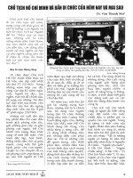
Fig. 1. Structural features of RNAP clasping
minimal nucleic acid sca ffold. (A) Minimal
nucleic acid scaffold (MS) used in th is study.
(B) Model of MS in c om plex with Taq core
RNAP [9]. MS (ball and stick representation:
nontemplate DNA strand, black; template
DNA strand, light violet; RNA, red) is placed
inside the main RNAP c hannel. Also shown
are active site m agnesium (light blue),
rifampicin (orange) clashing w ith RNA,
b¢ F-bridge h elix (green), rudder (red), b¢
coiled-coil r-subunit binding protrusion
(dark violet), b flexible flap (blue; region cor-
responding to Eco amino acids 885–914 is
showninred),andb¢ elements from the
downstream part of the main channel (j aw and
a part of t he clamp, light blue; W217His
6
insertion site is red). The b2(lobe)domainof
the b-subunit (amino acids 174–314, corres-
ponding to Eco186–433) located above the
MS is outlined by a thick brown line. The
secondary channel is located just behind the
F-bridge. The location of the GreB binding
site [15] is s hown schematically as a yellow
oval. The ochre contour corresponds to the
r-subunit, the position of which was taken
from the T. thermophilus holoenzyme struc-
ture [8]. r-Induced conformational changes
are not shown. The semitransparent area
shows the position of r region 3.2.
4922 A. Kulbachinskiy et al. (Eur. J. Biochem. 271) Ó FEBS 2004
mechanism of transcription and conformational flexibility
of RNAP.
Materials and methods
Proteins
Eco core RNAP with a H is
6
tag in t he C terminus of the
b¢-subunit and the r
70
-subunit were purified as described
[35,36]. Eco core RNAP bearing the insertion of six histidine
residues at position 217 of the b¢-subunit was reconstituted
in vitro from individual subunits [37]. Mutant Eco core
enzymes with deletions of the b2(bD186–433) and the
flexible flap ( bD885–914) domains were kindly provided by
K. Severinov and K. Kuznedelov [38,39]. Thermus aquaticus
(Taq) core RNAP was purified from Eco cells expressing all
four core subunits from plasmid pET28ABCZ as described
[40]. The GreB protein was a generous gift of S. Borukhov
(The State University of Ne w York).
Selection of aptamers to Eco core RNAP
A ssDNA library (Fig. 2 A) was purchased from Operon
Technologies Inc. The amounts of ssDNA and the core
enzyme varied f rom 5 nmol and 100 pmol, respectively, in
the first ro und of selection to 100 pmol and 10 pmol in
subsequent rounds. Prior to each round of selection, a
10-pmol aliquot of ssDNA was labeled with -[
32
P]ATP[cP]
(7000 Ci Æmmol
)1
,ICN,CostaMesa,CA,USA)andT4
polynucleotide kinase (New England BioLabs, Beverly,
MA, USA), purified by 10% P AGE and added to t he bulk
DNA sample to monitor the binding of the library to
RNAP. ssDNA was then diluted in 1 mL binding buffer
(20 m
M
Tris/HCl pH 7.9, 10 m
M
MgCl
2
,300m
M
NaCl,
30 m
M
KCl; in subsequent rounds NaCl and K Cl concen-
trations were increased to 400 and 40 m
M
, respectively),
heated for 5 min at 95 °C and cooled rapidly to 0 °C. The
DNA solution was passed through a 50-lLNi
2+
–nitrilo-
triacetic acid–agarose (Qiagen) microcolumn p re-equili-
brated with the binding buffer. The core enzyme w as then
added to the solution and the mixture was incubated for
15 min at room temperature. Thirty microliters Ni–nitrilo-
triacetic acid–agarose was added and the incubation was
continued for a further 20 m in with occasional shaking. The
solution containing unbound DNA was r emoved and t he
sorbent was washed two to four times with 1 mL of binding
buffer (for a total time of 30–60 min). ssDNA–RNAP
complexes w ere e luted with 300 lL binding buffer contain-
ing 200 m
M
imidazole. The solution was treated with
300 lL phenol and 300 lL chloroform. DNA was ethanol
precipitated, dissolved in water and amplified using V ent
DNA polymerase (New England BioLabs) and primers
corresponding to fixed regions of the initial library
(5¢-GGGAGCTCAGAATAAACGCTCAA-3¢ and BBB-
5¢-GATCCGGGCCTCATGTCGAA-3¢, w here B is a bio-
tin r esidue). Two DNA strands were separated b y s ize o n
10% denaturing PAGE, the nonbiotinilated strand was
eluted an d used for the next SELEX round. In the Rif-
directed SELEX experiment each round of the selection
included two successive partitioning steps. The initial
selection of oligonucleotides was carried out as described
above. DNA eluted f rom the complexes w ith RNAP w as
treated with phenol and chloroform and ethanol precipi-
tated. The resulting enriched library was incubated with the
core enzyme (taken in twofold excess relative to the first
selection step) in the p resence of 2 0 lgÆmL
)1
Rif (rifamycin
SV, S igma, St Louis, MO, U SA). DNA–protein complexes
were adsorbed on Ni
2+
–agarose and discarded, while
unbound oligonucleotides remaining in the solution were
ethanol precipitated, PCR amplified and used in the next
SELEX round. After the final round of selection, the
enriched libraries were amplified with primers containing
EcoRI and HindIII sites and cloned into the pUC19
plasmid. The sequences of individual aptamers were
determined using the standard sequencing protocol. Indi-
vidual ssDNA aptamers were obtained by PCR with the
primers corresponding to aptamer flanks; the DNA strands
Fig. 2. Selection of aptamers to Eco core RNAP. (A) Random ssDNA
library used in selection experiments. (B) The effect of Rif on the
binding of rou nd 11 librarie s (0.1 n
M
)toEco core RNAP (10 n
M
)in
binding bu ffer containing 440 m
M
salt. Binding was measured as
described i n M ate rials an d methods. One hundred pe r c ent corres-
ponds to t he binding in t he absence of Rif. ( C) Sequences of repre-
sentative aptamers f rom 13 different c lasses described. Shown are the
central 32-nt-lo ng r egio ns of the aptam ers. Ap tam er E 3 contains a
T fi A change at the first position of the right constant region;
aptamer E13 contains a single nucleotide deletion at the same site. The
sequence motif identical in aptamers E9 and E12 is underlined.
Ó FEBS 2004 Aptamer probes of RNA polymerase structure (Eur. J. Biochem. 271) 4923
were separated on denaturing PAGE as described above.
Control experiments demonstrated that aptamers did not
bind to the Ni-affinity sorbent and therefore the SELEX
protocol was highly efficient in selecting specific aptamer
sequences.
Quantitation of the binding of aptamers to RNAP
Determination of K
d
values f or the binding of oligo-
nucleotides to RNAP was achieved b y using the nitro-
cellulose filtration method as described [41]. All
measurements were performed in binding b uffer contain-
ing 400 m
M
NaCl and 4 0 m
M
KCl unless o therwise
indicated. A 5 ¢-end labeled o ligonucleotide ( 0.003 n
M
)
was incubated with a series of dilutions of core RNAP
(from 0.0 1 to 100 n
M
) in binding buffer containing
50 lgÆmL
)1
BSA for 45–60 min at 22 °Candthen
filtered t hrough 0.45-lm nitrocellulose filters (HAWP,
Millipore) prewetted in the same buffer. The filters were
washed with 5 mL buffer and quantified on a Phosphor-
Imager (Molecular Dynamics, Sunnyvale, CA, USA). K
i
measurements were car ried out at fixed core (1–3 n
M
)
and aptamer (0.1 n
M
) concentrations. Rif, r
70
or GreB
were included in the binding reactions 5 m in prior to the
addition of oligonucleotides; the samples were incubated
for 1 h a t room temperature and passed through
nitrocellulose filters. K
d
and K
i
values were calculated
from the binding curves using
KALEIDAGRAPH
software
(Synergy, Reading, PA, USA). To measure dissociation
kinetics of RNAP–aptamer complexes, the core polym-
erase (3 n
M
) was preincubated with a labeled aptamer
(0.1 n
M
) for 60 min, the complex was c hallenged with the
corresponding unlabeled aptamer (100 n
M
), minimal
nucleic acid scaffold (500 n
M
), Rif ( 2 lgÆmL
)1
), r
70
-
subunit (1 l
M
)orGreB(3l
M
) and aliquots of the
sample were filtered a fter increasing time intervals.
Control e xperiments demonstrated that the level of
RNAP–aptamer binding did not change if the measure-
ments were done in the absence of the inhibitors.
Minimal nucleic acid scaffold (MS)
The sequences of DNA and RNA oligos used to recons-
tituteMSareshowninFig.1A.MSwaspreparedas
described [9]. The RNA oligo (200 p mol, final concentra-
tion 10 l
M
) was labeled with 10 U T4 polynucleotide kinase
and 0.5 mCi [
32
P]ATP[cP], mixed with template and
nontemplate DNA oligonucleotides (final concentrations
of the oligonucleotides were 1, 1 and 2 l
M
, respectively) in
the binding buffer, heated to 65 °C and slowly cooled to
20 °C. Determination of K
d
for the binding of MS to
RNAP was performed as described above. In some cases,
the binding was measured i n the buffer containing 200 m
M
salt (20 m
M
Tris/HCl pH 7.9, 10 m
M
MgCl
2
, 160 m
M
NaCl, 40 m
M
KCl). When s tudying the inh ibitory effect
of aptamers on R NAP activity, Eco core enzyme (10 n
M
)
was added to the mixture of unlabeled MS (10 n
M
)and
aptamers (30 n
M
) in binding buffer containing 400 m
M
NaCl and 40 m
M
KCl. The samples were incubated for
30 min a t room temperature and supplemented with
[
32
P]UTP[aP] (0.1 l
M
,3000CiÆmmol
)1
, Perkin Elmer,
Wellesley, MA, USA). The reaction was stopped after
10 min by the addition of a formamide-containing stop
buffer a nd applied to 23% urea PAGE. The amount of
radioactively labeled 9-nt RNA product was quantified by
using a PhosphorImager.
Results
Selection of aptamers to Eco core RNAP – conventional
vs. site-directed SELEX
Both the c ore a nd holo enzymes of bacterial RNAP bind
nucleic acids [42–45]. While the holoenzyme is able to
recognize specific DNA sequences, the interactions of the
core enzyme with DNA and RNA are generally nonspecific.
There are numerous reports on interactions of the core with
total cellular DNA and RNA [43,46], tRNA [47,48], ssDNA
[43] and also some individual RNA sequences [49]. Repor-
ted K
d
values for some of t hese interactions are in the range
of 10
)8
to 10
)10
M
[43,48,49] and are comparable to the
affinities of known aptamers to their protein targets.
We used a library of 75-nt long ssDNA containing a 32-nt
central region of rand om sequence to select aptamers that
would s pecifically interact with the RNAP core ( Fig. 2A).
We found that at low ionic strength, molecules from the
unenriched library bound the core enzyme very tightly
(K
d
0.2 n
M
at 40 m
M
salt). Such a high level o f nonspe-
cific affinity of RNAP to nucleic acids could be a serious
obstacle for the selection of specific aptamer sequences.
However, we observed that the nonspecific binding of
ssDNA to core RNAP was considerably reduced at
increased ionic strength (K
d
> 100 n
M
at 300 m
M
salt).
Therefore, we performed all selection procedures at elevated
monovalent salt concentrations (300–440 m
M
).
We conducted two types of experiments to select
aptamers to Eco core RNAP. In the first type of experiment
(I), the SELEX procedure was performed in a conventional
way. In b rief, in each round of the selection the ssDNA
library was in cubated with c ore RNAP immobilized on a
Ni-affinity sorbent via th e h exahistidine tag p resent a t the
C-terminal end of the b¢-subunit. Then un bound DNA
was extensively washed out to select se quences that formed
stable complexes with RNAP. RNAP–DNA complexes
were eluted with imidazole, recovered oligonucleotides were
amplified by PCR and used in the next r ound of selection.
To avoid s election of nonspecific sequences that bind to the
affinity sorbent u sed in the reaction, the library was passed
through Ni–agarose column in the absence of RNAP before
each SELEX round.
The second type of experiment (II) aimed to identify
ligands th at bound specifically to the Rif-bin ding pocket of
RNAP. Rif is one of the most potent inhibitors of the
enzyme and is used as a drug in the therapy of several
infectious diseases. H owever, a l arge number o f mutations
in core RNAP conferring resistance to this drug have been
described. Identification of new ligands that can mimic the
effect of Rif is therefore of great importance. Each round of
site-directed SELEX consisted o f two consecutive binding
reactions. First, we selected sequences that bound to free
core RNAP. Seco nd, DNA mole cules that i nteracted with
RNAP were incubated w ith core R NAP in the presence of
excess Rif. DNA molecules that were unable to bind RNAP
in complex with Rif were used in the next round of selection.
4924 A. Kulbachinskiy et al. (Eur. J. Biochem. 271) Ó FEBS 2004
After 11 rounds of selection, the enriched libraries
obtained by both protocols bound core polymerase with
high affinity (K
d
5n
M
in binding buffer containing
440 m
M
salt) but exhibited s ubstantially different s ensitivity
to Rif addition (Fig. 2B). While the R NAP binding of the
ÔconventionallyÕ enriched library was essentially resistant to
Rif, binding of the site-specifically selected library was
severely inhibitedby the antibiotic.Both libraries were cloned
and 50 individual clones were sequenced in each case.
Analysis of individual clones allowed us to identify 13
different classes of sequences, designated E 1–E13 (Fig. 2C;
the total number of clones within each class is shown i n
Table 1). Each class consisted of several clones w ith identical
or closely r elated sequences. Sequences from classes E 1–E4
were found only in the conventionally enriched library,
sequences from classes E5–E8 were p resent in both types of
libraries and sequences from classes E9–E13 were unique to
the library obtained by Rif-directed selection. All of the
aptamers were predicted to f old into distinct secondary
structures, s uch as h airpins and G-quartets(e.g. aptamers E1,
E3, E4, E5) ( data not shown). O ne aptamer representative
of each class was chosen for further investigation (Fig. 2C).
Aptamers bind Eco core RNAP with high affinity and
inhibit the enzyme’s activity
Individual aptamers from all 1 3 classes proved to be high-
affinity ligands to Eco core RNAP with apparent K
d
values
ranging from 0.13 n
M
for aptamer E1 to 6.3 n
M
for aptamer
E8 at 440 m
M
salt (Table 1). These affinities are comparable
to the affinity of Rif to Eco core polymerase [50,51] and
greatly exceed those of other small molecule ligands of
RNAP such as streptolydigin [52], microcin J25 [20] and
CBR703 [21]. Neither the initial library nor any other
nonspecific oligonucleotide t ested appreciably bound
RNAP at these conditions. All of the a ptamers c ompeted
with each other for the binding to core RNAP which
indicated that they interacted with overlapping sites o n the
RNAP surface (data not shown).
We compared the RNAP–aptamer complexes with a
complex o f the RNAP core bound to the minimal nucleic
acid scaffold (MS) (Fig. 1 A) – a model of the elongation
complex [34]. The contacts of MS with Eco core RNAP
were mapped previously by nucleic acid–protein crosslink-
ing t echniques a nd the results were used to position MS on
the t hree-dimensional structure of Taq core RNAP
(Fig. 1 B) [9]. The interaction of MS with RNAP was
shown to be independent of the MS sequence [9,13]. The
MS used in our study consisted of an 18-nt-long down-
stream DNA duplex and an 8 -nt-long RNA–DNA hetero-
duplex separated by two unpaired DNA bases (Fig. 1A).
Unlike the aptamers, MS bound both Eco and Taq core
RNAPs with comparable affinities (with a K
d
value of
1n
M
in binding buffer containing 40 m
M
salt). The
complex of MS with Eco core polymerase was transcrip-
tionally active at both low (40 m
M
) a nd high (440 m
M
)salt
concentrations (data not shown). Remarkably, the a ffinity
of MS to RNAP at 440 m
M
salt (K
d
‡ 50 n
M
)waslower
than the affinities of the a ptamers at these conditions.
All selected a ptamers competed w ith MS f or binding to
core RNAP and efficiently inhibited R NAP activity in t he
transcription assay (most probably b y p reventing the
formation of the RNAP–MS complex, see below) (Fig. 3).
The inhibition of the core polymerase activity by aptamers
was specific as much weaker inhibition was observed in the
case of the initial random library (Fig. 3).
Aptamers interact with distinct sites inside the main
channel of core RNAP
In order to locate the aptamer binding sites more p recisely
we checked the ability of t he aptamers to interact with Eco
core RNAP bearing insertion–deletion mutations in several
sites o n the periphery of the main channel (Table 1 and
Fig. 1B). The mutations were a d eletion of the flexible fl ap
domain in the b-subunit (bD885–914), a deletion of the
domain b2intheb-subunit (bD186–433) and an insertion of
six histidine residues at position 217 of the b¢-subunit
Table 1. Properties of the aptamers to Eco core RNAP. K
d
values were measured in the binding buffer contain ing 400 m
M
NaCl and 40 m
M
KCl.
Aptamer SELEX
Clones (n)
K
d
(n
M
)
Binding to mutant RNAPs
a
Inhibition by
III Dflap D186–433 WHis
6
Rif r
b
GreB
c
E1 I 10 – 0.13 +/– – + – 8.1 2.5
E2 I 16 – 1.04 + – +/– – 19.9 3.2
E3 I 12 – 1.23 + – – – 28.8
E4 I 6 – 2.23 + – +/– + + 6.3
E5 I + II 2 3 0.72 + +/– +/– + 10.4 3.3
E6 I + II 2 4 1.62 + – +/– + +
E7 I + II 2 1 2.21 + + + + + 1.1
E8 I + II 1 4 6.32 + – + + +
E9 II – 8 1.43 + – – + 14.2 5.5
E10 II – 4 2.82 + – +/– + + 6.5
E11 II – 5 3.56 +/– – + + +
E12 II – 9 4.00 + – +/– + + 5.8
E13 II – 7 4.93 + – + + +
a
The increase in K
d
for aptamer binding to mutant variants of core RNAPs over K
d
values for the wild-type enzyme: +, 1–5 times; +/),
5–20 times; –, more than 20 times.
b
The increase in K
d
for aptamer binding to the core polymerase in the presence of 0.5 l M r-subunit: +,
approximate change in K
d
is 10–30 times.
c
The increase in K
d
for aptamer binding in the presence of 1.5 lM GreB. Blank cells, no data.
Ó FEBS 2004 Aptamer probes of RNA polymerase structure (Eur. J. Biochem. 271) 4925
(b¢W217His
6
). The aptamers differed in their affinity to the
mutants (Table 1 ). The flap deletion had the least pro-
nounced effect on the interactions of the aptamers with
RNAP, significantly affecting the binding of only two of
them, E1 and E11 (their K
d
values were increased 5.6- and
11.2-fold, respectively). In c ontrast, the binding of mos t of
the aptamers was disturbed severely by the b2 domain
deletion (for example, K
d
for E9 increased about 250-fold)
and the only a ptamer that bound this mutant with
considerable affinity was E7. The most interesting results
were obtained with t he b¢W 217His
6
insertion mutant. While
some of the aptamers (E13, E8) interacted with the mutant
with unchanged affinity, binding of the others was weak-
ened to different degrees (Table 1). The strongest effect was
for aptamer E3 (K
d
increased 10 0-fold). The simplest
interpretation of the observed effects is that the regions of
RNAP changed by the mutations are parts of the aptamers’
binding sites.
Effect of rifampicin on the binding of aptamers
Rif binds near the RNAP active center at the so-called Rif-
binding pocket of the b-subunit and sterically prevents the
synthesis of RNAs longer than a dinucleotide (Fig. 1B). Rif
also prevents the binding of MS to the core enzyme [13]. We
confirmed this result and found that Rif inhibited MS
binding with an apparent K
i
of < 0.5 n
M
(Fig. 4 ). This
value is in g ood agreement with earlier r eports on Rif K
d
for binding to RNAP (0.5–2 n
M
) [50,51].
Rif exhibited different effects on the interaction of various
aptamers with RNAP (Table 1) . The binding of all the
aptamers obtained through Rif-directed selection (E5–E13)
was inhibited by Rif with th e same e fficiency as the binding
of MS (these aptamers were therefore c alled RifS, for R if-
sensitive, aptamers , T able 1 and Fig. 4). In c ontrast, m ost
of the a ptamers unique to the conventional selection
procedure (E1–E3) were insensitive to Rif (RifR, for Rif-
resistant, aptamers, Table 1 and Fig. 4) and only one of
them (E4) was found to be RifS. RifS sequences from classes
E5–E8 which were identified in both selection experiments
comprised only a small fraction of all sequences in the first
SELEX population (Table 1). Thus, conventional S ELEX
produced mainly RifR aptamers whe reas Rif-directed
SELEX succeeded in identifying only RifS sequences. T he
high efficiency of the site-directed SELEX protocol used in
our work suggests that similar procedures can be used to
obtain h igh a ffin ity a ptamers to antibiotic-binding sites of
many proteins of interest.
We repeated the binding assay using Rif-resistant core
RNAP carrying an S531F substitution in the b-subunit.
In this case, the effect of Rif was much weaker with
K
i
0.5 l
M
(Fig. 4). At the same time, the mutation did
not affect the binding of aptamers. Thus, t he core mutation
conferring Rif resistance w eakened Rif binding to RN AP
by more than three orders of magnitude while having little
or no effect on RNAP–aptamer i nteractions.
The r
70
-subunit and GreB suppress the interaction
of the core RNAP with aptamers
The r
70
-subunit inhibited the binding of all the aptam ers to
the core polymerase. Apparent K
d
s for the binding of
different a ptamers t o the holoenzyme of RNAP were
increased in the range 8–30 times in comparison with those
for the core enzyme (Table 1). When th e binding of the E2
aptamer was measured at fixed core and increasing r
70
-
subunit concentrations, r inhibited the interaction w ith an
observed K
i
of 10 n
M
(Fig. 5 A). This value apparently
corresponded to K
d
for the r
70
–core interaction at these
conditions. The r
70
-subunit also suppressed the interaction
of the core enzyme with MS (Fig. 5A). This result is in
agreement with previous studies which demonstrated that
the binding of r and RNA in the elongation complex was
Fig. 3. Inhibition of th e Eco core polymerase activity by aptamers.
RNAP activity was measured as described in Materials and methods.
The core enzyme was added to the mixture of MS and aptamers in
binding buffer containing 440 m
M
salt and transcription was initiated
by add ing [
32
P]UTP[aP]. T he amount of radio active ly l abele d
9-nt-long RNA product was quantified and normalized to the activity
in the a bsence of the i nhib itor. I, Aptamers found o nly in the con -
ventional selection experiment; II, aptamers unique to the Rif-directed
experiment; I + II, aptamers identifiedinbothselections;N,the
initial library.
Fig. 4. Effect of Rif on the binding of aptamers and MS to core RNAP.
Binding reactions contained 10 n
M
ofthecoreenzyme,0.1n
M
oligo-
nucleotides and varied amounts of Rif. Monovalent salt concentration
in the binding buffer was 440 m
M
inthecaseofaptamersand200m
M
in the case o f MS. Bind ing w as measured as described in Materials and
methods and n ormalized t o t he binding in the absence of Rif. T he
experiment was performed with the wild-type core enzyme (S) a nd Rif-
resistant mutant R NAP (S531F, R).
4926 A. Kulbachinskiy et al. (Eur. J. Biochem. 271) Ó FEBS 2004
mutually exclusive [53,54]. In the three-dimensional struc-
ture of the holoenzyme polymerase, region 3.2 of r seems to
clash with the 5¢ end of growing RNA during initiation
(Fig. 1 B) [7,8]. Thus, it is po ssible that r
70
interferes w ith
MS binding by competing with its RNA component for the
same site on core RNAP.
GreB exerts its effect on the elongation complex in a
backtracked state stimulating the nuclease activity of the
RNAP active center [55]. We f ound that the b inding of MS
to the core polymerase was not affected by GreB. GreB also
failed to stimulate the cleavage of the RNA c omponent of
MS (data not shown). This, as well as resistance of MS to
pyrophosphorolysis (N. Korzheva, personal communica-
tion), suggested that MS was captured b y RNAP in a post-
translocated state. At the same t ime, GreB suppressed the
interaction of Eco RNAP with all the aptamers tested except
E7, increasing their apparent K
d
values three- to sixfold,
when present at 1.5 l
M
(Table 1). The weaker effect of
GreB in comparison with the r-subunit is p robably due to
its lower affinity to core RNAP. Indeed, t he increase of
GreB concentration resulted i n complete inhibition of
aptamer binding (Fig. 5B). The apparent K
i
value for GreB
action calculated from the inhibition curve was 100 n
M
.
This value is in goo d agreement with K
d
reported for the
GreB–core interaction [56].
MS, Rif and the r
70
-subunit do not affect the stability
of RNAP–aptamer complexes while GreB promotes their
rapid dissociation
To investigate the nature of the effects of MS, Rif, r
70
and
GreB on RNAP–aptamer interactions, we measured the
dissociation k inetics o f several RNAP–aptamer comple xes
in the p resence of these ligands (Fig. 6 ). When the
complexes containing radioactively labeled aptamers were
Fig. 5. Inhibition of aptamer b in ding by the r
70
-subunit a nd GreB. (A)
Inhibition of t he binding of aptamer E2 and MS (0.1 n
M
)tothecore
polymerase (1 and 2 n
M
, respectively) by increasing concentrations of
the r-subunit. Binding buffer cont ained 440 m
M
salt in the case of the
aptamers and 200 m
M
salt in the case o f MS. (B) Inhibition of t he
binding of aptamer E9 (0.03 n
M
) to the core enzyme (3 n
M
)by
increasing am ounts o f G reB. Binding was measure d in buffer con-
taining 440 m
M
salt.
Fig. 6. Dissociation kinetics of RNAP–aptamer complexes in the pres-
ence of various competitors. The core enzyme was preincubated with a
labeled aptamer and the complex was challenged with the corres-
ponding unlabeled aptamer, MS, Rif, the r
70
-subunit or GreB.
Aptamer binding was measu red in buffer containing 440 m
M
salt. The
dissociationkineticsisshownforaptamersE4(A),E7(B)andE10(C).
Ó FEBS 2004 Aptamer probes of RNA polymerase structure (Eur. J. Biochem. 271) 4927
incubated with an excess of the corresponding unlabeled
aptamers, they dissociated with half-life times of more than
1 h . T he dissociation k inetics of t he RNAP–aptamer
complexes measured in the presence of MS, Rif (in case of
RifS aptamers) o r the r-subunit followed the kinetics
observed when the unlabeled aptamer was used as a
competitor (Fig. 6). Control e xperiments demonstrated
that when these ligands were added to RNAP before the
aptamers, they completely suppressed complex formation
(data not shown).
In contrast, GreB greatly reduced the s tability of several
RNAP–aptamer complexes (Fig. 6 and data not shown). In
agreement with K
d
measurements, GreB d id not affect the
stability of t he E7–RNAP complex (Fig. 6B). At the same
time, when GreB w as added to the preformed complexes of
RNAP with E4 and E10 aptamers, it caused their rapid
dissociation; half-life times of the complexes were reduced
by more than 10 times (5 min in comparison with > 1 h
when the kinetics was measured without GreB) (Fig. 6A
and C). The residual binding of aptamers measured at large
time intervals corresponded to the maximum inhibition
observed when GreB w as added b efore the aptamers
(Fig. 5 B a nd data not shown).
Specific and nonspecific interactions of aptamers
with the RNAP main channel
The interaction of the a ptamers with RNAP w as found to
be highly dependent on the ionic s trength o f t he solution.
At elevated ionic strength (440 m
M
), the binding of the
aptamers was very sequence specific as even point mutations
of aptamers’ sequences disrupted their interaction with
RNAP. The aptamers were also specific to Eco core RNAP
and neither of them bound Taq RNAP (data not shown).
At lower ionic strength (< 200 m
M
), RNAP still bound
the aptamers but sequence s pecificity w as apparently lost.
Under these conditions all the sequences tested, including
the random DNA libr ary, bound the core enzyme with equal
affinities (K
d
1n
M
). MS suppressed the binding of all the
oligonucleotides which suggested that the nonspecific bind-
ing of s sDNA also oc curred a t RNAP sites involved in the
interaction with RNA and DNA in the elongation complex.
At elevated ionic strength, Rif a nd r
70
suppre ssed
RNAP–aptamer interactions (above). Under low ionic
strength conditions, Rif and r
70
hadnoeffectonthe
binding of RifS aptamers to core RNAP (data not shown).
Therefore, the structure of nonspecific complexes of RNAP
with the aptamers differs from the structure of the
complexes formed at high ionic s trength.
Discussion
The principal result of this work is that the aptamers sense
the interaction of RNAP with various ligands, including
nucleic acids, an tibiotics and protein f actors. Based on the
mechanism of the inhibition of aptamer binding, these
ligands can be divided into two groups. The minimal
nucleic acid scaffold, Rif and the r
70
-subunit seem to
inhibit RNAP–aptamer interactions by steric blocking of
the aptamer binding sites on the RNAP molecule, while
GreB is likely to affect aptamer binding in an allosteric
manner.
Several facts indicated that the aptamers interact with the
main channel of RNAP where nucleic acids in natural
transcription complexes are held. All of t he aptamers
competed with MS for binding to RNAP and inhibited core
polymerase activity. Binding of the aptamers w as affected
by mutations at several sites in th e main channel t hat were
previously implicated in the interactions with nuc leic acids
in transcription c omplexes. F urthermore, the binding of 10
out of 13 aptamers was sensitive to Rif. As Rif does not
cause any sign ificant conformational changes of the core
polymerase [13], its effect must result from direct compe-
tition with aptamers for the Rif pocket of the b-subunit.
Finally, the dissociation kinetics of t he RNAP–aptamer
complexes measured in the presence of MS and Rif followed
thesametimecourseasthekineticsmeasuredinthe
presence of the unlabeled aptamers. This indicated that
these ligands acted by simple trapping of free RNAP and
preventing reassociation o f the complexes. Thus, both MS
and R if are likely to compete with the aptamers for the
binding sites in the main channel.
The r-subunit also binds within the main channel of
RNAP. The main docking sites of r on the core polymerase
include the clamp domain of b¢ and the flexible flap domain
of the b-subunit (Fig. 1B) [ 7,8]. I n addition, the N-terminal
region of r, which is not visible in the holoenzyme structure,
was shown to occupy the downstream portion of the m ain
channel [57]. The binding of r to the core polymerase causes
repositioning of several structural modules of the core,
including the c lamp, b1, b2 and flap domains, w hich results
in partial c losure of the main channel [ 7]. T hus, the
inhibition of aptamer binding by r could occur by both
steric and allosteric mechanisms. We found that, similarly to
MS and Rif, r did not affect the dissociation rate of
RNAP–aptamer complexes. Thus, the most likely inter-
pretation of t he inhibitory effect of r is that it also directly
blocks RNAP sites involved i n a ptamer binding. The steric
competition between aptamers and r is not surprising, when
taking into account the extensive interaction interface
between r and the core polymerase. Hopefully, further
studies of mutant variants of r as well as testing various
alternative r-subunits will help to establish the regions of r
which are responsible for the inhibition of aptamer binding.
In contrast to MS, Rif and the r-subunit, GreB
dramatically increased the dissociation rate of RNAP–
aptamer complexes and therefore actively disrupted
RNAP–aptamer interactions. As opposed to r
70
,GreB
binds RNAP from the s econdary channel side of t he
enzyme, i.e. at the side opposite to the aptamers (see
Fig. 1B) [15]. The binding site of the C-terminal do main of
GreB near the entrance of the secondary channel is located
outside of the enzyme’s catalytic cleft and s eems unlikely t o
be involved in aptamer binding. The GreB N-terminal
coiled-coil domain protrudes deep i nto the secondary
channel, providing two conserved acidic residues which
play a k ey role in the RNA cleavage reaction [15,16,58].
Based on these observations, one could s uggest two
mechanisms of GreB action on the binding of the aptamers.
One possibility is that the aptamers bound in the main
channel might occupy the mouth o f the secondary channel
and directly interfere w ith GreB b inding. Alternatively, t he
aptamers could sense GreB-induced conformational chan-
ges inside the RNAP main channel.
4928 A. Kulbachinskiy et al. (Eur. J. Biochem. 271) Ó FEBS 2004
The strong stimulatory effect of GreB on the disso-
ciation of RNAP–aptamer complexes provides serious
evidence in support of the allosteric mechanism of GreB
action. Sensing of G reB binding by several aptamers,
each interacting with RNAP in a different way, as well
as different strengths of GreB effect on various aptamers
(Table 1) is also consistent with the allosteric mechanism.
Our data thus give evidence that the interaction of GreB
with RNAP may result in structural changes of the core
polymerase. The resolution o f c urrent structural data
does not allow us to verify such changes [15]. However,
conformational rearrangements in the main channel were
observed in the complex of yeast RNAPII with elonga-
tion factor TFIIS, which also protrudes into the secon-
dary channel and se ems to utilize very similar
mechanisms to stimulate RNA cleavage [59]. GreB-
induced conformational changes of RNAP detected with
the aptamers may be essential for the stimulation of the
endonuclease activity o f the enzyme.
Recent studies demonstrated that other protein factors
(e.g. DksA) and antibiotics (microcin) also bind RNAP
within the secondary channel and seem to affect RNAP
conformation [60–62]. We propose that the aptam ers
could be used to study the conformational changes of
RNAP induced by the b inding of these r egulatory
factors. The aptamers could also be useful in studies of
various RNAP mutations that are thought to change the
conformation of th e enzyme. The examples of such
mutations i nclude the substitution at position 934 near
the F-bridge helix in the b¢-subunit that was proposed to
shift the conformation of the F-bridge toward the bent
form [14] and mutations on the surface of the b-subunit
that impair Q-pro tein med iated anti-termination ( pre-
sumably by changing the conformation of the interior of
the main channel) [63]. It should be noted that such
hypothetical conformational c hanges of RNAP are
usually very difficult to verify. The aptamers thus
represent a very useful tool to probe RNAP structure
in many experimental systems.
Acknowledgements
We thank K. Severinov for protein and plasmid samples and for
reading the manuscript, K. Kuzned elov and S. Borukhov f or materials,
A. Stolyarenko for reading the m anuscript. A.K. is especially grateful
to N. Korzheva, V. Epshtein and A. Mustaev for help in doing some
experiments. This work was supported by the NIH grant GM30717 to
A.G. and by the Russian Foundation for Basic Research grant 02-04-
48525.
References
1. Gelles, J. & Landick, R. (1998) RNA polymerase as a molecular
motor. Cell 93, 13–16.
2. Erie, D.A. (2002) The many conformational states of RNA
polymerase elongation complexes and their roles in the regulation
of transcription. Biochim. Biophys. Acta 1577, 224–239.
3. Korzheva, N. & Mustae v, A. (2003) RNA and DN A p olymerases.
In Molecular Motors (Schliwa, M., ed.), pp. 153–177. Wiley, John
& Sons Inc., Hoboken, NJ, USA.
4. Zhang, G., Campbell, E.A., Minakhin, L., Richter, C., Severinov,
K. & D arst, S.A. (1999) Crysta l structure of Thermus aquaticus
core RNA polymerase at 3.3 A
˚
resolution. Cell 98 , 811–824.
5. Cramer, P., Bushnell, D.A. & Kornberg, R.D. ( 2001) Structural
basis of transc ription: RNA polymerase II at 2.8 angstrom
resolution. Science 292 , 1863–1876.
6.Gnatt,A.L.,Cramer,P.,Fu,J.,Bushnell,D.A.&Kornberg,
R.D. (2001) Structural basis of transcription: an RNA po lymerase
II elongation complex at 3.3 A
˚
resolution. Science 292, 1876–
1882.
7. Murakami,K.S.,Masuda,S.&Darst,S.A.(2002)Structuralbasis
of transcription initiatio n: RNA p olymerase h oloenzyme at 4 A
˚
resolution. Science 296 , 1280–1284.
8. Vassylyev, D.G., S ekine, S., Laptenko, O., L ee, J., Vassylyeva,
M.N., Borukhov, S. & Yokoyama, S. (2002) Crystal structure
of a bacterial RNA polymerase holoenzyme at 2.6 A
˚
resolution.
Nature 417, 7 12–719.
9. Korzheva, N., Mustaev, A., K ozlov, M., M alhotra, A., Nikiforov,
V., Goldfarb, A. & Darst, S.A. (2000) A structural model of
transcription elongation. Science 289, 619–625.
10. Naryshkin, N., Revyakin, A., Kim , Y. , M ekler, V. & Ebright,
R.H. (2000) Structural organization o f the RNA polymerase-
promoter open complex. Cell 101, 601–611.
11. Murakami, K.S., Masuda, S., Campbell, E.A., Muzzin, O. &
Darst, S.A. (2002) Structural basis of transcription initiation: an
RNA polymerase holoenzyme-DNA complex. Science 296, 1285–
1290.
12. Darst, S.A. (2001) Bacterial RNA polymerase. Curr. Opin. Struct.
Biol. 11, 155–162.
13. Campbell, E.A., Korzheva, N., M ustaev, A ., Mur akami, K., Nair,
S., Goldfarb, A. & Darst, S.A. (2001) Structural mechanism for
rifampicin inhibition of bacterial RNA polymerase. Cell 104,
901–912.
14. Epshtein, V., Mustae v, A., Markovtsov, V., Bereshchenko, O .,
Nikiforov, V. & Goldfarb, A. (2002) Swing-gate model of
nucleotide entry i nto the R NA po lymerase a ctive cente r. Mol. Cell
10, 623–634.
15. Opalka, N., Chlenov, M., Chacon, P., Rice, W.J., Wriggers, W. &
Darst, S.A. (2003) Structure and function of the transcription
elongation factor GreB bound to bacterial RNA polymerase. Cell
114, 335–345.
16. Sosunova, E., Sosunov, V., Kozlov, M., Nikiforo v, V., Goldfarb,
A. & M ustaev, A. (2003) Don ation of catalytic r esidues to RNA
polymerase active c enter by transcription factor Gre. Proc. Natl
Acad. Sci. USA 100 , 15469–15474.
17. Darst, S.A., Opalka, N., Chacon, P., Polyakov, A., Richter, C.,
Zhang, G. & Wriggers, W. (2002) Conformational flexibility
of bacterial RNA polymerase. Proc. Natl Acad. Sci. USA 99,
4296–4301.
18. Landick, R. (2001) RNA polymerase clamps down. Cell 105,
567–570.
19. Bushnell, D.A., Cramer, P. & Kornberg, R.D. (2002) Structural
basis of transcription: alpha-amanitin-RNA polyme rase II
cocrystal at 2.8 A
˚
resolution. Proc. Natl Acad. Sci. USA 99,
1218–1222.
20. Yuzenkova, J., D elgado, M., Nechaev, S ., Savalia, D., Epshtein,
V.,Artsimovitch,I.,Mooney,R.A.,Landick,R.,Farias,R.N.,
Salomon, R. & Severinov, K. (2002) Mutations of bacterial RNA
polymerase leading t o resistance to microcin j25. J. Biol. Chem.
277, 50867–50875.
21. Artsimovitch, I., Chu, C., Lynch, A.S. & Landick, R. (2003) A
new class of bacterial RNA polymer ase in hibitor affects nucle otide
addition. Science 302, 6 50–654.
22. Gold, L., Polisky, B., Uhlenbeck, O. & Y arus, M. ( 1995) Diversity
of oligonucleotide function s. Annu. Rev. Biochem. 64, 763–
797.
23. Tuerk, C., MacDougal, S. & Gold, L. ( 1992) RNA pseudoknots
that inhibit h uman immunodeficie ncy virus type 1 reverse tran-
scriptase. Proc.NatlAcad.Sci.USA89, 6988–6992.
Ó FEBS 2004 Aptamer probes of RNA polymerase structure (Eur. J. Biochem. 271) 4929
24. Jaeger, J., Restle, T. & Steitz, T.A. (1998) The structure of HIV-1
reverse tran scriptase c omplexed with an RNA pseudoknot
inhibitor. EMBO J. 17 , 4535–4542.
25. Dang, C . & Jayasena, S.D . (1996) Oligonucleotide inhibitors o f
Taq DNA polymerase facilitate detection of low copy n umber
targets by PCR. J. Mol. Biol. 264, 2 68–278.
26. Allen,P.,Worland,S.&Gold,L.(1995)Isolationofhigh-affinity
RNA ligands to HIV-1 integrase from a random pool. Virology
209, 327–336.
27. Rowsell, S., Stonehouse, N.J.,Convery,M.A.,Adams,C.J.,
Ellington, A.D., Hirao, I.,P eabody, D.S., Stockley, P.G. &Phillips,
S.E. (1998) Crystal structures of a series of RNA aptamers
complexed to the same protein t arget. Nat. Struct. Biol. 5, 970–
975.
28. Huang, D.B., Vu, D., Cassiday, L.A., Zimmerman, J.M., Maher,
L.J. & 3rd & Ghosh, G. (2003) Crystal structure of NF-kappaB
(p50) 2 complexed to a high-affinity RNA aptamer. Proc. Natl
Acad. Sci. USA 100 , 9268–9273.
29. Gold, L. (1995) Oligonucleotides as research, diagnostic, and
therapeutic agents. J. Biol. Chem. 270, 13581–13584.
30. Brody, E.N. & Gold, L. (2000) Aptamers as therapeutic and
diagnostic agents. J. Biotechnol. 74, 5–13.
31. Hale, S .P. & Schimmel, P. (1996) Pr otein synthesis editing b y a
DNA aptamer. Proc. Natl Acad. Sc i. USA 93, 2755 –2758.
32. Biroccio, A ., Hamm, J ., Incitti,I.,DeFrancesco,R.&Tomei,
L. (2002 ) Se lection of RNA aptamers that are specific and high-
affinity ligands of the hepatitis C virus RNA-dependent RNA
polymerase. J. Virol. 76, 3688–3696.
33. Hamm, J., Alessi, D.R. & Biondi, R.M. (2002) Bi-functional,
substrate mimicking RNA inhibits MSK1-mediated cAMP-
response element-bindin g p rotein p hosphorylation and reveals
magnesium ion-dependent conformational changes of the k inase.
J. Biol. Chem. 277, 45793–45802.
34. Korzheva, N., M ustaev, A., Nudler, E ., Nikiforov, V. & Gold-
farb, A. ( 1998) Mechanistic model of t he elo ngation c omplex o f
Escherichia coli RNA polymerase. Cold Sprin g H arb. Symp Q uant.
Biol. 63, 337–345.
35. Kashlev, M., Martin, E., Polyakov, A., Severinov, K., Nikiforov,
V. & Goldfarb, A. (1993) Histidine-tagged RNA polymerase:
dissection o f t he transcription cycle using immobilized e nzym e.
Gene 130, 9–14.
36. Borukhov, S. & Goldfarb, A. ( 1993) Recombinant Escherichia coli
RNA polymerase: purification o f individually overexpressed
subunits and in vitro asse mbly. Protein Expr. Purif. 4, 503–511.
37. Kulbachinskiy, A.V., Ershova, G.V., K orzh eva, N.V., Brodolin,
K.L. & N ikiforov, V .G. ( 2002) Mutations i n b¢ subunit of the
Escherichia coli R NA polymerase i nfluence i nteraction with the
downstream DNA d u plex i n the elongation comple x. Genetika
[Russian] 38, 1207–1211.
38. Severinov, K. & Darst, S.A. (1997) A mutant RNA polymerase
that forms unusual open promote r complexes. Proc. Natl Acad.
Sci. USA 94, 13481–13486.
39. Kuznedelov, K., Minakhin, L., Niedziela-Majka, A., Dove, S.L.,
Rogulja, D., Nickels, B.E., H ochschild,A.,Heyduk,T.&Sever-
inov, K. (2002) A role for interaction of the RNA polymerase flap
domain with the sigma subunit i n p romoter recognition. Science
295, 855–857.
40. Minakhin, L., Nechaev, S., Campbell, E.A. & Severinov, K.
(2001) Recombinant Thermus aquaticus RNA polymerase, a new
tool for structure-based analysis of transcription. J. Bacteriol. 183,
71–76.
41. Carey, J., Cameron, V., de Haseth, P.L. & Uhlenbec k , O.C. ( 1983)
Sequence–specifi c interaction o f R17 c oat protein w ith its ribo-
nucleic acid binding site. Biochemistry 22 , 2601–2610.
42. Yefimova, L.Y., Knorre, V.L., Savinkova, L.K. & Salganik, R.I.
(1975) Selective binding of oligoribonucleotides by E. coli RNA
polymerase and their effect on DNA-dependent RNA synthesis.
FEBS Lett. 58, 359–362.
43. deHaseth, P.L., Lohman, T .M., Burgess, R.R. & R ecord, M.T.
Jr (1978) Nonspecific interactions of Escherichia coli RNA
polymerase with native and d enatured DNA: diffe rences in the
binding behavior of core and holoenzyme. Biochemistry 17, 1612–
1622.
44. Strauss, H.S., Burgess, R.R. & Record, M.T. Jr (1980) Binding of
Escherichia coli ribo nucleic acid polymerase holoe nzyme to a
bacteriophage T7 promoter-containing fragment: selectivity exists
over a wide range of solution conditions. Biochemistry 19, 3496–
3504.
45. Wheeler, A.R., Woody, A.Y. & Woody, R.W. (1987) Salt-
dependent binding of Escherichia c oli RNA polymerase to D NA
and s pec ific transcription b y the core enzyme and holoenzyme.
Biochemistry 26, 3322–3330.
46. Tissieres, A., Bourgeois, S. & G ros, F. (1963) Inhibition o f RNA
polymerase by RNA. J. Mol. Biol. 7, 100–103.
47. Bremer, H., Yegian, C. & Konrad, M. (1966) Inactivation
of purified E scherichia c oli RNA p olymerase by t ransfer R NA.
J. Mol. Biol. 16, 94– 103.
48. Spassky, A., Busby, S.J., Danchin, A. & Buc, H. (1979) On the
binding of tRNA to Escherichia coli RNA polym eras e. Eur.
J. Biochem. 99, 187–201.
49. Altmann, C.R., Solow-Cordero, D.E. & Chamberlin, M.J. (1994)
RNA cleavage and chain elongation by Escherichia coli DNA-
dependent RNA polymerase in a binary en zyme. RNA complex.
Proc.NatlAcad.Sci.USA91, 3784–3788.
50. Handschin, J.C. & Wehrli, W. (1976) On the kinetics of the
rifampicin-RNA-polymerase complex. Differences between crude
and purified enzyme fractions. Eur. J. Biochem. 66, 309–317.
51. Yarbrough, L.R., Wu, F.Y. & Wu, C.W. (1976) Molecular
mechanism o f t he rifa mpicin–RNA polyme rase interaction. Bio-
chemistry 15, 2669–2676.
52. Heisler, L.M., Suzuki, H., Landick, R. & Gross, C.A. (1993) Four
contiguous amino acids define the target for streptolydigin
resistance in the b eta subunit of Esc herichia coli RNA p olymeras e.
J. Biol. Chem. 268, 25369–25375.
53. Sidorenkov, I., Komissarova, N . & K ashlev, M. ( 1998) Crucial
role of the RNA: DNA hybrid in the processivity of transcription.
Mol. Cell 2, 55–64.
54. Daube, S.S. & von Hippel, P.H. (1999) Interactions of Escherichia
coli sigma (70) within the transcription elongation complex. Proc.
NatlAcad.Sci.USA96, 8390–8395.
55. Borukhov, S., Laptenko, O. & Lee, J. (2001) Escherichia coli
transcript cleavage factors GreA and GreB: functions and
mechanisms of action. Methods Enzymol. 342, 64–76.
56. Koulich, D., Orlova, M., M alhotra, A., Sali, A., Darst, S.A. &
Borukhov, S. (1997) Domain organization of Escherichia coli
transcript cleavage factors GreA a nd GreB. J. Biol . Chem. 272,
7201–7210.
57. Mekler, V., Kortkhonjia, E., Mukhopadhyay, J., Knight, J.,
Revyakin, A., Kapanidis, A.N., Niu,W.,Ebright,Y.W.,Levy,R.
& Ebright, R.H. (2002) Structural organization of bacterial RNA
polymerase holoenzyme and the RNA polymerase-promoter open
complex . Cell 108, 599 –614.
58.Laptenko,O.,Lee,J.,Lomakin,I.&Borukhov,S.(2003)
Transcript cleavage factors GreA and GreB act as transient
catalytic components of RNA polymerase. EMBO J. 22, 6322–
6334.
59. Kettenberger, H., Armache, K.J. & Cramer, P. (2003) Archi-
tecture of t h e RNA polymerase II-TFIIS co mple x and implica-
tions for mRNA cleavage. Cell 114, 347–357.
60. Perederina, A., Svetlov, V., Vassylyeva, M.N., Tahirov, T.H.,
Yokoyama, S., Artsimovitch, I. & Vassylyev, D.G. (2004)
Regulation through t he se co ndary c hannel – structural framework
4930 A. Kulbachinskiy et al. (Eur. J. Biochem. 271) Ó FEBS 2004
for ppGpp -DksA s ynergism during transcription. Cell 118,297–
309.
61. Adelman,K.,Yuzenkova,J.,LaPorta,A.,Zenkin,N.,Lee,J.,
Lis, J.T., Borukhov, S., Wang, M.D. & Severinov, K. (2004)
Molecular mechanism of transcription inhibition by peptide
antibiotic microcin j25. Mol. Cell 14, 753–762.
62. Nickels, B.E. & Hochschild, A. (2004) Regulation of RNA poly-
merase through t he secondary channel. Cell 11 8 , 281–284.
63. Santangelo, T.J., Mooney, R.A ., Landick, R. & Roberts, J .W.
(2003) RN A polymerase m utations that impair co nversion to a
termination-resistant complex by Q antiterminator proteins. Genes
Dev. 17, 1281–1292.
Ó FEBS 2004 Aptamer probes of RNA polymerase structure (Eur. J. Biochem. 271) 4931