Báo cáo khoa học: Trans-splicing of a mutated glycosylasparaginase mRNA sequence by a group I ribozyme deficient in hydrolysis pptx
Bạn đang xem bản rút gọn của tài liệu. Xem và tải ngay bản đầy đủ của tài liệu tại đây (329.68 KB, 7 trang )
Trans
-splicing of a mutated glycosylasparaginase mRNA sequence
by a group I ribozyme deficient in hydrolysis
Eirik W. Lundblad
1
, Peik Haugen
1
and Steinar D. Johansen
1,2
1
Department of Molecular Biotechnology, RNA Research group, Institute of Medical Biology, University of Tromsø, Norway;
2
Department of Fisheries and Natural Sciences, Bodø Regional University, Norway
RNA reprogramming represents a new concept in correcting
genetic defects at the RNA level. However, for the technique
to be useful for therapy, the level of reprogramming must be
appropriate. To i mprove the efficiency of g roup I ribozyme-
mediated RNA reprogramming, when using the Tetrahy-
mena ribozyme, regions complementary to the target RNA
have previously been extended in length and accessible sites
in the target RNAs have been identified. As an alternative to
the Tetrahymena model ribozyme, the D iGIR2 group I
ribozyme, derived from a mo bile group I intron i n rDNA
of the myxomycete Didymium iridis , represents a new and
attractive tool in RNA reprogramming. We r eported
recently that the deletion of a structural element within the
P9 domain of DiGIR2 turns off hydrolysis at the 3¢ splice site
(side reaction) without affecting self-splicing [Haugen, P.,
Andreassen, M., Birgisdottir, A
˚
.B.&Johansen,S.D.(2004)
Eur. J. Biochem. 271 , 1015–1024]. Here we a nalyze the
potential of the modified ribozyme, deficient in hydrolysis at
the 3¢ splice site, for applicatio n in group I ribozyme-medi-
ated trans-splicing of RNA. The improved ribozyme cata-
lyses both cis-splicing and trans-splicing in vitro of a human
glycosylasparaginase mRNA sequence with the same effi-
ciency as the original DiGIR2 ribozyme, but without
detectable levels of the unwanted hydrolysis.
Keywords: glycosylasparaginase mRNA; group I i ntron;
ribozyme hydrolysis; RNA reprogramming; trans-splicing.
Group I ribozyme-mediated RNA r eprogramming by
trans-splicing, has been successfully carried out using the
Tetrahymena ribozyme and various target RNAs [1–5]. The
trans-splicing reaction is similar to the self-splicing reaction
normally catalysed by group I introns [6], except that the 5¢
exon is presented in trans and a corrected 3¢ exon is attached
to the r ibozyme. Ligation o f these exons produces the
chimerical transcript that can be translated into a functional
protein. RNA r eprogramming is guided by a region
complementary to the target RNA (internal guide sequence,
IGS) located within the ribozyme, and the specificity and
efficiency of trans-splicing have mainly been improved by
extending the IGS [2,7–9]. In addition, group I ribozymes
with randomised IGSs are used to identify regions on the
target RNAs that are accessible [1,4,10–13]. I n spite of
recent advances and significant efforts to optimize trans-
splicing reactions, the RNA reprogramming in cells remains
inefficient. Moreover, group I ribozymes, including the
Tetrahymena ribozyme, catalyze additional reactions that
directly c ompete with sp licing and prob ably lower the
efficiency of trans-splicing. Most pronounced is the 3¢ splice
site hydrolysis of precursor RNAs [14–16], which is
catalysed by the Tetrahymena ribozyme at a relatively high
rate [16,17]. Hydrolysis results i n the formation of full-
length intron RNA circles, which are commonly detected
both in vitro and in vivo in a number of group I introns [17–
19]. Designing ribozymes that catalyse little or no competing
side reactions can therefore prove valuable in the search for
better ribozyme tools that can be used in RNA reprogram-
ming.
DiGIR2 i s the splicing ribozyme derived from the
twin-ribozyme g roup I intron D ir.S956-1 i n Didymium
ribosomal DNA (Fig. 1A) [20,21]. DiGIR2 represents the
group IE intron subgroup with clear distinction in
structure c ompared t o t he distantly related Tetrahymena
group IC1 intron [16,18,19]. We recently reported t hat
deletion of the P9.2 paired element in the DiGIR2
ribozyme (Fig. 1B) s ignificantly reduces hydrolytic clea-
vage at the 3¢ splice site without affecting the self-splicing
activity in cis-splicing constructs [16]. The remarkable
loss of unwanted side reactions, apparently without
compromising splicing, identifies the new ribozyme con-
struct (denoted DiGIR2DP9.2) as a potential improved
tool in group I ribozyme-mediated trans-splicing of
RNA. Here we set out to investigate t he ability and
efficiency of DiGIR2 a nd DiGIR2DP9.2 to trans-splice
RNA molecules. Trans-splicing ribozymes were construc-
ted and targeted against a mutated glycosylasparaginase
Correspondence to S. Johansen, Department of Molecular Biotech-
nology, Institute of Medical Biology, University of Tromsø, 9037
Tromsø, Norway. Fax: + 47 77 64 53 50, Tel.: + 47 77 64 53 67,
E-mail:
Abbreviations: AGU, aspartylglycosaminuria; EGS, extended guide
sequence; GA, glycosylasparaginase; IGS, internal guide sequence;
nt, nucleotide; RPA, ribonuclease protection analysis.
Note: The oligonucleotide sequences used in this work are available on
request and as a supplement at the RNA Research Group’s website
at />(Received 15 April 2004, revised 9 August 2004,
accepted 25 October 2004)
Eur. J. Biochem. 271, 4932–4938 (2004) Ó FEBS 2004 doi:10.1111/j.1432-1033.2004.04462.x
(GA) mRNA sequence. Mutations in GA cause the
human lysosomal storage disease aspartylglycosaminuria
(AGU) [22].
Experimental procedures
Plasmid constructions and
in vitro
mutagenesis
The cis -splicing construct p DiGIR2 AGU was made by
combining two different PCR products. The first product
contains the T7 promoter, 15 nucleotides (nt) from the
human GA open reading frame (ORF) and the DiGIR2
splicing ribozyme, and was generated from the pDiGIR2
template [20] using the primer combination OP340/341.
The second product (108 nt) was amplified from a cloned
human GA cDNA template using the primer combination
OP342/346. The two PCR products were blu nted, phos-
phorylated and ligated using the Sure Clone Ligation Kit
(Amersham Biosciences, Piscataway, NJ, USA). Fin ally, a
new PCR product w as generated f rom the ligation mix
using the oligo primers OP341/346 and subsequently cloned
into pUC18. The P9.2 hairpin was deleted from the
pDiGIR2 AGU by using the Quick Change site-directed
mutagenesis kit (Stratagene, Cedar Creek, TX, USA) and
OP296/297, generating pDiGIR2DP9.2 AGU. The trans-
splicing constructs were made by PCR amplification with
Pfu DNA polymerase (Promega, Madison, WI, USA)
using pDiGIR2 AGU and pDiGIR2DP9.2 AGU as
templates, generating the trans-splicing plasmid versions
of pDiGIR2 AGU and pDiGIR2DP9.2 AGU, respectively.
Here, the primer combinations OP1191/1192 and OP1191/
1202 were used. The forward primer w as designed with two
sequences complementary to the target RNA [8 nt IGS and
9 nt extended guide sequence (EGS)] separated by a 5 nt
wobble region. The reverse primers were designed with
alternative codons, of which the first 5 nt in the 3¢ exon are
able to form a P10 helix with nucleotides in the IGS-wobble
region. The PCR products were digested with NotIand
BamHI, gel extracted (QIAquick gel extraction kit;
QIAGEN, Gmbh, Germany), and ligated downstream of
Fig. 1. Constructs and structural features of
the DiGIR2 ribozyme. (A) Organization of
the twin-ribozyme intron (Dir.S956-1) into
group I ribozyme motifs (DiGIR1 and
DiGIR2) and the I-DirI homing endonuclease
gene, as w ell as the two versions of the
DiGIR2 ribozyme used in th is study. The 5¢
and 3¢ splice sites (SS) are indicated, and
flanking exon sequences are shown as open
boxes. (B) Secondary structure of the DiGIR2
ribozyme [16]. Boxed nucleotides in P9.2 are
deletedinDiGIR2DP9.2. Intron RNA nucle-
otides and exon nucleotides are presented as
uppercase and lowercase letters, respectively.
Ó FEBS 2004 Hydrolysis deficient trans-splicing group I ribozyme (Eur. J. Biochem. 271) 4933
the CMV- and T7-promoters into corresponding sites in a
pDNA3.1(–) vector (Invitrogen, Norway), which had the
nucleotide s equences between the NheIandXbaIsites
deleted to bring the inserts closer to the T7 RNA
polymerase t ranscription initiation site. The target GA
RNA, containing the prevalent Finnish mutation (Fig. 2 A),
was PCR amplified with Pfu ultra HF DNA polymerase
(Stratagene) using the primer combination OP1219/1220
(containing NheIandBamHI sites, respectively). The PCR
product was digested with NheIandBamHI, gel extracted
(Qiagen gel extraction kit), a nd ligated downstream o f t he
CMV- and T7-promoters into corresponding sites i n the
pDNA3.1(–) vector (Invitrogen). All constructs were con-
firmed by automatic sequencing by the ABI PRISM
BigDyeTerminator Cycle Sequencing Ready Reaction Kit
(PerkinElmer, Norway) running on an ABI Prism 377
system (PerkinElmer). Oligonucleotide sequences used in
this work are available on request or as a supplement at the
RNA Research Group’s website at med.
uit.no/info/imb/amb.
In vitro
transcription, splicing reactions and RT-PCR
analysis
PrecursorRNAsforcis-splicing analyses were transcribed
from T7 promoters off BamHI-linearized pDiGIR2,
pDiGIR2 AGU and pDiGIR2DP9.2 AGU plasmids.
[
35
S]CTP[aS] (10 lCiÆlL
)1
; Amersham Biosciences) was
uniformly incorpora ted into the RNA transcripts. R NA
splicing was performed under self-splicing conditions
essentially as described [20]. Samples were separated on
8
M
urea/5% polyacrylamide gels, followed by autoradio-
graphy. To obtain the sufficient amounts o f RNA the
constructs were transcribed at 8 m
M
MgCl
2
, resulting in
some splicing activity a t time point 0. To analyse the
ligated exon sequences from pDiGIR2 AGU and pDi-
GIR2DP9.2 AGU RNAs, RNA corresponding to ligated
exons was g el isolated and e luted in 400 lLofelution
buffer (0.3
M
NH
4
Ac, 0.1% SDS, 10 m
M
Tris pH 8 and
2.5 m
M
EDTA pH 8) overnight on a rotary mixer at
4 °C. RNA w as subsequently filtered through a 0.45 l
M
A
B
Fig. 2. Cis-splicing experiments of DiGIR2-
derived ribozymes inserted into GA RNA
sequences. ( A) Top; schematic map of the
human GA ORF indicating the intron inser-
tion site at position 436. Mutations at posi-
tions 488, 800 and 916 are frequently
associated with the most common lysosomal
degradation disorder AGU found in the Fin-
nish, Spanish/American and American popu-
lations, respectively [22]. Middle; sche matic
drawing of RNA transcripts generated from
constructs containing the DiGIR2 or
DiGIR2DP9.2 ribozymes. The ribozyme
internal guide sequence (IGS) sequence was
adapted to the heterologous exon sequence.
Bottom; similarities between flanking 5¢ and 3¢
exon sequences are noted between the
Didymium rDNA and human GA ORF.
Underlined positions are identical. (B) Left;
time course cis-splicing experiment (0–30 min)
of DiGIR2 [in small subunit (SSU) rRNA]
and the two GA ORF intron constructs
DiGIR2 AGU and DiGIR2DP9.2 AGU. The
RNA species is present at time point 0 due to
some splicing activity during transcription
(Experimental procedures). Right; represen-
tative result of a ligated exon sequence ladder
obtained from an RT-PCR analysis of RNA
5. The DNA sequence is similar to the RNA
sequence shown below. The ligated exon
junction is marked by an arrowhead.
4934 E. W. Lundblad et al.(Eur. J. Biochem. 271) Ó FEBS 2004
single use filter (Millipore, Ireland), ethanol precipitated
and subjected to reverse transcription using t he First
Strand Synthesis kit (Amersham Biosciences) and OP346.
Ligated exons (120 bp) were amplified with OP346/421,
separated on a high percentage agarose gel, eluted using
the Agarose Gel Extraction kit (Boehringer Mannheim,
Mannheim, Germany), and finally cloned into pUC18.
Two independent ligated exon cDNA clones from each
of the pDiGIR2 AGU and pDiGIR2DP9.2 AGU were
manually sequenced using the Thermo Sequenase sequen-
cing kit (Amersham Biosciences) and [
33
P]ddNTPs
(GATC; 450 lCiÆmL
)1
) as the label. Precursor RNAs
for trans-splicing analyses (Fig. 3) were in vitro tran-
scribed from T7 promoters off Bam HI-linearized plas-
mids without [
35
S]CTP l abeling. The t arget G A RNA
transcript was 1050 n t. A similar RT-PCR experiment,
as described a bove, was performed on the t rans-spliced
products, but using the primer OP1194 in the RT
reaction and OP1193/1194 in amplification.
Trans-splicing and ribonuclease protection analyses
In trans-splicing experiments, unn labeled DiGIR2 AGU
or DiGIR2DP9.2 AGU RNAs and PAGE-purified GA
RNA were mixed in a 3 : 1 ratio. Two microliters of 5·
low-salt buffer ( 40 m
M
Tris/HCl pH 7.5 , 200 m
M
KCl,
2m
M
spermidine, 5 m
M
dithiothreitol, 10 m
M
MgCl
2
,
0,2 m
M
GTP) was added and the volume was adjusted
to 10 lLwithwater.Thetrans-splicing mix was incuba-
ted at 3 7 °C for 3 h. Ribonuclease protection a nalysis
(RPA) was performed on 5 lLoftrans-splicing RNA-
mix by the RNase p rotection kit (Roche Applie d Science,
Penzberg, Germany) according to the manufacturer’s
instructions. The RPA probe was generated from the
RT-PCR product of in vitro trans-spliced GA RNA (see
above) cloned i nto the pGEM-T easy vector (Promega).
This plasmid was linearized and transcribed from the
SP6 promoter, labelling with [
35
S]CTP as described
above, to get a RPA probe of larger size than the
probe fragment protected by trans-spliced RNA in
analysis by RPA. The transcribed RPA probe was
500 nt (Fig. 4B). RPA samples were separated on 8
M
urea/5% polyacrylamide gels, followed by autoradio-
graphy (Fig. 3B) and phosphoimager quantitation (Fuji
BAS 5000 system;
IMAGE GAUGE
4.0 software). The
cytosine content in the part of the RPA probe protected
by the different sized RNAs was calculated and included
as a theoretical value to make the intensities of different
sized bands comparable. The amount of reprogrammed
product (RNA 2) was calculated as a fraction (in
A
B
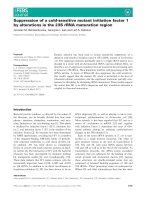
Fig. 3. Design of trans-splicing ribozyme c on-
struct s. (A) The ribozyme contains the internal
guide sequence (IGS) and extended guide
sequence (EGS), which base-pairs to the
complementary sequence in GA mRNA
upstream of the mutation. The ribozyme
catalyzes the coupled cleavage of m utated
mRNA and the ligation of the restorative 3¢
exon to the remaining 5¢ exon. (B) The ribo-
zyme constructs used contain silent mutations
(underlined) introduced by alternative codons,
andasequencetagusedinRT-PCRdetection
(boxed nucleotides) [4].
Ó FEBS 2004 Hydrolysis deficient trans-splicing group I ribozyme (Eur. J. Biochem. 271) 4935
percentage) of reprogrammed product (RNA 2) + target
GA RNA (RNA 3). The amount of trans-splicing
ribozymes that had undergone the r eaction [ calculated
as the amount o f reprogrammed product (RNA 2) as a
fraction (in percentage) of reprogrammed product (RNA
2) + tra ns-splicing ribozyme (RNA 4)] was similar fo r
the t wo different ribozyme c onstructs (data not sh own).
Three parallels of RPA experiments were performed.
AB
Fig. 4. Trans-splicing experiments of DiGIR2-derived ribozymes including the GA RNA as target sequence. RT-PCR and ribonu clease protection
analysis (RPA) of trans-spliced GA mRNA generated by DiGIR2 AGU and DiGIR2DP9.2 AGU ribozymes. (A) Top; RT-PCR products from
in vitro trans-splicing analyses with DiGIR2 AGU and D iGIR2DP9.2 AGU corresponding to reprogrammed products of the expected size (361 nt).
The negative control (Neg. Ctrl.) contains fi rst-strand synthesis master mix. Below; representative re sult of trans-spliced GA RNA sequence
obtained from RT-PCR. The trans-splicing junction is marked by an arrow. (B) Top; representative result of major RNA-species (numbered 1–4)
detected in RPA. RNA 1, undigested probe; RNA 2, trans-spliced GA mRNA; RNA 3, major GA band; RNA 4, major DiGIR2 AGU and
DiGIR2DP9.2 AGU band. Additional bands result from degradation of RNA during incubation at trans-splicing and RPA hybridization
conditions. Below; quantitation of RPA of trans-spliced GA mRNA generatedbyDiGIR2AGUandDiGIR2DP9.2 AGU. Comparative
quantitative data were collected from three indepen dent assays. The trans -splicing efficie ncy (perce ntage) was c alculated b y dividing the trans-
splicing band (RNA 2) ·100bythesumofthetrans-splicing band (RNA 2) and the major GA band (RNA 3). As the amount of DiGIR2 AGU and
DiGIR2DP9.2 AGU r iboz ymes ad ded in t he trans -splicing reactions was a pproximately identical, the a ddition of t he ribozyme b and (RNA 4 ) in t he
fractions denominator gave approximately identical comparative results (data not shown).
4936 E. W. Lundblad et al.(Eur. J. Biochem. 271) Ó FEBS 2004
Results and Discussion
In vitro
self-splicing of DiGIR2 and DiGIR2DP9.2
ribozymes from heterologous transcripts
To test the potential of DiGIR2 and DiGIR2DP9.2 in RNA
reprogramming, the ribozymes were first inserted in cis into
heterologous exons that represent therapeutic, relevant RNA
sequences. The ribozymes were inserted between positions
436 and 437 of the human GA ORF sequence (Fig. 2A) and
tested for splicing a ctivity in vitro. The most common
disorder of glycoprotein degradation, AGU, is caused by
mutations in GA [22]. The nucleotides flanking the intron
insertion site in GA RNA, upstream of the prevalent Finnish
AGU mutation, is similar to the nucleotides flanking the wild
type Didymium rDNA insertion site (Fig. 2A). In the
corresponding splicing constructs, t he IGS was modified
from GGCCGCfiGGUCUU in order to adapt the ribo-
zymes to the GA 5¢ exon. Figure 2B shows that the IGS-
modified DiGIR2 ribozyme excised itself from the precursor
RNA, and in the same process correctly ligated the
surrounding exons (DiGIR2 AGU, Fig. 2B). Bands that
represent intron circle (RNA 1), precursor RNA (RNA 2), 5¢
exon–intron (RNA 3), f ree intron (RNA 4), ligated exons
(RNA 5), and free 3¢ exons (RNA 7), are visible. The small
5¢ exon (RNA 6) was run off the gel. The IGS-modified
DiGIR2DP9.2 AGU transcript generated a band pattern
analogous to DiGIR2 AGU, except for the hydrolysis-
dependent RNA species (RNAs 1, 3 and 7) that were absent
in the reaction (Fig. 2B). In conclusion, these results show
that both the DiGIR2 and DiGIR2DP 9.2 ribozymes accu-
rately self-splice when inserted into foreign exons in cis.
In vitro trans
-splicing of mutated
GA
mRNA sequences
using DiGIR2 and DiGIR2DP9.2 ribozymes
To test whether the DiGIR2 and DiGIR2DP9.2 ribozymes
are able to splice foreign exons also in trans, we targeted the
ribozymes to position 436 (uracil) in the mutated GA
mRNA (same site as in the cis-splicing experiment; Fig. 3)
located upstream of the most common AGU mu tations
(Fig. 2). The ribozymes were designed with modifications
known to increase trans-splicing efficiency and specificity
[1,4,9]; an IGS of 8 nt was used, and based on work by
Sullenger and coworkers [5], a 9 nt EGS complementary to
the GA mRNA target was added (Fig. 3) to further increase
specificity and efficiency. Furthermore, a P10 helix of 5 nt
were included as this i s shown to substantially increase
trans-splicing efficiency of the Tetrahymena riboz yme [9].
Finally, the 3¢ exon that contains the c orrected AGU
sequence was degenerated by alternative codons (Fig. 3) in
order t o avoid strong inter molecular base-pairing to the
region complementary to the target RNA [4].
The trans-splicing ribozymes and RNA targets were
mixed and subjected to conditions that favour splicing (see
above). In a n RT-PCR a pproach the trans-ligated exon
products were amplified and DNA sequen ced to verify
correct splicing (Fig. 4A). In order to quantify the amount
of trans-spliced product, and compare the trans-splicing
efficiency between DiGIR2 AGU and DiGIR2DP9.2 AGU,
we performed analysis by RPA. The RPA probe was
designed to hybridize to a 312 n t region located upstream of
U436 in mutated GA mRNA and to a 49 nt region of the 3¢
exon in the ribozymes, resulting in a 361 nt protected region
for the trans-spliced RNA. The probe was in vitro
transcribed containing additional vector sequences in order
to easily separate full-length probe from RNA fragments
protected in analysis by RPA. Gel analyses of RPA
products (Fig. 4B) confirmed the RT-PCR based experi-
ment presented above of in vitro trans-splicing. The amount
of trans-spliced products were ap proximately similar for
DiGIR2AGU and DiGIR2DP9.2 AGU (1.8% and 2.0%,
respectively).
In summary, t he DiGIR2DP9.2 ribozyme deficient i n
hydrolysis is able to perform trans-splicing with high fidelity
in vitro at comparable rate compared to the wild-type
derived DiGIR2 ribozyme. The former ribozyme is smaller
in size and lacks the hydrolytic processing known to
compete with intro n splicing [18]. Previous works on RNA
reprogramming have focused on using the Tetrahymena
ribozyme as t he tool. Our findings demonstrate that t he
DiGIR2 ribozyme (and its derivatives), in which 3¢ splice
site hydrolysis can be assigned to defined structures within
the intron [18], represent an interesting alternative to the
Tetrahymena ribozyme. Although the 3¢ splice site hydro-
lysis side reaction is under control we realize that the
challenge for the future will be to increase the fraction of
reprogrammed mRNA. Here, experiments that involve
selection for better target accessibility a nd re programming
[4,13,23] will be crucial.
Acknowledgements
This work was f unded by grants f rom The Norwegian Research
Council, The Norwegian Cancer Society, The Aakre Foundation for
Cancer Research, and Simon Fougner Hartmanns Foundation. We
thank Dr Ole K. Tollersrud and Dr Hilde Monica Frostad Riise for the
glycosylasparaginase cDNA plasmids.
References
1. Watanabe, T. & Sullenger, B.A. (2000) RNA repair: a novel
approach to gene therapy. Adv. Drug. Delivery Rev. 44, 109–118.
2. Rogers, C.S., Vanoye, C.G., S ullenger, B.A. & George, A.L.
(2002) Functional repair of a mutant chloride channel using a
trans-splicing ribozyme. J. Clin. Invest. 110, 1783–1789.
3. Long, M.B., Jones, J.P., Sullenger, B.A. & Byun, J. (2003) Ribo-
zyme-mediated re vision o f RNA and DNA. J. Clin. In vest. 112,
312–318.
4. Einvik,C.,Fiskaa,T.,Lundblad,E.W.&Johansen,S.(2004)
Optimization and a pplicat ion of the group I ribozym e trans-spli-
cing reaction. Methods Mol. Biol. 252, 359–372.
5. Byun, J., Lan, N., Long, M. & Sullenger, B.A. (2003) Efficient and
specific repair of s ickle beta-globin RNA by trans- splicing ribo-
zymes. RNA 9, 1254–1263.
6. Cech, T.R. (1990) Self-splicing of group I introns. Annu. Rev.
Biochem. 59, 543–568.
7. Zarrinkar, P.P. & Sullenger, B.A. (1999) Optimizing the substrate
specificity of a group I intron ribozyme. Biochemistry 38, 3426–
3432.
8. Ayre,B.G.,Kohler,U.,Goodman,H.M.&Haseloff,J.(1999)
Design of highly specific cytotoxins b y using trans-splicing ribo-
zymes. Proc. Natl Acad. Sci. USA 96, 3507–3512.
9. Kohler,U.,Ayre,B.G.,Goodman,H.M.&Haseloff,J.(1999)
Trans-splicing ribozymes for targeted gene delivery. J. Mol. Biol.
285, 1935–1950.
Ó FEBS 2004 Hydrolysis deficient trans-splicing group I ribozyme (Eur. J. Biochem. 271) 4937
10. Sullenger, B.A. (1995) Colocalizing r ibozymes with substrate
RNAs to increase their efficacy as gene inhibitors. Appl. Biochem.
Biotechnol. 54, 57–61.
11. Lan, N., Howrey, R.P., Lee, S.W., Smith, C.A. & Sullenger, B.A.
(1998) Ribozyme-mediated repair of sickle beta-globin mRNA’s
in erythrocyte precursors. Science 280, 1593–1596.
12. Lan,N.,Rooney,B.L.,Lee,S.W.,Howrey,R.P.,Smith,C.A.&
Sullenger, B.A. ( 2000) Enhancing RNA repair efficiency by com-
bining trans-splicing ribozymes that recognize different accessible
sites on a target RNA. Mol. Ther. 2, 245–255.
13. Ayre, B.G., Kohler, U., Turgeon, R. & Haseloff, J. (2002) Opti-
mization of trans-splicing ribozyme efficiency a nd specificity by
in vivo genetic selection. Nucleic Acids Res. 30,e141,1–9.
14. Golden, B.L. & Cech, T.R. (1996) Conformational switches
involved in orchestrating the successive steps of group I RNA
splicing. Biochemistry 35, 3754–3763.
15. Zarrinkar, P.P. & Sullenger, B.A. (1998) Probing the interplay
between the two steps of group I intron splicing: competition of
exogenous guanosine with xG. Biochemistry 37, 18056–18063.
16. Haugen, P., An dreassen, M., Birgisdottir, A
˚
.B. & Johansen, S.
(2004) Hydrolytic cleavage by a group I intron ribozym e is
dependent on RNA structures not important for splicing. Eur. J.
Biochem. 271, 1015–1024.
17. Nielsen, H., Fiskaa, T., Birgisdottir, A
˚
.B., Haugen, P., Einvik, C.
& Johansen, S. (2003) The ability to form full-length intron RNA
circles is a general property of nuclear group I intro ns. RNA 9,
1464–1475.
18. Haugen, P., De Jonckheere, J.F. & Johansen, S. (2002) Char-
acterization of the self-splicing products of two complex Naegleria
LSU rDNA group I introns containing homing endonuclease
genes. Eur. J. Biochem. 269, 1641–1649.
19. Lundb lad, E.W., Einvik, C., Rønning, S., Haugli, K. & Johansen,
S. (2004) Twelve group I introns in the same pre-rRNA transcript
of the myxomycete Fuligo septica: RNA processing and evolution.
Mol. Biol. Evol. 21 , 1283–1293.
20. Decatur, W.A., Einvik, C., Johansen, S. & Vogt, V.M. (1995) Two
group I ribozymes with different func tions in a nu clear rDNA
intron. EMBO J. 14, 4558–4568.
21. Einvik, C., Elde, M. & Johansen, S. (1998) G roup I twintrons:
genetic elements in myxomycete and schizopyrenid amoebo-
flagellate ribosomal DNAs. J. Biotechnol. 17 , 63–74.
22. Mononen,I.,Fisher,K.J.,Kaartinen,V.&Aronson,N.N.Jr
(1993) Aspartylglycosaminuria: protein chemistry and mole cular
biology o f the most common lysosomal storage disorder of gly-
coprotein degradation. FASEB J. 7, 1247–1256.
23. Jones, J.T. & Sullenger, B.A. (1997) Evaluating and e nhancing
ribozyme reaction efficiency in mammalian cells. Nat. Biotechnol.
15, 902–905.
4938 E. W. Lundblad et al.(Eur. J. Biochem. 271) Ó FEBS 2004