Báo cáo khoa học: Stabilities and activities of the N- and C-domains of FKBP22 from a psychrotrophic bacterium overproduced in Escherichia coli pptx
Bạn đang xem bản rút gọn của tài liệu. Xem và tải ngay bản đầy đủ của tài liệu tại đây (262.55 KB, 11 trang )
Stabilities and activities of the N- and C-domains
of FKBP22 from a psychrotrophic bacterium overproduced
in Escherichia coli
Yutaka Suzuki
1
, Kazufumi Takano
1,2
and Shigenori Kanaya
1
1 Department of Material and Life Science, Graduate School of Engineering, Osaka University, Suita, Osaka, Japan
2 PRESTO, JST, Suita, Osaka, Japan
When polypeptides are synthesized at ribosomes, pep-
tide bonds are connected in trans form. In the case of
peptide bonds N-terminal of the proline residues, how-
ever, some of them form cis peptide bonds in correctly
folded proteins [1]. Consequently, trans-to-cis conver-
sions of these peptide bonds (prolyl isomerizations)
should occur during protein folding reactions. As dem-
onstrated in some refolding experiments [2,3], prolyl
isomerizations are relatively slow and can be the rate
limiting step in protein folding reactions. The cis-trans
isomerizations of peptide bonds N-terminal of the pro-
line residues are catalyzed by peptidylprolyl cis-trans
isomerases (PPIases; EC 5.2.1.8) [4]. Three structurally
unrelated families of PPIases are known. They are
cyclophilins, parvulins, and FK506-binding proteins
(FKBPs) [5].
We have previously shown that the cellular content
of FKBP22 (SIB1 FKBP22) from a psychrotrophic
Keywords
domain structure; FKBP22; PPIase;
psychrotrophic bacterium; thermal stability
Correspondence
S. Kanaya, Department of Material and Life
Science, Graduate School of Engineering,
Osaka University, 2-1, Yamadaoka, Suita,
Osaka 565-0871, Japan
Tel ⁄ Fax: +81 6 6879 7938
E-mail:
(Received 27 September 2004, revised 23
October 2004, accepted 29 October 2004)
doi:10.1111/j.1742-4658.2004.04468.x
FKBP22 from a psychrotrophic bacterium Shewanella sp. SIB1, is a dimer-
ic protein with peptidyl prolyl cis-trans isomerase (PPIase) activity. Accord-
ing to homology modeling, it consists of an N-terminal domain, which is
involved in dimerization of the protein, and a C-terminal catalytic domain.
A long a3 helix spans these domains. An N-domain with the entire a3 helix
(N-domain
+
) and a C-domain with the entire a3 helix (C-domain
+
) were
overproduced in Escherichia coli in a His-tagged form, purified, and their
biochemical properties were compared with those of the intact protein.
C-domain
+
was shown to be a monomer and enzymatically active. Its opti-
mum temperature for activity (10 °C) was identical to that of the intact
protein. Determination of the PPIase activity using peptide and protein
substrates suggests that dimerization is required to make the protein fully
active for the protein substrate or that the N-domain is involved in sub-
strate-binding. The differential scanning calorimetry studies revealed two
distinct heat absorption peaks at 32.5 °C and 46.6 °C for the intact protein,
and single heat absorption peaks at 44.7 °C for N-domain
+
and 35.6 °C
for C-domain
+
. These results indicate that the thermal unfolding transi-
tions of the intact protein at lower and higher temperatures represent those
of C- and N-domains, respectively. Because the unfolding temperature of
C-domain
+
is much higher than its optimum temperature for activity,
SIB1 FKBP22 may adapt to low temperatures by increasing a local flexibil-
ity around the active site. This study revealed the relationship between the
stability and the activity of a psychrotrophic FKBP22.
Abbreviations
ALPF, N-succinyl-Ala-Leu-Pro-Phe-p-nitroanilide; CD, circular dichroism; DSC, differential scanning calorimetry; FKBP, FK506-binding protein;
MIP, macrophage infectivity potentiator; PPIase, peptidyl prolyl cis-trans isomerase.
632 FEBS Journal 272 (2005) 632–642 ª 2005 FEBS
bacterium Shewanella sp. SIB1 increases at 4 °C, as
compared to that at 20 °C [6]. This protein is a member
of the macrophage infectivity potentiator (MIP)-like
FKBP subfamily proteins and shows amino acid
sequence identities of 56% to Escherichia coli FKBP22
[7], 43% to E. coli FkpA [8], and 41% to Legionella
pneumophila MIP [9]. SIB1 FKBP22 exists as a homo-
dimer and exhibits the PPIase activity like other MIP-
like FKBP subfamily proteins. However, the optimum
temperature of this protein for activity (10 °C) is much
lower than that of E. coli FKBP22 (> 25 °C). We pro-
pose that this activity facilitates efficient folding of pro-
teins containing cis prolines in psychrotrophic bacteria
at low temperatures.
According to the crystal structures of L. pneumophila
MIP [10] and E. coli FkpA [11], these proteins are com-
posed of N- and C-domains, which are spanned by a 40
amino acid long a3 helix. The N-domain consists of a1
and a2 helices and an N-terminal region of a3 helix.
The C-domain consists of six b-strands (b1–b6), a4
helix, and a C-terminal region of a3 helix. The
N-domain is unique to the MIP-like FKBP subfamily
proteins. This domain is involved in dimerization of the
protein and the interface between two monomers is sta-
bilized by the hydrophobic interactions of a1 and a2
helices. In contrast, the C-domain (except a3 helix) is
conserved in all FKBP family proteins and contains the
entire PPIase active-site, suggesting that all FKBP fam-
ily proteins share a common catalytic mechanism. The
a3 helix seems to be required to control the positions of
the two C-domains of the homodimer, such that these
domains are located with an appropriate distance and
orientation. Because of the high similarity in the amino
acid sequence of SIB1 FKBP22 with that of L. pneumo-
phila MIP and E. coli FkpA, SIB1 FKBP22 might have
a similar three-dimentional structure.
The stability–activity relationships of MIP-like FKBP
subfamily proteins remain to be analyzed. Because the
prolyl isomerization is a spontaneous reaction and the
rate for this reaction increases as the reaction tempera-
ture increases, the PPIase activity cannot be accurately
determined at > 30 °C. Therefore, it seems difficult to
analyze the stability–activity relationships of PPIases
from mesophilic and thermophilic organisms. SIB1
FKBP22 seems to be an excellent model to analyze these
relationships because its optimum temperature for activ-
ity is 10 °C. In addition, because this protein is expected
to consist of N- and C-domains, it would be informative
to construct the SIB1 FKBP22 variants containing
either one of these domains and compare their activities
and stabilities with those of the intact protein.
In this report, the N- and C-domains of SIB1
FKBP22 were overproduced in E. coli and purified in
an amount sufficient for physicochemical studies. By
comparing their activities and stabilities with those of
the intact protein, we showed that the unfolding tem-
perature of SIB1 FKBP22 is much higher than the
optimum temperature for activity. Based on these
results, we discuss a role of each domain of SIB1
FKBP22 and a cold-adaptation mechanism of this
protein.
Results
Design
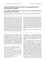
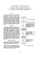
A model for the three-dimensional structure of the
His-tagged form of SIB1 FKBP22 (SIB1 FKBP22*)
was constructed based on the crystal structure of
L. pneumophila MIP [10] (Fig. 1). According to this
model, SIB1 FKBP22 consists of N- and C-domains.
The N-domain of one molecule interacts with that of
another molecule to form a homodimer. The
C-domain represents a catalytic domain. Based on this
model, three types of the SIB1 FKBP22 variants con-
taining either one of these two domains were designed.
They are N-domain
+
, C-domain
+
, and C-domain
–
.
The primary structures of these variants are schemati-
cally shown in comparison with that of the intact pro-
tein in Fig. 2. Because a long a3 helix spans both the
N- and C-domains, and because the region containing
only a1 and a2 helices seems to be too short to fold
correctly, N-domain
+
was designed such that
it contains the entire a3 helix. Likewise, C-domain
+
and C-domain
–
were designed such that the former
Fig. 1. A tertiary model of SIB1 FKBP22 homodimer. The a3 helix
(Val52–Arg93), which spans both the N- and C-domains, is most
deeply shaded. The N-domain without a3 helix (Met1–Ala51), which
is involved in dimerization, is moderately shaded, and the C-domain
without a3 helix (Asp94–Ile205), which is involved in catalytic func-
tion, is most lightly shaded.
Y. Suzuki et al. Stability and activity of SIB1 FKBP22 domains
FEBS Journal 272 (2005) 632–642 ª 2005 FEBS 633
contains the entire a3 helix and the latter does not
contain it.
Overproduction and purification
Upon induction for overproduction at 10 °C,
N-domain
+
and C-domain
+
accumulated in the cells
in a soluble form, whereas C-domain
–
accumulated in
the cells in inclusion bodies (Fig. 3). C-domain
–
was
solubilized in the presence of 6 m urea and refolded by
removing urea with a yield of nearly 100%. All pro-
teins were purified to give a single band on
SDS ⁄ PAGE (data not shown).
The molecular masses of N-domain
+
, C-domain
+
,
and C-domain
–
were estimated to be 15 kDa, 26 kDa,
and 17 kDa, respectively, by SDS ⁄ PAGE (Fig. 3).
These values are slightly larger than the calculated
ones from their amino acid sequences including a His-
tag (12 042 for N-domain
+
, 19 149 for C-domain
+
,
and 14 085 for C-domain
–
). The molecular mass of
SIB1 FKBP22* estimated by SDS ⁄ PAGE (29 kDa)
has been reported to be larger than that determined by
EMI-MS, which is identical to the calculated one
(23 947) [6]. Slow migration in the gel may be a char-
acteristic common to SIB1 FKBP22* and its variants.
The molecular masses of N-domain
+
and C-domain
+
were also estimated to be 39 kDa and 23 kDa, respect-
ively, by gel filtration column chromatography. The
former and latter values are larger than the calculated
ones by 3.2 and 1.2 times, respectively, suggesting that
N-domain
+
exists as a trimer and C-domain
+
exists
as a monomer. However, the molecular mass of a
dimeric form of SIB1 FKBP22* estimated by gel filtra-
tion column chromatography has been reported to be
larger than that determined by sedimentation equilib-
rium analytical ultracentrifuge by 1.5 times [6]. This
discrepancy is probably caused by the unusual mole-
cular shape of the protein, which is cylindrical rather
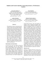
ABC
Fig. 3. Estimation of the amount of the proteins in soluble and insoluble forms by SDS ⁄ PAGE. N-domain
+
(A), C-domain
–
(B), and C-domain
+
(C) were overproduced in E. coli as described for SIB1 FKBP22* [6]. The soluble (lane S) and insoluble (lane P) fractions after sonication lysis
were analyzed by 15% (for C-domain
+
) and 17% (for N-domain
+
and C-domain
–
) SDS ⁄ PAGE. The gel was stained with Coomassie Brilliant
Blue. Arrows indicate the recombinant proteins overproduced in the cells. The positions of the standard proteins contained in a low mole-
cular mass marker kit (Pharmacia Biotech, Piscataway, NJ, USA) are shown alongside the gels, together with their molecular masses.
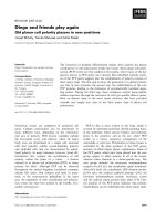
Fig. 2. Schematic representations of the pri-
mary structures of SIB1 FKBP22* and its
variants. A His-tag attached to the N-termini
of the proteins is represented by shaded
box. The a-helices and b-strands are repre-
sented by cylinders and arrows, respect-
ively. These secondary structures are
arranged based on a tertiary model of SIB1
FKBP22. Numbers indicate the positions of
the residues relative to the initiator methio-
nine residue. The ranges of the N- and
C-domains are also shown.
Stability and activity of SIB1 FKBP22 domains Y. Suzuki et al.
634 FEBS Journal 272 (2005) 632–642 ª 2005 FEBS
than globular. Because N-domain
+
is expected to
assume a similar cylindrical structure, sedimentation
equilibrium analytical ultracentrifugation was per-
formed to determine its molecular mass in solution.
The data fitted well to a single-species model with no
evidence of aggregation, and the molecular mass was
determined to be 23 431 Da. This value is 1.9 times
larger than that calculated from the amino acid
sequence, indicating that N-domain
+
exists as a dimer.
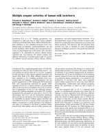
CD spectra
The far-UV CD spectra of N-domain
+
, C-domain
+
,
C-domain
–
, and SIB1 FKBP22* were measured at
10 °C (Fig. 4A). The spectrum of N-domain
+
, which
gave a broad trough with a double minimum at 208
and 222 nm, was similar to that of SIB1 FKBP22*,
although the depth of the trough in this spectrum is
larger than that in the SIB1 FKBP22* spectrum.
The helical content was calculated to be 51% for
N-domain
+
and 38% for SIB1 FKBP22* from these
spectra using the method of Wu et al. [12]. These val-
ues were comparable to those calculated from a ter-
tiary model of SIB1 FKBP22* (60% for N-domain
+
and 34% for SIB1 FKBP22*), suggesting that
N-domain
+
assumes a similar helical structure to that
of the N-domain in the intact molecule. On the other
hand, the CD spectra of C-domain
+
and C-domain
–
gave a broad trough with a single minimum at 207 nm
and one without any clear minimum, respectively. The
depths of these troughs were considerably smaller than
that in the SIB1 FKBP22* spectrum.
The near-UV CD spectra of these proteins were also
measured at 10 °C (Fig. 4B). These spectra reveal the
three-dimensional environments of aromatic residues
such as Trp and Tyr. SIB1 FKBP22* contains one
tryptophan residue (Trp157), which is conserved in the
FKBP family proteins and required for PPIase activity,
and seven tyrosine residues. Because most of these
residues (one tryptophan and six tyrosine residues) are
located in its C-domain, the near-UV CD spectrum of
SIB1 FKBP22* may reflect the conformation of the
C-domain. The spectrum of C-domain
+
was similar to
that of SIB1 FKBP22*, suggesting that C-domain
+
assumes a similar structure to that of the C-domain
in the intact molecule. In contrast, the spectrum of
C-domain
–
was quite different from those of
C-domain
+
and SIB1 FKBP22*, suggesting that the
structure of C-domain
–
is considerably different from
that of the C-domain in the intact molecule.
PPIase activity
When the PPIase activity was determined at 10 °C
by the protease coupling assay using N-succinyl-Ala-
Leu-Pro-Phe-p-nitroanilide (ALPF) as a substrate,
C-domain
+
exhibited PPIase activity, whereas
C-domain
–
did not. The catalytic efficiency (k
cat
⁄ K
m
)
of C-domain
+
was estimated to be 1.43 lm
)1
Æs
)1
,
which was 1.6 times higher than that of SIB1
FKBP22*. The temperature dependence of the PPIase
activity of C-domain
+
was nearly identical to that of
SIB1 FKBP22* (Fig. 5A). In contrast, when the PPI-
ase activity was determined by the RNase T
1
refolding
assay, C-domain
+
exhibited much less activity as com-
pared to that of SIB1 FKBP22*. The acceleration
effect of C-domain
+
on the RNase T
1
refolding reac-
tion was not detected at 21 nm, but detected at 210 nm
(Fig. 5B). The acceleration effect similar to that detec-
ted in the presence of 210 nm C-domain
+
was detected
in the presence of 19 nm SIB1 FKBP22*. The k
cat
⁄ K
m
values were estimated to be 0.5 lm
)1
Æs
)1
for SIB1
FKBP22* and 0.015 lm
)1
Æs
)1
for C-domain
+
.
Thermal stability
Heat induced unfolding of N-domain
+
, C-domain
+
,
and SIB1 FKBP22* were analyzed by differential
Fig. 4. CD spectra of SIB1 FKBP22* and its
variants. The far-UV (A) and near-UV (B) CD
spectra of SIB1 FKBP22* (dashed line),
N-domain
+
(heavy thick line), C-domain
–
(thin
line), and C-domain
+
(moderately thick line)
are shown. All spectra were measured at
10 °C as described under Experimental
procedures.
Y. Suzuki et al. Stability and activity of SIB1 FKBP22 domains
FEBS Journal 272 (2005) 632–642 ª 2005 FEBS 635
scanning calorimetry (DSC) (Fig. 6, Table 1). All DSC
curves were reproduced by repeating thermal scans,
indicating that thermal unfoldings of these proteins are
highly reversible. The denaturation curve of SIB1
FKBP22* clearly showed two well separated transi-
tions. Deconvolution of the thermogram according to
a non-two-state denaturation model gives melting tem-
perature (T
m
) values of 32.5 °C and 46.6 °C for these
transitions. These T
m
values are nearly equal to those
of C-domain
+
(35.6 °C) and N-domain
+
(44.7 °C),
suggesting that the thermal unfolding transitions of
SIB1 FKBP22* at lower and higher temperatures rep-
resent those of its C-domain and N-domain, respect-
ively. For unfolding of N-domain
+
, the van’t Hoff
enthalpy (DH
vH
) was roughly two times larger than the
calorimetric enthalpy (DH
cal
). Because N-domain
+
exists as a dimer, this result possibly reflects a coupling
of the unfolding of N-domain
+
to dissociation of
the homodimer. Similarly, the unfolding reaction of
C-domain
+
seems to contain complex processes, as
indicated by the DH
cal
⁄DH
vH
ratio far from unity.
Comparison of thermal stability of SIB1 FKBP22*
and E. coli FKBP22*
To examine whether SIB1 FKBP22* is less stable than
its mesophilic counterpart, heat induced unfolding of
E. coli FKBP22* was analyzed by DSC. However,
thermodynamic parameters including T
m
could not be
obtained because of the poor reversibility of this pro-
tein in thermal unfolding. Therefore, thermal stabilities
of SIB1 FKBP22* and E. coli FKBP22* were analyzed
by circular dichroism (CD). The far-UV CD spectra of
SIB1 FKBP22* and E. coli FKBP22* were measured
at various temperatures and the spectra of SIB1
FKBP22* at 10 and 50 °C are shown in comparison
with those of E. coli FKBP22* at 20 and 80 °Cin
Fig. 7. The spectrum of SIB1 FKBP22* at 10 °Cis
identical to that shown in Fig. 4A. The spectra of
Table 1. Thermodynamic parameters for heat induced unfolding of
SIB1 FKBP22*, C-domain
+
and N-domain
+
recorded by microcalori-
metry. The melting temperature (T
m
), calorimetric enthalpy (DH
cal
),
and van’t Hoff enthalpy (DH
vH
) were obtained from the DSC curves
shown in Fig. 6, using
ORIGIN software (MicroCal, Inc.).
Protein T
m
(°C) DH
cal
(kJÆmol
)1
) DH
vH
(kJÆmol
)1
)
SIB1 FKBP22* 32.5 82.8 404.2
46.4 194.8 303.9
C-domain+ 35.6 171.8 232.4
N-domain
+
44.7 140.9 259.2
Fig. 6. DSC curves of N-domain
+
, C-domain
+
, and SIB1 FKBP22*.
The DSC curves of N-domain
+
(thick line), C-domain
+
(thin line), and
SIB1 FKBP22* (dashed line), which were measured at a scan rate
of 1 °CÆmin
)1
, are shown. These proteins were dissolved in 20 mM
sodium phosphate (pH 8.0) at 0.6 mgÆmL
)1
.
Fig. 5. PPIase activities of C-domain
+
. (A) The temperature dependence of the PPIase activity of C-domain
+
(–d–), which was determined
by protease coupling assay using ALPF as a substrate, is shown in comparison with that of SIB1 FKBP22* (–s–). The catalytic efficiency,
k
cat
⁄ K
m
, was calculated according to Harrison & Stein [34]. The experiment was carried out in duplicate. Each plot represents the average
value and errors from the average values are shown. (B) The increase in tryptophan fluorescence at 323 nm during refolding of RNase T
1
(0.2 lM) is shown as a function of the refolding time. Refolding reaction was carried out at 10 °C in the absence (dotted line), or presence of
21 n
M of C-domain
+
(thick solid line), 210 nM of C-domain
+
(thin solid line) or 19 nM of SIB1 FKBP22* (dashed line).
Stability and activity of SIB1 FKBP22 domains Y. Suzuki et al.
636 FEBS Journal 272 (2005) 632–642 ª 2005 FEBS
SIB1 FKBP22* at 10 °C and E. coli FKBP22* at
20 °C, which represent the spectra of these proteins in
a native form, were similar to each other, suggesting
that the tertiary structures of these proteins are similar
to each other. With a temperature shift from 10 to
50 °C, the spectrum of SIB1 FKBP22*, which gave a
broad trough with double minimum [h] values of
)11 200 at 209 nm and )12 100 at 222 nm, was
greatly changed so that it exhibits a trough with a
minimum [h] value of )7800 at 207 nm, which is
accompanied by a shoulder with a [h] value of )5700
at 220 nm. A similar spectral change was observed for
E. coli FKBP22* when the temperature was shifted
from 20 to 80 °C. The spectra of SIB1 FKBP22* at
50 °C and E. coli FKBP22* at 80 °C were not seri-
ously changed at higher temperatures, indicating that
these spectra represent the spectra of these proteins in
a denatured form. In these conditions, SIB1 FKBP22*
was fully reversible in thermal denaturation, whereas
E. coli FKBP22* was not. The reversibility of E. coli
FKBP22* was roughly 70%.
The thermal denaturation curves of SIB1 FKBP22*
and E. coli FKBP22* were measured by monitoring a
change in the CD values at 222 nm (Fig. 8). SIB1
FKBP22* apparently unfolded through an intermedi-
ate state. The T
m
values for the first and second transi-
tions were roughly estimated to be 32 and 44 °C,
respectively, which were comparable with those
determined by DSC. As compared to SIB1 FKBP22*,
E. coli FKBP22* unfolded at higher temperatures,
indicating that it is more stable than SIB1 FKBP22*.
However, it is unclear whether this protein unfolds
through an intermediate state as well, because this
intermediate state was not clearly detected. The ther-
mal unfolding curve of this protein did not fit the the-
oretical curve, which was drawn on the assumption
that the protein unfolds in a single cooperative fashion
(data not shown).
Discussion
Unfolding of SIB1 FKBP22*
In this study, SIB1 FKBP22* was shown to unfold in
a complex non-two-state mechanism with two peaks
apparent in the DSC curve. Construction of the
N-domain
+
and C-domain
+
, which lack the C- and
N-domains, respectively, followed by DSC analyses,
clearly showed that two peaks of heat capacity
observed in thermal unfolding of SIB1 FKBP22* rep-
resent unfoldings of its N- and C-domains. In this
thermal unfolding process, the C- and N-domains
unfold at lower and higher temperatures, respectively.
It has been reported that a phosphoglycerate kinase
[13] and a chitobiase [14] from psychrophilic bacteria
consist of a heat labile domain and a heat stable
domain. Bentahir et al. [13] have proposed that a heat
labile domain provides a sufficient flexibility around
the active site, and a heat stable domain provides a
sufficient rigidity to the substrate-binding site, so that
Fig. 8. Thermal denaturation curves of SIB1 FKBP22* and E. coli
FKBP22*. The [h] values of SIB1 FKBP22* (trace 1) and E. coli
FKBP22* (trace 2) at 222 nm are shown as a function of tempera-
ture. The proteins were dissolved in 20 m
M sodium phosphate
(pH 8.0) at 0.30 mgÆmL
)1
for SIB1 FKBP22* and 0.29 mgÆmL
)1
for
E. coli FKBP22*. A cell with an optical path length of 2 mm was
used. Temperature was linearly raised at 1 °CÆmin
)1
.
Fig. 7. Far-UV CD spectra of SIB1 FKBP22* and E. coli FKBP22*.
The CD spectra of SIB1 FKBP22* measured at 10 °C (thick line) and
50 °C (thick dashed line), and those of E. coli FKBP22* measured at
20 °C (thin line) and 80 °C (thin dashed line) are shown. The spectra
were measured as described under Experimental procedures.
Y. Suzuki et al. Stability and activity of SIB1 FKBP22 domains
FEBS Journal 272 (2005) 632–642 ª 2005 FEBS 637
the enzymatic reaction is efficiently achieved at low
temperatures. Because the C-terminal catalytic domain
of SIB1 FKBP22 represents a heat labile domain, the
instability of this domain may be required to increase
the flexibility of the active-site at low temperatures.
Stability and activity of SIB1 FKBP22*
SIB1 FKBP22* was shown to be much less stable than
E. coli FKBP22*. Its optimal temperature for activity
has been reported to be greatly shifted downward as
compared to that of E. coli FKBP22* [6]. Cold-adap-
tation has been specified by the increase in the cata-
lytic efficiency at low temperatures, the downward
shift in the optimum temperatures for activity, and the
reduction in the conformational stability [15]. There-
fore, SIB1 FKBP22 can be defined as a cold-adapted
enzyme, although it is less active than E. coli
FKBP22* even at low temperatures [6]. Several cold-
adapted enzymes have also been reported to be less
active than their mesophilic counterparts [16–19].
Analyses of the thermal stability of SIB1 FKBP22*
by DSC (Fig. 3) and CD (Fig. 8) indicate that unfold-
ing of this protein is initiated at > 25 °C. In fact, the
CD spectrum of SIB1 FKBP22* at 20 °C was nearly
identical to that at 10 °C (data not shown), suggesting
that the conformation of this protein is not seriously
changed upon temperature shift from 10 to 20 °C.
Thermal unfolding of C-domain
+
is also initiated
at > 25 °C. Nevertheless, SIB1 FKBP22* and
C-domain
+
both exhibit the maximal PPIase activity
at 10 °C and their activities are greatly reduced at
20 °C. These results suggest that a subtle conforma-
tional change around the active-site causes a great
reduction of the enzymatic activity. The large differ-
ence in the temperatures for enzymatic inactivation
and structural unfolding has been observed for
cold-adapted a-amylase and family 8 xylanase from an
Antarctic bacterium [20,21]. The apparent optimal
temperatures of these proteins for enzymatic activities
are much lower than the temperatures at which any
significant conformational event occurs. In contrast,
the optimal temperatures for the activities of their
mesophilic and thermophilic counterparts closely cor-
relate with the temperatures for their structural transi-
tions. Thus, the large difference in the temperatures
for enzymatic inactivation and structural unfolding
seems to be a characteristic feature of cold-adapted
enzymes. It has been proposed that this difference is
caused by a cold-adaptation strategy termed ‘localized
flexibility’ [20]. Although an increase in flexibility
around the active site increases k
cat
by reducing the
energy cost of conformational change during the cata-
lytic reaction, it should increase K
m
concomitantly. By
restricting the increase of flexibility within small areas,
cold-adapted enzymes prevent unfavorable increases in
K
m
[22]. SIB1 FKBP22 probably adopts a similar
strategy for cold-adaptation.
Structural importance of a3 helix
Two types of the SIB1 FKBP22* variants, which con-
tain the C-domain, were designed based on its tertiary
model. C-domain
+
contains an entire a3 helix,
whereas C-domain
–
does not contain it. These two
proteins differ greatly in their biochemical properties.
C-domain
+
was overproduced in E. coli in a soluble
form and exhibited the PPIase activity. Its near-UV
CD spectrum was similar to that of SIB1 FKBP22*.
In contrast, C-domain
–
was overproduced in E. coli in
inclusion bodies and exhibited little PPIase activity. Its
near-UV CD spectrum was quite different from that of
SIB1 FKBP22*. These results strongly suggest that a3
helix is required to facilitate folding of the C-domain,
or to stabilize it, so that the C-domain assumes a
native conformation. It has previously been reported
that limited proteolysis of L. pneumophila MIP allows
the separation of their N- and C-domains such that
the C-domain contains the C-terminal half of the a3
helix [23,24]. In addition, the C-domain of E. coli
FkpA shows a high tendency to form inclusion bodies
when it is overproduced in E. coli in a form without
a3 helix [25]. These results are consistent with our
results. According to the crystal structure of L. pneu-
mophila MIP, there are three distinct contacts between
the C-terminal region of a3 helix and the C-domain
[10]. These contacts may also be conserved in the
structure of SIB1 FKBP22.
Role of N- and C-domains
Most organisms contain multiple PPIases within a sin-
gle cell. They are usually composed of several domains;
one is common to the members of each family and
specifies the family to which that PPIase belongs, and
the others are unique to the particular PPIase
and thought to be related to the protein’s distinct
function. The C- and N-domains of MIP-like FKBP
subfamily proteins represent the former and latter
domains, respectively. Therefore, biochemical charac-
terizations of N-domain
+
and C-domain
+
will facili-
tate understanding of the roles of these domains in the
intact molecule.
The observation that N-domain
+
exists as a dimer,
whereas C-domain
+
exists as a monomer supports a
tertiary model of SIB1 FKBP22, in which the a1 and a2
Stability and activity of SIB1 FKBP22 domains Y. Suzuki et al.
638 FEBS Journal 272 (2005) 632–642 ª 2005 FEBS
helices form the dimerization core of the protein. In
addition, we showed that the PPIase activity of
C-domain
+
determined by the RNase T
1
refolding assay
was greatly reduced as compared to that of the intact
protein. These results suggest that a dimeric structure of
SIB1 FKBP22 is responsible for its high PPIase activity
for protein substrates. Alternatively, N-domain contains
a binding site for protein substrates. Similar results have
been reported for other MIP-like FKBP subfamily pro-
teins. For example, The C-domain of L. pneumophila
MIP produced upon limited proteolysis has been repor-
ted to exist as a monomer and exhibit weak PPIase
activity for protein substrate [23]. Likewise, the
C-domain of E. coli FkpA is devoid of chaperone-like
function, although it shows PPIase activity [11,25]. Fur-
thermore, it has been reported that human FKBP12
which intrinsically consists of a single domain, exhibited
lower activity for RNase T
1
substrate and higher activity
for tetrapeptide substrates than E. coli FkpA [25]. How-
ever, the reason why C-domain alone exhibits a weak
activity for protein substrates remains to be clarified.
Further structural and functional studies of these pro-
teins will be required to clarify this reason.
Experimental procedures
Cells and plasmids
Psychrotrophic bacterium Shewanella sp. SIB1 was isolated
from water deposits in a Japanese oil reservoir [26]. E. coli
JM109 [recA1 , supE44, endA1, hsdR17, gyrA96, relA1, thi,
D(lac-proAB) ⁄ F¢, traD36, proAB
+
, lacI
q
lacZDM15] was
obtained from Toyobo Co., Ltd. (Kyoto, Japan). E. coli
BL21(DE3) [F
–
, ompT, hsdS
B
(r
B
–
,m
B
–
), gal, dcm (DE3)]
and plasmid pET-28a were obtained from Novagen (Madi-
son, WI, USA). Plasmid pUC18 was obtained from Takara
Shuzo Co., Ltd. (Kyoto, Japan). The E. coli transformants
were grown in Luria–Bertani medium containing 50 mgÆL
)1
ampicillin or 35 mgÆL
)1
kanamycin.
Plasmid construction
Plasmid pSIB1-Nd, pSIB1-Cd, and pSIB1-a3+Cd for over-
production of a His-tagged form of the N-domain of SIB1
FKBP22 with entire a3 helix (N-domain
+
), C-domain with-
out a3 helix (C-domain
–
), and C-domain with entire a3 helix
(C-domain
+
), respectively, were constructed by ligating a
part of the SIB1 FKBP22 gene amplified by PCR into pET-
28a as follows. Genomic DNA was prepared from a Sarkosyl
lysate of the Shewanella sp. SIB1 cells [27] and used as a tem-
plate. The gene encoding Met1–Asp94 of SIB1 FKBP22 was
amplified by PCR and ligated into the NdeI–SacI sites of
pET-28a to produce plasmid pSIB1-Nd. Likewise, the genes
encoding Gly95–Ile205 and Gly47–Ile205 of SIB1 FKBP22
were amplified by PCR and ligated into pET-28a to produce
plasmids pSIB1-Cd and pSIB1- a3+Cd, respectively. The
sequences of the 5¢ PCR primers were 5¢-AGAGAGAA
TT
CATATGTCAGATTTGTTCAG-3¢ for N-domain
+
,5¢-
CTGAAAACGCTAAG
CATATGGGTATTACGA-3¢ for
C-domain
–
, and 5¢-CTTGCTGATGCACATATGGGGAA
AGAAAGC-3¢ for C-domain
+
, where underlined bases
show the position of the NdeI site. The sequences of the 3¢
PCR primers were 5¢-GACTCT
GAGCTCGTAATCTAGT
CACGCTTA-3¢ for N-domain
+
, where underlined bases
show the position of the SacI site, and 5¢- GGCCACT
GGATCCAACTACAGCAATTCTCA-3¢ for C-domain
–
and C-domain
+
, where underlined bases show the position
of the BamHI site. PCR was performed with GeneAmp PCR
system 2400 (PerkinElmer, Tokyo, Japan) using KOD
polymerase (Toyobo Co., Ltd) according to the procedures
recommended by the supplier.
Overproduction and purification
His-tagged forms of SIB1 FKBP22 (SIB1 FKBP22*) and
E. coli FKBP22 (E. coli FKBP22*) were overproduced
and purified as described previously [6]. N-domain
+
,
C-domain
–
, and C-domain
+
were overproduced in the
E. coli BL21(DE3) cells transformed with plasmids pSIB1-
Nd, pSIB1-Cd, and pSIB1-a3+Cd, respectively, and puri-
fied, as described for SIB1 FKBP22* [6], except for the
purification of C-domain
–
. For purification of C-domain
–
,
which was overproduced in inclusion bodies, the cells were
disrupted by sonication and centrifuged at 15 000 g for
30 min at 4 °C. The pellet was dissolved in 20 mm sodium
phosphate (pH 8.0) containing 6 m urea and 0.5% (w ⁄ v)
Triton X-100, and incubated overnight at 4 °C. After cen-
trifugation at 15 000 g for 30 min at 4 °C to remove insol-
uble materials, the protein was refolded by dialysis against
20 mm sodium phosphate (pH 8.0), and purified as des-
cribed for SIB1 FKBP22* using metal chelating affinity
chromatography and gel filtration chromatography [6].
Production of the recombinant proteins in the E. coli
cells, as well as their purities, were analyzed by SDS ⁄ PAGE
[28] on a 15 or 17% polyacrylamide gel, followed by stain-
ing with Coomassie Brilliant Blue.
Protein concentration
Protein concentrations were determined from the UV
absorption on the basis that the absorbance at 280 nm of
a 0.1% solution is 0.68 for SIB1 FKBP22*, 0.12 for
N-domain
+
, 1.01 for C-domain
–
, 0.75 for C-domain
+
and
0.69 for E. coli FKBP22*. These values were calculated by
using ¼ 1576 m
)1
Æcm
)1
for Tyr and 5225 m
)1
Æcm
)1
for
Trp at 280 nm [29]. For N-domain
+
, which contains only
one tyrosine residue and no tryptophan residues, a method
Y. Suzuki et al. Stability and activity of SIB1 FKBP22 domains
FEBS Journal 272 (2005) 632–642 ª 2005 FEBS 639
of Scopes [30] was used to confirm the accuracy of its
concentration. In this method, the protein concentration
(mgÆmL
)1
) is calculated from A
205nm
⁄ (31 · b), where
A
205nm
represents absorbance at 205 nm and b represents
an optical path length (cm).
Molecular mass
The molecular masses of purified proteins were estimated by
gel filtration column chromatography using a Superdex 200
16 ⁄ 60 gel filtration column (Amersham Biosciences, Piscat-
away, NJ, USA) equilibrated with 50 mm Tris ⁄ HCl (pH 8.0)
containing 50 mm NaCl. Elution was performed at a flow
rate of 0.5 mLÆmin
)1
. Bovine serum albumin (67 kDa),
ovalbumin (44 kDa), chymotrypsinogen A (25 kDa), and
RNase A (14 kDa) were used as standard proteins.
The molecular mass of N-domain
+
in solution was deter-
mined by sedimentation equilibrium analytical ultracentri-
fugation. Sedimentation equilibrium experiments were
performed at 10 °C for 20 h with a Beckman Optima XL-A
Analytical Ultracentrifugate using an An-60 Ti rotor at a
speed of 28 000 r.p.m. Before measurements, the protein
solutions were dialyzed overnight against 20 mm sodium
phosphate (pH 8.0) at 4 °C. The initial loading concentra-
tion of the protein was 1.8 mgÆmL
)1
. The protein concen-
tration distribution within the cell was monitored by the
absorbance at 280 nm. Analysis of the sedimentation equili-
bria was performed using the program xlavel (Beckman,
Tokyo, Japan, version 2).
Enzymatic activity
The PPIase activity was determined by protease-coupling
assay [31,32] and RNase T
1
refolding assay [33]. For the
protease-coupling assay, chymotrypsin was used as the
protease and N-succinyl-Ala-Leu-Pro-Phe-p-nitroanilide
(ALPF; Wako Chemicals, Osaka, Japan) was used as the
substrate. The reaction mixture (2.1 mL) contained 35 mm
Hepes buffer (pH 7.8), 25 lm tetrapeptide substrate, and
the appropriate amount of the enzyme. The reaction mix-
ture was incubated at reaction temperature (4, 10, 15 or
25 °C) for 3 min prior to the addition of chymotrypsin.
The reaction was initiated by the addition of 30 lLof
0.76 mm chymotrypsin. The isomerization reaction cata-
lyzed by PPIases was measured by monitoring the change
in the concentration of p-nitroaniline, because p-nitroaniline
is not released from the substrate when the peptide bond
N-terminal of the proline residue is in the cis conformation.
The concentration of p-nitroaniline was determined from
the absorption at 390 nm with the molar absorption coeffi-
cient value of 8900 m
)1
Æcm
)1
using a Hitachi U-2010
UV ⁄ VIS spectrophotometer (Hitachi Instruments, Tokyo,
Japan). The catalytic efficiency (k
cat
⁄ K
m
) was calculated
from the relationship k
cat
⁄ K
m
¼ (k
p
– k
n
) ⁄ E, where E repre-
sents the concentration of the enzyme, and k
p
and k
n
represent the first-order rate constants for the release of
p-nitroaniline from the substrate in the presence and
absence of the enzyme, respectively [34].
For the RNase T
1
refolding assay, RNase T
1
was first
unfolded by incubating the solution containing 50 mm
Tris ⁄ HCl (pH 8.0), 1 mm EDTA, 5.6 m guanidine hydro-
chloride, and 16 lm RNase T
1
(Funakoshi, Tokyo, Japan) at
10 °C overnight. Refolding was then initiated by diluting this
solution 80-fold with 50 mm Tris ⁄ HCl (pH 8.0) containing
SIB1 FKBP22* or C-domian
+
. The final concentrations of
RNase T
1
, SIB1 FKBP22*, and C-domian
+
were 0.2 lm,
19 nm, and 21 or 210 nm, respectively. The refolding reaction
was monitored by measuring the increase in tryptophan
fluorescence with an F-2000 spectrofluorometer (Hitachi
Instruments). The excitation and emission wavelengths were
295 and 323 nm, respectively, and the band width was
10 nm. The refolding curves were analyzed with double expo-
nential fit [35]. The k
cat
⁄ K
m
values were calculated from the
relationship described above, where k
p
and k
n
represent the
first-order rate constants for the faster refolding phase of
RNase T
1
in the presence and absence of the enzyme,
respectively.
Circular dichroism
The CD spectra were recorded on a J-725 automatic spec-
tropolarimeter from Japan Spectroscopic Co., Ltd. (Tokyo,
Japan). The proteins were dissolved in 20 mm sodium phos-
phate (pH 8.0) and incubated for 30 min at the temperatures
indicated prior to the CD measurement. For measurement
of the far-UV CD spectra (200–260 nm), the protein concen-
tration was approximately 0.2 mgÆmL
)1
and a cell with an
optical path length of 2 mm was used. For measurement of
the near-UV CD spectra (240–320 nm), the protein concen-
tration was 0.4–1.0 mgÆmL
)1
and a cell with an optical path
length of 10 mm was used. The mean residue ellipticity, h,
which has units of degÆcm
2
Ædmol
)1
, was calculated by using
an average amino acid molecular mass of 110.
Differential scanning calorimetry
DSC measurements were carried out on a high-sensitivity
VP-DSC controlled by the vpviewer
TM
software package
(Microcal, Inc., Northampton, MA, USA) at a scan rate of
1 °CÆmin
)1
. Prior to the measurements, samples were fil-
tered through 0.22 lm pore size membranes and then de-
gassed in a vacuum. The protein concentrations during the
measurements were 0.5 mgÆmL
)1
. The reversibility of
thermal denaturation was verified by reheating the samples.
Homology modeling
A model for dimeric structure of SIB1 FKBP22 was built
by SWISS-MODEL (Swiss Institute of Bioinfomatics)
Stability and activity of SIB1 FKBP22 domains Y. Suzuki et al.
640 FEBS Journal 272 (2005) 632–642 ª 2005 FEBS
[36,37] using the structure of L. pneumophila MIP (PDB
ID: 1fd9) as a template.
Acknowledgements
We thank K. Ogasahara (Institute for Protein Research,
Osaka University) for use of Hitachi U-2010 UV ⁄ VIS
spectrophotometer and microcal DSC, and Dr M.
Morikawa for helpful discussions. This work was sup-
ported in part by a Grant-in-Aid for National Project
on Protein Structure and Functional Analyses and by a
Grant-in-Aid for Scientific Research (No. 16041229)
from the Ministry of Education, Culture, Sports,
Science and Technology of Japan, and by a research
grant from the Noda Institute for Scientific Research.
References
1 Kay JE (1996) Structure-function relationships in the
FK506-binding protein (FKBP) family of peptidylprolyl
cis-trans isomerases. Biochem J 314, 361–385.
2 Brandts JF, Halvorson HR & Brennan M (1975) Con-
sideration of the possibility that the slow step in protein
denaturation reactions is due to cis-trans isomerism of
proline residues. Biochemistry 14, 4953–4963.
3 Kiefhaber T, Quaas R, Hahn U & Schmid FX (1990)
Folding of ribonuclease T1. 2. Kinetic models for the
folding and unfolding reactions. Biochemistry 29, 3061–
3070.
4 Schiene C & Fischer G (2000) Enzymes that catalyse
the restructuring of proteins. Curr Opin Struct Biol 10,
40–45.
5 Gothel SF & Marahiel MA (1999) Peptidyl-prolyl cis-
trans isomerases, a superfamily of ubiquitous folding
catalysts. Cell Mol Life Sci 55, 423–436.
6 Suzuki Y, Haruki M, Takano K, Morikawa M &
Kanaya S (2004) Possible involvement of an FKBP
family member protein from a psychrotrophicbacterium
Shewanella sp. SIB1 in cold-adaptation. Eur J Biochem
271, 1372–1381.
7 Rahfeld JU, Rucknagel KP, Stoller G, Horne SM, Schi-
erhorn A, Young KD & Fischer G (1996) Isolation and
amino acid sequence of a new 22-kDa FKBP-like pepti-
dyl-prolyl cis ⁄ trans-isomerase of Escherichia coli. Simi-
larity to Mip-like proteins of pathogenic bacteria. J Biol
Chem 271, 22130–22138.
8 Horne SM & Young KD (1995) Escherichia coli and
other species of the Enterobacteriaceae encode a pro-
teinsimilar to the family of Mip-like FK506-binding
proteins. Arch Microbiol 163, 357–365.
9 Engleberg NC, Carter C, Weber DR, Cianciotto NP &
Eisenstein BI (1989) DNA sequence of mip, a Legionella
pneumophila gene associated with macrophage infectiv-
ity. Infect Immun 57, 1263–1270.
10 Riboldi-Tunnicliffe A, Konig B, Jessen S, Weiss MS,
Rahfeld J, Hacker J, Fischer G & Hilgenfeld R (2001)
Crystal structure of Mip, a prolylisomerase from Legio-
nella pneumophila. Nat Struct Biol 8, 779–783.
11 Saul FA, Arie JP, Vulliez-le Normand B, Kahn R,
Betto NJM & Bentley GA (2004) Structural and func-
tional studies of FkpA from Escherichia coli,acis ⁄ trans
peptidyl-prolyl isomerase with chaperone activity. J Mol
Biol 335, 595–608.
12 Wu CS, Ikeda K & Yang JT (1981) Ordered conforma-
tion of polypeptides and proteins in acidic dodecyl sul-
fate solution. Biochemistry 20, 566–570.
13 Bentahir M, Feller G, Aittaleb M, Lamotte-Brasseur J,
Himri T, Chessa JP & Gerday C (2000) Structural,
kinetic, and calorimetric characterization of the cold-
active phosphoglycerate kinase from the antarctic
Pseudomonas sp. TACII18. J Biol Chem 275, 11147–
11153.
14 Lonhienne T, Zoidakis J, Vorgias CE, Feller G, Gerday
C & Bouriotis V (2001) Modular structure, local flexibil-
ity and cold-activity of a novel chitobiase from a psy-
chrophilic Antarctic bacterium. J Mol Biol 310, 291–
297.
15 Feller G & Gerday C (2003) Psychrophilic enzymes: hot
topics in cold adaptation. Nat Rev Microbiol 1, 200–
208.
16 Ohtani N, Haruki M, Morikawa M & Kanaya S (2001)
Heat labile ribonuclease HI from a psychrotrophic bac-
terium: gene cloning, characterization and site-directed
mutagenesis. Protein Eng 14, 975–982.
17 Birolo L, Tutino ML, Fontanella B, Gerday C, Mainolfi
K, Pascarella S, Sannia G, Vinci F & Marino G (2000)
Aspartate aminotransferase from the Antarctic bacter-
ium Pseudoalteromonas haloplanktis TAC 125. Cloning,
expression, properties, and molecular modelling. Eur J
Biochem 267, 2790–2802.
18 Di Fraia R, Wilquet V, Ciardiello MA, Carratore V,
Antignani A, Camardella L, Glansdorff N & Di Prisco G
(2000) NADP
+
-dependent glutamate dehydrogenase in
the Antarctic psychrotolerant bacterium Psychrobacter
sp. TAD1. Characterization, protein and DNA sequence,
and relationship to other glutamate dehydrogenases. Eur
J Biochem 267, 121–131.
19 Gerike U, Danson MJ, Russell NJ & Hough DW
(1997) Sequencing and expression of the gene encoding
a cold-active citrate synthase from an Antarctic bacter-
ium, strain DS2-3R. Eur J Biochem 248, 49–57.
20 D’Amico S, Marx JC, Gerday C & Feller G (2003)
Activity-stability relationships in extremophilic enzymes.
J Biol Chem 278, 7891–7896.
21 Collins T, Meuwis MA, Gerday C & Feller G (2003)
Activity, stability and flexibility in glycosidases adapted
to extreme thermal environments. J Mol Biol 328,
419–428.
Y. Suzuki et al. Stability and activity of SIB1 FKBP22 domains
FEBS Journal 272 (2005) 632–642 ª 2005 FEBS 641
22 Fields PA & Somero GN (1998) Hot spots in cold
adaptation: localized increases in conformational flex-
ibility in lactate dehydrogenase A4 orthologs of
Antarctic notothenioid fishes. Proc Natl Acad Sci USA
95, 11476–11481.
23 Kohler R, Fanghanel J, Konig B, Luneberg E, Frosch
M, Rahfeld JU, Hilgenfeld R, Fischer G, Hacker J &
Steinert M (2003) Biochemical and functional analyses
of the Mip protein: influence of the N-terminal half
and of peptidylprolyl isomerase activity on the viru-
lence of Legionella pneumophila. Infect Immun 71,
4389–4397.
24 Arie JP, Sassoon N & Betton JM (2001) Chaperone
function of FkpA, a heat shock prolyl isomerase, in the
periplasm of Escherichia coli. Mol Microbiol 39, 199–210.
25 Ramm K & Pluckthun A (2001) High enzymatic activity
and chaperone function are mechanistically related fea-
tures of the dimeric E. coli peptidyl-prolyl-isomerase
FkpA. J Mol Biol 310, 485–498.
26 Kato T, Haruki M, Imanaka T, Morikawa M &
Kanaya S (2001) Isolation and characterization of psy-
chotrophic bacteria from oil-reservoir water and oil
sands. Appl Microbiol Biotechnol 55, 794–800.
27 Imanaka T, Tanaka T, Tsunekawa H & Aiba S (1981)
Cloning of the genes for penicillinase, penP and penI, of
Bacillus licheniformis in some vector plasmids and their
expression in Escherichia coli, Bacillus subtilis, and
Bacillus licheniformis. J Bacteriol 147, 776–786.
28 Laemmli UK (1970) Cleavage of structural proteins dur-
ing the assembly of the head of bacteriophage T4.
Nature 227, 680–685.
29 Goodwin TW & Morton RA (1946) The spectrophoto-
metric determination of tyrosine and tryptophan in pro-
teins. Biochem J 40, 628–632.
30 Scopes RK (1974) Measurement of protein by
spectrophometry at 205 nm. Anal Biochem 59, 277–
282.
31 Fischer G, Wittmann-Liebold B, Lang K, Kiefhaber T
& Schmid FX (1989) Cyclophilin and peptidyl-prolyl
cis-trans isomerase are probably identical proteins.
Nature 337, 476–478.
32 Takahashi N, Hayano T & Suzuki M (1989) Peptidyl-
prolyl cis-trans isomerase is the cyclosporin A-binding
protein cyclophilin. Nature 337, 473–475.
33 Schonbrunner ER, Mayer S, Tropschug M, Fischer G,
Takahashi N & Schmid FX (1991) Catalysis of protein
folding by cyclophilins from different species. J Biol
Chem 266, 3630–3635.
34 Harrison RK & Stein RL (1990) Mechanistic studies of
peptidyl prolyl cis-trans isomerase: evidence for catalysis
by distortion. Biochemistry 29, 1684–1689.
35 Ramm K & Pluckthun A (2000) The periplasmic
Escherichia coli peptidylprolyl cis,trans-isomerase FkpA.
II. Isomerase-independent chaperone activity in vitro.
J Biol Chem 275, 17106–17113.
36 Schwede T, Kopp J, Guex N & Peitsch MC (2003)
Swiss-Model: an automated protein homology-modeling
server. Nucleic Acids Res 31, 3381–3385.
37 Guex N & Peitsch MC (1997) Swiss-Model and the
Swiss-PdbViewer: an environment for comparative pro-
tein modelling. Electrophoresis 18, 2714–2723.
Stability and activity of SIB1 FKBP22 domains Y. Suzuki et al.
642 FEBS Journal 272 (2005) 632–642 ª 2005 FEBS