Ebook Laboratory outlines in plant pathology: Part 1
Bạn đang xem bản rút gọn của tài liệu. Xem và tải ngay bản đầy đủ của tài liệu tại đây (7.84 MB, 112 trang )
sh
sith
-*
Recast sta?
re
oo
Gas
Ob 75/
ceo lie, AA eae
2?
CopyrightN°
COPYRIGHT DEFOSE:
PabORATORY
OUTLINES
IN
PLANT
PATHOLOGY
BY
H. H. WHETZEL
Professor of Plant Pathology
LEX
R. HESLER
CHAS.
Assistant Professor of Plant Pathology
Ty GREGORY
Assistant Professor of Plant Pathology
W. HOWARD
RANKIN
Assistant Professor of Plant Pathology
NEW
YORK
STATE
COLLEGE
OF
AGRICULTURE
AT
CORNELL
UNIVERSITY
ITHACA,
Nox
£OT6
PUBLISHED
BY
THE
AUTHORS
COPYRIGHT, 1916
H. H. WHETZELL
-_—
set
ae
OCT 30 1916
*
W. F. Humphrey, Printer, Geneva, N. Y.
©cra445394 ©
PREFACE
/
‘HESE outlines are designed solely for the purpose of most effectively
acquainting the student with the laboratory materials presented
in this course.
They are the result of several years of experience
and test in actual practice work. They have been frequently revised.
It is expected that they will now be revised and reprinted at least every
two years.
Although the acquisition of a body of facts is an important and necessary part of the work in such a course a more vital feature is the training
in logical methods of acquiring them.
The student is urged to follow
his outline carefully, making sure at each step, that the outline and the
materials before him agree.
‘The same sequence of treatment is followed
throughout all the outlines. This sequence in procedure should be mastered promptly.
The term papers will afford opportunities for determining
how well the student has grasped the logic of this procedure.
The grouping of diseases here presented is, we believe, an important
step in diverting attention from the domination of systematic mycology
in phytopathological teaching and writing, and of directing it toward
the more logical classification and study of diseases on the basis of the
pathological phenomena exhibited.
At the same time the subgrouping
of the diseases, according to the chief etiologic factor involved, provides
for a point of view still generally presented in the teaching of plant pathology.
It is not expected that all the diseases herein outlined will be covered
in a three hours’ course.
The instructor will make such selections from
the different groups as will best serve his purpose in illustrating the fundamentals of the subject in the case of the students in his classes.
No
laboratory practice in the methods of control of the diseases studied is
provided.
That phase of the subject is fully treated in the course based
upon and following this, namely, The Principles of Plant Disease Control.
These outlines are designed specifically for the work as given at Cornell
University and without any attempt to adapt them for use in other institutions.
It is hoped, however,
that teachers elsewhere may
at least find
them helpful and possibly usable in their classes.
Acknowledgments are due Mr. Chas. Chupp, instructor in the department of Plant Pathology for the preparation of a number of the outlines.
We also gratefully acknowledge the friendly advice and assistance of the
Comstock Publishing Company in our effort to make the cost of these
outlines to the student as reasonable as possible.
\
THE
AUTHORS.
CONTENTS
;
PAGE
ieldie (rip: ssi ceee neces as EMOT
Witeratuneror PlanteWiseases! %
GOIDISE OIC CE
EOI
ee eae
4.520 .se%n s She ste © Seal ae Senso eele eis
NECROTIC
Caused
a
10
DISEASES
by bacteria
EYDRE, EF ie Sy
ea
Len
SN
eT
re
Streak of Sweet Peas ............. NTRS
CIE Olin GORA Se Soon eee
TByervay J
Bieofee, alee
eA rch a Bibs sei ha SES cae ee ae een ee em
ee
Vie kagern NS PATOLLE See cee ici aL anatattc Saleh) Gye 0.545 Nis alo disisie sy volSlew aveievla
EAL OVO
SOU le ORI
eid ois tale's Seine aici s
15
19
21
23
25
Caused by fungi
BBsifiapet ol eg CCCI
a. alanine Sic asl Sale Sh Ants + aoe sie es ce ak.
DO
abereneae Ob miaOestn ee eA SRE S SE Ate.
Ue bh asl Cheaters deses
31
Dawiny, Wildewr Or CAICUD MIS. = Srasri aco. as cies osc 8 « d Sale ois Selene ores cise eee
34
Sclerotinia Rot ..... pee
A ee es ST
CICS
IEG
RG MCE
IGS. creat
36
Eereaut selsSMO COMIES 5 4 15-4 21fie eine ne attsOe wiSke ald ales « » afavw die ma stele teres
40
Benue OteeStOMe Ulises. mci de w/o sees was renee 0 ok a ees CE oes
44
ENCEHGY JeSOULE CAG SUNN MO
Ae
9 Oe a
re
ee ee
48
PMN TACHOSe GE Sy CAmMOres ANG OAKS. )/2 624. Scu locke ute eons cee ne oe che as
51
eeeaett txOta Mere APCS pee Se) AR Saco eh es A oh okey ooh ae Mush Oince Wie eke suds
55
TRB REL Leora ass (Gs126
110 TS Bien inayat
a
ane
ae
58
SAPS
TG OUSirigy LZ
VS 16)Gi apt pea ea oe Re
a a
ee
61
Pseudepezica Leaf-spot of Alfalfa and Clover...
0... ..05 0.00% see0s enc oes
64
REELEOSPOTAVIZeAl-SDOU ML ISCELS Wa cide os co Midiw cas iat ele oes we law Ricwen eS wie bee
66
Sepuotialleat-cpot Ol © ClCry. oa na joes ote slag acs o4.c ate DOR
en sdcae
68
SEAGRASS POG GEIR OSES ain an 5,6. creteysaeciaic Deegan = aes td SR
RE
eet
a cache
70
HECAL SBOE WE ISCASESE pik Piih total Vein Oe aes eie BOTs RISE ok poh
IOS BO
Se
72
BStice OPO
AP
OPIES: Byte) sano oie
wth one AEE oss ener om BE
ORR
vi
74
JSCDVOUEU BPG
1G] OA RES ARE 2 SV a ES Sa
eee
Oya ge
Soke | hilt
eA eas Gin OUGES GNAL Acie.
abwicme isha So eee
Fe ae obi valeate
80
Bache on Canker ot Apple...
sists carci Sh iwislsie dae ¥ cece eiseuilnle tence sete
82
Badmiimameagker oh @Hestnitt v.45 suit sas ae isi Sins A
Gn es Cee a nee alte
85
MAACO
lita UEMIALOL ee Soi ct hiss ieee Sisk es ona
ee habe oca ca sae
90
POM DORACCOUS MVOOU-LGOES =«rents
2 ee 2c, cas hetore a's Sa ate OS Seo a ce ae Be
94
SOE
SES HEN GIES) paHTSOI
Necanaee i
RIO
a
98
Esaibe olSIGs sere 0 eee
[ABIDE TROIS
FERn a 2 AW
SM
i
cl
ae
oe
BA ae
oe
ae
eR
ee
Cet
101
104
Cause, not an organism
SUCVCTE
SIRES 9 Sec
prey tea cy ts COENEN
ICE eae
ea
ek
107
6
HYPOPLASTIC
DISEASES
Caused by fungi
Downy Mildew of Grapes i'(.00. Ps seyete cite Satie aoe neh
te
Downy Maldews of the Ranuneula
cee i, ag on nays aes tte eee
Powdery Mildews of Plorists Cropakens.3y05.)
02 ce ee ee ee
er
Powdery Mildew of Cereals and Grasses............... RE PS CRIA. Sisters
\ Powdery Mildews of Treestand raits: ay.)ie o.8 bee) ct ce coe cee
Apple Scaoueecsc,
det oe
eae cr eae
eetTie
Sted alent tead ea
PAGE
109
113
IAF
120
123
128
Bireotiot Rwyiea ee see 7s casously: hate Siceseeucia inde SeSto eats Ce eUS at elaae ee aR
a
@ntoniSmut <) secte Poe
ee Gt
cee
Eee
emery Paar sale legosatateye
132
135
Loose Smiutofl Wheat. G2e.ak clek beter eae atone nel ake See eet eee
Loose Siti of Odtiss os.6 25S cince fie ie ee shone aes Se eee
ec
137
139
Sowa wba yoann On WANE,
oe ng ac kaomdeanoa
fhe wisn,
duakedohert tepaces Chole nae peee
ASPATACUSURUStt
cen eye eee
nC Ree ge
en Aree EN AIAG
Black Stem=rustot’GerealsvandsG@rassesti,
.t-7 sete eet eee tetcetera
Carnation Rusia is ood ct
eae sale cine pie diel ei ieee ee eee
eee
141
143
148
153
Caused by phanerogams
Deodder |. gee iyulacs ocd ty civ is6G csllel LP ae eeehtc tale aR
Cause, not an organism
Tobacco Mosaicee
i) x..o8)aisciaye (1! s/s ao sett
METAPLASTIC
Caused
en
ee
eons inneree
155
158
DISEASES
by slime-molds
Spongospora Scab. of Potatoes. ..4.< s.././smisi oa wielae kala oteee
Club-reetofCruciiers: 2.70% 2 creek, aes 'ao-2,otter sla et
nee
Caused by bacteria
Legume’ Pubercles
ss 2c. sivayoue aligns cetiae wa.ce ae RODE
ee
ee
162
166
oan
170
Caused by fungi
Peach’ -Leaf-curl.o;.)
ch
boii
aoe td cae we ad Ree
Ree
eee
= -LLeai-blister of -Oaks.4
o2.4) celts" s ae barnes aw tbs ee ie erent ee
Black Knot of Plumsand!@herries\)
3 S450.
eee See ee ae
eee
Corn Sits 6,2 a4 sae ae
oe Sos
eee
PPPaP
TEs se ht ilies
cH5
Hollyhock (Rust. ofsn so als dostece sn eiere olsavers
eeote Soe eee
) *Risstiok Cedar and Apple. : 2csuee
see 2 eS Ee oie oe Thea tee ee eee
wBlister-rust of White Pine...
2). s asc vacie tee vee eee
173
176
178
181
184
187
190
Caused by phanerogams
Mistletoe of Junipers ..... 22.25 6.5
194
see
Cause, not an organism
Oedema, foie. Oe ess wes acs meee lsSOUS
eee Se ene
nee ee
Cree ann SEU er Skates
195
MISCELLANEOUS
Term-paper Subjects.............-GLOSSATYe Ss os 2 ds:
ae aie
ete ees
ee
sis. bg. Phe Eee
Pee
SRE
he os rade nae
196
205
FIELD
TRIP
This first exercise consists of a field trip taken to points close to the
laboratory.
The purpose is to introduce the student to a variety of plant
diseases as they appear in the field. Attention will be directed to such
diseases of all sorts of plants as are found.
The student himself should seek to discover diseased plants. He will
be expected to collect at least ten specimens of diseased plants for study
and report. Healthy specimens for comparison should also be taken.
(See REPORT at end of exercise.)
For field identification of diseases one must depend largely upon
symptoms and signs.
By symptoms is meant those changes induced
in the diseased plant which distinguish it from the healthy. Signs of
disease are incidental or experimental evidences of disease and not the
direct results or expressions of the diseased conditions.
SYMPTOMS
Symptoms are usually the expression of structural changes of one kind
or another (histological or morphological).
Diseases in plants are of three
general types within each of which are exhibited a considerable variety
of symptoms.
These three types of disease with some of their more
characteristic symptoms are :—
NECROTIC DISEASES—those in which the most striking effect on
the host is the death of the affected tissues resulting in such symptoms as:—
Rot—the term applied when the killed tissues become discolored
and decayed.
If the tissue in such lesions are dry and firm it is called dry
rot; if soft and mushy, soft rot; if firm or tough but very watery or soggy,
wet rot; if white in color, white rot; if black, black rot. The term rot, coupled
with the name of the part or organ affected serves to designate a variety of
symptoms such as, stem-rot, bud-rot, crown-rot, collar-rot, root-rot, heart-rot,
foot-rot.
Blight—the term applied when there is rapid killing of the affected
parts often accompanied by wilting and withering of the foliage. This
term coupled with the name of the host organs gives, twig-blight, blossomblight, body-blight, leaf-blight, and the like.
Wilt—the term applied where all or a portion of a plant becomes
limp due to loss of turgor. Wilt differs from blight in that the causal
factor does not directly affect the wilting or dying organs.
The injury
results from the activities of the causal factor usually in the vascular
system somewhere below the wilting organs.
Spot—the term used to designate necrotic areas, usually those in
leaves, fruits and herbaceous stems.
This term is used in numerous
combinations to designate a variety of spot-symptoms as, leaf-spot,
fruit-spot, pod-spot, and the like.
Shot-hole—the term applied to limited necrotic spots on foliage
where the dead tissue drops out leaving a hole.
Peculiar largely to the
foliage of peaches, plums and cherries.
'
Damping-off—used to designate the symptom resulting from the
rotting of the stems of seedlings at the base and the consequent falling-over
of the tops. A form of stem-rot.
a
8
Canker—the
term
applied to definitely delimited necrotic lesions
in the bark of woody plants or the cortex of herbaceous stems;
or roughened,
sunken
smooth
or raised.
HYPOPLASTIC DISEASES—those in which the most striking effect
on the plant is a halting in some feature of its normal growth and development resulting in such symptoms as:—
Dwarfing—the term applied in those cases where the plant or organ
does not reach normal size. Special terms like, curly-dwarf, leaf-roll,
spindling-sprouts, and little-peach, are but disease names used to designate
peculiar forms of dwarfing.
Chlorosis—used to designate the failure of or insufficient development
of chlorophyl.
Especially characteristic forms of chlorosis have been
designated by such names as, mosaic, calico, frenching, and yellows.
METAPLASTIC DISEASES—those in which the most striking effect
of the disease is the overgrowth or overdevelopment of the affected tissues
or organs.
Stimulation of growth and development beyond the normal
results in such symptoms as :—
Hypertrophy—the term used in its broadest sense to designate all
abnormal overgrowths in size of diseased parts or organs. Among the
specially designated forms of hypertrophy are, galls, knots, and tubercles,
which are more or less globose swellings of leaves, stems, fruits or roots
resulting from the stimulating activities of such causal factors as insects,
fungi, bacteria and mechanical injuries. Combinations with the names of
the plant-organs affected give the terms, crown-gall, root-gall, root-knot, and
root-tubercles.
Witches’-brooms—broom-like growths resulting from the dense
fasciculation or clustering of branches due to the forcing of adventitious
buds from or about the diseased tissues.
Hairy root—-abnormal root development of the same character and
nature as witches’-brooms on the limbs.
Scab—definite areas on fruits, tubers and other organs usually
due to injury followed by abnormal cork-development resulting in a rough
raised or sunken spot.
Curl—a term applied usually to overgrowths of leaves resulting in
a thickening and fluting or puffing of the diseased areas, usually accompanied by abnormal coloration.
SIGNS
The more striking and diagnostic signs of disease are the characteristic
fruiting structures of the pathogene.
Some of these have received pathologic designations, as:—
Smut—the black powdery spore-masses developed by the so-called
smut-fungi.
Found usually in the fruiting structures of the host, although
sometimes in leaves and stems.
Rust—the yellow, red, brown or black powdery spore-masses of the
rust-fungi usually produced in small pustules on leaves and stems.
Mildew—the superficial mycelial or conidial structures developed
by certain fungi on the lesions. When such growths are mealy or silky
they are designated powdery mildews, when fluffy, downy muldews.
The
former are usually white and on the upper surface of the leaves, while the
latter are usually grey or purplish and on the under surface.
9
Mould—egrey or black fluffy growths, mycelial or conidial structures,
usually accompanying necrotic lesions; sometimes used synonomously
with mildew.
Ooze—sticky gummy or fluid exudates from certain lesions.
Punks—sporophores of basidiomycetous shelf-fungi whose presence
on the surface of apparently healthy trees is evidence of heart- or sap-rot.
Attempt to identify with one of the above, each symptom or sign of
disease observed on the trip. Classify the signs and symptoms represented
in the collected materials.
The instructor in charge will explain the cause, stages of development
and other points of interest concerning some of the diseases met with.
REPORT
1. Describe and sketch the signs and symptoms of ten diseases,
illustrating the different types observed during the trip.
THE
LITERATURE
OF
PLANT
DISEASES
The literature on plant diseases is widely scattered. Abstracts of the
current literature on the subject (American and some of the foreign)
appear regularly in each number of the EXPERIMENT STATION RECORD.
Other journals and publications in which such reviews and abstracts
of phytopathological papers are to be found, follow:—
Phytopathology. (Official organ of the American Phytopathological
Society.)
Zeitschrift fir Pflanzenkrankheiten.
Botanisches Centralblatt.
Centralblatt fur Bacteriologie und Parasitenkunde.
Just’s Botanische Jahresbericht.
Index au Bulletin Bibliographique Herbdomadaire Institute International d’Agriculture.
Hollrung’s Jahresbericht uber das Gebeit der Pflanzenkrankheiten.
Mycologia, formerly Journal of Mycology.
Texts and reference books in which references to the literature of the
subject are more or less brought together, are :—
Duggar, B. M.
Fungous Diseases of Plants.
1909.
Stevens, F. L. The Fungi Which Cause Plant Disease.
1913.
Sorauer, P. Handbuch der Pflanzenkrankheiten.
1909.
Smith, E. F. Bacteria in Relation to Plant Diseases, vol. I-III.
1905-1911—1914.
Massee, G.
Diseases of Cultivated Plants and Trees.
1910.
PROCEDURE IN PREPARING A BIBLIOGRAPHY
Choose from the list of term-paper subjects, according to directions
there given, the disease on which a term paper is to be written.
Write in three different columns on one of the reference-sheets provided;
(a) the names of the host-plants;
(b) different names applied to the
disease; (c) different names applied to the pathogene.
Note:—Begin each column with the names given in the subjectlist, adding to each; other hosts, other names of the disease and other names
of the pathogene, discovered as the work proceeds.
These will constitute ~
key-words under which references to the disease in hand will be looked
for in the indices.
To find and copy references
:-—
A. Select the latest volume of the Experiment Station Record
available and, turning to the index, locate under key-words the references
to the disease chosen.
1. Write on a sheet of paper the number of the volume in hand.
2. Record
under
this volume
number,
in numerical
pages on which references to the disease are abstracted.
roa Je)
order, the
(See Information
Suppose for example the key-words as found in the subject-list for the
Ler
paper are: —
;
Apple
Bitter Rot
Glomerella cingulata
First consult the index under apple, noting the page of every item
that may possibly refer to the disease bitter rot, and record as directed
above.
10
ah
Then consult the index under bitter rot, for you may find there
references to the disease not listed under apple.
Finally consult the index under the names of the pathogene.
3. Examine each reference in the text carefully, and copy
according to directions under B below, any of the references which refer
in any way to the disease.
B. Copy the references on the 5x8 sheets of paper provided
using one sheet for one reference only.
Begin at the very top.
Write
lengthwise of the sheet and arrange the data as follows :—
1. Record the source of the reference in the very upper righthand corner thus:—E.S.R. 18:748.
(See Information on,—Source of
reference p. 12.)
2. Place the surname of the author, followed by his initials
in the upper left-hand corner on the line below that of the source of the
reference.
3. Directly following the author’s name record in order:—
(title)
(publication)
(series) (vol.) (part) (pages)
(plates)
(figures)
(year)
Binertot. Jour Aer oc, — 2. 247 2. 3. :3/-47, pl. 1-4, fig. 1-9. 1906.
(See sample sheet below.)
Note:—No matter what the arrangement and punctuation given
in the source of the reference or in the original, it should be arranged and
punctuated as above.
When in doubt on any point, consult “Information” under the
proper heading following the sample-sheet, page 12.
C. Key-words.
These should always appear on the referencesheet.
(See sample-sheet below.)
First the name
of the host
or hosts,
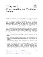
SAM PLE-SHEET
E.S.R. 18: 874
HASSELBRING, H. Bitter rot. Jour. Agr. Science.
2:47: 3: 37All, pl. 1-4, fig. 1-9. 1906.
Apple
Grape
Pear
Bitter rot
Anthracnose
Ripe rot
[Glomerella cingulata]
Glomerella rufomaculans
Gloeosporium fructigenum
Abstract from original.
HOSTS
Hosts listed are apple, pear, etc. p. 37.
VARIETAL SUSCEPTIBILITY
Most susceptible, Greening, Baldwin;
Winesap, etc. p. 39.
least
susceptible,
DISEASE
NAMES
Following
names
listed;
bitter rot, anthracnose,
ripe rot.
The first most commonly applied. p. 40.
(Continue on other sheets under the pertinent heads
subheads as used in the outline for the term paper.)
and
12
especially dealt with in the article should be written close to the left
margin of the sheet and one or two spaces lower than the last line of the
reference, in a column one above the other.
In the same manner place the names of the disease at the middle
of the sheet and the names of the pathogene at the right of the sheet.
These key-words should be for each sheet, only those which appear
in that particular reference.
If none appear in the reference (for example,
if the name of the pathogene is entirely omitted), write the name generally
accepted and enclose in brackets thus :—
Apple
Bitter rot
The chief purpose of these key-words
and identifying the references later.
[G. cingulata]
is to assist in assorting
INFORMATION
Source of reference.
Having this with each reference will enable
one to readily verify it or correct errors made in copying.
It should
include the abbreviated name of the publication, volume-numeral
(or of
year or number as case may be), colon (:) followed by the page on which
the reference
occurs.
If the article
is first discovered
in the original,
indicate thus :—“‘Orig.”’
Author.
Surname, comma,
initials, period, is the order of the arrange-
ment.
Where there are two or more authors, arrange each name in the
same way connecting the names with ‘‘and” or its foreign equivalent,
or with “commas” and ‘‘and” in the case of more than two authors.
In the case of several authors one may write for example,—Stewart,
F. C.
and others, or et al. In the case of anonymous articles, write in place
of the author’s name the word, Anonymous.
Title. The title of an article follows directly after the author’s name
and should always be in the language of the original if possible. When
only the translation of the title is given precede it with the name of the
original language and the word, title, in brackets, thus:—[Italian title]
“Concerning the influence, etc.’”’ When both the original and the translation of the title are given, the translation follows the original and
is to
be inclosed in brackets.
No abbreviations of the title should be used
except such as appear in the original. Only the first word and proper
names in the title may be capitalized, except when the title is in German.
The title terminates with a period.
Name of society or organization publishing the work follows directly
after the title, the second line beginning directly under the fourth letter
of the author’s surname.
It is followed by a period; abbreviation
allowed.
Name of publication.
Journal, Berichte, bulletin (properly abbreviated) follows directly after the name of the society or organization, or
where these are wanting, directly after the title. When the last word
is not an abbreviation, no punctuation-mark
follows.
Volume-numerals are always to be in arabic and are to be underscored with a wavy line, indicating bold-faced type in printing. The word
volume or its abbreviation (vol.) should not appear in a reference.
Reports are often issued for a given year as for example the Alabama
Agr. Exp. Sta. Report for 1896, no volume-number being given.
In such
15
a case the year-numerals take the place of volume-numerals and the above
would appear thus:—Alabama Agr. Exp. Sta. Rept. 1896:1-87.
Number-numerals when indicating consecutively paged parts of a
volume (as for example, vol. 27, No. 6 pp. 70-83) should not appear in
the reference.
When not paged consecutively the number-numeral
appears between volume-numeral and page-numeral.
Bulletins, circulars, memoirs, leaflets and the like are usually numbered
and are not considered as constituting consecutively paged numbers of
avolume.
The numerals appear therefore in place of the volume-numerals,
but are not to be underscored.
The proper abbreviation for the word
Bulletin is Bul. with a capital B. Where qualified, as for example, Technical bulletin, it is abbreviated but not capitalized.
Series, Abteilungen.
If a periodical is issued in two or more separate
series the serzes-numeral precedes the volume-numeral, separated from it
by a colon and is not to be underscored, as:—3:46: 23-87.
Sometimes
the series is indicated thus:—Science n. s. 42: 47-56.
The n.s. = New
Series.
Parts.
If parts of a volume are paged separately, the part-numeral
follows the volume-numeral and is separated from it by a colon:—thus,
82 :2:241-256.
Pages.
The page-numerals when preceded by volume- or numbernumerals and a colon are to be cited inclusively, 1.e. first and last pages
separated by a dash thus:—47-83, and followed by a period.
Tables, plates and figures follow directly after the pages, properly
abbreviated, followed by the proper numerals in arabic.
Year.
This must be the actual year of publication.
It may not
always be determined with certainty except by consulting the original.
Exercise care on this point and leave the space blank until the original
can beconsulted, if not absolutely sure of it from the reference.
Books.
In cases where no volume is given, cite the pages thus:—p. 27
or p. 1441.
Citation from books.
Following the date, give the title of the chapter
or paragraph, especially referred to, then, Ju, then the title of the book
followed by the letter p. to indicate page, then the page number and finally
the date thus :—
Ellis, J. B. Dothidea pomigena. Schw. Jn The North American
Pyrenomycetes. p. 605.
1892.
Government publications are always to be cited in the following form;—
Wes. sec, Dept. Yearbook: or U.S. Plant Ind. Bur. Bul. 37:
State Experiment Station publications are always to be cited thus:—
Alabama Agr. Exp. Sta. Bul.; or New York (Geneva) Agr. Exp. Sta.
Rept.; Cornell Univ. Agr. Exp. Sta. Bul. 237:46-91.
Note:—For further details and examples of correct citation see
pamphlet ‘‘Notes for guidance of author’s.’”” A copy may be checked from
the departmental library.
PREPARATION
OF AN
ABSTRACT
OF AN
ARTICLE
The abstract should always be made from the original, unless for some
good reason this is impossible.
Do not copy the abstract of the article
as it appears in the Experiment Station Record or elsewhere.
14
Begin the abstract on the reference-sheet directly beneath the keywords.
Use both sides of the paper or use additional sheets or both if
desired.
One should abstract as he reads, assorting and entering data under
the proper head as indicated in the outline for writing term paper. (See
term—paper outline and sample-sheet in this outline p. 11.)
Follow each entry of data with the number of the page on which it
was found in the original. This will enable one, when writing the
term paper to readily cite the source of any statement made.
The abstract should be full enough that it will not usually be necessary
to refer again to the original in preparing the term paper.
REPORT
Hand
in the bibliography of the subject chosen, arrange as follows:—
1. The
references,
each
on
a
separate
sheet.
Arrange
the
sheets in the manilla folder provided, in the order of importance of the
references,—the most important first.
2. One or more of the most important references carefully
abstracted as above described.
3. The folder labeled thus:—
(Name of host)
(Name of disease)
Bibl.
(Name of student).
NECROTIC
FIRE
DISEASES
BLIGHT
This is the most common and best known bacterial disease of plants
occurring in this country.
It affects apples, pears, quinces and occasionally plums, apricots and a few ornamental and wild plants related to the
apple family.
SYMPTOMS
The symptoms of this disease will be studied in the order in which
they manifest themselves during the season on different parts of the tree,
beginning with the appearance of the cankers in the spring.
Hold-over cankers.
These are the sources of inoculum for the first
infection of the blossoms in the spring. Study the typical cankers on
the limbs of apple and pear trees provided and OBSERVE :—
1. The smooth, more or less sunken area in the bark,—the canker;
its margin sharply defined by a definite crack.
In cankers in which
the pathogene is active this margin is not sharply defined.
(See illustration specimens if available; photograph 1; Cornell Bul. 272, fig. 16, or
329, fig. 114.)
2. The margin.
Note that it is irregular, the crack being
formed by the drying away of the diseased tissue from the healthy when
the active progress of the pathogene is suddenly checked.
Dry or cold
weather may thus check the enlargement of the canker.
These specimens
were collected in the autumn or winter.
3. The surface of the canker.
Note
that it is smooth,
seldom
roughened or wrinkled.
It is often checked at the margin by drying.
Compare with the healthy bark in this respect.
Locate the lenticles.
What is their structure and function?
Make a V-shaped cut across the margin of the canker and DETERMINE :—
4. How deeply the disease penetrates.
What
tissues are
affected?
Make a DRAWING
of the canker studied.
Label fully. These cankers
are formed during the summer and early autumn and in many of
them the bacteria pass the winter dormant, or only slightly active
in the partially living tissues along the margin.
With the increased
temperature and the beginning of growth-activities in the spring, these
bacteria become active, work rapidly into the adjoining healthy tissue,
increase the area of the canker and ooze out through the lenticles to the
surface in sticky, milky drops. Study photograph 1; Cornell. Bul. 272,
fig. 16, or 329, fig. 114.
OBSERVE :—
5. That the advancing margin of the canker is not distinctly
evident here. The diseased area covers nearly all the surface shown in the
illustration, except on the extreme left.
6. The large viscid milky drops ooz ng out and running down the
limb; above to the left, two small globules just oozing out from the lenticles. Read Cornell Bul. 272:40-41, also Ontario Bul. 176: 15-238.
15
16
Make a DRAWING from photograph 1; Cornell Bul. 272, fig. 16, or 329,
fig. 114. Label fully.
Blossom-blight. Bees and flies visit these active cankers in the spring,
to feed on the exuding sap and then visit the opening blossoms where they
leave behind them some of the bacteria which adhered to their bodies.
Herein the nectar and in the injuries made by the insects’ claws in the tender
tissue of the flower, the bacteria multiply rapidly killing the blossoms.
Study the specimens provided; Ontario Bul. 176, frontispiece; Cornell Bul.
329, fig. 112; or photograph 2. OBSERVE :——
7. The dead and blackened flowers.
The leaves of the spur are
also dead and brown.
The bacteria have spread down the pedicles into
the spur.
These dead and blackened blossom-spurs are usually the first
striking evidence of the disease in the spring. The oozing cankers are
usually overlooked.
Make an outline DRAWING of a blighted blossomspur.
Fruit-blight.
Frequently only one blossom on a spur is infected and
by the time the bacteria have killed it and worked their way down the
pedicle to the spur itself, the uninfected blossoms have developed fruit
of a considerable size. From the spur the bacteria now work into the base
of these fruit-pedicles and by way of them into the growing fruit. Study
photograph 3; Ontario Bul. 176, fig. 15-19; or illustration specimen.
OBSERVE -—
'
8. The blackened pedicle and discolored lower half of the fruit.
A fruit affected in this way usually shrivels and drops from the tree. It
may cling to the twig as a blackened and shriveled mummy.
The loss
from fruit-blight is sometimes heavy. The curculio and aphids frequently introduce the bacteria into the fruit through their punctures.
The pathogene does not always enter the fruit by way of the pedicle.
Note that the leaves of the spur are also dead and shriveling.
In rainy,
muggy weather the bacteria ooze from these blighted fruits and blossoms
in sticky drops as they do from the hold-over cankers.
Study photographs
4a and 4b. OBSERVE :—
9. The discolored and slightly sunken tissues about the base of
the stem and extending toward the blossom end. The sticky, milky
drops oozing from the diseased area. : A large one on the pedicle. Make
DRAWINGS (from specimens or photographs) showing these different phases
of fruit-blight.
Twig-blight.
The bacteria from the diseased blossoms and fruits
are carried by sucking insects to the tips of the growing shoots and watersprouts and are there introduced through the wounds or punctures made
by the insects, into the tender succulent tissues.
Here they multiply
rapidly killing the shoot, causing the form of the disease known
as twig-
blight. Blighted and healthy twigs (fresh and preserved) are provided.
Examine and OBSERVE :—
10. The contrast between the diseased and healthy portions
of both the twigs and leaves.
You may be able to find the dried ooze.
(See illustration specimen.)
SKETCH to show contrast between diseased
and healthy twigs.
11. That in some of the specimens the dormant buds in
the axils of the leaves just below the blighted portion have been prematurely forced.
Explainthis. (See illustration specimen or photograph 6.)
17
Origin of the cankers.
Examine the specimens of cankers again
very carefully and OBSERVE :—
12. That the cankers almost always surround the base of the
spur, twig or watersprout, or that there is at least one or more within
Sometimes it is a fruit-spur, the bacteria had entered by way
the area.
of the blossoms; more frequently it is a water-sprout, the tender succulent growth of which is very favorable for the rapid development
of the disease. The bacteria kill the water-sprout down to the trunk
or limb and spread out into the bark often for a considerable distance
around the base of the sprout. (See illustration specimens and photograph
8.) If the previous DRAWING of the canker does not show the dead spur
or water-sprout, DRAW to show it.
Sometimes the pruningCankers occasionally originate in other ways.
Cankers may start from insect-wounds
knife carries the inoculum.
in the bark.
(See Ontario Bul. 176:33-38.)
ETIOLOGY
The organism that causes this disease is Bacillus amylovorus
(Burrill)
Trevisan.
Laboratory studies in the life-history of this pathogene,
Life-history.
aside from the facts brought out in the above study of symptoms, are
necessarily limited to a short study of the morphology of the bacillus.
Before proceeding with the following observations the student should
have read carefully that portion of the text* dealing with the life-history
of the organism.
If pure cultures are available, examine them carefully and OBSERVE :—
13. The character of the growth on the surface of the solid
How long has it taken to
media. The effect on the bouillon. Why?
produce this effect? What does that indicate as to the rapidity of multiMake amount froma pure culture and before covering introplication?
duce a bee’s foot. OBSERVE:—
14. The size and motility of the organism; cell-unions.
Compare it in size with the claw of the bee’s foot. It may now be understood
how a bee or fly might carry thousands of these on its feet. DRAW the
bee’s claw and some bacteria beside it. Maintain relative proportions.
Study photograph 9 or the stained mount under the demonstration
microscope and MAKE OUT:—
15. The cilia; their number, length and distribution over the
thallus. They are peritrichic. It is this character that places the organism
in the genus Bacillus.
If fresh material is available crush a bit of the most recently affected
tissue in water on a slide, cover and try to DEMONSTRATE :—
16. The living bacteria. How do they compare in size, form and
abundance with those from the pure culture? Are they motile?
Pathological Histology.
Cross-sections of fresh growing twigs showing both healthy and diseased tissues are to be studied (supplemented with
stained section provided).
Examine the healthy portion and OBSERVE :—
17. The reddish brown outer coat, made up of the epidermis
and the several layers of cork-cells beneath.
*This refers to any text which is available.
18
18. The large globose or oval cortex-cells, closely packed next
the cork-layer but with looser arrangement toward the wood.
Their
color and contents.
19. Just beneath and partly surrounded by the cortex-cells,
bundles of white densely packed sclerenchyma-fibers (bast).
20. Toward the center in the following order, the phloem,
cambium, and xylem.
21. At the center of the cross-section,—the pith; the medullary
rays distributed radially in the vascular cylinder.
Study the diseased portion of the twig, comparing the blighted tissues
with the healthy tissues already examined.
OBSERVE :—
22. The affected area. Which tissues are involved?
How
recognized? Are all the cells within the diseased region killed?
23. The effect of the pathogene on the epidermis, cork, cortex,
sclerenchyma-fibers, medullary rays, phloem, cambium, xylem and pith.
Compare with the healthy condition of the cells as to form, size, color and
contents.
24. The relatively large cavities scattered throughout the affected
portion.
How may these be accounted for?
25. The stratified appearance just outside of the phloem.
To
what due?
26. The apparent absence of the bacillus. Why not evident?
DRAWINGS:—(a) Make a diagrammatic drawing to show the healthy
and diseased tissues..
(b) A much larger and detailed drawing of a portion
of a cross-section of the affected twig. Make the drawing include both
healthy and diseased bark.
Study closely the margin of the lesion. In case the section was taken
from a twig in which the pathogene was inactive the diseased and healthy
tissues will be separated by a cork-layer.
If the pathogene was active
no cork-layer will have yet been formed.
Determine which was the
condition in the twig from which the section provided wascut.
In theapex
of growing twigs, the parasite may invade the xylem-ducts and thus
migrate some distance in them down the twig.
REPORT
1. In working out methods of control, of what importance are
the following facts about fire-blight :—
a. It occurs only in North America.
b. The bacteria causing the disease pass the winter in holdover cankers in any of its numerous hosts.
c. The bacteria get into the host through wounds.
d. The chief agents of inoculation are certain insects.
e. The bacteria are usually introduced into the young and
growing parts of the host where in these succulent tissues they multiply
and develop the disease very rapidly.
STREAK
This bacterial disease
legumes including clover.
OF SWEET
PEAS
affects not only sweet
peas but many
SYMPTOMS
Examine the specimens provided (fresh, dry, or in liquid).
other
OBSERVE :—
On the stems.
1. The streak-like lesions; color, extent, part of stem affected.
Look for young isolated lesions; old lesions.
How do they differ as to
extent and color.
pRAaw to show variation in stem-lesions.
Label to
indicate colors.
On the leaves and tendrils.
2. The similarity of the lesions to those on the stem.
Find a
stem-lesion that has extended along the petiole into the blade at its base.
Note the reddening of the affected veins and the dead blade-tissues between.
DRAW.
3. Locate isolated lesions or spots in the leaf. Study carefully.
DRAW and label fully as to markings and colors.
On the flower.
4, The character of the lesions on the petals.
in dark flowers.
DRAW.
Especially striking
ETIOLOGY
The cause of this disease, long unknown, has recently been shown to be
Bacillus lathryi Manns and Taubenhaus.
This pathogene appears to
- be widely distributed in England and United States.
Life-history.
Little is certainly known of the habits of this parasite.
It is supposed to pass the winter in the soil and decaying plant parts which
were killed the previous year.
The Primary Cycle is initiated about the time the peas begin to
blossom.
Pathogenesis.
The bacteria in the soil are splashed by the rain
upon the plants and gain entrance to the tissue through the stomata.
Peel the epidermis from the stem of a healthy sweet pea. Mount outside
up and OBSERVE :—
5. The epidermal cells; their shape and arrangement.
The
stomata; numbers
and structure.
pDRAw.
These stomata
are the infec-
tion-courts.
The bacteria in the moisture on the stem pass through the
stomatal opening into the substomatal cavity. Here they multiply and
by their toxic secretions kill the adjoining cortical cells. The disorganized
juices of these cells diffuse into the substomatal cavity affording food for
further growth and multiplication of the pathogene.
Make sections through a young lesion on the stem (or use prepared
slides).
Examine and if possible DETECT :—
6. The bacteria in the tissues. Are they between or in the cells?
DRAW to show the relation of the bacteria to the cells.
Saprogenesis. Whether the pathogene is able to live and multiply in the soil is an open question.
It is, however, readily cultivated
19
20
:
on nutrient media where it can be better studied than in the tissues of the
host. Examine the plate-colonies of Bacillus lathryi. OBSERVE :—
7. Their form, size, color and consistency.
DRAW.
Mount a bit of one of the colonies in a drop of water.
OBSERVE :—
8. The
size, shape,
motility
and
cell-unions
Cover and
of the bacteria.
DRAW; and copy also from Delaware Bul. 108:22, fig. 1.
Secondary Cycles originate from bacteria oozing from the primary
lesions and spattered by rain to nearby leaves and stems.
They do not
differ in any other way from the primary.
Pathological Histology. Study the prepared sections (cross- and longisections) through lesions on the stem.
OBSERVE :—
9. That at first the bacteria occupy the intercellular spaces,
adjacent to the substomatal cavity.
10. That these soon break down the cell-walls and invade
the cell-cavities.
(See Deleware Bul. 108, pl. I.) To what is the color
in the lesion due?
11. That not all the tissues of the stem are invaded.
Determine
which.
Show this in a DRAWING of the section through the stem-lesion.
Pathogenicity Studies.
Under the direction of the instructor the
student
will, if fresh material
is available,
make
isolations
from
stem-
lesions on nutrient agar or potato-agar in a petri-dish. Set the petri-dish
away until the next exercise at which time compare with the pure culture
studied above.
If growing sweet peas and clover are available, inoculate under the
direction of the instructor, with these pure cultures of the pathogene.
Examine the plants after two days and again later for evidences of infection.
Record in your notes the length of the incubation-period.
REPORT
1. Describe fully how the pathogene was isolated, character of its
growth in the media used, how the inoculations were made and the results.
BEAN
BLIGHT
This is the common bacterial disease of beans.
It apparently affects
all varieties of Phaseolus vulgaris L., the field- and garden-bean, as well
as the Lima bean, Phaseolus lunatus L.
SYMPTOMS
This disease affects all parts of the host above the ground,
stems, pods and seeds.
On the leaves.
Examine the leaves provided and OBSERVE :—
leaves,
1. The location, size, color and general appearance of the lesions
on both sides of the leaf.
2. The zonate character of the spots. How do you account
for each zone, especially the opaque, green water-soaked zone which shows
best when the leaf is held to the light; also the pale green outermost zone?
Draw to show these characters and indicate fully in the labeling, the
color and appearance.
On the stems. The affected stems show no distinct lesions since only
the vascular system, as arule, is usually involved.
Diseased stems finally
shrivel following the wilting of the leaves.
This symptom is not common.
On the pods. Examine the pods provided and OBSERVE :—
3. The location, size, color and water-soaked appearance of
the lesions.
Pod-lesions are sometimes red-bordered.
(See illustration
specimens. )
4. Depth to which the lesions penetrate.
Determine by cutting
across the pod through a lesion.
Draw to show form and depth of the
lesion.
Label fully as to color and character.
On the seed. Examine the lesion with a hand-lens and OBSERVE :—
5. The discolored, roughened surface.
To what is this due?
What relation do the lesions on the seed bear to the pod-lesions?
Make a
DRAWING showing the character of the lesions on the seed.
Examine the passe-partouts provided and compare the symptoms of
this disease with those of the anthracnose, a fungous disease of the bean
which is also very prevalent.
Make prawincs of a pod, seed and leaf
affected with anthracnose, showing how the symptoms of that disease
differ from those of the bean blight.
ETIOLOGY
The cause of this disease is Bacterium Phaseoli E. F. Smith
monas Phaseolt EFS.), a monotrichic
bactertum
(=Pseudo-
which produces yellow
colonies on agar.
Life-history.
This organism is a restricted parasite, normally passing
its entire life within the bean.
It probably has no true saprogenic phase
in its life-history.
The Primary Cycle is initiated in the spring from the cotyledons
of diseased seedlings.
Pathogenesis. Diseased seed have been soaked a short time in water.
Peal the seed-coat
from
the lesion.
Make
two
mounts,
one
from
the
diseased seed-coat and one from the diseased cotyledon, chopping and
crushing the tissues. OBSERVE :—
6. The minute, short rod-shaped bacteria to be detected only
with the high-power.
Are they inside or outside the cells? DRAw.
21