Báo cáo khoa học: Structures of Phanerochaete chrysosporium Cel7D in complex with product and inhibitors ppt
Bạn đang xem bản rút gọn của tài liệu. Xem và tải ngay bản đầy đủ của tài liệu tại đây (695.6 KB, 13 trang )
Structures of Phanerochaete chrysosporium Cel7D
in complex with product and inhibitors
Wimal Ubhayasekera
1
, Ine
´
s G. Mun
˜
oz
1,
*, Andrea Vasella
2
, Jerry Sta
˚
hlberg
1
and Sherry L. Mowbray
1
1 Department of Molecular Biology, Swedish University of Agricultural Sciences, Uppsala, Sweden
2 Laboratory of Organic Chemistry, ETH, Ho
¨
nggerberg, Zu
¨
rich, Switzerland
Cellulose is the most abundant polymer on earth. It
has been estimated that as much as 15% of all atmo-
spheric carbon dioxide is fixed yearly, resulting in vast
quantities of plant biomass, mostly as a complex mix-
ture of cellulose and lignin [1]. The recycling of this
carbon is critically dependent on the action of micro-
bial organisms, primarily fungi and bacteria. An
understanding of the processes at work is obviously of
enormous environmental importance. The enzymes
involved are also useful for applications that include,
among others, their use in commercial laundry pow-
ders, as well as in the de-inking of recycled paper and
the synthesis of fine chemicals.
Cellulases, the enzymes that hydrolyse cellulose,
have been broadly characterized as cellobiohydrolases
(1,4-b-d-glucan cellobiohydrolase, EC 3.2.1.91) and
Keywords
Cellulase; cellobiohydrolase; glycoside
hydrolase; Trichoderma reesei;
Phanerochaete chrysosporium
Correspondence
J. Sta
˚
hlberg, Department of Molecular
Biology, Swedish University of Agricultural
Sciences, Biomedical Centre, PO Box590,
SE-751 24 Uppsala, Sweden
Fax: +46 18 536971
Tel: +46 18 471 4566
E-mail:
*Present address
Structural Biology and Biocomputing
Programme, Spanish National Cancer Centre
(CNIO), Melchor Ferna
´
ndez Almagro 3,
28029 Madrid, Spain
(Received 6 December 2004, revised 15
February 2005, accepted 22 February 2005)
doi:10.1111/j.1742-4658.2005.04625.x
The cellobiohydrolase Pc_Cel7D is the major cellulase produced by the
white-rot fungus Phanerochaete chrysosporium, constituting 10% of the
total secreted protein in liquid culture on cellulose. The enzyme is classified
into family 7 of the glycoside hydrolases and, like other family members,
catalyses cellulose hydrolysis with net retention of the anomeric carbon
configuration. Previous work described the apo structure of the enzyme.
Here we investigate the binding of the product, cellobiose, and several
inhibitors, i.e. lactose, cellobioimidazole, Tris ⁄ HCl, calcium and a thio-
linked substrate analogue, methyl 4-S-b-cellobiosyl-4-thio-b-cellobioside
(GG-S-GG). The three disaccharides bind in the glucosyl-binding subsites
+1 and +2, close to the exit of the cellulose-binding tunnel ⁄ cleft.
Pc_Cel7D binds to lactose more strongly than cellobiose, while the oppos-
ite is true for the homologous Trichoderma reesei cellobiohydrolase
Tr_Cel7A. Although both sugars bind Pc_Cel7D in a similar fashion, the
different preferences can be explained by varying interactions with nearby
loops. Cellobioimidazole is bound at a slightly different position, displaced
2A
˚
toward the catalytic centre. Thus the Pc_Cel7D complexes provide
evidence for two binding modes of the reducing-end cellobiosyl moiety; this
conclusion is confirmed by comparison with other available structures. The
combined results suggest that hydrolysis of the glycosyl-enzyme intermedi-
ate may not require the prior release of the cellobiose product from the
enzyme. Further, the structure obtained in the presence of both GG-S-GG
and cellobiose revealed electron density for Tris at the catalytic centre.
Inhibition experiments confirm that both Tris and calcium are effective
inhibitors at the conditions used for crystallization.
Abbreviations
GG-S-GG, methyl 4-S-b-cellobiosyl-4-thio-b-cellobioside; IBTG, o-iodo-benzyl-b-
D-thio-glucoside; Pc_Cel7D, cellobiohydrolase Cel7D from
Phanerochaete chrysosporium; PDB, Protein Data Bank; pNP-Lac, p-nitrophenyl-b-
D-lactoside; Tr_Cel7A, cellobiohydrolase Cel7A from
Trichoderma reesei.
1952 FEBS Journal 272 (2005) 1952–1964 ª 2005 FEBS
endoglucanases (1,4-b-d-glucan glucanohydrolase,
EC 3.2.1.4) [2]. Cellobiohydrolases tend to act proces-
sively from the end of a cellulose chain, that is, they
cleave off a number of cellobiose units in succession
before the enzyme is released [3,4]. Endoglucanases cut
cellulose at random positions within the chains, thus
creating new ends from which cellobiohydrolases can
work. Efficient degradation of cellulose requires a
synergistic balance between the two types of activities.
Cellulases and other glycoside hydrolases have been
classified into structurally related families, based on
sequence homology as well as the patterns of hydro-
phobic residues [5,6]. To date nearly 100 glycoside
hydrolase families are defined in the CAZY database
( Efficient cellulose-
degrading fungi generally have at least one member
of glycoside hydrolase family 7. The enzymes in this
family perform hydrolysis with net retention of the
anomeric configuration, in a double-displacement
mechanism through a covalent glycosyl-enzyme inter-
mediate [7,8]. Most, but not all, members have a small
cellulose-binding module connected to the catalytic
module by a presumably flexible linker. The catalytic
core of this family is a b-sandwich composed of two
large, mainly antiparallel, b-sheets packed onto each
other (Fig. 1). A long cellulose-binding site is defined
by loops on one face of the sandwich. It has been
demonstrated, for this and some other structural famil-
ies, that a very important difference between an endo-
glucanase and a cellobiohydrolase is the size of such
loops. In a cellobiohydrolase, they are generally lon-
ger, and form a tunnel that encloses the catalytic resi-
dues. Substrate usually reaches the active site by
threading itself in from the end of the tunnel. In con-
trast, an endoglucanase has shorter loops that define a
more open binding cleft, and allow more direct access
of an intact cellulose chain.
Among the fungi that have a family 7 cellobiohydro-
lase, it is the major enzyme in the cellulase mixture
secreted. The first member of the family for which the
structure was determined was the cellobiohydrolase of
Trichoderma reesei (a clonal derivative of Hypocrea
jecorina), Tr_Cel7A, formerly called CBH 1 [9]. Three
acidic residues (Glu212, Asp214 and Glu217) were
shown to be responsible for cleavage of the cellulose
chain. Further studies allowed a complete mapping of
cellulose binding along the 50 A
˚
-long active site tunnel
[10,11]. Tr_Cel7A binds 10 glucosyl units in subsites
)7 to +3 (numbering starts from the point of glycosi-
dic bond cleavage, between )1 and +1; negative num-
bers indicate the nonreducing end of the cellulose
chain, and positive numbers, the reducing end [12]).
The +1 and +2 sites are often designated as the
‘product sites’, as they bind the cellobiose unit that will
-7
-4
-2
-1
+1
+2
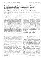
Fig. 1. Binding of disaccharides to Pc_Cel7D. Overall structure of Pc_Cel7D with cellobiose bound in the +1 ⁄ +2 sites. Backbone of the
enzyme’s catalytic domain is coloured terracotta, aromatic side chains that form cellulose-binding subsites are green, and the three acidic
residues involved in catalysis are red. Cellobiose is indicated by a ball-and-stick model coloured light blue. Numbers indicate the position of
some of the glucosyl-binding subsites.
W. Ubhayasekera et al. Cel7D ⁄ saccharide complex structures
FEBS Journal 272 (2005) 1952–1964 ª 2005 FEBS 1953
be cleaved off at the reducing end of the chain
(Fig. 1). These sites are placed close to the exit of the
binding site cleft⁄ tunnel, which should simplify the
subsequent release of the disaccharide. However, prod-
uct inhibition is commonly observed for cellobiohydro-
lases [13–16].
Structures are also known for three endoglucanases
of family 7, from T. reesei [17], Humicola insolens [18]
and Fusarium oxysporum [19]. The active sites of these
enzymes are very similar and the enzymes are believed
to use the same catalytic mechanism as the cellobio-
hydrolases. As expected, the loops flanking the cellu-
lose-binding cleft in each case are significantly shorter,
leaving the active sites completely open to solvent.
Cellobiohydrolase Cel7D (previously called CBH 58)
is the major cellobiohydrolase produced by the basi-
diomycete Phanerochaete chrysosporium under most
growth conditions [20]. We recently solved the struc-
ture of Pc_Cel7D, and showed that it is similar to
Tr_Cel7A [21]. The catalytic residues were identified as
Glu207, Asp209 and Glu212. Nearly all interacting
residues of Tr_Cel7A are conserved, which suggested
that Pc_Cel7D would bind cellulose in much the same
way. However, several deletions make the binding
tunnel slightly more open in Pc_Cel7D.
A recent comparative study revealed striking differ-
ences in the activity on insoluble model substrates:
although Pc_Cel7D had only slightly higher activity on
cellotetraose, it hydrolysed amorphous and bacterial
microcrystalline cellulose eight times and 4.4 times fas-
ter, respectively, than Tr_Cel7A. Enzyme kinetics on
p-nitrophenyl lactoside gave similar k
cat
values for the
two enzymes; however, Pc_Cel7D showed a threefold
higher K
m
(and hence threefold lower k
cat
⁄ K
m
) as well
as reduced cellobiose inhibition (eight times higher K
i
)
[22]. Furthermore, estimation of specificity constants
(k
cat
⁄ K
m
) for dinitrophenyl-cellooligosaccharides with
2–5 glucose units, pointed at differences between the
enzymes in the relative contribution of intrinsic bind-
ing energy to catalysis at subsites )3to)5. Another
study revealed differences in the binding specificity for
cellobiose and lactose, presumably at the product sites
+1 ⁄ +2. While Tr_Cel7A prefers binding of cellobiose
to lactose, the opposite is true for Pc_Cel7D [23].
As part of global efforts to replace fossil fuels with
renewable energy sources, cellulases have received
increasing attention as a possible means of converting
cellulosic biomass to fermentable sugars for ethanol
production [24]. However, the enzyme cost is a critical
factor, and improvements in the efficiency of the pro-
cess will directly influence whether such ‘bioethanol’
can effectively compete with petroleum [25]. The major
industrial source of cellulase enzymes at present is
T. reesei [26]. Deletion of individual cellulase genes in
T. reesei showed that Tr_Cel7A was rate limiting in
the degradation of crystalline cellulose in the fungal
system [27]. Understanding the molecular details of
how the Cel7 enzymes work thus lies at the heart of
finding the best solution in future applications.
In the present paper, we report three structures of
Pc_Cel7D in complex with disaccharides: the product
(cellobiose) and two inhibitors (lactose and cellobio-
imidazole). These structures provide a picture of two
different glycosyl binding modes, as well as explaining
the differences in affinity between the two natural
sugars. A structure obtained in the presence of cellobi-
ose, methyl 4-S-b-cellobiosyl-4-thio-b-cellobioside (GG-
S-GG), Tris ⁄ HCl and calcium revealed that Tris binds
in the active site. In kinetic studies, we show that
Pc_Cel7D is in fact inhibited by both Tris and calcium
at the concentrations used in the crystallization; this
is the first report of such behaviour within the family.
Results
Overall structures
Deglycosylated Pc_Cel7D catalytic module was crystal-
lized in the presence of two natural disaccharides, cell-
obiose and lactose, as well as with cellobioimidazole, a
compound that mimic the transition state of some cel-
lulases [28]. The crystals were isomorphous with pre-
vious ones [21] and complete diffraction data sets to
1.7 A
˚
resolution or better could be collected using syn-
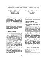
chrotron radiation. In all three cases, clear electron
density was found for the bound ligand prior to its
inclusion in the models (Fig. 2). Statistics relating to
the diffraction data and the final refined models are
summarized in Table 1. Each model contains the com-
plete catalytic module of Pc_Cel7D (residues 1–431),
an N-acetylglucosamine residue bound to Asn286, one
molecule of the respective ligand and a number of
bound waters. The protein structures are very similar
to each other and to the published structure of
Pc_Cel7D {Protein Data Bank (PDB) [29] entry code
1GPI [21]} with overall r.m.s. differences of 0.2–0.3 A
˚
when all Ca atoms are compared pair-wise.
Binding of cellobiose to Pc_Cel7D
Product inhibition in Pc_Cel7D is consistent with the
observed binding of cellobiose in the +1 ⁄ +2 (pro-
duct) sites of Pc_Cel7D (Figs 1, 2A and 3A). The
nonreducing end of the disaccharide is in the +1 site;
this glucosyl unit shows the ‘classical’ stacking on a
tryptophan residue that is a feature in many proteins
Cel7D ⁄ saccharide complex structures W. Ubhayasekera et al.
1954 FEBS Journal 272 (2005) 1952–1964 ª 2005 FEBS
interacting with carbohydrates [30]. The hydrophobic
B-face of the sugar thus makes a number of nonpolar
contacts with the indole ring of Trp373 (Fig. 3A). The
interactions on the opposite (A) face of this carbohy-
drate unit are more polar. O2 interacts with the main-
chain carbonyl oxygen of Asp248. O3 and O4 are
linked to the catalytic acid Glu212 via hydrogen bonds
with water. In addition, the guanidino group of
Arg240 forms hydrogen bonds with O5 and O6. The
electron density for the side-chain atoms of Arg240 is
slightly weaker than average. Both the structural set-
ting and the density suggest that the interactions of
Arg240 with sugar compete with a salt link to Asp248,
and a hydrogen bond to Gln172. Fewer interactions
are seen in subsite +2, and electron density of this
glycosyl unit is also somewhat weaker than that
observed for the +1 sugar. The observed interactions
are on the same side of the cleft as those in the +1
subsite. The guanidino group of Arg391 is within
hydrogen bonding distance to O1, O5 and O6 of the
sugar. O1 also interacts with Asp336, and O6 with a
solvent molecule. There is, however, no aromatic
stacking in the +2 subsite.
Both glucosyl rings adopt a regular
4
C
1
chair, i.e. a
favourable conformation in solution. The planes of
the two sugar units have opposite orientations, with
torsion angles (/ ¼ )78°, w ¼ +120°) that deviate
slightly from those observed in the small-molecule
crystal structure of cellotetraose ()93, +96, and )93,
+86) [31]. Of the inter-residue interactions that stabil-
ize cellulose chains, only the O3
i+1
–O5
i
hydrogen
bond is present; that between O6
i+1
and O2
i
is lack-
ing. The less common gauche-gauche conformation of
the exocyclic C6–O6 bond is apparently stabilized by
its interaction with Arg391, which is preferred to an
intramolecular one with O2. The sugar is tightly sand-
wiched between the walls of the product sites by the
interactions with protein described above. The hydro-
xyls along one edge of the disaccharide point into the
binding cleft, where several water molecules are found;
hydroxyls along the other edge point out toward the
bulk solvent.
Binding of lactose to Pc_Cel7D
Lactose is an effective competitive inhibitor of
Pc_Cel7D (Table 2). The only chemical difference
between this disaccharide and cellobiose is the confi-
guration at C4 in the galactosyl unit: the hydroxyl
group is equatorial in cellobiose, and axial in lactose.
As might be expected, lactose binds in the +1 ⁄ +2
subsites in a manner very similar to that described
+2
+1
-1-2
-3
-4
-5
A cellobiose B lactose C cellobioimidazole
D GG-S-GG + TRIS + cellobiose
Fig. 2. Electron density for ligands bound in Pc_Cel7D. Final 2F
o
-F
c
maps contoured at 1 r, are shown for (A) cellobiose (B) lactose (C) cello-
bioimidazole and (D) GG-S-GG, Tris and cellobiose. The numbers for the glucosyl-binding subsites indicate the location within the substrate
binding tunnel of Pc_Cel7D.
W. Ubhayasekera et al. Cel7D ⁄ saccharide complex structures
FEBS Journal 272 (2005) 1952–1964 ª 2005 FEBS 1955
above for cellobiose (Figs 3B and 4A). However, the
axial placement of O4 allows the disaccharide to make
a direct hydrogen bond to Arg240. The guanidino
group of that side chain has rotated slightly, so that
it interacts with O4 and O5, instead of O5 and O6
(Fig. 3B). At the same time, Arg240 can make more
favourable interactions with Gln172 and Asp248. O6
now appears to lack a fixed hydrogen-bonding part-
ner. The interactions of the glucosyl unit in the +2
site are the same as those observed for cellobiose.
However, the electron density for both sugar and
protein in the immediate area is significantly better
than that observed for cellobiose (Fig. 2A and B),
and the temperature factors for the ligand are corre-
spondingly lower (Table 1). These differences provide
a structural basis for the observation that Pc_Cel7D
binds more tightly to lactose than to cellobiose
(Table 2).
Binding of cellobioimidazole in Pc_Cel7D
Monosaccharide-derived imidazoles such as cellobio-
imidazole (Fig. 2C), feature an sp
2
-hybridized ano-
meric centre and a charge distribution that mimics the
transition state of some exoglycosidase reactions
[32,33]. Cellobioimidazole is apparently not sufficiently
compatible with the transition state of Pc_Cel7D to
promote its binding in the catalytic substrate sites
)2 ⁄ )1. The preferred binding is instead in sites
+1 ⁄ +2, as was seen for the other two disaccharides
(Fig. 3). The cellobioimidazole is, however, shifted
more than 2 A
˚
along the cleft, toward the catalytic
centre (Fig. 4A).
The glucosyl unit in the +1 site continues to make
good stacking interactions with Trp373, although it
now lies more directly against the six-membered ring
of the indole. O2 maintains the hydrogen bond to 248-
O, but the sugar oxygen is displaced 1A
˚
from its
position in the cellobiose and lactose complexes; this is
the only one of the polar interactions that is preserved
in this site. The hydrogen-bonding capacity of O3 is
saturated by interactions with the side chains of
His223, Asp209 and Glu212, and with a solvent mole-
cule. O4 also makes a hydrogen bond with Glu212,
but O6 appears to have no hydrogen-bonding partner
in the enzyme. Like the sugars of cellobiose and lac-
tose, the glucosyl unit in cellobioimidazole adopts a
regular
4
C
1
chair conformation.
In the +2 site, the glucoimidazole ring makes
hydrogen bonds to Arg240 and Arg391, as well as to
several solvent molecules. Under the crystallization
conditions (pH 7.0), only a small fraction of the cello-
bioimidazole will be protonated (pK
a
6.1 [34]), and
charge–charge repulsion is apparently not a problem.
Unlike the glucosyl and galactosyl units, the glucoimi-
dazole moiety cannot adopt a
4
C
1
chair conformation
because of its C1–N5 double bond. The most favour-
able solution conformation is the
4
H
3
half-chair
observed in the crystal and NMR structures of
glucoimidazole alone, although several other confor-
mations are also possible within its pseudo-rotational
sequence [35]. In the complex with Pc_Cel7D, the
A
cellobiose
212
391
240
207
373
C
cellobioimidazole
His223
212
207
209
240
373
Arg391
Asp336
Asp248
Arg240
Gln172
Glu207
nucleophile
Asp209
Trp373
Arg256
Glu212
acid/base
B
lactose
+2
+1
Fig. 3. Interactions between the disaccharides and Pc_Cel7D. Hydrogen-bonding interactions are shown for (A) cellobiose (B) lactose and (C)
cellobioimidazole. In each case, the protein is shown with gold carbon atoms, while those of the ligand are yellow. Hydrogen bonds are indi-
cated by cyan-coloured ‘bubbled’ lines. Water molecules interacting with protein and ligand are small light-blue spheres.
Cel7D ⁄ saccharide complex structures W. Ubhayasekera et al.
1956 FEBS Journal 272 (2005) 1952–1964 ª 2005 FEBS
conformation is closest to an envelope form, with C3
out of plane.
The electron density (Fig. 2C) and temperature fac-
tors (Table 1) for cellobioimidazole, as well as its good
interactions with protein, suggest that it should be an
effective cellobiohydrolase inhibitor. No affinity meas-
urements are yet available for this compound with
Pc_Cel7D, but the related Tr_Cel7A is indeed inhibited
by cellobioimidazole, although, at least at pH 5.7, bind-
ing is weaker than for cellobiose (Table 2) [36]. Due to
the close proximity of the imidazole ring to the guanidi-
no groups of arginines 256 and 391, it seems likely that
the ligand will be a better inhibitor at pH 7 or higher,
when the glucoimidazole ring is unprotonated, than at
lower pH. More biochemical data are obviously needed.
Binding of thio-linked substrate analogue
GG-S-GG and cellobiose
Crystals of Pc_Cel7D were soaked with a combination
of cellobiose and the thio-linked sugar GG-S-GG, in
hopes of obtaining a complex that included sugars
bound in both ends of the active-site cleft. As seen
from the electron density in Fig. 2D, the product sites
+1 ⁄ +2 were again completely occupied with disac-
charide, in the position observed with cellobiose alone.
Table 2. Selected binding and kinetic constants for Pc_Cel7D and Tr_Cel7A.
Enzyme
K
d
, cellobiose
(l
M)
K
d
, lactose
(l
M)
K
i
, cellobio-imidazole
(l
M)
K
m
, pNP-Lac
(l
M)
k
cat
, pNP-Lac
(s
)1
)
Pc_Cel7D 115
a
77
a
NA 5100
b
0.17
b
Tr_Cel7A 20
c
310
c
130
d
900
b
0.10
b
a
K
d
values at pH 5.0 and 25 °C from displacement chromatography experiments with Pc_Cel7D immobilized on silica, as published by Hen-
riksson et al. [23].
b
Michaelis–Menten kinetic parameters at pH 5.0 and 25 °C [23].
c
K
d
at pH 5.0 and 25 °C determined by protein differ-
ence spectroscopy [16].
d
Non-competitive K
i
at pH 5.7 and 30 °C from inhibition experiments using 2-chloro-nitrophenyl b-lactoside as
substrate [36]. NA, data not available.
Table 1. Data collection and refinement statistics. The space group was C2. Statistics for the highest resolution shell are given in paren-
theses. A stringent boundary Ramachandran plot was used [47]. Data collection statistics were taken from
TRUNCATE [48]. Other values for
the refined structures were calculated using
MOLEMAN2 [49].
Data collection
Complex
Cellobiose Lactose Cellobioimidazole
Cellobiose, Tris,
GG-S-GG
Environment ESRF, ID14 : 4 ESRF, ID14 : 4 ESRF, ID14 : 2 ESRF, ID14 : 3
Wavelength 0.9370 0.9322 0.9330 0.9310
Cell dimensions 87.4, 46.8, 99.5 86.9,46.7, 99.1 87.4, 46.6, 98.5 86.47, 46.47, 98.36
(A
˚
, °) b ¼ 103.0° b ¼ 102.8° b ¼ 102.6° b ¼ 102.49
Resolution (A
˚
) 50–1.70 (1.73–1.70) 50–1.60 (1.63–1.60) 43–1.70 (1.79–1.7) 96.321–1.7 (1.79–1.70)
Unique reflections 43 328 50 272 39 785 50967
Average multiplicity 4.3 3.4 2.3 3.6
Completeness (%) 99.6 (93.1) 99.3 (99.6) 93.2 (92.4) 96.6
R
merge
9.8 (32.1) 7.2 (23.8) 5.2 (19.4) 10.9
<I⁄ rI > 11.0 (5.8) 14.8 (8.1) 14.0 (3.8) 11.6 (4.3)
Refinement
Number of reflections 40 782 (100.0) 47 274 (100.0) 37 334 (100.0) 37 329 (96.32)
(completeness,%)
Resolution range (A
˚
) 40.0–1.70 39.2–1.61 36.3–1.70 95.35–1.72
R-factor ⁄ R-free (%) 17.2 (20.2) 15.7 (20.2) 17.8 (22.7) 17.1 (23.24)
Number of protein atoms (Average B, A
˚
2
) 3198 (24.7) 3198 (22.2) 3198 (17.5) 3198 (20.6)
Number of water molecules (Average B, A
˚
2
) 106 (27.9) 278 (31.2) 180 (21.9) 389 (30.8)
Number of N-acetyl-glucosamine atoms 14 (37.2) 14 (29.1) 14 (26.5) 14 (34.0)
(Average B, A
˚
2
)
e
Number of ligand atoms (Average B, A
˚
2
) 23 (33.7) 23 (24.0) 25 (16.9) 31 (24.2)
r.m.s bond length (A
˚
) 0.027 0.010 0.006 0.013
r.m.s. bond angle (°) 2.08 1.51 1.21 1.50
No. Ramachandran plot outliers (%) 4 (1.1) 3 (0.8) 3 (0.8) 3 (0.8)
W. Ubhayasekera et al. Cel7D ⁄ saccharide complex structures
FEBS Journal 272 (2005) 1952–1964 ª 2005 FEBS 1957
The other side of the cleft contains additional electron
density not observed in the other Pc_Cel7D structures,
which may indicate binding of the longer sugar at low
occupancy. Although the shape of the density in sites
)4 and )5 bears strong resemblance to glucose rings,
on the whole it is too weak to allow unambiguous
modelling of this ligand within the active site. The low
occupancy is almost certainly due to the limited crystal
soaking time used (2 min); the crystals deteriorated at
longer soaking times. Co-crystallization could not be
used because the long exposure time would lead to
hydrolysis of the O-glycosidic bonds in the ligand. We
are currently seeking other solutions to this problem.
However, clear electron density in the immediate
vicinity of the catalytic residues of this structure was
only compatible with Tris, among the known crystal-
lization reagents. Inhibition by Tris had not been
reported previously for Cel7 enzymes.
Inhibition experiments
The discovery of Tris density in the active site promp-
ted us to undertake a systematic study of the com-
ponents of the crystallization solution. Inhibition
experiments at pH 7.0 using p-nitrophenyl lactoside as
substrate (summarized in Fig. 5) showed that both
10 mm Tris ⁄ HCl and 5 mm CaCl
2
individually inhibit
Pc_Cel7D. As there was no inhibition with 10 mm
NaCl, the Tris and the calcium ions are the inhibiting
species.
Comparison with ligand binding in Tr_Cel7A
To date, five structures have been published for the
related cellobiohydrolase of T. reesei (Tr_Cel7A) with
carbohydrates bound in sites +1 ⁄ +2: the wild-type
enzyme with the inhibitor o-iodo-benzyl-b-d-thio-
glucoside (IBTG; PDB entry 1CEL [9]), an inactive
B
A
C
Fig. 4. Superposition of sugar residues bound in the +1 ⁄ +2 sites of
Pc_Cel7D and Tr_Cel7A. Selected residues of Pc_Cel7D are shown
with gold carbon atoms and of Tr_Cel7A with blue carbon atoms.
(A) Superposition of the three disaccharides as bound by Pc_Cel7D.
Cellobiose (magenta), lactose (lilac) and cellobioimidazole (cyan)
are shown as ball-and-stick representations. Only cellobioimidazole
enters directly into the active site, forming hydrogen bonds to the
catalytic acid Glu212. The interactions of O2 with 248-O in the +1
site, and Arg391 with O6 in the +2 site, are the only direct polar
interactions found in all three complexes (Results). Two additional
conserved interactions are shown, in which water molecules medi-
ate links between sugar hydroxyls and protein residues at the deep-
est point of the binding cleft. The O6 hydroxyl in subsite +2 thus
also interacts with a water molecule bound to Asp251 OD2. A sec-
ond water molecule links Thr221-OG1 to O2 in site +1. These inter-
actions hold O2 of the +1 subsite and O6 of the +2 subsite in very
similar positions in all three complexes. (B) Cellobiose binding near
the catalytic residues in Pc_Cel7D (magenta ligand) is shown
together with the complex of cellobiose with Tr_Cel7A (yellow lig-
and). In this binding mode there is room for water (pale green
spheres) between the O4 hydroxyl of the hexose in site +1 and the
catalytic acid ⁄ base (Glu212 in Pc_Cel7D). (C) Superposition of avail-
able cellobiohydrolase structures with sugars bound in the +1 ⁄ +2
sites highlighting the existence of two discrete binding modes. The
bound sugar residues are colour-ramped using a rainbow, with blue
indicating the position closest to the point of cleavage, and red,
that farthest away. The identity of each complex is indicated by
coloured boxes. Conserved interactions involving water are also
shown.
Cel7D ⁄ saccharide complex structures W. Ubhayasekera et al.
1958 FEBS Journal 272 (2005) 1952–1964 ª 2005 FEBS
E212Q mutant with cellobiose (3CEL [10]), cellotetra-
ose (5CEL [11]) and cellopentaose (6CEL [11]), and a
second inactive mutant (E217Q) with cellobiose
(together with cellohexaose bound in sites )7to)2;
7CEL [11]). The catalytic domains of Tr_Cel7A and
Pc_Cel7D share 55% amino-acid sequence identity,
which provides a good basis for detailed comparison
of the two enzymes.
The complex of Tr_Cel7A with cellobiose (3CEL)
can be superimposed on that of Pc_Cel7D using a
cut-off of 0.7 A
˚
, giving an r.m.s. difference of 0.4 A
˚
for 267 matching Ca atoms ( 60% of the total).
The most similar portions of the enzymes represent
the core b-sheet structure as well as the highly con-
served residues of the active site. Cellobiose is bound
in an equivalent position in the two enzymes
(< 0.8 A
˚
difference for all atoms); the position of
O6 within the +2 site is identical (Fig. 4B).
Although this Tr_Cel7A structure actually represents
an inactive mutant, the mutated residue (equivalent
to Pc_Cel7D’s Glu207) is not directly involved in lig-
and binding, and does not appear to complicate the
comparison. The most significant differences between
the two complexes result from a deletion in one of
the active-site loops of Pc_Cel7D (Fig. 6). The role
of Arg240 in binding to O5 and O6 in the +1 site
is thus assumed by Arg251 in Tr_Cel7A. The main-
chain atoms of Arg251 are 3A
˚
away from those
of Arg240, but the functional guanidino groups are
similarly placed and serve a similar purpose in the
two enzymes. However, Arg251 in Tr_Cel7A is sup-
ported by a better local network of hydrogen bonds,
including Thr246 and Asp259. The longer loop in
Tr_Cel7A also provides one additional direct hydro-
gen bond to the ligand: Thr246 interacts with O6 in
the +1 site. On the other hand, a deletion in
Tr_Cel7A results in the loss of an interaction at O1
in site +2, which can be provided by Asp336 in
Pc_Cel7D (Fig. 6). All of the other interactions that
Pc_Cel7D makes with cellobiose are found intact in
the complex with Tr_Cel7A. Better hydrogen bond-
ing, together with the more enclosed Tr_Cel7A active
site, provides a reasonable explanation for why cello-
biose binds more tightly to this enzyme than to
Pc_Cel7D (Table 2), and so there is less product
inhibition in the latter enzyme.
Fig. 6. Comparison of the Pc_Cel7D and Tr_Cel7A structures near
the exit of the cellulose-binding tunnel. The complex of Pc_Cel7D
with cellobiose is in gold and coral carbons, while Tr_Cel7A is in
green. The sugar is embraced in Tr_Cel7A by the 245–250 loop
(seen at the upper left), but not in Pc_Cel7D that has a six-residue
deletion here. Pro258 in Tr_Cel7A may hold Arg251 out of reach for
O4 of lactose. At the bottom of the figure it is seen that the inser-
tion of Asp336 in Pc_Cel7D results in differences in main-chain con-
formation and provides an additional hydrogen bond with the
reducing-end hydroxyl of a bound cellulose chain.
B
A
Fig. 5. Inhibition of Pc_Cel7D by Tris and other compounds.
Absorbance at 400 nm was measured after a 30-min incubation of
Pc_Cel7D with pNP-Lac at 30 °C, pH 7.0, as described in Experi-
mental procedures. (A) h, No inhibitor; ·,10m
M NaCl; +, 10 mM
Tris ⁄ HCl; –, 5 mM CaCl
2
;*,5mM CaCl
2
and 10 mM Tris ⁄ HCl. (B)
h, No inhibitor; n, 0.1 m
M cellobiose; s, 0.1 mM cellobiose and
10 m
M Tris ⁄ HCl; e, 0.1 mM cellobiose, 10 mM Tris ⁄ HCl and 5 mM
CaCl
2
.
W. Ubhayasekera et al. Cel7D ⁄ saccharide complex structures
FEBS Journal 272 (2005) 1952–1964 ª 2005 FEBS 1959
There is presently no structure available for
Tr_Cel7A in complex with lactose, but some points
seem clear in a comparison of the two enzymes. In
Pc_Ce7D, the flexibility of the Arg240 side chain is an
important factor in binding of the various disaccha-
rides. In the lactose complex particularly, Arg240 has
adapted its conformation to give a good hydrogen-
bonding network that includes the axial O4 of the
galactosyl unit. Possibilities are different for the
Arg251 in Tr_Cel7A, since both steric and hydrogen-
bonding options will be altered locally. In Pc_Cel7D,
lactose also has an additional H-bond from O1 to
Asp336 in the +2 site. Apparently, the combined
differences do not favour the binding of lactose over
cellobiose by Tr_Cel7A (Table 2).
Discussion
Our structural results provide a good framework for
understanding why Pc_Cel7D binds lactose more
tightly than cellobiose, and why Tr_Cel7A exhibits the
opposite behaviour. Furthermore, the distinct mode of
binding exhibited by the complex of Pc_Cel7D with
cellobioimidazole prompted a closer look at the avail-
able structural data on binding in the +1 and +2 sites
in these enzymes (Fig. 4C). The nonreducing end of
the disaccharide in each of the available complex struc-
tures is in the +1 site, and the reducing end in the +2
site. However, two quite distinct binding modes are
clearly present. In one scenario, the hexose in site +1
is close to the active centre, with its O4 hydroxyl
bound to the catalytic acid Glu212 (equivalent to 217
in Tr_Cel7A). This type of binding is found in the
Pc_Cel7D ⁄ imidazole complex, and in the complexes of
Tr_Cel7A with IBTG, cellopentaose, and cellobiose +
cellohexaose (1CEL, 6CEL, 7CEL). In the second
mode, the sugar is shifted 2A
˚
away from the cata-
lytic centre, leaving room for a water molecule between
the sugar and the catalytic acid. This type of binding
is observed for cellobiose and lactose in Pc_Cel7D,
and for complexes of cellobiose or cellotetraose with
Tr_Cel7A (3CEL and 5CEL, respectively). The loca-
tion of O2 in the +1 site, and of O6 in the +2 site, is
very similar in all structures; the two primary binding
modes appear to result from a pivoting motion around
these points. As the disaccharide moves away from the
catalytic residues, the sugar in the +1 site moves out
toward the bulk solvent, and the sugar in the +2 site
moves deeper into the cleft ⁄ tunnel of the enzymes.
Processive hydrolysis of cellulose requires that the
enzyme can slide along a cellulose chain. The tunnels
of Pc_Cel7D and Tr_Cel7A are wide enough to allow
the passage of the chain, apparently without need for
conformational changes in the protein [11,21]. When
the reducing end of the chain has passed the active
centre and entered into the product binding sites, the
glucose residue at site )1 still has sufficient space to
remain in the most stable
4
C
1
chair conformation.
However, in order for hydrolysis to take place the )1
glucosyl must approach the catalytic nucleophile at the
bottom of the catalytic centre; this requires a flip from
the chair into the boat conformation, and a concomit-
ant bending of the cellulose chain at this position.
Our observations hint at events near the active site
during catalysis. We propose that the docking mode
where the disaccharide unit is placed immediately at
the active site (as observed, e.g. for the complex of
Pc_Cel7D with cellobioimidazole) represents a ‘cut’
mode. We will refer to the other docking position, that
where the disaccharide is slightly further away (as for
the cellobiose and lactose complexes), as the ‘slide’
mode.
For the cellulose chain to be cleaved, it must first
dock in the ‘cut’ mode, and the )1 glucosyl must flip
from a chair to a boat conformation. The O3 hydroxyl
in site +1 then points down towards the deepest part
of the cleft, and interacts directly with the acid ⁄ base
Glu212-OE1. It is now also within hydrogen-bonding
distance of the catalytically important Asp209 and
His223. The other carboxylate oxygen of Glu212
hydrogen-bonds to O4. In a true enzyme–substrate
complex, this O4 hydroxyl would actually be the gly-
cosidic oxygen that links the sugar in site +1 to that
in site )1; the hydrogen bond between Glu212 and O4
is suggestive of the protonation of the glycosidic oxy-
gen in the transition state. In the transition state, the
reducing end of the cellulose substrate (i.e. the cello-
biosyl unit in sites +1 ⁄ +2) remains bound in the ‘cut’
mode, with the glycosidic oxygen bound to Glu212.
Once the cellulose chain is cleaved, the cellobiose
product can remain bound, but pivots into the ‘slide’
mode. Now, the positions previously occupied by the
O3 and O4 hydroxyls are filled by water molecules to
which O3 and O4 bind. The water molecule that lies
between O4 of the hexose in site +1 and the acid ⁄ base
Glu212 is compatible with the existence of the inter-
mediate, and well positioned to perform a nucleophilic
attack on its anomeric carbon. Therefore, it seems
possible that cleavage of the intermediate is followed
by release of the product, rather than the product
necessarily leaving prior to hydrolysis, as has always
been proposed. Indeed, the cellobiose could be essential
to proper positioning of the catalytic water. After the
water attack, the glucosyl unit at the new reducing end
of the cellulose chain (that in the )1 site) will have a
new hydroxyl group in the b-configuration. When the
Cel7D ⁄ saccharide complex structures W. Ubhayasekera et al.
1960 FEBS Journal 272 (2005) 1952–1964 ª 2005 FEBS
sugar unit flips back from the boat conformation to an
ordinary
4
C
1
chair, this would be expected to create
steric problems that would force the cellobiose product
(rather than the longer and more firmly bound cellulose
chain on the substrate side) to leave the site. Thus the
energy released when the high-energy glycosyl-diester
bond is cleaved would provide kinetic energy used to
drive product release. Such a mechanism might explain
why cellobiose is a much weaker inhibitor of Tr_Cel7A
( 80-fold difference in K
i
) when it acts processively
on cellulose than with soluble substrates [37].
One might expect that the enzyme had evolved for
optimal interactions with the cellobiosyl unit in the
‘cut’ mode, in order to maximize transition-state stabil-
ization. Since cellobiose clearly makes more favourable
interactions with Pc_Cel7D in the ‘slide’ mode, stabil-
izing the transition state cannot be the sole considera-
tion for this enzyme. A key residue in stabilizing the
‘slide’ mode in Pc_Cel7D, Asp336, is located at the
very end of the binding cleft ⁄ tunnel (Fig. 6). The cor-
responding residue is deleted in Tr_Cel7A. An align-
ment of 53 nonredundant sequences retrieved from the
ProDom server ( />2004.1/html/home.php) indicates that family 7 endo-
glucanases lack this structural motif, although it is
rather well conserved in the cellobiohydrolases. In 31
out of 41 cellobiohydrolase sequences, the segment has
the same length, and the aspartate is conserved. In
another five the length is conserved, but not the aspar-
tate. In two of these, the aspartate is replaced with glu-
tamate that might play a similar role. Four sequences
are shorter (by one residue), including three enzymes
of Trichoderma species (T. reesei, T. viride, T. harzia-
num) and one from Thermoascus aurantiacus (Swiss-
Prot accession code Q96UR5). There is also a single
Cel7 sequence with a one-residue insertion; this seg-
ment includes two glutamates, but no aspartate (Lep-
tosphaeria maculans Cel2, Q9P8K7). We anticipate
that the differences will be indicative of different kin-
etic properties in the respective enzymes. Among the
enzymes that can stabilize the ‘slide’ mode in this way,
one might also expect a reduction of transglycosylation
activity, since the product would be too distant to per-
form the reverse reaction.
The Pc_Cel7D structure was used previously for
homology modelling of the other five family 7 iso-
enzymes in P. chrysosporium [21]. We predicted that
the catalytic properties of Pc_Cel7C, E and F would
be very similar to those of Pc_Cel7D ( 80% identity),
while Pc_Cel7A and B were expected to be more
distinct (66% identity). Re-evaluation of the models
with the present structural data indicates that the two
residues (Arg240 and D336) implicated as important
for binding in the product sites are conserved in C, E
and F, but not in A and B isozymes. Arg240 is
replaced by Ser (in A) or Ala (in B), while Asp336 is
replaced by Glu (in A) or Gly (in B). The differences
would be expected to reflect different kinetic proper-
ties, and possible endoglucanase activity.
Our data also provide the new information that
Pc_Cel7D is inhibited by both Tris and calcium. As
these conditions resemble those in the crystallization
solutions used here, it is not surprising that Tris is
observed in the active site of the complex with GG-S-
GG, although the tetrasaccharide is observed at only
low occupancy (Fig. 2D). The position of Tris near the
nucleophile Glu207 and its partner Asp209, as well as
the acid ⁄ base Glu212, provides a clear explanation for
the inhibition. Re-inspection of previous data con-
firmed that Tris is not present in the structures with
apo enzyme or the disaccharides alone, indicating that
some degree of synergy exists in its binding with the
thio-linked sugar. Comparison of the catalytic-site
regions suggests that these compounds will also bind
to and inhibit Tr_Cel7A. Such inhibition has not pre-
viously been reported for enzymes in glycoside hydro-
lase family 7, although unrelated proteins with similar
catalytic sites, such as the family 13 amylase [38] are
known to be inhibited by Tris. Although the amylase
has a completely different structure, based on a (b ⁄ a)
8
barrel, it too is a retaining enzyme that binds Tris in
the )1 site in the immediate vicinity of the nucleophile
and catalytic acid.
Experimental procedures
Preparation of protein, crystallization and data
collection
Intact Cel7D protein from P. chrysosporium was the kind
gift of Gunnar Johansson, Department Biochemistry,
Uppsala University. Preparation of the deglycosylated cata-
lytic module of Cel7D has been described previously [21].
Hanging-drop vapour diffusion experiments included
18 mgÆmL
)1
protein, 10 mm Tris ⁄ HCl pH 7.0, 5 mm CaCl
2
,
15–22.5% polyethylene glycol 5000 and 12% glycerol. Sin-
gle-soaking experiments of Cel7D crystals were performed
with 10 mm cellobiose, 10 mm lactose (Sigma, St. Louis,
MO, USA) and 5 mm cellobioimidazole {(5R,6R,7S,8S)-6-
(b-d-glucopyranosyloxy)-5,6,7,8-tetrahydro-5-[(hydroxy)methyl]
imidazo[1,2-a] pyridine-7,8-diol [36]}, respectively. A dou-
ble-soak experiment was performed with 10 mm cellobiose
followed by 0.5 mm of the thio-linked cellotetraoside, GG-
S-GG. Soaking time of the shorter ligands was almost
10 min, but even after 1 day these crystals were stable.
However, in the case of the GG-S-GG soaks, crystals were
W. Ubhayasekera et al. Cel7D ⁄ saccharide complex structures
FEBS Journal 272 (2005) 1952–1964 ª 2005 FEBS 1961
flash-frozen after 2 min due to the instability of the crystals
at longer time points. Complete X-ray diffraction data sets
were collected at 100 K from single crystals. Statistics for
the crystallographic data are summarized in Table 1.
Structure solution, model-building and
refinement
Approximately 5% of the reflections were set aside for free
R-factor calculations [39,40] during refinement. Initial phases
were obtained using CCP4 [41] using the protein coordinates
for apo Pc_Cel7D (PDB entry code 1GPI [21]). Refinement
was carried out with REFMAC5 [42,43] and included rigid-
body refinement as the first step. As with previous data sets,
the complex data were rather anisotropic, and the most
successful refinement strategy made use of Babinet’s bulk
solvent correction. Several rounds of rebuilding using the
program o [44] and the placement of water, a covalently
bound residue of N-acetylglucosamine and ligands into the
electron density, resulted in the four structures described
here. Statistics after crystallographic refinement are summar-
ized in Table 1. Coordinates for the final models, and the
corresponding structure factor data, have been deposited in
the PDB with entry codes 1Z3T, 1Z3V, and 1Z3W for the
cellobiose, lactose and cellobioimidazole complexes, respect-
ively. Additional cellulase structures were obtained from the
Protein Data Bank, and were aligned and compared with
the present ones using the programs lsqman [45] and o [44].
Figures were prepared using o and molray [46].
Inhibition studies
Inhibition of intact Pc_Cel7D was tested at pH 7 with 2 lm
enzyme in 50 mm sodium morpholine ethane sulphonic acid
and 10 mm Tris ⁄ HCl, 5 mm CaCl
2
, 0.1 mm cellobiose and
10 mm NaCl, alone and in combinations that mimic the
crystallization conditions. Ten different substrate (4-nitro-
phenyl b-d-lactoside, pNP-Lac) concentrations were used,
ranging from 0.2 to 7 mm in a reaction volume of 500 lL.
Samples were incubated for 30 min at 30 °C, after which
the reaction was stopped by adding 500 lL of 0.2 m
NaOH. Absorbance was measured at 400 nm in a Beckman
DU 640 spectrophotometer. Controls were included to
account for possible background absorbance of the enzyme,
substrate and inhibitor solutions. The amount of 4-nitro-
phenol released from pNP-Lac was calculated using an
extinction coefficient of 16590 m
)1
Æcm
)1
. Results were fit
using nonlinear regression with the programs ultrafit
(Biosoft, Cambridge, UK) and microsoft excel.
Acknowledgements
We are grateful to Dr Gunnar Johansson, Department
of Biochemistry, Uppsala University, for providing us
with Pc_Cel7D protein, to Prof. Hugues Driguez,
CNRS-CERMAV, Grenoble, France, for providing
the GG-S-GG ligand, and to Sabah Mahdi and Gun-
nar Berglund for some of the crystallization and data
collection work. Financial support is acknowledged
from the Swedish Research Council (SM), the Swedish
Foundation for Strategic Research (SSF) via the Gly-
coconjugates in Biological Systems Network (GLIBS;
WU ⁄ SM) and the Swedish Structural Biology Network
(SBNet; IM ⁄ JS), the Centre for Forest Biotechnology
and Chemistry (JS), as well as from Bo Rydins Foun-
dation for Scientific Research (JS) and the Swedish
Council for Forestry and Agricultural Research (JS).
References
1 Gottschalk G (1988) Cellulose degradation and the car-
bon cycle. In Biochemistry and Genetics of Cellulose
Degradation (Aubert J-P, Beguin P & Millet J, eds),
pp. 3–8. Academic Press, London.
2 International Union of Biochemistry and Molecular
Biology (1992). Enzyme Nomenclature, Academic Press,
San Diego.
3 Boisset C, Fraschini C, Schulein M, Henrissat B &
Chanzy H (2000) Imaging the enzymatic digestion of
bacterial cellulose ribbons reveals the endo character of
the cellobiohydrolase Cel6A from Humicola insolens and
its mode of synergy with cellobiohydrolase Cel7A. Appl
Environ Microbiol 66, 1444–1452.
4 Kipper K, Valjamae P & Johansson G (2005) Processive
action of cellobiohydrolase Cel7A from Trichoderma
reesei is revealed as ‘burst’ kinetics on fluorescent poly-
meric model substrates. Biochem J 385, 527–535.
5 Henrissat B & Bairoch A (1996) Updating the sequence-
based classification of glycosyl hydrolases. Biochem J
316, 695–696.
6 Henrissat B & Davies G (1997) Structural and
sequence-based classification of glycoside hydrolases.
Curr Opin Struct Biol 7, 637–644.
7 Koshland DE Jr (1953) Stereochemistry and the mech-
anism of enzymatic reactions. Biol Rev 28, 416–436.
8 Sinnott ML (1990) Catalytic mechanism of enzymic
glycosyl transfer. Chem Rev 90, 1171–1202.
9 Divne C, Sta
˚
hlberg J, Reinikainen T, Ruohonen L, Pet-
tersson G, Knowles JK, Teeri TT & Jones TA (1994)
The three-dimensional crystal structure of the catalytic
core of cellobiohydrolase I from Trichoderma reesei.
Science 265, 524–528.
10 Sta
˚
hlberg J, Divne C, Koivula A, Piens K, Claeyssens
M, Teeri TT & Jones TA (1996) Activity studies and
crystal structures of catalytically deficient mutants of
cellobiohydrolase I from Trichoderma reesei. J Mol Biol
264, 337–349.
Cel7D ⁄ saccharide complex structures W. Ubhayasekera et al.
1962 FEBS Journal 272 (2005) 1952–1964 ª 2005 FEBS
11 Divne C, Sta
˚
hlberg J, Teeri TT & Jones TA (1998) High-
resolution crystal structures reveal how a cellulose chain
is bound in the 50 A long tunnel of cellobiohydrolase I
from Trichoderma reesei. J Mol Biol 275, 309–325.
12 Davies GJ, Wilson KS & Henrissat B (1997) Nomencla-
ture for sugar-binding subsites in glycosyl hydrolases.
Biochem J 321, 557–559.
13 Tuohy MG, Walsh DJ, Murray PG, Claeyssens M,
Cuffe MM, Savage AV & Coughlan MP (2002) Kinetic
parameters and mode of action of the cellobiohydro-
lases produced by Talaromyces emersonii. Biochim Bio-
phys Acta 1596, 366–380.
14 Igarashi K, Samejima M & Eriksson KE (1998) Cello-
biose dehydrogenase enhances Phanerochaete chrysos-
porium cellobiohydrolase I activity by relieving product
inhibition. Eur J Biochem 253, 101–106.
15 Schmidhalter DR & Canevascini G (1993) Purification
and characterization of two exo-cellobiohydrolases from
the brown-rot fungus Coniophora puteana (Schum ex
Fr) Karst. Arch Biochem Biophys 300, 551–558.
16 Claeyssens M, Van Tilbeurgh H, Tomme P, Wood TM
& McRae SI (1989) Fungal cellulase systems. Compari-
son of the specificities of the cellobiohydrolases isolated
from Penicillium pinophilum and Trichoderma reesei.
Biochem J 261, 819–825.
17 Kleywegt GJ, Zou JY, Divne C, Davies GJ, Sinning I,
Sta
˚
hlberg J, Reinikainen T, Srisodsuk M, Teeri TT &
Jones TA (1997) The crystal structure of the catalytic
core domain of endoglucanase I from Trichoderma ree-
sei at 3.6 A resolution, and a comparison with related
enzymes. J Mol Biol 272, 383–397.
18 MacKenzie LF, Sulzenbacher G, Divne C, Jones TA,
Woldike HF, Schulein M, Withers SG & Davies GJ
(1998) Crystal structure of the family 7 endoglucanase I
(Cel7B) from Humicola insolens at 2.2 A resolution and
identification of the catalytic nucleophile by trapping of
the covalent glycosyl-enzyme intermediate. Biochem J
335, 409–416.
19 Sulzenbacher G, Schulein M & Davies GJ (1997) Struc-
ture of the endoglucanase I from Fusarium oxysporum:
Native, cellobiose, and 3,4-epoxybutyl beta-d-cellobio-
side-inhibited forms, at 2.3 angstrom resolution. Bio-
chemistry 36, 5902–5911.
20 Eriksson KE & Pettersson B (1975) Extracellular
enzyme system utilized by the fungus Sporotrichum
pulverulentum (Chrysosporium lignorum) for the break-
down of cellulose 3. Purification and physico-chemical
characterization of an exo-1,4-beta-glucanase. Eur
J Biochem 51, 213–218.
21 Mun
˜
oz IG, Ubhayasekera W, Henriksson H, Szabo
´
I,
Pettersson G, Johansson G, Mowbray SL & Sta
˚
hlberg J
(2001) Family 7 Cellobiohydrolases from Phanerochaete
chrysosporium: Crystal structure of the catalytic module
of CBH58 (Cel7D) at 1.32 A
˚
resolution and homology
models of the isozymes. J Mol Biol 314, 1097–1111.
22 von Ossowski I, Stahlberg J, Koivula A, Piens K,
Becker D, Boer H, Harle R, Harris M, Divne C, Mahdi
S et al. (2003) Engineering the exo-loop of Trichoderma
reesei cellobiohydrolase, Cel7A: A comparison with
Phanerochaete chrysosporium Cel7D. J Mol Biol 333,
817–829.
23 Henriksson H, Mun
˜
oz IG, Isaksson R, Pettersson G &
Johansson G (2000) Cellobiohydrolase 58 (P.c. Cel 7D)
is complementary to the homologous CBH I (T.r. Cel
7A) in enantioseparations. J Chromatogr A 898, 63–74.
24 Sun Y & Cheng J (2002) Hydrolysis of lignocellulosic
materials for ethanol production: a review. Bioresour
Technol 83, 1–11.
25 Sheehan J & Himmel M (1999) Enzymes, energy, and
the environment: a strategic perspective on the US
Department of Energy’s research and development
activities for bioethanol. Biotechnol Prog 15, 817–827.
26 Bajpai P (1999) Application of enzymes in the pulp and
paper industry. Biotechnol Prog 15, 147–157.
27 Suominen PL, Mantyla AL, Karhunen T, Hakola S &
Nevalainen H (1993) High frequency one-step gene
replacement in Trichoderma reesei. II. Effects of dele-
tions of individual cellulase genes. Mol Gen Genet 241,
523–530.
28 Varrot A, Schulein M, Pipelier M, Vasella A & Davies
GJ (1999) Lateral protonation of a glycosidase inhibi-
tor. Structure of the Bacillus agaradhaerens Cel5A in
complex with a cellobiose-derived imidazole at 0.97 ang-
strom resolution. J Am Chem Soc 121, 2621–2622.
29 Berman HM, Westbrook J, Feng Z, Gilliland G, Bhat
TN, Weissig H, Shindyalov IN & Bourne PE (2000)
The Protein Data Bank. Nucleic Acids Research 28,
235–242.
30 Vyas NK (1991) Atomic features of protein–carbohy-
drate interactions. Current Opinion Structural Biol 1,
732–740.
31 Raymond S, Heyraud A, Qui DT, Kvick A & Chanzy
H (1995) Crystal and molecular structure of beta-d-cel-
lotetraose hemihydrate as a model of cellulose II.
Macromolecules 28, 2096–2100.
32 Panday N, Canac Y & Vasella A (2000) Very strong
inhibition of glucosidases by C (2)-substituted tetrahy-
droimidazopyridines. Helvetica Chimica Acta 83, 58–79.
33 Heightman TD & Vasella AT (1999) Recent insights
into inhibition, structure, and mechanism of configura-
tion-retaining glycosidases. Angewandte Chemie-Int
Edition 38, 750–770.
34 Panday N & Vasella A (1999) Synthesis of glucose- and
mannose-derived N-acetylamino imidazopyridines and
their evaluation as inhibitors of glycosidases. Synthesis
(Stuttgart) 1459–1468.
35 Granier T, Panday N & Vasella A (1997) Structure-
activity relations for imidazo-pyridine-type inhibitors of
beta-d-glucosidases. Helvetica Chimica Acta 80, 979–
987.
W. Ubhayasekera et al. Cel7D ⁄ saccharide complex structures
FEBS Journal 272 (2005) 1952–1964 ª 2005 FEBS 1963
36 Vonhoff S, Piens K, Pipelier M, Braet C, Claeyssens M
& Vasella A (1999) Inhibition of cellobiohydrolases
from Trichoderma reesei. Synthesis and evaluation of
some glucose-, cellobiose-, and cellotriose-derived
hydroximolactams and imidazoles. Helvetica Chimica
Acta 82, 963–980.
37 Gruno M, Valjamae P, Pettersson G & Johansson G
(2004) Inhibition of the Trichoderma reesei cellulases by
cellobiose is strongly dependent on the nature of the
substrate. Biotechnol Bioeng 86, 503–511.
38 Brzozowski AM, Lawson DM, Turkenburg JP, Bisg-
aard-Frantzen H, Svendsen A, Borchert TV, Dauter Z,
Wilson KS & Davies GJ (2000) Structural analysis of a
chimeric bacterial alpha-amylase. High-resolution analy-
sis of native and ligand complexes. Biochemistry 39,
9099–9107.
39 Bru
¨
nger AT (1992) Free R value: a novel statistical
quantity for assessing the accuracy of crystal structures.
Nature 355, 472–475.
40 Kleywegt GJ & Bru
¨
nger AT (1996) Checking your ima-
gination: applications of the free R value. Structure 4,
897–904.
41 Collaborative Computing Project Number 4 (1994) The
CCP4 Suite. Programs for Protein Crystallography.
Acta Crystallogr D50, 760–763.
42 Murshudov GN, Vagin AA & Dodson EJ (1997)
Refinement of macromolecular structures by the maxi-
mum-likelihood method. Acta Crystallogr D53, 240–255.
43 Murshudov GN, Vagin AA, Lebedev A, Wilson KS &
Dodson EJ (1999) Efficient anisotropic refinement of
macromolecular structures using FFT. Acta Crystallogr
D Biol Crystallogr 55, 247–255.
44 Jones TA, Zou J-Y, Cowan SW & Kjeldgaard M (1991)
Improved methods for building protein models in elec-
tron density maps and the location of errors in these
models. Acta Crystallogr A47, 110–119.
45 Kleywegt GJ (1996) Use of non-crystallographic sym-
metry in protein structure refinement. Acta Crystallogr
D52, 842–857.
46 Harris M & Jones TA (2001) Molray – a web interface
between O and the POV-Ray ray tracer. Acta Crystal-
logr D57, 1201–1203.
47 Kleywegt GJ & Jones TA (1996) Phi ⁄ Psi-cology: Rama-
chandran revisited. Structure 4, 1395–1400.
48 French GS & Wilson KS (1978) On the treatment of
negative intensity observations. Acta Crystallogr A34,
517–525.
49 Kleywegt GJ (1997) Validation of protein models from
Calpha coordinates alone. J Mol Biol 273, 371–376.
1964 FEBS Journal 272 (2005) 1952–1964 ª 2005 FEBS
Cel7D ⁄ saccharide complex structures W. Ubhayasekera et al.


![Tài liệu Báo cáo khoa học: Expression of two [Fe]-hydrogenases in Chlamydomonas reinhardtii under anaerobic conditions doc](https://media.store123doc.com/images/document/14/br/hw/medium_hwm1392870031.jpg)