Simple and fast determination of tetrodotoxin in human plasma based on hydrophilic-interaction/ion-exchange mixed-mode solid phase extraction combined with liquid chromatography-ta
Bạn đang xem bản rút gọn của tài liệu. Xem và tải ngay bản đầy đủ của tài liệu tại đây (1.87 MB, 9 trang )
Journal of Chromatography A 1684 (2022) 463567
Contents lists available at ScienceDirect
Journal of Chromatography A
journal homepage: www.elsevier.com/locate/chroma
Simple and fast determination of tetrodotoxin in human plasma based
on hydrophilic-interaction/ion-exchange mixed-mode solid phase
extraction combined with liquid chromatography-tandem mass
spectroscopy
Liang Xin a,b,c,1 , Yan Liang a,b,c,1 , Shuangshuang Yang a,b,c,1 , Fengli Jiang a,b,c , Fan Yu a,b,c ,
Meiwei Zhang a,b,c , Wei Chang d , Wei Wang d , Chen Yu a,b,c , Gangyi Liu a,b,c,∗ , Youli Lu a,b,c,∗
a
Central Laboratory, Shanghai Xuhui Central Hospital/Zhongshan-Xuhui Hospital, Fudan University, Shanghai, China
Shanghai Engineering Research Center of Phase I Clinical Research & Quality Consistency Evaluation for Drugs, Shanghai, China
c
Shanghai Institute of Clinical Mass Spectrometry, Shanghai, China
d
Department of Emergency Medicine, Shanghai Xuhui Central Hospital/Zhongshan - Xuhui Hospital, Fudan University, Shanghai, China
b
a r t i c l e
i n f o
Article history:
Received 26 August 2022
Revised 11 October 2022
Accepted 12 October 2022
Available online 18 October 2022
Keywords:
Tetrodotoxin
Hydrophilic-interaction/ion-exchange
Mixed-mode solid phase extraction
LC-MS/MS
Human plasma
a b s t r a c t
In this study, we developed and validated a simple, fast and sensitive LC-MS/MS method for the measurement of tetrodotoxin (TTX) in human plasma. Three HILIC-type solid phase extraction (SPE) carriers
(PSA, silica, Siphila i HILIX) with different stationary phase functional groups were compared. The Siphila
i HILIX SPE plate containing multi-carboxyl groups was finally selected due to obviously better extraction
recovery of TTX (about 80% of recovery from plasma samples) than the other two and no significant matrix effects were observed, which was speculated to have mixed-mode synergistic effects of hydrophilic
interaction and ion exchange. 100μL plasma sample was precipitated rapidly with acetonitrile containing
1% trichloroacetic acid, and filtrates were loaded onto Siphila i HILIX 96 well SPE plate. After washed
with 95% acetonitrile, TTX was eluted with 200μL of 50% acetonitrile containing 1% trichloroacetic acid.
2μL of elution solution was directly injected into LC-MS/MS and the total run time on a BEH amide column was 4.5 min. The method avoids the evaporation and ultrafiltration processes which is simple and
timesaving (<30 min). TTX and internal standard (arginine-15 N4 ) were monitored in positive mode using
m/z 320.3→162.2 (quantification transition for TTX), 320.3→284.1 (confirmation transition for TTX) and
179.2→63.0 (transition for IS), respectively. The method was linear in the range of 0.1–20 ng/mL for TTX
with the low limit of quantification (S/N > 10) of 0.1 ng/mL; the intra- and inter-assay accuracies were in
the range of 98.5%-99.8% (relative standard deviations, RSDs ≤ 5.92%) and 98.8–99.5% (RSDs ≤ 6.23%), respectively. Biases of spiking analysis were ranged from -7.00% to 7.43% for healthy human plasma samples
(RSDs ≤ 8.83%) and from -5.00% to 3.93% for hemolytic, high triglyceride, high cholesterol and high bilirubin plasma samples (RSDs ≤ 6.40%), which proved the good anti-interference property of the method. The
results showed that the method is sensitive, accurate, specific, reliable, and can be used to monitor the
concentration of TTX in plasma to meet the needs of clinical research and poisoning screening.
© 2022 The Authors. Published by Elsevier B.V.
This is an open access article under the CC BY-NC-ND license
( />
Introduction
Tetrodotoxin (TTX) poses a serious threat to human health in
coastal areas where marine food is the main food source [1–2]. Incidents of food poisoning caused by TTX were reported almost every year [3–7]. Compared to other marine toxins, TTX intoxication
∗
Corresponding authors.
E-mail addresses: (G. Liu),
(Y. Lu).
1
These authors are considered as co-first authors.
leads to more severe symptoms and higher mortality [8]. The grade
of TTX poisoning depends on the ingested amount, the time since
ingestion, and the health status before poisoning [9–10]. There is a
strong correlation between the symptoms and the levels of TTX in
the blood of victims [11].
As a potent toxin, TTX selectively blocks voltage-gated sodium
(NaV ) channels, such as NaV 1.1, NaV 1.2, NaV 1.3, NaV 1.6, and NaV 1.7
[12–13], which are widely distributed in the human central and peripheral nervous system [14], making it a very promising drug for
the treatment of pain [15–16]. The therapeutic doses reported in
current clinical trials were 15–45 μg with Cmax of approximately
/>0021-9673/© 2022 The Authors. Published by Elsevier B.V. This is an open access article under the CC BY-NC-ND license ( />
L. Xin, Y. Liang, S. Yang et al.
Journal of Chromatography A 1684 (2022) 463567
1 ng/ml in plasma [17]. While, the lethal dose of TTX in human is
about 1.5–2.0 mg, with a blood concentration of 9 ng/ml [11,18].
Therefore, to ensure drug safety and study the relationship between drug concentration and efficacy, a highly sensitive and accurate assay is needed to monitor TTX blood or plasma levels in
human.
Although various methods used for the determination of TTX
have been published, such as mouse bioassay [19], immunoassay [20], nuclear magnetic resonance (NMR) [21], and liquid chromatography with fluorescence detection (LC-FL) [22], most of them
are for shellfish or fish tissue samples. Dealing with human biological samples is much more challenging because the amount of
TTX in the urine and plasma is generally extremely low. The liquid
chromatography-tandem mass spectroscopy (LC-MS/MS) method
was considered the most popular method for the identification and
quantification of TTX because of its high sensitivity, selectivity, and
specificity [23–24].
Since TTX is a highly polar substance, there would be poor retention on the conventional reverse-phase column and low ionization efficiency in mass spectrometry [25]. Hydrophilic interaction
liquid chromatography (HILIC) could help to address these issues
and showed good retention capability and improved ionization efficiency in LC-MS [26–27]. While ion suppression (one kind of matrix effect) was another challenge in analyzing TTX in biological
samples. Ion suppression usually results from the coelution of nondetected interferences [28–29]. In general, ion suppression can be
reduced by sample preparation, chromatographic separation, and
MS analysis [30–32].
The most daunting analysis bottleneck is sample preparation.
Because its effective extraction and enrichment from complicated
human biological samples are challenging [33]. The most popular technique for the cleanup of TTX samples is solid phase extraction (SPE), usually using cation exchange extraction [34–35].
The positively charged guanidinium group of TTX strongly interacts with the sulfonic acid group in the stationary phase, allowing
TTX to be strongly retained on SPE cartridges. Only strong acids
such as hydrochloric acid can elute it, requiring additional solvent
evaporation and residue redissolution steps, which are both timeconsuming and labor-intensive. Therefore, strong cation exchange
extraction techniques are rarely used for the detection of TTX at
present.
Another SPE technique applied to TTX detection is the use of
graphitized carbon carriers. However, the TTX recoveries of urine,
serum, and plasma samples were 43%, 23%, and 20%, respectively [36]. Low recoveries can be attributed to toxin loss and/or
ion suppression during SPE cleanup. The limits of quantification
for serum and plasma samples were 21.8 and 25.4 ng/mL, respectively, which were not sensitive enough for pharmacokinetic
studies. Ochi [37] also used graphitized carbon carriers and optimized the method by ion-pair solid-phase extraction. The difference was that the use of ion-pair solvent in sample loading condition improve the retention of TTX on graphitized carbon. While the
method was only applied to the determination of toxins in bivalve
extracts, there was no data to prove its recovery and matrix effect
in human biological samples.
Hydrophilic interaction liquid chromatography-solid phase extraction (HILIC-SPE) can be a third option due to the hydrophilic
nature of TTX. However, few attempts have been made to extract
TTX from plasma using HILIC-SPE technology. One reason is the
limited impurity removal capability of a single HILIC-SPE. Considering this, double SPE using C18-HILIC had been used to improve
the efficiency of removing ion suppression [38].
In this study, we attempted to use three HILIC-type SPE carriers for sample preparation to compare their extraction recoveries and influence on matrix effects. A simple and fast SPE protocol based on hydrophilic-interaction/ion-exchange mixed-mode
was developed to extract TTX from human plasma without evaporation and ultrafiltration. The pretreatment method combined with
LC-MS/MS method was used for the quantitation of TTX in human
plasma with satisfactory sensitivity, recovery, and matrix effect,
which greatly simplified the sample preparation steps and shortened the total analytical time.
1. Material and methods
1.1. Reagents and materials
TTX (99.5%) was purchased from Standardpharm Co., Ltd. (New
York, USA). Arginine-15 N4 (90.4%) was purchased from TLC Pharmaceutical Standards Ltd. (Ontario, Canada). Voglibose (98%) was
purchased from Alta Scientific. Ltd. (Tianjin, China). Siphila i HILIX
96 well plates (30 mg/2 mL) were obtained from Siphila Technologies Co., Ltd. (Tianjin, China). PSA 96 well plates (30 mg/2 mL)
and silica 96 well plates (30 mg/2 mL) were obtained from TupLabs Co., Ltd. (Tianjin, China). The links for the three SPE plates
were shown in the supplementary materials. Protein precipitation plates (2 mL/well) were obtained from TupLabs Co., Ltd.
(Tianjin, China). Trichloroacetic acid (TCA, ≥99%) was supplied by
Sinopharm Chemical Reagent Co., Ltd. (Shanghai, China). HPLCgrade acetonitrile (ACN) and methanol were supplied by Merck
KGaA (Darmstadt, Germany). HPLC-grade formic acid (FA, 99%)
and ammonium formate (98%) were supplied by ACS (Wilmington,
USA). Water was purified using Simplicity system (Merck KGaA,
Darmstadt, Germany). Blank human plasma was obtained from
healthy donors who signed the Informed Consent Forms.
The standard stock solutions of TTX (100 μg/mL) were prepared
in deionized water and stored at −80 °C until further use. Working standard solutions were prepared by serial dilution of standard
stock solutions using deionized water at 5 different points (1, 2, 10,
50, 200 ng/mL) before they were added to human blank plasma to
create calibration curves with ranges from 0.1 to 20 ng/mL. The
same way was used to make quality control samples (QCs) at 4
different concentration levels (0.1, 0.5, 2, 10 ng/mL).
1.2. Sample pretreatment
One hundred microliters of human plasma sample (calibration
curves, QCs or real samples) and 10μL arginine-15 N4 (internal
standard, IS, 100 ng/mL) working solution were mixed and transferred to 96 well protein precipitation plate, then 700μL of 1%TCAACN was added for protein precipitation. After filtering with a positive pressure device, all filtrates were transferred to Siphila i HILIX
96 well SPE plate, which was previously activated with 1 mL of
methanol and equilibrated with 1 mL ACN. Wash once with 200μL
of 95% ACN and elute with 200μL of 1%TCA-50% ACN. 2μL of elution solution was directly injected into LC-MS/MS.
1.3. LC-MS/MS analysis
The LC-MS/MS system consisted of a triple quadrupole mass
spectrometer 6500 system (SCIEX, USA) and an ExionLC system
(SCIEX, USA). Data was acquired and statistically calculated by Analyst 1.6.3 (SCIEX, USA).
TTX and IS were monitored using multiple reaction monitoring
(MRM) in positive mode with the transitions of m/z 320.3→162.2
(quantification transition for TTX), 320.3→284.1 (confirmation
transition for TTX) and 179.2→63.0 (transition for IS), respectively.
Ion source parameters were also optimized: 550 °C source temperature, 60 psi ion source gas1, 70 psi ion source gas2, 30 psi curtain
gas, 40 0 0 V ion spray voltage, 120/43 V declustering potential, and
49/27 eV collision energy.
2
L. Xin, Y. Liang, S. Yang et al.
Journal of Chromatography A 1684 (2022) 463567
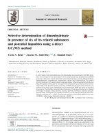
Fig. 1. Effects of ammonium formate (a) and formic acid (b) concentration in mobile phase for TTX.
Chromatographic separation was performed on an ACQUITY
UPLC BEH Amide column (50×2.1 mm, 1.7 μm, Waters, USA) at
room temperature. The mobile phase consisted of (A) deionized
water and (B) ACN, both containing 10μmol/L ammonium formate
and 0.01% FA. The following gradient program was used [mobile
phase B]: 85–55% at 0–2 min; held 55% at 2–3 min; 55–85% at 3–
3.01 min; held 85% at 3.01–4.5 min with a flow rate of 0.5 mL/min.
2.2. Selection and optimization of SPE
In the beginning, MCX and WCX cation exchange SPE plates
(Waters, USA) were tested to clean up the plasma samples and purify TTX. It was found that the WCX plate can hardly retain TTX
during loading, and the MCX plate can hardly elute TTX using organic solvent containing moderate acids like fomic acid or acetic
acid. Strong acid like hydrochloric acid wasn’t tried on MCX plate
due to its damage to liquid chromatography and mass spectrometry, even if it can be dried up, poor peak shapes and severe matrix
effect might not be avoided.
In turn, three different HILIC-type SPE plates were attempted
to separate TTX from the plasma matrix. PSA plate containing
polyamide material, silica plate containing silica gel material, and
Siphila i HILIX plate containing multi-carboxyl material were compared. The flow charts of comparison and optimization for three
different HILIC-SPE were shown in Fig. 2. All the loading, washing,
and elution samples were collected and analyzed to calculate the
loss or recovery of TTX.
HILIC-type SPE usually requires high concentrations of organic
solvents as loading solutions and water is used as a strong eluent [40]. In this study, pure ACN and ACN containing 1%TCA,
5%TCA, and 2%FA were investigated respectively as loading solutions, which also acted as protein precipitating agents, denaturing plasma proteins and releasing TTX. 100μL of standard solution
(prepared by deionized water) and plasma samples were precipitated with 700μL of loading solutions in 96-well protein precipitation plates respectively and then all filtrates were loaded onto
three different SPE plates, which were previously activated with
methanol and equilibrated with ACN. The results showed TTX was
most strongly retained on the Siphila i HILIX SPE plate during sample loading. ACN was the optimal loading solvent and facilitated
the retention of TTX on SPE plates, followed by 2%FA-ACN, 1%TCAACN, and 5%TCA-ACN (Fig. 3). The loss of TTX in plasma samples
were less than that in standard solution samples during loading,
which might be due to the poor buffering capacity of standard solution samples compared with plasma.
To further investigate the loss and recovery of TTX during washing and elution steps, 200μL of 95%ACN was used for washing followed by 200μL of 1%TCA-50%ACN for elution. In the 2%FA-ACN
and ACN loading groups, due to the low recoveries after elution,
another 200μL of 5%TCA-ACN was used for further elution to improve the recovery. The results were shown in Fig. 4.
Different retention and elution capabilities of TTX were observed on the same SPE plate under different loading conditions
during washing and elution. Siphila i HILIX was the most distinct
and optimal one (Fig. 4a). In the 1%TCA-ACN loading group, washing with 95% ACN resulted in a loss of 18% TTX, while elution with
1% TCA-50% ACN resulted in the recovery of 80% TTX in plasma
samples. While during the same loading and washing steps, the
corresponding standard solution samples would lose even more,
with less than 30% TTX recovered by elution. In the 5%TCA-ACN
loading group, both the loading and washing steps resulted in
1.4. Method validation
The lower limit of quantification (LLOQ), linearity, inter/intrabatch accuracy and precision, matrix effect, extraction recovery,
specificity, and interference of the established method were investigated according to the U.S. food and drug administration (FDA)
bioanalytical method validation guidelines [39].
2. Results and discussion
2.1. Development of chromatography and mass spectrometry method
TTX is difficult to retain on reversed-phase columns [26], and
most recent reports used an amide column with a length of 10 cm
to improve the retention of 10 TTX analogs [37]. Here, an ACQUITY
UPLC BEH C18 Amide column (50×2.1 mm, 1.7 μm, Waters, USA)
was used to separate TTX from its interferences, which performed
better compared with an ACQUITY UPLC BEH C18 HILIC column
(50×2.1 mm, 1.7 μm, Waters, USA). The retention times for TTX
and IS on the amide column were 2.41 min and 2.31 min, respectively, with a total analysis time of 4.5 min.
The pH value and the concentration of ammonium formate in
the mobile phase were considered to have significant effects on
retention time and peak area [37]. Therefore, different FA and ammonium formate concentrations were tested under the same separation conditions. It was observed that the retention time and peak
area of TTX increased with the decrease of FA (from 0.05% to 0.01%,
v: v) (see Fig. 1a) and ammonium formate minimized the retention
time shift between pure solvent and matrix samples but decreased
the peak area of TTX (Fig. 1b). In this study, 10 μmol per liter of
ammonium formate and 0.01%FA (v: v) were used in both mobile
phases to acquire better sensitivity and make the peaks more stable. The variation of 56 consecutive injections prepared by plasma
samples was tested (see Fig. S1) and the mean retention time was
2.42±0.1 min (2.37–2.47 min), which indicated that the chromatographic separation conditions performed well with excellent reproducibility. MS/MS parameters for TTX and IS were optimized by
direct infusion of individual standard solutions. The quantifier and
confirmation product ion for TTX were m/z 162.2 and 284.1, respectively, although the most abundant product ion was observed as
[M + H–H2 O]+ ion (m/z 302.2). The latter has higher baseline and
stronger interference in plasma samples.
3
L. Xin, Y. Liang, S. Yang et al.
Journal of Chromatography A 1684 (2022) 463567
Fig. 2. The flow charts of comparison and optimization for three different HILIC-SPE.
more loss of TTX, about 60% for plasma samples and even higher
for standard solution samples (>80%). In the 2%FA-ACN loading
group, no significant TTX loss was observed for plasma samples
after washing with 95%ACN, and the recovery was about 22% after
elution with 1%TCA-ACN. Compared to the two TCA-ACN loading
groups, the total recovery of TTX in plasma samples was reduced
throughout the SPE, so an additional 5%TCA-ACN elution was performed to look for the lost TTX and the total recovery was increased to 60% after the additional elution. This was quite different
from the total recovery of standard solution samples which was
close to 100%. The total recovery of TTX in plasma samples was significantly lower in the ACN loading group, no matter in the loading and washing solutions, or the eluted and twice-eluted samples,
while the recovery of standard solution samples was nearly 100%.
In the 2%FA-ACN and ACN loading groups, the lower recovery of
TTX from plasma samples compared to standard solution samples
may be due to the incomplete release of TTX from plasma proteins
upon precipitation of these two loading solutions. Similar results
were observed when using PSA and silica plates (Fig. 4b and 4c).
Strong acids such as TCA in the precipitating agent may be necessary for the release of TTX from plasma proteins. Excessive TCA
resulted in weak retention of TTX on SPE, which may be related
to the decreased polarity of the TTX-TCA conjugates in the presence of ion pairs solvents, thus reducing the retention of TTX on
HILIC-type SPE plates.
The results showed that the retention capacity among Siphila i
HILIX, PSA, and silica SPE were quite different. Compared to Siphila
i HILIX, PSA and silica had weaker retention of TTX under almost
all loading conditions. It was speculated that both Siphila i HILIX
and silica may have two interaction modes: hydrophilic interaction and ion exchange. Some SiOH-containing silica materials had
been shown to have cation-exchange properties [41]. While in our
study, we found that the ion exchange effect of silica SPE might be
very weak which could be easily inhibited by TCA or FA. In addition to hydrophilic interaction, Siphila i HILIX may also retain polar
basic compounds through cation-exchange interaction which was
similar or even stronger to that of weak cation-exchange material
with carboxylic acid moieties [42]. It seemed that PSA had only
a pure hydrophilic effect on TTX, and retention was the weakest.
Among the three HILIC materials, Siphila i HILIX was considered
to have better synergistic effect of hydrophilic interaction and ion
exchange, which was suitable for the retention of TTX.
After optimizing the loading conditions, the washing solvents
were selected among 95% ACN, 98% ACN, and ACN. No significant
difference was observed, so 95%ACN was chosen because it had the
strongest cleaning capability. The elution solvent remained 1%TCA50%ACN, which had no obvious ion suppression or enhancement
effects on TTX. Since stable isotope labeled TTX is very expensive and difficult to obtain, we used the reported voglibose and
the available arginine-15 N4 as candidate internal standards in this
4
L. Xin, Y. Liang, S. Yang et al.
Journal of Chromatography A 1684 (2022) 463567
Fig. 3. The loss of TTX under different sample loading conditions on three HILIC-SPE plates.
Table 1
The major validation parameters for TTX and IS in human plasma.
TTX
Spiked conc. (ng/mL)
Intra-assay (n = 6)
Inter-assay (n = 6 × 3)
Extraction recovery (n = 6)
IS-normalized MF (n = 6)
Mean accuracy (%)
RSD (%)
Mean accuracy (%)
RSD (%)
Mean (%)
RSD (%)
Mean (%)
RSD (%)
0.1
0.5
2
10
99.8
98.7
98.5
99.2
5.92
5.75
2.29
4.37
99.5
99.4
99.1
98.8
5.47
6.23
4.16
5.26
81.5
80.8
81.2
5.53
3.58
6.78
0.979
3.41
0.968
4.29
Spiked conc. (ng/mL)
arginine-15 N4
Extraction recovery (n = 6)
voglibose
Extraction recovery (n = 6)
Mean (%)
74.3
Mean (%)
71.5
10
RSD (%)
4.26
study, both of which showed similar chromatographic behavior to
TTX on the amide column.
The performance of the two candidate internal standards was
further compared in plasma matrix. Post-column injection experiment was used to investigate the different matrix effects of TTX
and the two candidate internal standards in pure solvent and
plasma samples. It was found that TTX was barely enhanced or
inhibited by plasma matrix, arginine-15 N4 was slightly enhanced,
while voglibose was severely enhanced (Fig. 5). By comparing the
recoveries, it can be found that both arginine-15 N4 and voglibose
had similar extraction recoveries as TTX. Therefore, arginine-15 N4
was finally selected as IS in this study (see Table 1).
RSD (%)
3.17
a 200-fold linear dynamic range (0.1–20 ng/mL) with the LLOQ
(signal-to-noise ratio, S/N>10) of about 0.1 ng/mL. Moreover, spikerecovery studies assessed for four different QCs (at 0.1, 0.5, 2, and
10 ng/mL, n = 6) at three consecutive days. The intra- and interassay accuracies (%) were 98.5%−99.8% and 98.8%−99.5%, respectively, calculated by comparing measured concentrations to theoretically spiked concentrations. The intra- and inter-assay precision investigated in terms of relative standard deviations (RSDs%),
were ≤ 5.92% and ≤ 6.23%, respectively. The results were summarized in Table 1, which showed acceptable accuracy and precision
for plasma TTX in the investigated concentration range. The validation of the method selection proved that samples were prepared
well by Siphila i HILIX. The representative chromatograms of blank
samples and LLOQ samples at concentration of 0.1 ng/mL of TTX
and 10 ng/mL of IS were shown in Fig. 6. No obvious interferences
coeluting with the TTX and the IS was observed and the baselines
of noise were acceptable. The LLOQ was about 0.3 ng/mL for confirmation transition (m/z 320.3→284.1) according to the criterion
of S/N>10, indicating that the method in this study would be suit-
2.3. Method validation
After determining the optimization of SPE conditions, validation was attempted to investigate the practicability of the present
method. The method displayed excellent linearity of TTX with
1/x as weighting factor, and R value was higher than 0.999 over
5
L. Xin, Y. Liang, S. Yang et al.
Journal of Chromatography A 1684 (2022) 463567
Fig. 4. The recoveries of TTX in each step on three HILIC-SPE plates under different sample loading conditions. a. Siphila i HILIX SPE, b. Silica, c. PSA.
6
L. Xin, Y. Liang, S. Yang et al.
Journal of Chromatography A 1684 (2022) 463567
Fig. 5. Plasma matrix effect of a. TTX, b. arginine-15 N4 and c. voglibose.
Fig. 6. The representative chromatograms of blank, LLOQ (0.1 ng/mL) of a. TTX and b. IS (10 ng/mL).
7
L. Xin, Y. Liang, S. Yang et al.
Journal of Chromatography A 1684 (2022) 463567
Table 2
Bias and precision of spiked samples from different sources.
sources
Plasma1
Plasma2
Plasma3
Plasma4
Plasma5
Plasma6
hemolytic plasma
high triglyceride plasma
high cholesterol plasma
high bilirubin plasma
0.1 ng/mL (n = 3)
10 ng/mL (n = 3)
Mean (bias%)
RSD (%)
Mean (bias%)
RSD (%)
3.67
−3.37
1.73
−6.00
−1.43
−2.67
−3.53
0.90
−3.67
−5.00
8.83
6.16
8.59
2.50
5.90
1.49
4.28
4.80
3.10
4.95
4.23
0.97
−0.37
3.20
−7.00
7.43
−2.50
−4.67
−2.50
3.93
0.32
1.73
2.99
2.51
4.07
1.04
3.86
2.92
6.40
2.37
alency factors [46]. Although their SPE and LC-MS behavior may
be similar to that of TTX, it is likely that they share the same or
similar parent or product ions with TTX, which will interfere the
detection of TTX and require further chromatographic separation.
More work is needed to prove whether interference exists from
TTX analogs or whether TTX analogs can also be detected by our
method or an improved method.
3. Conclusions
An LC-MS/MS method had been developed and validated for
the quantitation of TTX in human plasma. This method used multicarboxyl group SPE materials and did not require evaporation and
ultrafiltration steps. Compared with previous reports, the method
was highly sensitive, simple, and rapid in sample pre-treatment
and had short chromatographic time. The method presented in this
paper showed wide linearity, low LLOQ, good precision and accuracy, and high recovery characteristics which well satisfied the requirements of pharmacokinetic study and clinical diagnosis of TTX.
able for the identification and quantification of TTX at poisoned
concentration levels.
The extraction recovery was assessed by comparing the peak areas of spiked plasma samples with those of post-extraction spiked
samples at three QC levels. The quantitative matrix factors were
evaluated by comparing the peak areas of post-extraction spiked
samples from six sources at two QC levels with those of neat standard solutions. The method demonstrated excellent extraction recovery for TTX, ranging from 80.8% to 81.5% at three different concentrations (RSDs ≤ 6.78%). The mean IS normalized matrix factor
of samples from six sources at two levels were 0.979 and 0.968
respectively, which indicated no significant ion suppression or enhancement existed by the optimized method (Table 1). The lost extraction recovery of TTX was mainly in the washing step, as shown
in Fig. 4a, which was speculated to be improved by increasing the
amount of SPE packed material or reducing the washing volume.
Good specificity means that the analytical method can differentiate between the target analyte and interfering substances for
samples from different sources and the bias and precision were
acceptable. Ten blank human plasma samples including samples
from six healthy volunteers and pooled blank human plasma samples mixing with high concentrations of hemoglobin, triglyceride,
cholesterol, and bilirubin were prepared and investigated by spiking analysis. In detail, working standard solutions of TTX were
spiked to the above 10 blank plasma samples to make QCs at LLOQ
and High concentration levels (0.1, 10 ng/mL). The QCs were pretreated and analyzed. The biases and RSDs were listed in Table 2.
The biases of target value were ranged from −7.00% to 7.43% for
healthy human plasma samples (RSDs ≤ 8.83%) and from −5.00%
to 3.93% for hemolytic, high triglyceride, high cholesterol and high
bilirubin plasma samples (RSDs ≤ 6.40%), which proved the good
anti-interference property of the method.
Saxitoxin and its approximately 60 natural analogues, which
are also potent and specific voltage-gated sodium channel blockers and share the same binding site as that of TTX [43, 44], also
cause paralysis and even death in extreme cases. In our supplemental study, gonyautoxin5 (GTX5) was purchased for interference
test since its retention time was closest to TTX on amide column
[45, 37]. Under the optimized LC and SPE conditions described in
this study, the retention time of GTX5 was 2.53 min which was
close to TTX (see Fig. S2). No interferences were observed between
the TTX and GTX5 MRM transitions. Therefore, we believe that saxitoxin and its analogues may not interfere the detection of TTX.
Besides, in a preliminary study, the total recovery of GTX5 from
plasma samples was about 70% according to the peak area ratio
of spiked plasma samples and neat standard solutions which indicated that the described method in our study may be suitable
for quantitation of these compounds after optimization. In addition, the analogs of TTX like 4,9-anhydro TTX, 4–epi TTX and so
on, usually have similar chemical structures and lower toxic equiv-
Credit authorship contribution statement
Liang Xin: Conceptualization, Investigation, Methodology, Writing - Original Draft. Yan Liang: Data Curation, Investigation, Writing - Original Draft. Shuangshuang Yang: Methodology, Data
Curation, Visualization. Fengli Jiang: Investigation, Methodology.
Fan Yu: Investigation, Validation. Meiwei Zhang: Data curation,
Methodology. Wei Chang: Funding acquisition. Wei Wang: Data
curation, Validation. Chen Yu: Supervision. Gangyi Liu: Formal
analysis, Project administration, Writing - Review & Editing. Youli
Lu: Funding acquisition, Formal analysis, Project administration,
Writing - Review & Editing.
Declaration of Competing Interest
The authors declare that they have no known competing financial interests or personal relationships that could have appeared to
influence the work reported in this paper.
Data Availability
Data will be made available on request.
Acknowledgments
This research was funded by the Innovation Fund of Science and
Technology Commission of Shanghai Municipality (20Y1190 080 0)
and the General Project of Medical Research Fund of Xuhui District,
Shanghai (SHXH202121).
Supplementary materials
Supplementary material associated with this article can be
found, in the online version, at doi:10.1016/j.chroma.2022.463567.
References
[1] L. Biessy, M.J. Boundy, K.F. Smith, D.T. Harwood, I. Hawes, S.A. Wood,
Tetrodotoxin in marine bivalves and edible gastropods: a mini-review, Chemosphere 236 (Dec. 01, 2019), doi:10.1016/j.chemosphere.2019.124404.
[2] P. Gouel, C.M. iti Gatti, L. de Haro, A. Liautaud, J. Langrand, D. Boucaud-Maitre,
Tetrodotoxin poisoning in Mainland France and French overseas territories: a
review of published and unpublished cases, Toxins (Basel) 14 (5) (May 2022)
351, doi:10.3390/toxins14050351.
[3] D.F. Hwang, T. Noguchi, Tetrodotoxin Poisoning, Adv. Food Nutr. Res. 52 (2007)
141–236, doi:10.1016/S1043-4526(06)52004-2.
[4] J.F. Fernndez-Ortega, et al., Seafood intoxication by tetrodotoxin: first case in
europe, J. Emerg. Med. 39 (5) (Nov. 2010) 612–617, doi:10.1016/j.jemermed.
2008.09.024.
8
L. Xin, Y. Liang, S. Yang et al.
Journal of Chromatography A 1684 (2022) 463567
[5] J. You, Y.J. Yue, F. Xing, W. Xia, S.Y. Lai, F.L. Zhang, Tetrodotoxin poisoning
caused by goby fish consumption in southeast China: a retrospective case series analysis, Clinics 70 (1) (2015) 24–29, doi:10.6061/clinics/2015(01)05.
[6] P. Almeida, R. Diaz, F. Hernandez, G. Ferrer, Blow: a case of pufferfish intoxication in South Florida, BMJ Case Rep. 12 (6) (Jun. 2019), doi:10.1136/
bcr- 2019- 229272.
[7] B. Alhatali, et al., A cluster of tetrodotoxin poisoning in Oman, Clin. Toxicol. 60
(2) (2022) 262–266, doi:10.1080/15563650.2021.1917595.
[8] M.C.K. Geoffrey K Isbister, Neurotoxic marine poisoning, Lancet Neurol. 4
(2005) 219–228, doi:10.1016/S1474- 4422(05)70041- 7.
[9] Y. Bentur, et al., Lessepsian migration and tetrodotoxin poisoning due to Lagocephalus sceleratus in the eastern Mediterranean, Toxicon 52 (8) (Dec. 15,
2008) 964–968, doi:10.1016/j.toxicon.2008.10.001.
[10] V. Bane, M. Lehane, M. Dikshit, A. O’Riordan, A. Furey, Tetrodotoxin: chemistry,
toxicity, source, distribution and detection, Toxins (Basel) 6 (2) (2014) 693–
755, doi:10.3390/toxins6020693.
[11] Q.T. Islam, et al., Puffer fish poisoning in Bangladesh: clinical and toxicological
results from large outbreaks in 2008, Trans. R. Soc. Trop. Med. Hyg. 105 (2)
(Feb. 2011) 74–80, doi:10.1016/j.trstmh.2010.10.002.
[12] N. Ogata and Y. Ohishi, “Molecular diversity of structure and function of the
voltage-gated Na+ channels,” 2002. doi: 10.1254/jjp.88.365.
[13] H.L. Chong, P.C. Ruben, Interaction between voltage-gated sodium channels
and the neurotoxin, tetrodotoxin, Channels 2 (6) (2008) 407–412, doi:10.4161/
chan.2.6.7429.
[14] W.A. Catterall, A.L. Goldin, S.G. Waxman, International Union of Pharmacology. XLVII. Nomenclature and structure-function relationships of voltage-gated
sodium channels, Pharmacol. Rev. 57 (4) (Dec. 2005) 397–409, doi:10.1124/pr.
57.4.4.
[15] F.R. Nieto, E.J. Cobos, M.Á. Tejada, C. Sánchez-Fernández, R. González-Cano,
C.M. Cendán, Tetrodotoxin (TTX) as a therapeutic agent for pain, Mar Drugs
10 (2) (2012) 281–305, doi:10.3390/md10020281.
[16] R. González-Cano, M.C. Ruiz-Cantero, M. Santos-Caballero, C. Gómez-Navas,
M. Tejada, F.R. Nieto, Tetrodotoxin, a potential drug for neuropathic and cancer
pain relief? Toxins (Basel) 13 (7) (2021), doi:10.3390/toxins13070483.
[17] M. Kavoosi, et al., Safety, tolerability, pharmacokinetics, and concentration-QTc
analysis of tetrodotoxin: a randomized, dose escalation study in healthy adults,
Toxins (Basel) 12 (8) (Aug. 2020), doi:10.3390/toxins12080511.
[18] G.M. Bucciarelli, M. Lechner, A. Fontes, L.B. Kats, H.L. Eisthen, H.B. Shaffer,
From poison to promise: the evolution of tetrodotoxin and its potential as
a therapeutic, Toxins (Basel) 13 (8) (2021) MDPI AG, Aug. 01, doi:10.3390/
toxins13080517.
[19] G.S. Shephard, “Committee on natural toxins and food allergens mycotoxins,”
2007. doi: 10.1093/jaoac/90.1.1b.
[20] D. Neagu, L. Micheli, G. Palleschi, Study of a toxin-alkaline phosphatase conjugate for the development of an immunosensor for tetrodotoxin determination, Anal. Bioanal. Chem. 385 (6) (Jul. 2006) 1068–1074, doi:10.1007/
s0 0216-0 06-0522-2.
[21] R. Watanabe, et al., Quantitation of Tetrodotoxin and Its Analogues with a
Combination of Liquid Chromatography-Tandem Mass Spectrometry and Quantitative 1H-NMR Spectroscopy, J. Agric. Food Chem. 67 (46) (Nov. 2019) 12911–
12917, doi:10.1021/acs.jafc.9b06380.
[22] M.A. O’Leary, J.J. Schneider, G.K. Isbister, Use of high performance liquid chromatography to measure tetrodotoxin in serum and urine of poisoned patients,
Toxicon 44 (5) (Oct. 2004) 549–553, doi:10.1016/j.toxicon.20 04.07.0 08.
[23] K.S.Y. Leung, B.M.W. Fong, Y.K. Tsoi, Analytical challenges: determination of
tetrodotoxin in human urine and plasma by LC-MS/MS, Mar. Drugs 9 (11)
(2011) 2291–2303 MDPI AG, doi:10.3390/md9112291.
[24] H.K. Knutsen, et al., Risks for public health related to the presence of
tetrodotoxin (TTX) and TTX analogues in marine bivalves and gastropods, EFSA
J. 15 (4) (Apr. 2017), doi:10.2903/j.efsa.2017.4752.
[25] F. Long, M. Zhang, J. Yuan, J. Du, A. Ma, J. Pan, A simple, versatile, and automated pulse-diffusion-focusing strategy for sensitive milliliter-level-injection
HILIC-MS/MS analysis of hydrophilic toxins, J. Hazard. Mater. 392 (Jun. 2020),
doi:10.1016/j.jhazmat.2020.122318.
[26] A.L.N. van Nuijs, I. Tarcomnicu, A. Covaci, Application of hydrophilic interaction chromatography for the analysis of polar contaminants in food and environmental samples, J. Chromatogr. A 1218 (35) (Sep. 02, 2011) 5964–5974,
doi:10.1016/j.chroma.2011.01.075.
[27] D. Salas, F. Borrull, N. Fontanals, R.M. Marcé, Hydrophilic interaction liquid
chromatography coupled to mass spectrometry-based detection to determine
emerging organic contaminants in environmental samples, TrAC - Trends Analyt. Chem. 94 (Sep. 01, 2017) 141–149, doi:10.1016/j.trac.2017.07.017.
[28] H. Mei, et al., Investigation of matrix effects in bioanalytical high-performance
liquid chromatography/tandem mass spectrometric assays: application to drug
[29]
[30]
[31]
[32]
[33]
[34]
[35]
[36]
[37]
[38]
[39]
[40]
[41]
[42]
[43]
[44]
[45]
[46]
9
discovery, Rapid Commun. Mass Spectrom. 17 (1) (2003) 97–103, doi:10.1002/
rcm.876.
T.M. Annesley, “Ion suppression in mass spectrometry,” 2003. doi:
10.1373/49.7.1041.
B.K. Matuszewski, M.L. Constanzer, C.M. Chavez-Eng, Strategies for the assessment of matrix effect in quantitative bioanalytical methods based on HPLCMS/MS, Anal. Chem. 75 (13) (Jul. 2003) 3019–3030, doi:10.1021/ac020361s.
H. Trufelli, P. Palma, G. Famiglini, A. Cappiello, An overview of matrix effects
in liquid chromatography-mass spectrometry, Mass Spectrom. Rev. 30 (3) (May
2011) 491–509, doi:10.1002/mas.20298.
F. Gosetti, E. Mazzucco, D. Zampieri, M.C. Gennaro, Signal suppression/enhancement in high-performance liquid chromatography tandem mass
spectrometry, J. Chromatogr. A 1217 (25) (Jun. 2010) 3929–3937, doi:10.1016/j.
chroma.2009.11.060.
M.M. Khamis, D.J. Adamko, A. El-Aneed, Strategies and Challenges in Method
Development and Validation for the Absolute Quantification of Endogenous
Biomarker Metabolites Using Liquid Chromatography-Tandem Mass Spectrometry, Mass Spectrom. Rev. (2019) John Wiley and Sons Inc., doi:10.1002/mas.
21607.
H.C. Jen, S.J. Lin, Y.H. Tsai, C.H. Chen, Z.C. Lin, D.F. Hwang, Tetrodotoxin
poisoning evidenced by solid-phase extraction combining with liquid
chromatography-tandem mass spectrometry, J. Chromatogr. B Anal. Technol.
Biomed. Life Sci. 871 (1) (Aug. 2008) 95–100, doi:10.1016/j.jchromb.2008.06.
030.
H.E. Cho, et al., Determination and validation of tetrodotoxin in human whole
blood using hydrophilic interaction liquid chromatography-tandem mass spectroscopy and its application, Forensic Sci. Int. 217 (1–3) (Apr. 2012) 76–80,
doi:10.1016/j.forsciint.2011.10.026.
M. Rambla-Alegre, et al., Rapid screening and multi-toxin profile confirmation
of tetrodotoxins and analogues in human body fluids derived from a puffer
fish poisoning incident in New Caledonia, Food Chem. Toxicol. 112 (Feb. 2018)
188–193, doi:10.1016/j.fct.2017.12.039.
N. Ochi, Simultaneous determination of ten paralytic shellfish toxins and
tetrodotoxin in scallop and short-necked clam by ion-pair solid-phase extraction and hydrophilic interaction chromatography with tandem mass spectrometry, J. Chromatogr. A 1651 (Aug. 2021), doi:10.1016/j.chroma.2021.462328.
B.M.W. Fong, S. Tam, S.H. Tsui, K.S.Y. Leung, Development and validation of a
high-throughput double solid phase extraction-liquid chromatography-tandem
mass spectrometry method for the determination of tetrodotoxin in human
urine and plasma, Talanta 83 (3) (Jan. 2011) 1030–1036, doi:10.1016/j.talanta.
2010.11.014.
U.S. Food and Drug Administration, Bioanalytical Method Validation Guidance for Industry, />Bioanalytical- Method- Validation- Guidance- for- Industry.pdf#:∼:text=
Bioanalytical%20Method%20Validation%20Guidance%20for%20Industry%201%
20This,is%20not%20binding%20on%20FDA%20or%20the%20public, 2018.
R. Li, W. Sun, X. Xiao, B. Chen, Y. Wei, Retention of stevioside polar compounds on a sulfonic acid-functionalized stationary phase, J. Chromatogr. A
(1620) 2020, doi:10.1016/j.chroma.2020.460978.
T. Ikegami, A. Taniguchi, T. Okada, K. Horie, S. Arase, Y. Ikegami, Functionalization using polymer or silane? A practical test method to characterize hydrophilic interaction chromatography phases in terms of their functionalization method, J. Chromatogr. A 1638 (2021) 461850, doi:10.1016/j.chroma.2020.
461850.
D. Bratkowska, R.M. Marcé, P.A.G. Cormack, D.C. Sherrington, F. Borrull,
N. Fontanals, Synthesis and application of hypercrosslinked polymers with
weak cation-exchange character for the selective extraction of basic pharmaceuticals from complex environmental water samples, J. Chromatogr. A 1217
(10) (2010) 1575–1582, doi:10.1016/j.chroma.2010.01.037.
M. Wiese, P.M. D’Agostino, T.K. Mihali, M.C. Moffitt, B.A. Neilan, Neurotoxic alkaloids: saxitoxin and its analogs, Mar. Drugs 8 (7) (2010) 2185–2211, doi:10.
3390/md8072185.
T.W. Soong, B. Venkatesh, Adaptive evolution of tetrodotoxin resistance in animals, Trends Genet. 22 (11) (2006) 621–626, doi:10.1016/j.tig.2006.08.010.
S. Numano, Y. Kudo, Y. Cho, K. Konoki, M. Yotsu-Yamashita, Temporal variation
of the profile and concentrations of paralytic shellfish toxins and tetrodotoxin
in the scallop, Patinopecten yessoensis, cultured in a bay of East Japan, Mar.
Drugs 17 (12) (2019), doi:10.3390/md17120653.
I.J. Tamele, M. Silva, V. Vasconcelos, The incidence of tetrodotoxin and its
analogs in the Indian Ocean and the Red Sea, Mar. Drugs 17 (1) (2019) 28–
38, doi:10.3390/md17010028.