Báo cáo khoa học: The specific delivery of proteins to human liver cells by engineered bio-nanocapsules doc
Bạn đang xem bản rút gọn của tài liệu. Xem và tải ngay bản đầy đủ của tài liệu tại đây (431.65 KB, 10 trang )
The specific delivery of proteins to human liver cells
by engineered bio-nanocapsules
Dongwei Yu
1
, Chie Amano
1
, Takayuki Fukuda
1,2
, Tadanori Yamada
3
, Shun’ichi Kuroda
3,4
,
Katsuyuki Tanizawa
3,4
, Akihiko Kondo
4,5
, Masakazu Ueda
4,6
, Hidenori Yamada
1
, Hiroko Tada
1
and Masaharu Seno
1,4,7
1 Graduate School of Natural Science and Technology, Okayama University, Japan
2 Kobe R & D Center, Katayama Chemical Industries Co. Ltd, Kobe, Japan
3 Institute of Scientific and Industrial Research, Osaka University, Japan
4 Research and Development Division, Beacle, Inc., Okayama, Japan
5 Faculty of Engineering, Kobe University, Japan
6 Keio University, School of Medicine, Tokyo, Japan
7 Research Center for Biomedical Engineering, Okayama University, Japan
A drug and gene delivery system has long been consid-
ered important for drug discovery and pharmaceutical
development. In particular, the establishment of a cell-
or tissue-specific targeting method is the latest area of
focus. Although, viral vectors, such as those utilizing
adenovirus or adeno-associated virus, have been devel-
oped for gene therapy, the cell specificity must be
ameliorated. Some other problems, such as inflamma-
tion, neutralizing antibodies, the dangers of mass pro-
duction, and insertional mutagenesis, limit the use of
Keywords
bio-nanocapsule; L fusion protein; protein
delivery vector; specific infection; topology
Correspondence
M. Seno, Graduate School of Natural
Science and Technology, Okayama
University, 3.1.1 Tsushima-Naka, Okayama
700-8530, Japan
Tel ⁄ Fax: +81 86 251 8216
E-mail:
(Received 20 March 2005, revised 9 May
2005, accepted 24 May 2005)
doi:10.1111/j.1742-4658.2005.04790.x
A bio-nanocapsule (BNC), composed of the surface antigen (sAg) of the
hepatitis B virus, is an efficient nanomachine with which to accomplish the
liver-specific delivery of genes and drugs. Approximately 110 molecules of
sAg are associated to form a BNC particle with an average diameter of
130 nm. The L protein is an sAg peptide composed mainly of preS and S
regions. The preS region, with specific affinity for human hepatocytes, is
localized in the N-terminus. The S region following the preS has two trans-
membrane regions responsible for the formation of particles. In this study,
the fusion of emerald green fluorescent protein (EGFP) at the C-terminus
of the S region was designed to deliver proteins to human hepatocytes.
Truncation of the C-terminus of the S region was required to obtain suffi-
cient expression levels in Cos7 cells. The nanoparticles that were produced
delivered EGFP to human hepatoma cells, displaying the EGFP moiety
outside, or enclosing it inside. However, only a single orientation character-
izes the particle, so that either type of L fusion particle could be effectively
and independently separated by an antibody affinity column. The dual
C-terminal topologies of the L fusion particles designed in this study could
be applied to various proteins for the C-terminal moiety of the L fusion
proteins, depending on the character of the proteins, such as cytoplasmic
proteins, as well as cytokines or ligands to cell surface receptors. We sug-
gest that this fusion design is the most efficient way to prepare a BNC that
delivers proteins to specific cells or tissues.
Abbreviations
BNC, bio-nanocapsule; DDS, drug delivery system; DMEM, Dulbecco’s modified Eagle’s medium; EGFP, emerald green fluorescent protein;
ER, endoplasmic reticulum; FBS, fetal bovine serum; HBV, hepatitis B virus; IFN, interferon; PMSF, phenylmethanesulfonyl fluoride; sAg,
surface antigen.
FEBS Journal 272 (2005) 3651–3660 ª 2005 FEBS 3651
viral vectors in humans. By contrast, liposomes as a
drug delivery vector have no limits to mass production
and do not have the drawbacks of viral vectors. How-
ever, liposomes have a lower transfection efficiency and
cell ⁄ tissue specificity compared with viral vectors, even
though they have the ability to transfer proteins into
cells. To find a material that has good tranfection effi-
ciency free from the hazards of viral infection, we come
up with the idea of using a hollow particle derived from
recombinant viral envelope proteins. The hepatitis B
virus (HBV) is a human liver-specific virus whose
3.2-kb genome codes three envelope proteins in a single
open reading frame (ORF). These encoded surface anti-
gens (sAgs) are called small (S), medium (M) and large
(L) proteins [1]. In 1982, Valenzuela et al. succeeded in
preparing recombinant sAg from yeast as S protein
particles with a diameter of 22 nm [2]. Around 1990,
recombinant L proteins were also found to be pro-
duced in yeast cells as hollow virus-like particles and
were developed as immunogens for hepatitis B vac-
cines, which were proven safe for use in humans [3,4].
The recombinant L proteins formed a virus-like particle
with a diameter of approximately 200 nm when pro-
duced in Saccharomyces cerevisiae [3,5]. We reported
that this particle was extremely useful as a vector to
target human hepatocyte in vivo exploiting the charac-
ter of infectivity of HBV limited to human liver [6].
Because this recombinant particle is empty, we named
this hollow nanoparticle a bio-nanocapsule (BNC) as it
represents an efficient nanomachine for achieving liver-
specific delivery of genes and drugs.
Although electroporation is proposed as a conveni-
ent procedure for enclosing substances in the particle
[6], fusion to the C-terminus of L protein appears to
be the most efficient way to prepare a BNC to convey
foreign proteins. Thus, fusing a suitable protein to the
tail should be a convenient way of preparing a BNC
that delivers proteins to human hepatocytes.
Results
Evaluation of hepatitis B L fusion particles
The HBV L envelope protein is composed of three
major regions. The preS1 region of 108 or 119 amino
acids at the N-terminus directly recognizes human
hepatocytes [1,7–9]. The preS2 region of 55 amino acids
following the preS1 region has an affinity with poly-
merized albumin-mediated interactions. The major 226
amino acids of the S region occupy the C-terminal half
of the L protein. Current models for the transmem-
brane structure of the S region assume that both the
N- and C-termini are at external positions in mature
particles [10,11]. Although it is predicted that four trans-
membrane-like a helices are present in the S region,
only the two in the N-terminus have been shown to be
transmembrane helices [12–14]. These two domains,
which correspond to amino acids 8–22 and 80–98,
respectively, are separated by a hydrophilic region that
is exposed to the internal side of the mature particle.
The C-terminal topology of the S region has not been
challenged experimentally and remains to be investi-
gated [12,15], whereas the preS region has been shown
to protrude into the lumen of the endoplasmic reti-
culum (ER) [16]. To enclose protein inside the BNC,
we designed emerald green fluorescent protein (EGFP)
fusion at the C-terminus of the S region, with or with-
out truncation, using 32, 45 and 54 amino acid residues
from the C-terminus, respectively, to obtain four types
of L fusion particles, L-FLAG–EGFP, L(D32)-FLAG–
EGFP, L(D45)-FLAG–EGFP and L(D54)-FLAG–
EGFP, with an intervening sequence of FLAG-tag
(Fig. 1). Truncations were designed to shorten the
C-terminus as much as possible for a single transmem-
brane spanning region. Because proline often functions
as a helix disruptor, the proline residues at 177 and 187
are considered in the truncation.
According to the amino acid sequence, the mole-
cular mass of L-FLAG–EGFP, which is the fusion
protein between the full length of the L protein and
EGFP, is calculated to be 68.5 kDa. The apparent
molecular mass of L-FLAG–EGFP is 71.5 kDa due to
the glycosylated Asn146 in the S region on SDS ⁄
PAGE [15,17]. Taking the sizes of C-terminal
truncation into account, the molecular mass of each
L(D32)-FLAG–EGFP, L(D45)-FLAG–EGFP and
L(D54)-FLAG–EGFP is calculated to be 68, 66.5 and
65.7 kDa, respectively.
These L proteins fused to EGFP were transiently
expressed in Cos7 cells (Fig. 2A). During the culturing
period, EGFP fluorescence was observed from the
transfected Cos7 cells along the ER and Golgi, but not
in the nuclei, indicating that the fused protein was pro-
duced in the secretional path. Fusion proteins were
detected with both anti-S and anti-GFP IgG in the
cell lysates of transfected cells by western blotting at
their expected sizes, as calculated (Fig. 2B). Immuno-
precipitates of the conditioned media, with either anti-
S, anti-FLAG or anti-GFP IgG, showed bands of
fusion proteins, in addition to those in the cell lysates
(Fig. 2C). Secretion of L-FLAG–EGFP and L(D54)-
FLAG–EGFP from Cos7 cells appeared limited,
although it was, in fact, produced in sufficient quan-
tity. Expression and secretion of L(D32)-FLAG–EGFP
were both low. Secretion of L(D45)-FLAG–EGFP was
obviously the highest of the four fusion constructs.
Bio-nanocapsules for protein delivery D. Yu et al.
3652 FEBS Journal 272 (2005) 3651–3660 ª 2005 FEBS
The conditioned media were further assessed for the
secretion of these different constructs of particles using
enzyme immunoassay and fluorescence (Fig. 3). When
the level of L-FLAG–EGFP was considered to be
100%, that of L(D45)-FLAG–EGFP was > 300% in
EIA. The fluorescence from the EGFP moiety of
L fusion protein showed that L(D45)-FLAG–EGFP
was secreted at the highest ratio. The analyses revealed
that L(D45)-FLAG–EGFP was suitably optimized for
production as the fusion protein.
L(D45)-FLAG–EGFP particle formation was assessed
by sucrose gradient ultracentrifugation (Fig. 4). When
cell extracts were subjected to ultracentrifugation, both
fluorescence and immunoreactivity were seen in the
same fraction at a density of 1.11 gÆmL
)1
, whereas the
native EGFP fluorescence peak was found to have a
density of 1.05 gÆmL
)1
. Peak fractions were analyzed by
western blotting to confirm the presence of fusion pro-
tein at 66.5 kDa. The density of the particles obtained
from both cell extracts and the conditioned media was
equivalent to that of the BNC. Whether from the condi-
tioned media or the cell extracts, L(D45)-FLAG–EGFP
has the potential to form a particle.
Topology analysis of the C-terminus of L fusion
protein
Consisting of 56 amino acids, the primary structure of
the C-terminus of the S region is rich in hydrophobic
residues. Therefore, the computer-assisted prediction
assumes that there should be two a helices, which
traverse the ER membrane twice [11,18]. In this con-
text, EGFP fused to L protein with an inter-
vening FLAG sequence may provide an excellent
means of judging the C-terminal topology. Because
L-FLAG–EGFP could be immunoprecipitated by
anti-FLAG and anti-GFP IgG (Fig. 2C), the L pro-
tein C-terminus should be located on the external side
of the particle. This is consistent with results reported
by Eble et al. [12].
Because generation of its fluorophore depends on
the correct formation of a tertiary structure and can
easily be detected by fluorescence microscopy, EGFP
has quickly become a powerful research tool for gene
expression and subcellular protein localization in liv-
ing cells and organisms. It was useful in this study,
not only to estimate the level of L-EGFP fusion pro-
tein expression, but also to observe the infection of
particles by its fluorescence. Within the structure of
EGFP, chromophores are insulated by tightly woven
barrel formations of b sheets, which offer strong
resistance against proteolytic attack [19,20]. This
resistance was confirmed by incubation with protein-
ase K at 60 °C for 6 h (data not shown). As a result
of this resistance, a convenient protease protection
assay was available to determine the topology of the
fused protein, as described below. However, slight
digestion after 1 h of incubation was observed, indi-
cating that the terminal sequences might be sensitive
to proteinase K (Fig. 5).
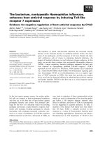
Fig. 1. Fusion proteins between L and EGFP. The 720 bp egfp gene was ligated to the 3¢ side of the L gene of the hepatitis B virus flanking
a 39 bp FLAG as a spacer sequence in an open reading frame. The resultant gene coding L-FLAG–EGFP was inserted downstream of a SRa
promoter. The C-terminus of the L protein was truncated by 32 (96 bp), 45 (135 bp) and 54 amino acid residues (162 bp) to optimize expres-
sion of the L fusion protein. The resultant fusion proteins are designated as L-FLAG–EGFP, L(n32)-FLAG–EGFP, L(n45)-FLAG–EGFP, and
L(n54)-FLAG–EGFP. TM1 and TM2 represent transmembrane regions of the L protein.
D. Yu et al. Bio-nanocapsules for protein delivery
FEBS Journal 272 (2005) 3651–3660 ª 2005 FEBS 3653
L(D45)-FLAG–EGFP particles were immunoprecipi-
tated with two different antibodies and treated with
proteinase K. Western blots with anti-GFP IgG
showed two digested EGFP bands of I and II
(Fig. 5A) when immunoprecipitated with anti-S IgG,
but only one band of II when immunoprecipitated
with anti-GFP IgG. The only difference between these
two bands of I and II should be due to the size of
N-terminal sequence of EGFP protected from protein-
ase K. This result indicates that the C-terminus of the
L-EGFP fusion protein may exhibit dual topology.
Because an anti-S IgG recognizes the immunodomi-
nant a-epitope, which is common to all six genotypes
of HBV as the major surface region of the HBsAg
envelope protein [21,22], this antibody will immuno-
precipitate all of the L(D45)-FLAG–EGFP particles.
By contrast, anti-GFP IgG selectively immunoprecipi-
tate particles that display the EGFP moiety at the
C-terminus on their surface. With the immunoprecipi-
tates, the EGFP moiety was processed to band II, the
N-terminus of which was not protected from protein-
ase K (Fig. 5A). Anti-S IgG immunoprecipitates inclu-
ded another topology, which protects the N-terminus
of the EGFP moiety from proteinase K. There is no
A
B
C
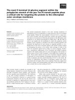
Fig. 2. Expression of L fusion proteins in Cos7 cells. (A) Cells
were transfected with plasmids to express (a) EGFP, (b) L protein,
(c) L-FLAG–EGFP, (d) L(n32)-FLAG–EGFP, (e) L(n45)-FLAG–EGFP,
and (f) L(n54)-FLAG–EGFP, by electroporation. Two days after elec-
troporation, the green fluorescence was observed under a confocal
microscope at a 63-fold magnification. The bar scale shows 50 lm.
After 3 days, the Cos7 cells were harvested and disrupted by sonica-
tion. (B) Cell lysates were probed in western blots with anti-S or anti-
GFP IgG. (C) Conditioned media were collected, immunoprecipitated
with anti-S, anti-GFP or anti-FLAG IgG, respectively, and subjected to
western blotting with anti-S IgG. Lane 1, EGFP; lane 2, L protein; lane
3, L-FLAG–EGFP; lane 4, L(n32)-FLAG–EGFP; lane 5, L(n45)-FLAG–
EGFP; lane 6, L(n54)-FLAG–EGFP.
Fig. 3. Secretion of L fusion particles evaluated by enzyme immuno-
assay and fluorescence. The HBsAg immunoreactivity (A) and the
fluorescence (B) in the conditioned medium of the transfected Cos7
cells were measured and the percentages were calculated with the
level of L-FLAG –EGFP expression assumed to be 100%. n32, n45
and n54 denote L(n32)-FLAG–EGFP, L(n45)-FLAG–EGFP and
L(n54)-FLAG–EGFP, respectively. Each SD was calculated from
three independent evaluations.
Bio-nanocapsules for protein delivery D. Yu et al.
3654 FEBS Journal 272 (2005) 3651–3660 ª 2005 FEBS
possibility that the dual C-terminal topologies coexist
in a particle. However, there should be two types of
L(D45)-FLAG–EGFP particles, because EGFP moiety-
displaying particles immunoprecipitated with anti-GFP
IgG showed only single digested product band II. If
the two types of C-terminal topology coexist in a parti-
cle there should be two digested bands following
immunoprecipitation by anti-GFP IgG, as revealed by
the anti-S IgG. Thus, we concluded that the EGFP
moiety was protected from proteinase K by the mem-
brane when it was located inside the particle, and that
it was slightly digested when located on the external
side of a particle without membrane protection.
To confirm this finding, we subtracted the L(D45)-
FLAG–EGFP particles from the conditioned medium
with anti-GFP IgG and protein G conjugated to
agarose by immunoprecipitation. The residual super-
natant was subjected to repeated immunoprecipitation
using the same procedure until the L fusion protein
could not be detected by western blotting. When the
A
B
C
Fig. 4. Particle formation of L(n45)-FLAG–EGFP evaluated by
sucrose gradient ultracentrifugation. (A) Cell extracts of the Cos7
cells transfected with L(n45)-FLAG–EGFP were subjected to
sucrose gradient ultracentrifugation and each fraction was evalu-
ated for immunoreactivity (e) and fluorescence (h). Native EGFP
was subjected to centrifugation and the fluorescence from EGFP
was simultaneously monitored (d). (B) Fractions that showed both
immunoreactivity and fluorescence in (A) were analyzed by western
blotting with anti-S IgG. (C) L(n45)-FLAG–EGFP particles prepared
from conditioned medium (open triangle) and cell extracts [fraction
11 in (B); h] were compared with the BNC prepared from recom-
binant yeast [6] (s) for immunoreactivity. The density of each frac-
tion was plotted by d.
A
B
Fig. 5. Evaluation of the C-terminal orientation of the EGFP moiety in
L fusion particle. (A) Two different L fusion particles were immuno-
precipitated by either anti-S or anti-GFP IgG, and the immunoprecipi-
tates were treated by proteinase K (pK). n32 and n45 denote
L(n32)-FLAG–EGFP and L(n45)-FLAG–EGFP, respectively. Digested
products were detected with anti-GFP IgG. The produced bands
were indicated by arrows with I (28 kDa) and II (25 kDa). The native
EGFP was treated with or without proteinase K and shown as the
reference. In (B) the conditioned medium containing L(n45)-FLAG–
EGFP particles was immunoprecipitated by anti-GFP IgG (lane 1), and
the process was repeated until no L fusion protein could be detected
by anti-S IgG (lane 2). The residual supernatant was immunoprecipi-
tated by anti-S IgG (lane 3).
D. Yu et al. Bio-nanocapsules for protein delivery
FEBS Journal 272 (2005) 3651–3660 ª 2005 FEBS 3655
final subtracted fraction of the supernatant was further
immunoprecipitated with the anti-S IgG conjugated to
a microparticle, the L fusion protein band was still
detected in the fraction, thereby indicating the presence
of L fusion particles, which contained EGFP moieties
inside (Fig. 5B).
Human hepatocyte specific delivery of EGFP
by infection
The preS1 peptide displayed on the surface of L parti-
cles recognizes the specific receptor present on human
hepatocytes and is essential for HBV infectivity [7,8].
This specific infectivity of the L particle should be
independent of the tolerable C-terminus truncation.
Cell type-specific infection of L(D45)-FLAG–EGFP
particles was assessed on various human cancer cells
(Fig. 6). After 9 h of incubation with L(D45)-FLAG–
EGFP particles, EGFP fluorescence was specifically
observed in human hepatocellular carcinoma HepG2
cells and NuE cells, whereas no EGFP fluorescence
was observed from either human colon adenocarcinoma
WiDr cells or human epidermoid carcinoma A431 cells
(Fig. 6A). The EGFP fluorescence was not observed in
HepG2 cells when incubated with BNC, EGFP or a
mixture of BNC and EGFP (Fig. 6B).
Discussion
In this study we attempted to establish a nanocapsule
that efficiently delivers protein for tissue- or cell-type-
specific targeting. Based on our technology of BNC as
a delivery vector, we used a fusion strategy that
ensures that the protein of interest is produced as a
component of the particle. The fusion proteins between
L protein and EGFP (L-FLAG–EGFP) were expressed
with or without C-terminal truncation of L protein. It
was necessary to truncate the C-terminus of the L pro-
tein by 45 amino acids for it to be efficiently secreted
from the cells. A EGFP moiety fused to the C-termi-
nus of the L protein appears to prevent secretion,
although the mechanism is not currently clear. How-
ever, incorrect folding of each half moiety of the
fusion protein does not explain the low secretion,
because we were able to prepare the particle from both
conditioned media and cell extracts. Once prepared,
Fig. 6. Infection of L fusion particles in vitro. (A) Five nanograms of L(n45)-FLAG-EGFP particles obtained from the conditioned medium
were added to the culture media of 5 · 10
4
cells of HepG2 (a), NuE (b), WiDr (c) and A431 (d), respectively. (B) Conditioned medium of non-
transfected Cos7 cell (a), 5 ng of BNC from the conditioned medium (b), 100 ng of EGFP (c) and a mixture of 5 ng of BNC with 100 ng of
EGFP (d), and 5 ng of L(n45)-FLAG-EGFP particles from the conditioned medium (e) were added to 5 · 10
4
cells of HepG2, respectively.
(C) Extracts of nontransfected Cos7 cells (a), 5 ng of L-FLAG-EGFP particles from the extracts of transfected Cos7 cells (b) and 5 ng L(n45)-
FLAG-EGFP particles from the extracts of transfected Cos7 cells (c) were added to 5 · 10
4
cells of HepG2. The fluorescence was observed
under a confocal microscope at a 63-fold magnification after 9 h infection. Scale bar ¼ 50 lm.
Bio-nanocapsules for protein delivery D. Yu et al.
3656 FEBS Journal 272 (2005) 3651–3660 ª 2005 FEBS
particles displayed immunoreactivity in an enzyme
immunoassay consistent with the fluorescence intensity,
when kept for 3 weeks at 4 °C in the presence of phe-
nylmethanesulfonyl fluoride (PMSF). Furthermore, the
cell extract was able to directly infect HepG2 cells
(Fig. 6C). These results indicate that the L-FLAG–
EGFP particle is stable, if properly prepared, and has
the potential to infect cells.
Following optimization of the C-terminal truncation,
we attempted to determine the topology of the fused
protein in the particle because the purpose of this
study was to design a nanoparticle that incorporated a
foreign protein using a fusion strategy. One of the pur-
poses of inserting a FLAG-tag between the C-terminus
of the L protein and EGFP was to study the topology
of the fused protein because we expected enterokinase
to specifically recognize and cleave FLAG peptide.
Unexpectedly, this protease cleaved other basic resi-
dues in the L protein moiety, displaying many degra-
ded products, which confused us. By contrast, the
strong resistance of EGFP to proteinase K was extre-
mely useful in studying the C-terminal topology of
L fusion protein. Proteinase K treatment of the
L(D32)-FLAG–EGFP particles showed results similar
to those obtained with the L(D45)-FLAG–EGFP parti-
cle. The hydrophobic sequence of approximately 20–30
amino acids in the C-terminus of the L(D32) or L(D45)
protein may traverse the membrane of lipid bilayer.
However, our results show that this terminus is not
sufficiently hydrophobic to anchor the C-terminus in
the membrane, although it is sufficient to exhibit dual
topology. Based on the results of the proteinase K pro-
tection assay, we proposed a model of the nanoparticle
of L fusion protein (Fig. 7). We designated the parti-
cle, whose N-terminal EGFP moiety was incorporated
within the membrane, as type I, whereas type II
denotes the EGFP moiety displayed on the surface of
the particle. Because the particle membrane protected
the N-terminal part of the EGFP moiety, proteinase K
digestion of type I showed the EGFP moiety to have a
slightly higher molecular mass than that produced by
the treatment of type II. We scanned the western blots
in Fig. 5A and analyzed the densities of the two bands.
We found the ratio of band I to band II was 39 : 61.
To confirm this ratio, we also compared the immuno-
reactivity of the conditioned medium containing both
type I and type II particles with that of the type
II-subtracted medium by the anti-GFP IgG, as shown
in Fig. 5B. The ratio of the result is 100 ) 36, which
means that the ratio of type I to type II is 36 : 64.
These different procedures used to estimate the ratio
of the particles in two topologies lead to almost equal
results. Therefore, we concluded that nearly 40% of
the particles are type I. The secretion enhanced by
C-terminal truncation might be explained by the fixed
topology because we could not detect a particle with a
mixed type I and type II topology in one particle.
However, it is difficult to find a determinant of the
topology of the C-terminal moiety that causes it to be
incorporated inside or displayed outside. This unfixed
Fig. 7. Proposed models of L fusion
particles. Type I, EGFP incorporating
particle. Type II, EGFP displaying particle.
D. Yu et al. Bio-nanocapsules for protein delivery
FEBS Journal 272 (2005) 3651–3660 ª 2005 FEBS 3657
pattern of topology might clarify the results of previ-
ous studies of the topology of the HBsAg protein. It is
suggested that the C-terminus of the envelope protein
protrudes from the particle in 1987 [12]. Localization
of HBV epitope by monoclonal antibodies revealed
that the residues 178–186 of the S peptide are exposed
on the surface of the virion particle [23]. Kuroda et al.
described that Asn146 was not glycosylated when the
recombinant L particle was prepared from yeast,
whereas it was glycosylated when expressed in mam-
malian cells [3]. This suggests that this aspargine resi-
due is located at the border of the external region and
the membrane-bound region. The C-terminal sequence
of 56 amino acids from 170 to 226 may be long
enough to traverse the membrane twice, although the
hydrophobicity is not sufficient to explain the topology
precisely. It may be possible to design the C-terminal
region as the clear transmembrane region by replacing
it with one of the transmembrane-type receptors to
limit the topology of the particle to type I.
Our BNC has the same tissue-specific infectivity as
HBV because of the N-terminal region of L protein,
preS1, which determines its narrow host range and dis-
tinct organ tropism. The region from 3 to 77 amino
acid residues of preS1 is essential for this specificity
[24]. To avoid impairing this selectivity, we fused
EGFP to the C-terminal of L protein, which was suc-
cessfully truncated in order to be secreted from mam-
malian cells and assembled to an L fusion particle.
We previously reported that BNC containing DNA of
interest yielded a very high transfection efficiency with a
high specificity of gene transfer to human liver-derived
cells [6]. The L fusion particle described here should
have equivalent specific transfection efficiency due to
the character of the preS1 region of the L protein. The
advantage of the fusion particle is that there is no need
to incorporate proteins using specialized methods, such
as electroporation, for which it is difficult to establish
the efficiency needed to transfer genes and drugs into
cells. Depending on the cell type, conditions vary and
optimization of the conditions may sometimes lead to a
10-fold increase in efficiency. This was also the case with
our BNC, and we had to optimize the electroporation
conditions, depending on the substances to be incorpor-
ated. An efficient procedure is required to eliminate
empty particles after electroporation in order to attain
the highest efficiency. As for the L fusion particle, all of
the particles are destined to convey the protein of inter-
est with a transfection efficiency of nearly 100% directed
to human-derived liver cells.
The L fusion particles designed in this study were
found to have dual C-terminal topologies, which could
easily be separated using antibodies. There was no dif-
ference in specific infectivity among them when monit-
ored using EGFP fluorescence (data not shown). This
means that it is possible to choose various proteins
for the C-terminal moiety of the L fusion proteins,
depending on the character of the proteins to be fused.
This possibility will include cytoplasmic proteins, as
well as cytokines or ligands, for the cell surface. In this
context, one of the candidates of the moiety might be
interferon (IFN) which is used with ribavirin to treat
hepatitis C virus-induced liver disease. This therapy
has many does-dependent side effects, such as depres-
sion and insomnia. The L–IFN fusion particle would
be extremely useful because it targets only the liver so
that the dose administered could be low so that the
side effects would not be a cause for concern.
Retargeting of BNC by replacing the preS1 region
with other targeting moieties or biorecognition mole-
cules, such as ligands, receptors and antibodies as pre-
viously proposed [6], should also be applicable to the
L fusion particles. The greatest problem in using the
particle is that people who have antibodies to HBV
are increasing in number due to the widespread hepati-
tis B vaccination program. Stealth mutations at
Gln129 and Gly145 to Arg would not only address this
problem, but also lead to a design of the delivery vec-
tor with extremely low immunogenicity [25,26]. Thus,
we are developing our BNC for its potential to become
a practical vector of protein delivery.
Experimental procedures
Cell cultures
Human hepatoma HepG2 cells, human squamous cell carci-
noma A431 cells and human colon adenocarcinoma WiDr
cells were cultured in Dulbecco’s modified Eagle medium
(DMEM), supplemented with 10% (v ⁄ v) fetal bovine serum
(FBS; PAA Laboratories, Pasching, Austria). Human hepa-
toma NuE cells were cultured in RPMI-1640 with 10%
(v ⁄ v) FBS. African green monkey kidney-derived Cos7 cells
for particle production were maintained in DMEM supple-
mented with 5% (v ⁄ v) FBS. These cells were maintained at
37 °C ⁄ 5%CO
2
.
Construct of plasmids
The HBV L gene was excised from the plasmid pGLD
LIIP39-RcT [7] and inserted into the XhoI site of
pEGFP-N1 vector (Clontech, Mountain View, CA). Then
the synthetic oligo-nucleotide coding FLAG-tag sequence
(5¢-ATATATTGATTACAAGGATGAC GACGATAAGA
TA-3¢) was inserted between the AccI site close to the
C-terminus of the L protein and the AgeI site at the N-ter-
Bio-nanocapsules for protein delivery D. Yu et al.
3658 FEBS Journal 272 (2005) 3651–3660 ª 2005 FEBS
minus of EGFP in pEGFP-N1. The Not I site after the ter-
mination codon of EGFP was changed to the XhoI site.
The resultant ORF of the L-FLAG–EGFP fusion protein
was excised with XhoI and inserted at the XhoI site down-
stream of the SRa promoter in the plasmid of pBO477,
which is a derivative of pTB1455 [27], to construct the
plasmid pBO572. We constructed three other expression
vectors pBO638, pBO637 and pBO822 for L proteins with
truncation by 32, 45 and 54 amino acid residues at the C-
terminus, respectively. The resulting three types of L fusion
particles were designated L(D32)-FLAG–EGFP, L(D45)-
FLAG–EGFP and L(D54)-FLAG–EGFP (Fig. 1).
Preparation of L fusion particles
Five micrograms of expression plasmid DNA were trans-
fected into 5 · 10
6
of Cos7 cells by electroporation at
300 V ⁄ 950 lF. Transfected cells were first cultured for
14–16 h in 8 mL of DMEM containing 5% (v ⁄ v) FBS in a
100 mm dish. The medium was replaced with 8 mL of
CHO-S-SFM II (Invitrogen, Carlsbad, CA) and the cells
were cultured for a further 72 h. The conditioned medium
was collected and condensed in a Vivaspin concentrator
tube (molecular mass cut-off at 100 kDa; Vivaspin, Sarto-
rius, Hannover, Germany) according to the manufacturer’s
instructions. Cells were harvested by a cell scraper and sus-
pended in 100 lL of NaCl ⁄ P
i
in DMEM per dish and then
sonicated for 30 s. The supernatant from the cell extracts
was collected by centrifugation. The concentration of L
fusion particles in the conditioned medium and in the cell
extracts was independently determined by IMx HBsAg
(Abbott Laboratories, Sligo, Ireland). The fluorescence of
EGFP from the particles was simultaneously measured by
an F-2000 fluorescence spectrophotometer (Hitachi, Tokyo,
Japan).
Sucrose gradient ultracentrifugation
L fusion particles were analyzed by sucrose gradient ultra-
centrifugation with himac CP70MX (Hitachi) as described
previously [3]. Briefly, transfected cells were harvested by a
cell scraper in the NaCl ⁄ P
i
containing EDTA. The wet cells
were suspended in buffer A [0.1 m sodium phosphate,
pH 6.8, 15 mm EDTA, 2 mm PMSF, 0.85% (w ⁄ v) NaCl
and 1% (v ⁄ v) Triton X-100], and then sonicated for 30 s
on ice. Cell extracts in the supernatant were subjected to
sucrose gradient ultracentrifugation at 103 600 g for 14 h at
4 °C in 27 mL of sucrose gradient of 10–50% (w ⁄ v) in
buffer A without Triton X-100. Fractions containing L
fusion particle were collected and dialyzed against buffer A
without Triton X-100. The dialyzed solution was again sub-
jected to sucrose gradient ultracentrifugation under the
same conditions. The conditioned medium was also subjec-
ted to sucrose gradient ultracentrifugation after condensa-
tion with Vivaspin. Each 1 mL, fractionated from the
top of the centrifugation tube, was analyzed for density,
immunoreactivity (IMx) and fluorescence. L fusion particle
fractions were collected and dialyzed against NaCl ⁄ P
i
for 16 h at 4 °C. The dialyzed solution was filtered through
a membrane filter (0.22 lm, MILLEX-HV, Millipore,
Cork, Ireland) and stored at 4 °C.
Protease protection assay
The L fusion particles were immunoprecipitated with
monoclonal anti-(S mouse epitope) IgG conjugated to
microbeads contained in the IMx kit or with monoclonal
anti-(GFP mouse epitope) IgG (Sigma, St Louis, MO) and
protein G agarose (Invitrogen). The immunoprecipitates
were washed five times with NaCl ⁄ P
i
, and resuspended in
10 lL of NaCl ⁄ P
i
. Proteinase K (New England Biolabs,
Beverly, MA) was then added to achieve a concentration of
100 lgÆmL
)1
. The suspension was incubated at 37 °C for
1 h. PMSF was added to 5 mm to stop the digestion.
Transfection of L fusion particle
About 5 · 10
4
cells of HepG2, NuE, WiDr and A431 were
seeded in each well of a Laboratory-Tek chamber slide
(Nunc, Naperville, IL) and cultured at 37 °Cin5%(v⁄ v)
CO
2
. After 12 h, the culture media were replaced with
300 lL of CHO-S-SFM II containing 5 ng of L(D45)-
FLAG–EGFP particles, and the cells were cultured for a
further 9 h. The chambers were subsequently detached and
the glass slide was washed with NaCl ⁄ P
i
. The cells were
covered with glass in the presence of NaCl ⁄ P
i
containing
10% (v ⁄ v) glycerol, and the specific EGFP fluorescence was
observed under a confocal microscope LSM 510 META
(Zeiss, Jena, Germany).
Acknowledgements
The authors thank Mrs Kumiko Soga for her excellent
technical assistance, and Mr Masayuki Kita for his
continuous encouragement. This project was supported
in part by Grants-in-Aid from the Ministry of Educa-
tion, Culture, Sports, Science and Technology, Japan,
and the Japan Science and Technology Corporation
(Research Fund for Patenting).
References
1 Heermann KH, Goldmann U, Schwartz W, Seyffarth T,
Baumgarten H & Gerlich WH (1984) Large surface pro-
teins of hepatitis B virus containing the pre-S sequence.
J Virol 52, 396–402.
2 Valenzuela P, Medina A, Rutter WJ, Ammerer G & Hall
BD (1982) Synthesis and assembly of hepatitis B virus
surface antigen particles in yeast. Nature 298, 347–350.
D. Yu et al. Bio-nanocapsules for protein delivery
FEBS Journal 272 (2005) 3651–3660 ª 2005 FEBS 3659
3 Kuroda S, Otaka S, Miyazaki T, Nakao M & Fujisawa Y
(1992) Hepatitis B virus envelope L protein particles,
synthesis and assembly in Saccharomyces cerevisiae,
purification and characterization. J Biol Chem 267,
1953–1961.
4 Fujisawa Y, Kuroda S, Van Eerd PM, Schellekens H &
Kakinuma A (1990) Protective efficacy of a novel hepa-
titis B vaccine consisting of M (pre-S2+S) protein parti-
cles (a third generation vaccine). Vaccine 8, 192–198.
5 Yamada T, Iwabuki H, Kanno T, Tanaka H, Kawai T,
Fukuda H, Kondo A, Seno M, Tanizawa K & Kuroda S
(2001) Physicochemical and immunological characteriza-
tion of hepatitis B virus envelope particles exclusively
consisting of the entire L (pre-S1+pre-S2+S) protein.
Vaccine 19, 3154–3163.
6 Yamada T, Iwasaki Y, Tada H, Iwabuki H, Chuah MK,
VandenDriessche T, Fukuda H, Kondo A, Ueda M,
Seno M et al. (2003) Nanoparticles for the delivery of
genes and drugs to human hepatocytes. Nat Biotechnol
21, 885–890.
7 Marion PL, Salazar FH, Alexander JJ & Robinson WS
(1979) Polypeptides of hepatitis B virus surface antigen
produced by a hepatoma cell line. J Virol 32, 796–802.
8 Neurath AR, Kent SB, Strick N & Parker K (1986)
Identification and chemical synthesis of a host cell recep-
tor binding site on hepatitis B virus. Cell 46, 429–436.
9 Neurath AR & Kent SB (1988) The pre-S region of
hepadnavirus envelope proteins. Adv Virus Res 34,
65–142.
10 Guerrero E, Gavilanes F & Peterson DL (1988) Model
for the Protein Arrangement in Hbsag Particles Based on
Physical and Chemical Studies, in Viral Hepatitis and
Liver Disease (Zuckerman AJ, ed.), pp. 606–613. Alan
R. Liss, New York.
11 Strik HJ, Thornton JM & Howard CR (1992) A topolo-
gical model for hepatitis B surface antigen. Intervirology
33, 148–158.
12 Eble BE, MacRae DR, Lingappa VR & Ganem D
(1987) Multiple topogenic sequences determine the
transmembrane orientation of the hepatitis B surface
antigen. Mol Cell Biol 7, 3591–3601.
13 Bruss V & Ganem D (1991) Mutational analysis of
hepatitis B surface antigen particle assembly and secre-
tion. J Virol 65 , 3813–3820.
14 Prange R, Nagel R & Streeck RE (1992) Deletions in
the hepatitis B virus small envelope protein: effect on
assembly and secretion of surface antigen particles.
J Virol 66, 5832–5841.
15 Bruss V, Gerhardt E, Vieluf K & Wunderlich G (1996)
Functions of the large hepatitis B virus surface protein
in viral particle morphagenesis. Intervirology 39, 23–31.
16 Eble BE, Lingappa VR & Ganem D (1990) The
N-terminal (pre-S2) domain of a hepatitis B virus
surface glycoprotein is translocated across membranes
by downstream signal sequences. J Virol 64,
1414–1419.
17 Sureau C, Fournier-Wirth C & Maurel P (2003) Role of
N glycosylation of hepatitis B virus envelope proteins in
morphogenesis and infectivity of hepatitis delta virus.
J Virol 77, 5519–5523.
18 Berting A, Hahnen J, Kroger M & Gerlich WH (1995)
Computer-aided studies on the spatial structure of the
small hepatitis B surface protein. Intervirology 38, 8–15.
19 Yang F, Moss LG & Phillips GN Jr (1996) The molecu-
lar structure of green fluorescent protein. Nat Biotechnol
14, 1246–1251.
20 Ormo M, Cubitt AB, Kallio K, Gross LA, Tsien RY &
Remington SJ (1996) Crystal structure of the Aequorea
victoria green fluorescent protein. Science 273, 1392–
1395.
21 Kennedy RC, Ionescu-Matiu I, Adler-Storthz K, Henkel
RD, Sanchez Y & Dreesman GR (1983) Characteriza-
tion of anti-hepatitis B surface antigen monoclonal anti-
bodies. Intervirology 19, 176–180.
22 Zuckerman JN & Zuckerman AJ (2003) Mutations of
the surface protein of hepatitis B virus. Antiviral Res 60,
75–78.
23 Paulij WP, de Wit PL, Sunnen CM, van Roosmalen
MH, Petersen-van Ettekoven A, Cooreman MP &
Heijtink RA (1999) Localization of a unique hepatitis B
virus epitope sheds new light on the structure of hepati-
tis B virus surface antigen. J Gen Virol 80, 2121–2126.
24 Le Seyec J, Chouteau P, Cannie I, Guguen-Guillouzo C
& Gripon P (1999) Infection process of the hepatitis B
virus depends on the presence of a defined sequence in
the pre-S1 domain. J Virol 73, 2052–2057.
25 Waters JA, Kennedy M, Voet P, Hauser P, Petre J,
Carman W & Thomas HC (1992) Loss of the common
‘A’ determinant of hepatitis B surface antigen by a vac-
cine-induced escape mutant. J Clin Invest 90, 2543–
2547.
26 Cooreman MP, van Roosmalen MH, te Morsche R,
Sunnen CM, de Ven EM, Jansen JB, Tytgat GN,
de Wit PL & Paulij WP (1999) Characterization of the
reactivity pattern of murine monoclonal antibodies
against wild-type hepatitis B surface antigen to G145R
and other naturally occurring ‘a’ loop escape mutations.
Hepatology 30, 1287–1292.
27 Tada H, Kurokawa T, Seita T, Watanabe T & Iwasa S
(1994) Expression and characterization of a chimeric
bispecific antibody against fibrin and against urokinase-
type plasminogen activator. J Biotechnol 33, 157–174.
Bio-nanocapsules for protein delivery D. Yu et al.
3660 FEBS Journal 272 (2005) 3651–3660 ª 2005 FEBS