Báo cáo khoa học: K4, K9 and K18 in human histone H3 are targets for biotinylation by biotinidase pdf
Bạn đang xem bản rút gọn của tài liệu. Xem và tải ngay bản đầy đủ của tài liệu tại đây (602.82 KB, 11 trang )
K4, K9 and K18 in human histone H3 are targets for
biotinylation by biotinidase
Keyna Kobza
1
, Gabriela Camporeale
1
, Brian Rueckert
1
, Alice Kueh
1
, Jacob B. Griffin
1
,
Gautam Sarath
2
and Janos Zempleni
3
1 Department of Nutrition and Health Sciences, University of Nebraska-Lincoln, Lincoln, NE, USA
2 USDA-ARS and Department of Entomology, University of Nebraska at Lincoln, Lincoln, NE, USA
3 Departments of Biochemistry, and Animal Sciences, University of Nebraska-Lincoln, Lincoln, NE, USA
Histones are small proteins (11–22 kDa) that mediate
the folding of DNA into chromatin. The following five
major classes of histones have been identified in euk-
aryotic cells: H1, H2A, H2B, H3 and H4 [1]. DNA is
wrapped around octamers of core histones, each con-
sisting of one H3–H3–H4–H4 tetramer and two H2A–
H2B dimers, to form the nucleosomal core parti-
cle. Histone H1 associates with the DNA connecting
nucleosomal core particles. Nucleosomes are stabilized
by electrostatic interactions between negatively charged
phosphate groups in DNA and positively charged
e-amino groups (lysine residues) and guanidino groups
(arginine residues) in histones.
Histones consist of a globular C-terminal domain
and a flexible N-terminal tail [1]. The amino terminus
of histones protrudes from the nucleosomal surface;
Keywords
biotin; biotinidase; histone H3; lysine
Correspondence
J. Zempleni, Department of Nutrition and
Health Sciences, University of Nebraska at
Lincoln, 316 Ruth Leverton Hall, Lincoln,
NE 68583-0806, USA
Fax: +1 402 472 1587
Tel: +1 402 472 3270
E-mail:
Note
K. Kobza and G. Camporeale contributed
equally to this work
Note
Presented in part at Experimental Biology
2004, Washington DC [Sarath G, Kobza K,
Rueckert B, Camporeale G, Zempleni J &
Haas E (2004) Biotinylation of human
histone H3 and interactions with biotinidase.
FASEB J 18, A103]
(Received 15 June 2005, accepted 1 July
2005)
doi:10.1111/j.1742-4658.2005.04839.x
Histones are modified post-translationally, e.g. by methylation of lysine
and arginine residues, and by phosphorylation of serine residues. These
modifications regulate processes such as gene expression, DNA repair, and
mitosis and meiosis. Recently, evidence has been provided that histones are
also modified by covalent binding of the vitamin biotin. The aims of this
study were to identify biotinylation sites in histone H3, and to investigate
the crosstalk among histone biotinylation, methylation and phosphoryla-
tion. Synthetic peptides based on the sequence of human histone H3 were
used as substrates for enzymatic biotinylation by biotinidase; biotin in pep-
tides was probed using streptavidin peroxidase. These studies provided
evidence that K4, K9 and K18 in histone H3 are good targets for biotiny-
lation; K14 and K23 are relatively poor targets. Antibodies were generated
to histone H3, biotinylated either at K4, K9 or K18. These antibodies
localized to nuclei in human placental cells in immunocytochemistry and
immunoblotting experiments, suggesting that lysines in histone H3 are biot-
inylated in vivo. Dimethylation of R2, R8 and R17 increased biotinylation
of K4, K9 and K18, respectively, by biotinidase; phosphorylation of S10
abolished biotinylation of K9. These observations are consistent with cross-
talk between biotinylation of histones and other known modifications of
histones. We speculate that this crosstalk provides a link to known roles
for biotin in gene expression and cell proliferation.
Abbreviation
DAPI, 4¢,6-diamidino-2-phenylindole.
FEBS Journal 272 (2005) 4249–4259 ª 2005 FEBS No claim to original US government works 4249
lysines, arginines, serines, and glutamates in the amino
terminus are targets for acetylation, methylation, phos-
phorylation, ubiquitination, poly(ADP-ribosylation)
and sumoylation [1–5]. These modifications play
important roles in chromatin structure, regulating pro-
cesses such as transcriptional activation or silencing of
genes, DNA repair, and mitotic and meiotic condensa-
tion of chromatin.
Recently, a novel covalent modification of histones
has been identified in human cells, namely the bio-
tinylation of lysine residues [6,7]. Two enzymes can
independently catalyze biotinylation of histones:
biotinidase [8] and holocarboxylase synthetase [9]. Bio-
tinidase belongs to the nitrilase superfamily of enzymes
[10]; biotinylation of histones by biotinidase depends
on the hydrolytic cleavage of biocytin (biotinyl-e-
lysine), coupled to the transfer of the biotinyl residue
to free amino groups in histones [11]. In contrast, bio-
tinylation of histones by holocarboxylase synthetase
depends on ATP and biotin [9]. Preliminary studies
suggest that biotinylation of histones might play a role
in processes such as gene silencing [12], cell prolifer-
ation [6,9], and DNA repair or apoptosis [12,13].
These observations could have important implications
for human health, based on the following lines of rea-
soning. First, preliminary evidence has been provided
that biotinylation of K12 in histone H4 decreases rap-
idly in response to double-stranded DNA breaks
caused by the cancer drug etoposide [13]. This observa-
tion is consistent with the hypothesis that alterations
in the biotinylation pattern of histones might be an
early signaling event in response to DNA damage.
Second, mutations of the genes encoding biotinidase
[14–16] and holocarboxylase synthetase [17] have been
documented; some of these mutations are fairly com-
mon [18,19]. Fibroblasts from individuals with mutated
holocarboxylase synthetase are deficient in histone
biotinylation [9]. Likewise, in vitro studies provided
evidence that mutated biotinidase is not capable of
catalyzing biotinylation of histones [8]. Future studies
might unravel abnormal patterns of gene silencing [12],
cell proliferation [6,9], and DNA repair or apoptosis
[12,13] in individuals carrying mutations of genes cod-
ing for biotinidase and holocarboxylase synthetase.
Although all five major classes of histones appear to
be biotinylated in human cells [6], only two biotinyla-
tion sites have been identified so far: K8 and K12 in
histone H4 [7]. This gap in our understanding of his-
tone biotinylation has created a significant obstacle for
investigating roles of biotinylated histones in cell bio-
logy, based on the following lines of reasoning. As
long as biotinylation sites remain unknown, no site-
specific antibodies to biotinylated histones can be
generated. Such antibodies are invaluable tools to: (a)
study the cross-talk among modifications of histones,
e.g. biotinylation and acetylation of lysine residues [7];
(b) investigate cellular distribution patterns of biotinyl-
ated histones by using immunocytochemistry; and (c)
investigate roles for biotinylation of histones in the
regulation of transcriptional activity of genes by using
chromatin immunoprecipitation assays.
Recently we have developed a peptide-based proce-
dure to identify biotinylation sites in histones [7]. In
this study we applied this procedure to identify bio-
tinylation sites in human histone H3. As a secondary
goal we investigated interactions among histone bio-
tinylation, methylation and phosphorylation. Histone
H3 was chosen as a model because of its pivotal role
in regulating gene expression [20–22].
Results
Biotinylation sites in histone H3
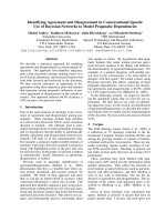
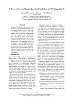
The N-terminal tail of histone H3 was efficiently bio-
tinylated by biotinidase. The binding of biotin was
substantially greater in peptide N
1)25
than in peptide
N
15)39
, if equal amounts of both peptides were incuba-
ted with biotinidase and biocytin for 45 min (Fig. 1;
Fig. 1. Biotinylation targets amino acids in the N-terminal tail of
human histone H3. Synthetic peptides based on the N- and C-ter-
minal region of histone H3 were biotinylated enzymatically, and bio-
tin was probed using gel electrophoresis and streptavidin
peroxidase. N
1)25
, peptide spanning amino acids 1–25 in histone
H3 (lanes 1a and b); N
15)39
, peptide spanning amino acids 15–39 in
histone H3 (lanes 2a and b); C
116)136
, peptide spanning amino acids
116–136 in histone H3 (lanes 3a and b). Duplicate analyses are
depicted.
Biotinylation sites in human histone H3 K. Kobza et al.
4250 FEBS Journal 272 (2005) 4249–4259 ª 2005 FEBS No claim to original US government works
compare lanes 1a and 1b with lanes 2a and 2b). The
peptide (C
116)136
) based on the C-terminus of histone
H3 was not biotinylated if incubated with biotinidase
(lanes 3a and 3b). This is consistent with previous
observations that biotinylation and other modifications
of histones cluster in the N-terminal region [2,7]. Also
these findings suggest that the primary targets for bio-
tinylation are located in the region spanning the 25
N-terminal amino acids. Thus, subsequent studies
focused on this region in the histone H3 molecule.
Previous studies suggested that lysine residues in
histones are targets for biotinylation [7]. Thus, we sub-
divided the N-terminal 25 amino acids into four syn-
thetic peptides to allow for easier identification of
biotinylated lysines in histone H3: N
1)9
(including K4
and K9), N
9)16
(including K9 and K14), N
16)23
(inclu-
ding K18 and K23), and N
18)25
(including K18 and
K23); subscripts denote the amino acid residues in the
histone H3 sequence. These peptides were incubated
with biotinidase and biocytin for up to 45 min; at
timed intervals aliquots were collected and biotinylated
peptides on transblots were probed using streptavidin
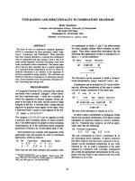
peroxidase. Apparently, peptide N
18)25
was a better
substrate for biotinylation than peptides N
1)9
,N
9)16
and N
16)23
(Fig. 2). The minor apparent differences
in biotinylation signal among the peptides loaded in
Fig. 2B lanes 2–4 are caused by intra-assay variation,
and are not observed if biotinylation of the same pep-
tides is quantified by gel densitometry using multiple
independent gels (Fig. 2A). Peptide N
1)25
was used as
a reference and was heavily biotinylated (Fig. 2B, lane
1): 100% relative biotinylation after 45 min of incuba-
tion. Peptide C
116)136
was used as a negative control
and was not biotinylated after 45 min (lane 6). These
findings suggest that either K18, K23, or both are
targets for biotinylation (see below). However, further
below we provide evidence that modifications of argi-
nines may substantially enhance the biotinylation of
histone H3 by biotinidase, and that K4 and K9 may
also be targets for biotinylation in vivo. All subsequent
enzymatic biotinylations were conducted for 45 min.
The next series of experiments focused on K4, K9
and K14. Peptide N
1)25
was used as a positive control
and was heavily biotinylated (Fig. 3, lane 1). As expec-
ted, if both lysines (K4 and K9) in a peptide spanning
amino acids 1–9 in histone H3 were substituted by
alanine (K4,9A
1)9
), no binding of biotin was detect-
able (lane 2). This is consistent with previous studies,
suggesting that lysines rather than other amino acids
are targets for biotinylation [7]. If K4 was substi-
tuted with alanine (K4A
1)9
), biotinylation of K9 was
barely detectable (lane 3). In contrast, if K9 was sub-
stituted with alanine (K9A
1)9
), K4 was biotinylated
considerably (lane 4). These findings suggest that K4 is
a target for biotinylation.
Next, variations of a peptide spanning amino acids
9–16 in histone H3 (i.e. including K9 and K14) were
tested. If both K9 and K14 were substituted with
alanine (K9,14A
9)16
), no binding of biotin was detect-
able (lane 5). If K14 was substituted with alanine
(K14A
9)16
), K9 was heavily biotinylated (lane 6).
This is in contrast to the findings described above,
which suggested that K9 is a poor target for bio-
tinylation (peptide K4A
1)9
in lane 3). We offer the
following explanation for these apparently contra-
dictory observations: peptide K14A
9)16
is lacking the
A
B
Fig. 2. Biotinylation of peptides based on the N-terminal tail in his-
tone H3. (A) Synthetic peptides were biotinylated enzymatically,
and biotin was probed using gel electrophoresis and streptavidin
peroxidase. N
1)9
, peptide spanning amino acids 1–9 in histone H3;
N
9)16
, peptide spanning amino acids 9–16 in histone H3;
N
16)23
, peptide spanning amino acids 16–23 in histone H3; and
N
18)25
, peptide spanning amino acids 18–25 in histone H3. Each
data point represents the mean of three independent measure-
ments. (B) Representative gels, depicting peptides that were incu-
bated with biotinidase and biocytin for 45 min. N
1)25
(lane
1), peptide spanning amino acids 1–25 in histone H3; N
1)9
,N
9)16
,
N
16)23
and N
18)25
(lanes 2–5) are as described for panel A; C
116)136
(lane 6), peptide spanning amino acids 116–136 in histone H3.
K. Kobza et al. Biotinylation sites in human histone H3
FEBS Journal 272 (2005) 4249–4259 ª 2005 FEBS No claim to original US government works 4251
positively charged and bulky arginine residue in
position 8; in contrast peptide K4A
1)9
includes R8.
Biotinylation of K14A
9)16
cannot be explained by
biotinylation of K14, given that K14 is a poor target
for biotinylation (peptide K9A
9)16
, lane 7). These
findings are consistent with the hypothesis that K9
might be a good target for biotinylation if R8 is
modified covalently; this hypothesis was further tested
in dimethylation experiments described below. Peptide
C
116)136
was used as a negative control; no biotinyla-
tion was detectable (lane 8).
The following series of experiments focused on K18
and K23. Peptide N
1)25
was used as a positive control
and was heavily biotinylated (Fig. 4, lane 1). As expec-
ted, if both lysines (K18 and K23) in a peptide based
on amino acids 16–23 in histone H3 were substituted
with alanine (peptide K18,23A
16)23
), no binding of
biotin was detectable (lane 2). Likewise, biotinylation
of K18 was weak if K23 was substituted with alanine
(K23A
16)23
; lane 3), and biotinylation of K23 was
weak if K18 was substituted with alanine (K18A
16)23
;
lane 4). This is in apparent contrast to the findings
presented in Fig. 2, which suggested that K18 or K23
are good targets for biotinylation (peptide N
18)25
in
Fig. 2). Based on the following lines of reasoning we
hypothesize that R17 in peptide K23A
16)23
interfered
with biotinylation of K18 in the experiments depicted
in Fig. 4: (a) Peptide N
18)25
(Fig. 2) starts with K18,
i.e. does not include R17; (b) peptide K23A
16)23
(Fig. 4) starts with A16, i.e. this peptide includes R17;
(c) experiments involving K9 suggested that arginine
residues may interfere with biotinylation (see above).
This hypothesis was tested as follows. Peptides were
synthesized that started with K18 in histone H3; hence,
these peptides did not include R17 but did include
both K18 and K23 unless noted otherwise. No biotiny-
lation was detected if both K18 and K23 were substi-
tuted with alanine (K18,23A
18)25
; lane 5). If K23 was
substituted with alanine (K23A
18)25
), K18 was heavily
biotinylated (lane 6). In contrast, if K18 was substi-
tuted with alanine (K18A
18)25
), biotinylation of K23
was barely detectable (lane 7). Peptide C
116)136
was
used as a negative control; no biotinylation was detect-
able (lane 8). These findings are consistent with the
Fig. 4. Biotinylation of K18 and K23 in the N-terminal tail in histone
H3. Synthetic peptides were biotinylated enzymatically, and biotin
was probed using gel electrophoresis and streptavidin peroxidase.
N
1)25
, peptide spanning amino acids 1–25 in histone H3 (lane 1);
K18,23A
16)23
, K23A
16)23
, K18A
16)23
, K18,23A
18)25
, K23A
18)25
and
K18A
18)25
, substitutions of K18 and K23 in histone H3; and
C
116)136
, peptide spanning amino acids 116–136 in histone H3.
Fig. 3. Biotinylation of K4, K9 and K14 in the N-terminal tail in his-
tone H3. Synthetic peptides were biotinylated enzymatically, and
biotin was probed using gel electrophoresis and streptavidin peroxi-
dase. N
1)25
, peptide spanning amino acids 1–25 in histone H3 (lane
1); K4,9A
1)9
, K4A
1)9
,K9A
1)9
, K9,14A
9)16
, K14A
9)16
and K9A
9)16
are substitutions of K4, K9 and K14 in histone H3; C
116)136
, peptide
spanning amino acids 116–136 in histone H3.
Biotinylation sites in human histone H3 K. Kobza et al.
4252 FEBS Journal 272 (2005) 4249–4259 ª 2005 FEBS No claim to original US government works
hypothesis that K18 is a target for biotinylation if R17
is modified; this hypothesis was further tested as des-
cribed below. Also, these findings suggest that K23 is a
poor target for biotinylation.
Arginine residues such as R2 and R17 in human his-
tone H3 are modified by mono- and di-methylation;
various lysine residues in histones are modified by
mono-, di-, or tri-methylation [2,23]. Here we deter-
mined whether naturally occurring modifications of
arginines render lysines a better target for biotinylation
in histone H3. Peptide N
16)23
was used as a control;
this peptide includes K18 and K23, and an arginine
residue (R17) that is not dimethylated. Peptide N
16)23
was a moderate target for biotinylation by biotinidase
(Fig. 5, lane 1), confirming findings presented above.
Likewise, peptides N
1)9
(including K4 and K9) and
N
9)16
(including K9 and K14) were relatively poor
targets for biotinylation (data not shown; see also
Fig. 2). Dimethylation of R2 and R8 (combined or
individually) moderately increased the enzymatic bioti-
nylation of K4 and K9 by biotinidase (compare lanes
2–4 to lane 1). Dimethylation of R17 (peptide
dmeR17
16)23
) substantially increased the enzymatic
biotinylation of K18 (compare lanes 1 and 5). Note
that peptide dmeR17
16)23
also contains K23; however,
studies presented above suggested that K23 is a poor
target for biotinylation.
Effects of arginine residues on biotinylation of
lysines were further corroborated in the following ser-
ies of experiments. The synthetic peptide N
6)13
(inclu-
ding R8 and K9) was used as a control; this peptide
was a moderate target for biotinylation (Table 1). If
R8 was substituted with an alanine (peptide R8A
6)13
)
biotinylation increased considerably, suggesting that
unmodified arginines interfere with biotinylation of
lysines by biotinidase. Substitution of arginine with
ornithine leaves intact the positive charge in position
8. If R8 was substituted with an ornithine (peptide
R8O
6)13
) biotinylation increased considerably, suggest-
ing that the positive charge of arginine is not respon-
sible for inhibiting biotinylation of lysines. If a
negative charge was introduced by phosphorylation of
S10 during peptide synthesis [S10S(p)
6)13
], K9 became
a poor target for biotinylation. This suggests that the
naturally occurring phosphorylation of S10 [2] may
play a role in decreasing the availability of K9 for bio-
tinylation. If K9 was substituted with an alanine (pep-
tide K9A
6)13
), no biotinylation was observed (negative
control). Finally, changing the sequence of amino acids
7 and 8 from AR to RA did not substantially affect
biotinylation of K9.
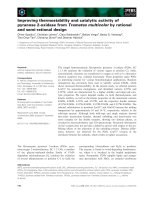
Polyclonal antibody
Polyclonal antibodies were generated to determine
whether histone H3 is biotinylated at K4, K9 and K18
in vivo. First, we determined whether the antibodies
were specific for biotinylation sites. Transblots of the
following biotinylated peptides were probed with the
Fig. 5. Effects of arginine dimethylation on the biotinylation of
lysine residues in the N-terminal tail in histone H3. Synthetic pep-
tides were biotinylated enzymatically, and biotin was probed using
gel electrophoresis and streptavidin peroxidase. N
16)23
, peptide
spanning amino acids 1–23 in histone H3 (lane 1); dmeR2R8
1)9
,
dmeR8
1)9
, dmeR2
1)9
and dmeR17
16)23
, dimethylation of R2, R8 or
R17 in histone H3 (lanes 2–5).
Table 1. Amino acid modifications affect biotinylation of K9 by bio-
tinidase. TARKSTGG represents the native unmodified peptide,
based on the amino acid sequence in position 6–13 in histone H3.
Identifier
Amino acid
sequence
Relative
biotinylation
N
6)13
TARKSTGG + +
R8A
6)13
TAAKSTGG + + +
R8O
6)13
TAOKSTGG + + +
S10S(p)
6)13
TARKS(p)TGG –
K9A
6)13
TARASTGG –
AR7,8RA
6)13
TRAKSTGG +
K. Kobza et al. Biotinylation sites in human histone H3
FEBS Journal 272 (2005) 4249–4259 ª 2005 FEBS No claim to original US government works 4253
newly developed antibodies in all possible combina-
tions: N
1)13
bioK4, N
1)13
bioK9 and N
13)25
bioK18 (see
Experimental procedures for sequence information).
The following observations were made with regard to
antibody specificities. The antibody raised against his-
tone H3 (biotinylated at K4) reacted with N
1)13
bioK4
and cross-reacted with N
1)13
bioK9, but bound only
very weakly to N
13)25
bioK18 (Fig. 6A, lanes 1–3). No
signal was detectable if nonbiotinylated peptide (N
1)25
)
was used as a target (lane 4), or if N
1)13
bioK4 was
probed using preimmune serum (lane 5). The antibody
raised against histone H3 (biotinylated at K9) reacted
with N
1)13
bioK9, but cross-reacted only very weakly
with N
1)13
bioK4 and N
13)25
bioK18 (lanes 6–8). No
signal was detectable if nonbiotinylated peptide (N
1)25
)
was used as a target (lane 9), or if N
1)13
bioK9 was
probed using preimmune serum (lane 10). The anti-
body raised against histone H3 (biotinylated at K18)
reacted with N
13)25
bioK18, but did not bind to
N
1)13
bioK4 and cross-reacted only very weakly with
N
1)13
bioK9 (lanes 11–13). No signal was detectable if
nonbiotinylated peptide (N
1)25
) was used as a target
(lane 14), or if N
13)25
bioK18 was probed using pre-
immune serum (lane 15). Peptides N
1)13
bioK4,
N
1)13
bioK9 and N
13)25
bioK18 produced equal signals
if biotin was probed with streptavidin–peroxidase (data
not shown). This is consistent with the notion that
equal amounts of peptide were loaded per lane in spe-
cificity experiments.
Finally, we titrated biotinylated and nonbiotinylated
peptides by using antibodies to biotinylated histones. If
biotinylated peptides were used as targets, the chemilu-
minescence signal paralleled the mass of peptide loaded
per lane (Fig. 6B, top row). In contrast, no signal was
detectable if nonbiotinylated peptides were used as tar-
get (Fig. 6B, bottom row). These findings suggest that
the affinities of antibodies for biotinylated peptides were
at least seven times greater than for nonbiotinylated
peptides: detection limits were about 1.9–3.8 lgÆlane
)1
for biotinylated peptides vs. > 15 lgÆlane
)1
for nonbio-
tinylated peptides if the autoradiography films were
exposed to membranes for about 5 s.
A
B
Fig. 6. Biotinylation site specificity of antibodies against histone H3, biotinylated at K4, K9 or K18. Synthetic peptides based on histone H3
were chemically biotinylated either at K4 (denoted N
1)13
bioK4), K9 (N
1)13
bioK9), or K18 (N
13)25
bioK18); a nonbiotinylated peptide spanning
amino acids 1–25 was used as a negative control (N
1)25
). (A) Peptides were probed using antibodies to histone H3, biotinylated at K4 (lanes
1–4), biotinylated at K9 (lanes 6–9), biotinylated at K18 (lanes 11–14), or preimmune sera (denoted Pre, lanes 5, 10 and 15) to test for cross-
reactivity. (B) Peptides were titrated using antibodies against biotinylated K4, K9, and K18; equal amounts of the nonbiotinylated peptide
N
1)25
were used as negative control.
Biotinylation sites in human histone H3 K. Kobza et al.
4254 FEBS Journal 272 (2005) 4249–4259 ª 2005 FEBS No claim to original US government works
Analysis of biotinylated histone H3 in human
cells by immunoblotting and
immunocytochemistry
Histone H3 in human JAr cell nuclei is biotinylated at
K4, K9 and K18, as judged by western blot analysis
(Fig. 7A, top row). Recombinant (nonbiotinylated)
histone H3 was used as a negative control and did not
produce a signal (bottom row). If human histone
extracts or recombinant histone H3 were probed with
preimmune sera no signal was detectable (data not
shown).
Biotinylation of histone H3 depended on biotinidase
and holocarboxylase synthetase. For example, biotiny-
lation of K18 in histone H3 was less abundant in
fibroblasts from biotinidase- and holocarboxylase
synthetase-deficient individuals compared with normal
fibroblasts (Fig. 7B, lanes a–c); equal loading of lanes
was confirmed by staining with Coomassie blue (lanes
d–f). Of note, the antibodies against biotinylated his-
tone H3 did not show significant cross-reactivity with
histones H1, H2A, H2B and H4.
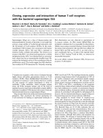
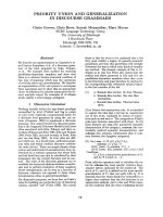
Finally, biotinylated species of histone H3 were visu-
alized in JAr cells by using immunocytochemistry.
Antibody to K4-biotinylated histone H3 localized pri-
marily to the cell nucleus (Fig. 8, image a–d); pre-
immune serum did not generate a detectable signal
(image e). Likewise, staining with antibodies to K9-
biotinylated and K18-biotinylated histone H3 was
consistent with nuclear localization of biotinylated
histones (images f–o). No signal was detectable if cells
were stained with secondary antibody alone (data not
shown). Staining with an antibody to K12-biotinylated
histone H4 [7] also produced a nuclear signal (positive
control; data not shown). Collectively, these findings
suggest that human cells contain histone H3, biotinyl-
ated at K4, K9 and K18.
Discussion
This study provides evidence (a) that K4, K9 and K18
in histone H3 are good targets for biotinylation by
human biotinidase; (b) that K14 and K23 are relatively
poor targets for biotinylation; (c) that human cells
contain histone H3, biotinylated in positions K4, K9
and K18; and (d) that dimethylation of arginine resi-
dues in histone H3 enhances biotinylation of adjacent
lysine residues, whereas phosphorylation of serine resi-
dues is likely to abolish biotinylation of adjacent lysine
residues.
The following observations suggest that biotinyla-
tion of K4, K9 and K18 in histone H3 is physiologi-
cally important. First, evidence has been provided that
biotinylation of histones might play a role in the cellu-
lar response to DNA damage [12,13]. Second, biotiny-
lation of histones might be associated with gene
silencing [12]. Third, K4 and K9 are targets for both
methylation [2] and biotinylation; methylation and
A
B
Fig. 7. Biotinylated histones H3 are present in extracts from human cell nuclei, but biotinylation is reduced in biotinidase- and holocarboxy-
lase synthetase-deficient cells. (A) Histones were extracted from JAr cell nuclei and biotinylated histones H3 were titrated using antibodies
against biotinylated K4, K9 and K18; equal amounts of recombinant (nonbiotinylated) histone H3 were used as negative control. (B) Histones
were extracted from biotinidase- and holocarboxylase synthetase-deficient human skin fibroblasts (lanes a and b, respectively); IMR-90 fibro-
blasts from a healthy human were used as a control (lane c). Histones were probed using an antibody to K18-biotinylated histone H3. Equal
loading was confirmed by staining with Coomassie blue (lanes d–f).
K. Kobza et al. Biotinylation sites in human histone H3
FEBS Journal 272 (2005) 4249–4259 ª 2005 FEBS No claim to original US government works 4255
biotinylation of the same lysine residue are mutually
exclusive. Methylation of K4 is associated with tran-
scriptionally active chromatin whereas methylation of
K9 is associated with transcriptionally silent chromatin
[3,24]. Thus, biotinylation of K4 and K9 is likely to
affect transcriptional activity of chromatin. Fourth,
K18 is a target for both acetylation [2,23] and biotiny-
lation. Acetylation of K18 is associated with transcrip-
tionally active chromatin [23]. It is unknown whether
biotinylation of K18 affects acetylation-dependent
activation of chromatin.
Modifications of arginine residues in histones affect
biotinylation of adjacent lysine residues. The following
lines of evidence support this notion. (a) Dimethyla-
tion of R2, R8 and R17 increased biotinylation of K4,
K9 and K18, respectively, by biotinidase. Dimethyla-
tion of R2 and R17 in histone H3 has been shown to
occur in vivo [2,23], suggesting that the findings presen-
ted here are physiologically relevant. (b) Substitution
of R8 with ornithine was associated with increased
biotinylation of K9. This is of potential physiologi-
cal significance, given that monomethyl- and dimethyl-
arginines in histones can be hydrolyzed to produce
citrulline and, perhaps, ornithine [25]. Formally, we
cannot exclude the possibility that free amino groups
in ornithine and citrulline are substrates for biotinyla-
tion rather than enhancing biotinylation of adjacent
lysines. However, our investigations of biotinylation
motifs suggested that ornithine is not biotinylated by
biotinidase, and that citrulline is only a relatively poor
target for biotinylation (A. Kueh and J. Zampleni,
unpublished data).
Finally, the present study provides evidence
that phosphorylation of serine residues may prevent
Fig. 8. Biotinylated histones H3 localize to the nucleus in JAr cells. Cells were stained with antibodies to K4-biotinylated histone H3 (top
panel), K9-biotinylated histone H3 (middle panel) and K18-biotinylated histone H3 (lower panel). The nuclear compartment was stained using
DAPI, and the cytoplasm was stained using rhodamine phalloidin. Images entitled ‘Merged’ were created by overlaying images obtained by
staining with antibody, DAPI and rhodamine phalloidin. Pre-immune sera were used as negative controls.
Biotinylation sites in human histone H3 K. Kobza et al.
4256 FEBS Journal 272 (2005) 4249–4259 ª 2005 FEBS No claim to original US government works
biotinylation of adjacent lysine residues. This may be
important for processes such as mitotic and meiotic
chromosome condensation (phosphorylation of S10
and S28 in histone H3), transcriptional activation of
chromatin (phosphorylation of S10 and S28 in histone
H3) and DNA repair (phosphorylation of S14 in his-
tone H2B) [23,26].
What are the limitations of the studies presented
here? First, it is unknown whether post-translational
modifications such as acetylation and methylation
coexist with biotinylation on the same histone mole-
cule. For example, does methylation of K9 coexist
with biotinylation of K4? These uncertainties are cur-
rently being addressed in our laboratory by MS ana-
lysis of histone extracts from human cells. Second, we
have identified some modifications that affect biotiny-
lation of histones, e.g. dimethylation of arginines and
phosphorylation of serines. It is unknown whether this
is a bidirectional interaction. For example, does bioti-
nylation of K9 prevent phosphorylation of S10? Third,
both biotinidase and holocarboxylase synthetase have
biotinyl histone transferase activity [8,9]. In this study
only biotinidase was used to identify biotinylation sites
in histone H3. Theoretically, holocarboxylase synthe-
tase might target distinct amino acid residues for bioti-
nylation.
Taken together, the present study has revealed three
new modifications of human histone H3: biotinylation
of K4, K9 and K18. Previous studies suggested that
K8 and K12 in histone H4 are also biotinylated [7],
bringing to a total of five the known biotinylation sites
in human histones. Undoubtedly, additional biotinyla-
tion sites will be identified in future studies, given
that all five major classes of human histones contain
streptavidin-reactive material [6]. The availability of
site-specific antibodies to biotinylated histones H3 and
H4 [7] is likely to generate novel insights into roles for
histone biotinylation in eukaryotic cells.
Experimental procedures
Peptide synthesis
Synthetic peptides were used as substrates for biotinidase to
identify biotinylation sites in histone H3; the amino acid
sequences in these peptides were based on human histone
H3 (GenBank accession number NP_066403). Peptides were
synthesized using Fmoc chemistry by a standard solid-
phase method [27] as described previously [7]; l-isomers of
amino acids were used in all syntheses. One-letter annota-
tion is used for denoting amino acids throughout this paper
[28]. Chemically modified peptides were synthesized by
using biotinylated, dimethylated, and phosphorylated
Fmoc-e-NH
2
-d-biotinyl-l-lysine, Fmoc-dimethyl-l-arginine,
and Fmoc-phospho-l-serine. Identities of synthetic peptides
were confirmed by MS [7].
Post-translational modifications
Post-translational modifications of histone H3 cluster in the
N-terminal region of the molecule (amino acids 1–36), e.g.
methylation of K4 and K9, acetylation of K9, K18, K23
and K36, phosphorylation of S10, and mono- or dimethyla-
tion of R17 [2]. In pilot studies we used the following syn-
thetic peptides to determine whether biotinylation of
histone H3 also takes place in the N-terminal region: (a)
N-terminus of histone H3, spanning amino acids 1–25
(ARTKQTARKSTGGKAPRKQLATKAA; this peptide
was denoted N
1)25
), and (b) a peptide based on amino
acids 15–39 in histone H3 (APRKQLATKAARKSAPA-
TGGVKKPH; denoted N
15)39
). As a negative control we
used a peptide spanning the C-terminus of histone H3, i.e.
amino acids 116–136 (KRVTIMPKDIQLARRIRGERA;
denoted C
116)136
). Pilot studies using these peptides and
previous studies of histone H4 [7] suggested that lysines
located in the N-terminus of histone H3 are the pri-
mary targets for biotinylation (see below). Thus, the
studies presented below focused on lysine residues in the
N-terminal region; the amino acid sequences of the syn-
thetic peptides used to identify biotinylation sites are
provided in Results.
Enzymatic biotinylation of peptides
Synthetic peptides were incubated with biotinidase for enzy-
matic biotinylation as described previously [7,8]; biocytin
(biotinyl-e-lysine) was used as a biotin donor.
Gel electrophoresis
After enzymatic biotinylation, peptides were resolved using
16% tricine ⁄ polyacrylamide gels according to the manufac-
turer’s instructions (Invitrogen, Carlsbad, CA, USA). Pep-
tides were electroblotted onto polyvinylidene fluoride
membranes (Millipore, Bedford, MA, USA); peptide-bound
biotin was probed with streptavidin–peroxidase [6,7]. In
previous studies both HPLC and MS were used to confirm
covalent biotinylation of peptides [7].
Polyclonal antibodies
The following polyclonal antibodies to human histone H3
were generated using a commercial facility (Cocalico Biolog-
icals, Reamstown, PA, USA): anti-H3 (biotinylated at K4),
anti-H3 (biotinylated at K9) and anti-H3 (biotinylated at
K18). To raise these antibodies, the following peptides
were custom-synthesized by the University of Virginia
K. Kobza et al. Biotinylation sites in human histone H3
FEBS Journal 272 (2005) 4249–4259 ª 2005 FEBS No claim to original US government works 4257
Biomolecular Research Facility (Charlottesville, VA, USA):
(a) N
1)13
bioK4, ARTK(biotin)QTARKSTGGC (amino
acids 1–13 in histone H3); (b) N
1)13
bioK9, ARTK-
QTARK(biotin)STGGC (amino acids 1–13); and (c)
N
13)25
bioK18, GKAPRK(biotin)QLATKAAC (amino acids
13–25). Peptide identities were confirmed by MS. Peptides
were conjugated to keyhole limpet hemocyanin by utilizing
the C-terminal cysteine [7]; these peptide conjugates were
injected into white New Zealand rabbits, following NIH and
USDA guidelines for animal care. All possible measures were
taken to minimize pain and discomfort to animals. Booster
injections were given after 14, 21 and 49 days. Serum was
collected before immunization (preimmune serum) and
2 days after each booster injection. Serum collected after the
third booster injection was used for the assays described
below; preimmune serum was used as a control. For assess-
ment of antibody specificities, electroblots of peptides
N
1)13
bioK4, N
1)13
bioK9 and N
13)25
bioK18 were probed
with the anti-(histone H3) Igs and a monoclonal mouse
anti-rabbit IgG peroxidase conjugate as described previously
[7]; nonbiotinylated peptide (N
1)25
) was used as a control.
Analysis of biotinylated histone H3 in human
cells by immunoblotting and
immunocytochemistry
JAr human choriocarcinoma cells were cultured as des-
cribed [29]. For western blot analysis, nuclear histones were
extracted by using hydrochloric acid as described [6].
Recombinant (nonbiotinylated) histone H3 was purchased
from Upstate Inc. (Lake Placid, NY, USA) and served as
negative control. Equal amounts of JAr cell histone H3 and
recombinant histone H3 (as judged by staining with Coo-
massie blue and gel densitometry) were loaded onto 18%
Tris ⁄ glycine gels (Invitrogen) for electrophoresis. Proteins
were electroblotted and probed with antibodies to biotinyl-
ated histone H3 as described [7]. Primary antibodies (rabbit
serum) were diluted 250-fold, and the secondary antibody
(mouse monoclonal anti-rabbit IgG peroxidase conjugate;
Sigma, St. Louis, MO, USA) was diluted 20 000-fold.
Biotinylation of histones is mediated by biotinidase and
holocarboxylase synthetase [8,9]. We obtained biotinidase-
deficient human skin fibroblasts (strain code WG1371) and
holocarboxylase synthetase-deficient human skin fibroblasts
(strain code WG2215) from the Cell Repository at Mon-
treal Children’s Hospital (Quebec, Canada) to determine
whether deficiency is associated with decreased biotinylation
of histone H3. Human IMR-90 fibroblasts were used as
control (ATCC clone CCL-186; Manassas, VA, USA).
Nuclear histones from fibroblasts were analyzed by immu-
noblotting as described above.
Finally, biotinylated histones H3 in JAr cells were visual-
ized by standard procedures of immunohistochemistry [26].
Primary antibodies (serum) were diluted 250-fold. Pre-
immune sera were used as negative controls. As secondary
antibody we used Cy2-conjugated AffiniPure donkey anti-
rabbit IgG (Jackson ImmunoResearch, West Grove, PA,
USA) at an 80-fold dilution. The nuclear compartment was
stained using 4¢,6-diamidino-2-phenylindole (DAPI), and
the cytoplasm was stained using rhodamine phalloidin
(Molecular Probes, Eugene, OR, USA). Images were
obtained using Olympus FV500 confocal microscope
equipped with an oil immersion lens.
Acknowledgements
This work was supported by NIH grants DK 60447
and DK 063945, by a grant from the Nebraska
Tobacco Settlement Biomedical Research Enhance-
ment Funds and in part by NIH Grant Number 1 P20
RR16469 from the BRIN Program of the National
Center for Research Resources. This paper is a contri-
bution of the University of Nebraska Agricultural
Research Division, Lincoln, NE 68583 (Journal Series
no. 14924).
References
1 Wolffe A (1998) Chromatin, 3th edn. Academic Press,
San Diego, CA.
2 Fischle W, Wang Y & Allis CD (2003) Histone and
chromatin cross-talk. Curr Opin Cell Biol 15, 172–183.
3 Jenuwein T & Allis CD (2001) Translating the histone
code. Science 293, 1074–1080.
4 Boulikas T, Bastin B, Boulikas P & Dupuis G (1990)
Increase in histone poly (ADP-ribosylation) in mitogen-
activated lymphoid cells. Exp Cell Res 187, 77–84.
5 Shiio Y & Eisenman RN (2003) Histone sumoylation is
associated with transcriptional repression. Proc Natl
Acad Sci USA 100, 13225–13230.
6 Stanley JS, Griffin JB & Zempleni J (2001) Biotinylation
of histones in human cells: effects of cell proliferation.
Eur J Biochem 268, 5424–5429.
7 Camporeale G, Shubert EE, Sarath G, Cerny R &
Zempleni J (2004) K8 and K12 are biotinylated in
human histone H4. Eur J Biochem 271, 2257–2263.
8 Hymes J, Fleischhauer K & Wolf B (1995) Biotinylation
of histones by human serum biotinidase: assessment of
biotinyl-transferase activity in sera from normal indivi-
duals and children with biotinidase deficiency. Biochem
Mol Med 56, 76–83.
9 Narang MA, Dumas R, Ayer LM & Gravel RA (2004)
Reduced histone biotinylation in multiple carboxylase
deficiency patients: a nuclear role for holocarboxylase
synthetase. Hum Mol Genet 13, 15–23.
10 Brenner C (2002) Catalysis in the nitrilase superfamily.
Curr Opin Struct Biol 12, 775–782.
11 Zempleni J (2005) Uptake, localization, and noncarb-
oxylase roles of biotin. Annu Rev Nutr in press.
Biotinylation sites in human histone H3 K. Kobza et al.
4258 FEBS Journal 272 (2005) 4249–4259 ª 2005 FEBS No claim to original US government works
12 Peters DM, Griffin JB, Stanley JS, Beck MM &
Zempleni J (2002) Exposure to UV light causes
increased biotinylation of histones in Jurkat cells. Am J
Physiol Cell Physiol 283, C878–C884.
13 Kothapalli N & Zempleni J (2004) Double strand breaks
of DNA decrease biotinylation of lysine-12 in histone H4
in JAr cells. FASEB J 18, A103–A104 [abstract].
14 Swango KL, Demirkol M, Huner G, Pronicka E,
Sykut-Cegielska J, Schulze A, Mayatepek E & Wolf B
(1998) Partial biotinidase deficiency is usually due to the
D444H mutation in the biotinidase gene. Hum Genet
102, 571–575.
15 Wolf B, Jensen K, Huner G, Demirkol M, Baykal T,
Divry P, Rolland MO, Perez-Cerda C, Ugarte M, Str-
aussberg R, Basel-Vanagaite L, Baumgartner ER, Suor-
mala T, Scholl S, Das AM, Schweitzer S, Pronicka E &
Sykut-Cegielska J (2002) Seventeen novel mutations that
cause profound biotinidase deficiency. Mol Genet Metab
77, 108–111.
16 Moslinger D, Muhl A, Suormala T, Baumgartner R &
Stockler-Ipsiroglu S (2003) Molecular characterisation
and neuropsychological outcome of 21 patients with pro-
found biotinidase deficiency detected by newborn screen-
ing and family studies. Eur J Pediatr 162, S46–S49.
17 Yang X, Aoki Y, Li X, Sakamoto O, Hiratsuka M,
Kure S, Taheri S, Christensen E, Inui K, Kubota M,
Ohira M, Ohki M, Kudoh J, Kawasaki K, Shibuya K,
Shintani A, Asakawa S, Minoshima S, Shimizu N, Nari-
sawa K, Matsubara Y & Suzuki Y (2001) Structure of
human holocarboxylase synthetase gene and mutation
spectrum of holocarboxylase synthetase deficiency. Hum
Genet 109, 526–534.
18 Wolf B & Heard GS (1991) Biotinidase deficiency.
Advances in Pediatrics (Barness L & Oski F, eds), pp.
1–21. Medical Book Publishers, Chicago, IL.
19 Wolf B (1991) Worldwide survey of neonatal screening
for biotinidase deficiency. J Inher Metab Dis 14, 923–927.
20 Plath, K, Fang J, Mlynarczyk-Evans SK, Cao R, Wor-
ringer KA, Wang H, Cruz CC, d. 1 Otte AP, Panning B
& Zhang Y (2003) Role of histone H3 lysine 27 methy-
lation in X inactivation. Science 300, 131–135.
21 Santos-Rosa H, Schneider R, Bannister AJ, Sherriff J,
Bernstein BE, Emre NC, Schreiber SL, Mellor J & Kou-
zarides T (2002) Active genes are tri-methylated at K4
of histone H3. Nature 419, 407–411.
22 Schneider R, Bannister AJ, Myers FA, Thorne AW,
Crane-Robinson C & Kouzarides T (2004) Histone H3
lysine 4 methylation patterns in higher eukaryotic genes.
Nat Cell Biol 6, 73–77.
23 Lachner M, O’Sullivan RJ & Jenuwein T (2003) An epi-
genetic road map for histone lysine methylation. J Cell
Sci 116, 2117–2124.
24 Bird A (2001) Methylation talk between histones and
DNA. Science 294, 2113–2115.
25 Bannister AJ, Schneider R & Kouzarides T (2002)
Histone methylation: dynamic or static? Cell 109,
801–806.
26 Cheung WL, Ajiro K, Samejima K, Kloc M, Cheung P,
Mizzen CA, Beeser A, Etkin LD, Chernoff J, Earnshaw
WC & Allis CD (2003) Apoptotic phosphorylation of
histone H2B is mediated by mammalian sterile twenty
kinase. Cell 113, 507–517.
27 Fields GB (1998) Solid-phase peptide synthesis. Mole-
cular Biomethods Handbook (Rapley R & Walker JM,
eds), pp. 527–545. Humana Press Inc., Totowa, NJ.
28 Garrett RH & Grisham CM (1995) Biochemistry. Saun-
ders College Publishing, Fort Worth, TX.
29 Crisp SERH, Camporeale G, White BR, Toombs CF,
Griffin JB, Said HM & Zempleni J (2004) Biotin
supply affects rates of cell proliferation, biotinylation
of carboxylases and histones, and expression of the
gene encoding the sodium-dependent multivitamin
transporter in JAr choriocarcinoma cells. Eur J Nutr
43, 23–31.
K. Kobza et al. Biotinylation sites in human histone H3
FEBS Journal 272 (2005) 4249–4259 ª 2005 FEBS No claim to original US government works 4259