Báo cáo khoa học: The N-glycans of yellow jacket venom hyaluronidases and the protein sequence of its major isoform in Vespula vulgaris pdf
Bạn đang xem bản rút gọn của tài liệu. Xem và tải ngay bản đầy đủ của tài liệu tại đây (339.81 KB, 9 trang )
The N-glycans of yellow jacket venom hyaluronidases
and the protein sequence of its major isoform in
Vespula vulgaris
Daniel Kolarich
1
, Renaud Le
´
onard
1
, Wolfgang Hemmer
2
and Friedrich Altmann
1
1 Department of Chemistry, University of Natural Resources and Applied Life Sciences (BOKU), Vienna, Austria
2 Floridsdorf Allergy Center (FAZ), Vienna, Austria
Almost 50% of patients with suspected hymenoptera
allergy turn out to be sensitized to both honeybee and
yellow jacket venom [1]. Contrasting with these in vitro
results, only very few patients show adverse reactions
to both venoms. In two recent studies, protein-bound
carbohydrate was shown to be a major, but not the
only, cause for the simultaneous reactivity of patients’
IgE with honeybee and yellow jacket venoms [2,3].
Apart from cases where true double sensitization to
both venoms or antibodies against cross-reactive pro-
tein appears to be involved, a frequently found situ-
ation is that patients sensitized to one of the venoms
cross-react with the other venom on the sole basis of
protein-linked carbohydrate, which forms the so-called
cross-reactive carbohydrate determinants (CCDs) [2].
Probably, CCDs also cause some of the cross-reactivity
observed for hyaluronidases from hornet (Dolichoves-
pula sp.), paper wasp (Polistes sp.) [4] and even bumble
bee and fire ant venom [5,6].
The protein responsible for most of the carbohydrate
based reactivity on immunoblots of yellow jacket
venom migrates at 43 kDa under denaturing conditions
[2]. A rabbit serum raised against plant glycoproteins
(oilseed rape extract) likewise primarily bound to the
43 kDa protein [2]. This band is believed to represent
hyaluronidase for which, in the case of Vespula
vulgaris, the primary structure is known from
cDNA sequencing (Swissprot P49370) [4] and which is
known to be glycosylated [7]. However, in a recent
investigation, by MALDI-TOF MS, of the 43 kDa
Keywords
cross-reactive carbohydrate determinant;
hyaluronidase; Hymenoptera; insect sting
allergy; Vespula
Correspondence
F. Altmann, Department of Chemistry,
BOKU, Muthgasse 18, A-1190 Vienna,
Austria
Fax: + 43 136006 6059
Tel: + 43 136006 6062
E-mail:
Note:
The nucleotide sequence of Ves v 2b can
be found at: EMBL AJ920395 The protein
sequence for Ves v 2b (fragment) can be
found at ExPASy Q5D7H4.
(Received 11 May 2005, revised 22 June
2005, accepted 1 July 2005)
doi:10.1111/j.1742-4658.2005.04841.x
Hyaluronidase (E.C. 3.2.1.35), one of the three major allergens of yellow
jacket venom, is a glycoprotein of 45 kDa that is largely responsible for
the cross-reactivity of wasp and bee venoms with sera of allergic patients.
The asparagine-linked carbohydrate often appears to constitute the com-
mon IgE-binding determinant. Using a combination of MALDI MS and
HPLC of 2-aminopyridine-labelled glycans, we found core-difucosylated
paucimannosidic glycans to be the major species in the 43–45 kDa band
of Vespula vulgaris and also in the corresponding bands of venoms from
five other wasp species (V. germanica, V. maculifrons, V. pensylvanica,
V. flavopilosa and V. squamosa). Concomitant peptide mapping of the
V. vulgaris 43 kDa band identified the known hyaluronidase, Ves v 2
(SwissProt P49370), but only as a minor component. De novo sequencing
by tandem MS revealed the predominating peptides to resemble a differ-
ent, yet homologous, sequence. cDNA cloning retrieved a sequence
with 58 and 59% homology to the previously known isoform and to the
Dolichovespula maculata and Polistes annularis hyaluronidases. Close homo-
logues of this new, putative hyaluronidase b (Ves v 2b) were also the
major isoform in the other wasp venoms.
Abbreviations
CCD, cross-reactive carbohydrate determinant; MUF
3
F
6
and MMF
3
F
6
, N-glycan structures.
5182 FEBS Journal 272 (2005) 5182–5190 ª 2005 FEBS
band, this hyaluronidase P49370 constituted only a
minor component and the strong signals could not be
assigned to a known protein sequence [2]. In this study,
a mixture of venoms from V. vulgaris and V. germanica
was employed and thus the unknown peptide peaks
were interpreted as possibly arising from V. germanica
hyaluronidase. This explanation was wrong, as shown
below.
Irrespective of the nature of the protein, protein-
linked glycans can bind IgE, which turns many pro-
teins, especially those of higher molecular mass, into
apparent allergens. Even though some studies have
shown in vitro histamine release by plant glycoprotein
glycans [8–10], the contribution of such carbohydrate
determinants to clinical symptoms is unclear and is
believed, by many researchers, to be negligible [2,3,
5,11,12]. However, patients’ sera containing anti-
glycan immunoglobulin can bind to a variety of plant
and insect glycoproteins and even to human proteins
unrelated to any allergen in the peptide part when the
glycan has been modified with core a1,3-fucose, as
shown for patients 9–14 in a previous study [11].
The glycan-based cross-reaction of honeybee-allergic
patients’ sera with wasp venom, and vice versa for that
of wasp venom-allergic patients’ sera with bee venom,
could be inhibited by oilseed rape pollen extract [2].
Furthermore, the inhibition could be performed with
BSA, to which small glycopeptides containing core
a1,3-fucose (and xylose) had been attached [2]. Thus,
it can be speculated that yellow jacket hyaluronidase
(or, more precisely, the 43 kDa protein) carries core
a1,3-fucosylated N-glycans, such as the honeybee
venom hyaluronidase [13]. However, Vespula venom
and the proteins therein have never been subjected to
any structural analysis of protein-linked carbohydrate.
Moreover, not even the polypeptide of hyaluronidase
has been analyzed, to date, and the data on its
sequence have been obtained exclusively from cDNA
cloning [4].
Here we report on the identification of the major
polypeptide in the 43 kDa band of V. vulgaris as a
new isoform of hyaluronidase. Furthermore, we ana-
lyzed the hyaluronidase band in five other common
yellow jacket species and we investigated the N-glycan
structures of hyaluronidase from all six species.
Results
Immunological detection of venom glycoproteins
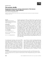
Electrophoretic separation of V. vulgaris venom under
reducing and denaturing conditions yielded three
major bands (Fig. 1). Antigen 5 migrated at 28 kDa,
phospholipase at 34 kDa and the putative hyaluroni-
dase formed a double band at 43–45 kDa. On immuno-
blots with a rabbit serum that binds complex plant
N-glycans (CCDs), one major band (at 43 kDa),
believed to be hyaluronidase, became visible. Faint
bands of lower molecular mass (34–40 kDa) were
believed to represent degradation products of hyalu-
ronidase. A sharp band at c. 105 kDa was shown by
MS to be a glycoprotein (data not shown) but could
not be identified. Only the 43 kDa band was subjected
to N-glycan analysis.
Analysis of N-glycans of the 43 kDa band
and of whole venom
The major glycan species found by MALDI-TOF MS
had masses indicating them to consist of two GlcNAc,
either two or three mannose residues and two fucose res-
idues (Fig. 2). Hence, they resembled the difucosylated
paucimannosidic N-glycans (Fig. 3), known from
honeybee venom phospholipase A
2
and hyaluronidase
[13,14]. Apart from quantitative differences, essentially
the same results were obtained when this analysis
was conducted with the hyaluronidase bands from
AB
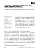
Fig. 1. Reducing SDS ⁄ PAGE (lane A) and immunoblot (lane B) of
the venom from Vespula vulgaris. The double band at 43–45 kDa,
usually considered to represent hyaluronidase, is the object of the
present study. In the immunoblot, the serum specific for cross-
reactive plant and insect N-glycans essentially only bound to the
presumed hyaluronidase. The 100 kDa glycoprotein could not be
identified. The diffuse bands of 40 kDa are regarded as degrada-
tion products of hyaluronidase.
D. Kolarich et al. Yellow jacket hyaluronidase
FEBS Journal 272 (2005) 5182–5190 ª 2005 FEBS 5183
V. maculifrons (Table 1). In this case, the structure of
the presumed N-glycan structure MMF
3
F
6
(Fig. 3) was
verified by 2D HPLC and fucosidase digestion. How-
ever, for this purpose, whole venom from V. maculifrons
was employed in order to obtain the necessary amount
of glycans, as this venom is the least expensive.
As hyaluronidase is the dominating glycoprotein in
the venom, we believe that the glycans from whole
venom essentially stem from, and therefore credibly
represent, those from hyaluronidase. First, pyridylami-
nated glycans were fractionated according to size
(Fig. 4). A large peak eluting at 21.7 min was collected
and further analyzed. Its elution time on reverse-phase
HPLC matched that of MMF
3
F
6
from honeybee
phospholipase. A moderate dose of bovine kidney
a-fucosidase caused a shift to a lower retention time,
which is consistent with the removal of an a1,6-linked
fucose residue from the reducing end GlcNAc [14,15].
The glycan mass and the removal of one fucose residue
were confirmed by MS (Fig. 4).
In summary, we conclude that wasp venom hyaluro-
nidases contain the difucosylated paucimannosidic
N-glycans MUF
3
F
6
and MMF
3
F
6
(Fig. 3) as the major
species. In addition, glycans containing one or two
GlcNAc residues at the nonreducing end and trace
amounts of other, probably hybrid-type, N-glycans
were present, but have not been further analyzed.
Proof that MMF
3
F
6
is, in fact, bound to hyaluroni-
dase came from tandem MS of a glycopeptide with a
mass of 2037.0 Da, which at first could not be assigned
to any known sequence. Only in retrospect was it
recognized as having the peptide sequence NGTYEIR
(which does not occur in P49370, see below), with a
glycan of the composition Man
3
GlcNAc
2
Fuc
2
. Among
the various glycopeptide and oligosaccharide frag-
ments, the fragment at m ⁄ z ¼ 1347.6, which still con-
tained two fucoses but only one GlcNAc residue,
exemplified best the difucosylation of the asparagine-
linked GlcNAc residue (Fig. 5).
Proteomic characterization of the 43 kDa band
As mentioned in a previous publication [2], peptide
mapping of the 43 kDa band revealed considerable
Fig. 2. Analysis of N-glycans from the Vespula vulgaris 43 kDa pro-
tein. Enzymatically released N-glycans from the 43 kDa gel band,
which was later shown to consist of two hyaluronidase isoforms,
were analyzed by linear MALDI-TOF MS. The major species repre-
sent difucosylated oligosaccharides (see Fig. 3 and Table 1).
Fig. 3. Structures of the major N-glycan species found on yellow
jacket hyaluronidases. Oligomannosidic structures are not depicted.
Abbreviations are made according to the ‘proglycan’ system (http://
www.proglycan.com). M, mannose; F, fucose; Gn, N-acetylglucosa-
mine. The distribution of the glycans in the different venoms is lis-
ted in Table 1. The two complex-type structures at the bottom
were only tentatively assigned; other isomers might also be pre-
sent.
Yellow jacket hyaluronidase D. Kolarich et al.
5184 FEBS Journal 272 (2005) 5182–5190 ª 2005 FEBS
inconsistency between the data bank entry for hyalu-
ronidase and the tryptic peptides obtained from the
43 kDa band of V. vulgaris venom (Fig. 6). Only a
few, smaller, signals could be assigned to SwissProt
entry P49370 (Fig. 6). Hence, the tryptic peptides were
subjected to nano-HPLC separation followed by tan-
dem mass spectrometric peptide sequencing. Blast
analysis of the sequences deduced from the fragment
spectra soon revealed moderate, but obvious, sequence
homologies with hyaluronidase P49370 (Fig. 7). We
Table 1. N-glycan analysis of the hyaluronidases from venoms of different wasp species. The structures are depicted in Fig. 3. The glycan
structures shown in parenthesis were tentatively assigned and other isomers may be present.
Mass
(Da) Glycan structure
V. vulgaris
%
V. maculifrons
%
V. germanica
%
V. pensylvanica
%
V. flavopilosa
%
V. squamosa
%
901.8 UUF
3
F
6
14.7 7.1
917.8 MUF
6
3.7 3.8 9.9 8.8 24.7
933.8 MM 2.6 3.0 4.5 8.6 1.5
1064.0 MUF
3
F
6
47.7 55.3 48.9 33.7 45.1 32.4
1080.0 MMF
6
11.5 10.2 9.0 7.0 13.8 8.4
1096.0 Man4 0.8 1.1
1226.1 MMF
3
F
6
28.1 22.3 32.1 23.5 23.9 26.9
1258.1 Man5 1.3
1420.3 Man6 0.6
1429.3 (MGnF
3
F
6
) 1.2 3.3 4.8 1.5 6.0
1753.5 (Man5GnF
3
F
6
) 4.3 2.0
Fig. 4. Structural analysis of the N-glycans from whole Vespula vulgaris venom. (A) Size fractionation of pyridylaminated glycans by normal
phase HPLC. (B) Analysis of the normal phase peak at 21.7 min by reversed phase HPLC. (C) Reanalysis of the same peak after treatment
with bovine kidney a-fucosidase, which preferentially cleaves the a1,6-linked fucose residue. (D) and (E) Chromatograms of reference gly-
cans MMF
3
and MMF
3
F
6
, respectively, from honeybee venom phospholipase [14]. (F) and (G), MALDI spectra of the normal phase peak at
21.7 min, before and after fucosidase digestion, respectively.
D. Kolarich et al. Yellow jacket hyaluronidase
FEBS Journal 272 (2005) 5182–5190 ª 2005 FEBS 5185
concluded that the 43 kDa band contained a second
isoform of hyaluronidase which – from the intensity of
MALDI and ESI peaks – actually constituted the
major form. To assess the true relative amount of the
isoforms, the band was subjected to Edman sequen-
cing, which revealed one sequence that deviated from
the sequence of P49370 (Fig. 7), but no clear indica-
tion for the presence of the known hyaluronidase (data
not shown). It may be added here that both potential
glycosylation sites were found as glycopeptides by
either MALDI-TOF MS (site 81, data not shown) or
ESI MS (site 66, see also previous chapter). Further-
more, the two glycopeptides were detected in both the
upper and the lower part of the double band, at
43–45 kDa, thus ruling out underglycosylation as a
reason for the different migration behaviour.
Molecular cloning of the new hyaluronidase
isoform, Ves v 2b
Aiming at the molecular cloning of the cDNA of
the newly discovered hyaluronidase isoform (referred
to as Ves v 2b in the present article, in contrast to
the known form, Ves v 2), the N-terminal sequence
and internal fragments near the C terminus were
translated into degenerate primers for PCR. Exten-
sion by 3¢-RACE led to identification of the entire
sequence. This new sequence was found to contain
two glycosylation sites. Site 66 yields a tryptic pep-
tide of mass 851.44 Da, which is identical to the
peptide mass observed for the (glyco-)peptide dealt
with in Fig. 5. It is notable that from the same
cDNA we could also isolate a clone of the ‘old’ iso-
form, Ves v 2 (which probably should be referred to
as Ves v 2a from now on).
Fig. 5. Fragment ion spectrum of a glycopeptide from the 43 kDa
band. The masses of (glyco-)peptides are written vertically, those
of free glycans horizontally, and that of the mother ion tilted. From
the loss of two fucose, of three mannose and, finally, of two Glc-
NAc residues, the composition of the glycan moiety of this peptide
could be deduced. The fragment at 1347.6 Da implied two fucose
residues to be linked to the reducing terminal GlcNAc. After acqui-
sition of the sequence of the new isoform, Ves v 2b, the peptide
moiety could be identified as comprising residues 66–72. Corres-
ponding peptides in the other hyaluronidases (see Fig. 7) lack the
glycosylation sequon.
Fig. 6. MALDI spectrum of tryptic peptides from the 43 kDa band. The upper and lower parts of the double band, at 43–45 kDa (Fig. 1),
were investigated separately but the spectra were identical. Only the three signals with mass numbers written horizontally match the data
bank entry, P49370.
Yellow jacket hyaluronidase D. Kolarich et al.
5186 FEBS Journal 272 (2005) 5182–5190 ª 2005 FEBS
Simple blast search with the new hyaluronidase
protein yielded 58% homology with P49370 and 59%
similarity with the hyaluronidases from P. annularis
and D. maculata. Sequence alignment with the cur-
rently known insect venom hyaluronidases is shown in
Fig. 7. Like its homologues, Ves v 2b contained several
hyaluronan-binding motifs (i.e. two basic amino acids
spaced by seven other residues) [16].
Ves v 2b-homologues in other Vespula species
The MALDI spectra of the hyaluronidase bands
from other wasp venoms (V. germanica, V. flavopi-
losa, V. maculifrons and V. pensylvanica) were almost
identical to that from V. vulgaris. This corroborates
the close relatedness of these wasp species and, at
the same time, identifies hyaluronidase b (possibly
Fig. 7. Alignment of insect venom hyaluronidases and of MS sequence tags: The partial sequences in upper lines represent those deduced
from fragment spectra of the unassignable peptides in the tryptic digest of the 43 kDa band of Vespula vulgaris venom. These pieces were
aligned with the known hyaluronidase Ves v 2a (or Ves v 2; P49370) and matching residues are printed white on black. While leucine (Leu)
is suggested by default in MS sequencing, isoleucine (Ile) is written when this residue occurs in the known protein sequence. The N termi-
nus was obtained by Edman sequencing. The peptide stretches chosen for primer design are underlined. In the actual protein alignment, the
new isoform, Ves v 2b, is shown at the top with the other sequences given in the order of decreasing similarity: Pol a 2 (Polistes annularis),
Ves v 2a (¼ Ves v 2; V. vulgaris), Dol m 2 (Dolichovespula maculata), Api c 2 (Apis cerana cerana), Api m 2 (A. mellifera), and even from
Anopheles gambiae. The highlighted positions correspond to amino acids that occur in Ves v 2b and in at least one of its homologues.
D. Kolarich et al. Yellow jacket hyaluronidase
FEBS Journal 272 (2005) 5182–5190 ª 2005 FEBS 5187
with a few species-specific differences) as the major
isoform in these species. In contrast, the hyaluroni-
dase band from V. squamosa yielded a totally differ-
ent MALDI peptide map, which suggests a low
sequence homology with the hyaluronidases from the
other species.
Discussion
The similarity of the glycan structures on wasp and
bee venom hyaluronidases explains – at least to a large
extent – their allergological cross-reactivity. The role
of venom hyaluronidases as major cross-reactive aller-
gens in honeybee and Vespula venom has been recog-
nized previously [17] and was thought to be a result
of the significant sequence identity ( 50%) between
these allergens [4]. Although there is some evidence for
cross-reactivity between honeybee and Vespula hyalu-
ronidases at the protein level [2,7], its significance in
relation to the cross-reactivity mediated by glycans
should be redefined in future studies.
The cross-species survey performed in this study
showed that the venoms from all six of the species
usually considered (V. vulgaris, V. germanica, V. flav-
opilosa, V. maculifrons, V. pensylvanica and V. squamo-
sa) contained MUF
3
F
6
and MMF
3
F
6
as the major
glycan structures (Fig. 3). Sera from patients with
antibodies directed to the core a 1,3-fucose determin-
ant will therefore inevitably react with the hyaluroni-
dase in venoms of all types of hymenoptera,
irrespective of the underlying protein. These sera may
be expected to bind also with other venom compo-
nents bearing similar glycans, such as phosphatases
[18], serine proteases [19], and honeybee and fire ant
phospholipases [6,14,20].
Moreover, in such cases it is not even clear whether
the original sensitizing agent was an insect venom or
a plant allergen, which also contain this carbohydrate
determinant [21]. Especially in the case of such
‘plant-allergic’ patients, a positive in vitro test against
insect venoms can be expected to be unassociated
with clinical symptoms from insect stings. Likewise,
IgE against carbohydrates induced by Hymenoptera
stings leads to a positive in vitro test with pollen
allergens but is not associated with symptoms of poll-
inosis [5]. This shows, once again, the importance of
a discrimination between carbohydrate- and protein-
based IgE binding.
Incidentally, analysis of glycans from the wasp
venom hyaluronidase led to the identification and
molecular cloning of Ves v 2b, the true major protein
in the 43 kDa band of V. vulgaris venom. It is currently
unknown whether Ves v 2b represents a ‘true’ allergen,
or binds with IgE only through its carbohydrate deter-
minants. The other isoform, Ves v 2 (or Ves v 2a, as we
would like to call it), was originally isolated by using
primers that had been designed for the hyaluronidase
of D. maculata [4]. In fact, the higher similarity of the
original Ves v 2 to the D. maculata homolog than to
Ves v 2b is depicted in a phylogenetic tree of insect
hyaluronidases (Fig. 8). As confirmed in the present
study, the cDNA amplified by these primers does code
for a hyaluronidase which occurs in V. vulgaris venom,
but as a minor isoform only. This example nicely dem-
onstrates the merits of proteomic analysis in parallel
with genomic work. As V. vulgaris does not stand high
on the list of organisms whose genomes will be fully
sequenced in the near future, the composition of the
43 kDa band would not have been revealed otherwise.
Cloning of the two isoforms may allow their expression
without immunogenic sugars, so that IgE binding
to the polypeptides alone can be measured, which
would clarify the question regarding the significance of
hyaluronidase-based cross-reactions between wasp and
honeybee venom. Furthermore, the individual role of
the two isoforms for binding of IgE of insect venom-
allergic patients can be studied.
Besides, the comparative analysis (e.g. by MALDI
peptide mapping of several wasp venoms) revealed
high homology between the hyaluronidases of all spe-
cies, except that of V. squamosa.
Fig. 8. Phylogenetic tree of hyaluronidases from several insect spe-
cies: Anopheles gambiae (A. gambiae), Apis cerana cerana (Api c
2), A. mellifera (Api m 2), Dolichovespula maculata (Dol m 2), Polis-
tes annularis (Pol a 2) and Vespula vulgaris (Ves v 2 and Ves v 2b).
The tree was obtained by using the program
MULTALIN (prodes.tou-
louse.inra.fr ⁄ multalin).
Yellow jacket hyaluronidase D. Kolarich et al.
5188 FEBS Journal 272 (2005) 5182–5190 ª 2005 FEBS
Experimental procedures
Materials
Vespula venoms in the form of venom sac extracts were
purchased from Sweden Diagnostics (Uppsala, Sweden).
Sequencing grade trypsin and peptide N-glycopeptidase A
were obtained from Roche (Basel, Switzerland). Rab-
bit anti-horseradish serum and bovine kidney a-fucosidase
were from Sigma-Aldrich (Vienna, Austria). Specimens
of V. vulgaris and V. germanica were collected in
Vienna with assistance of the beekeeper of the local fire
brigade.
Glyco-proteomic work
Vespula venom samples (approximately 10 lg of protein)
were separated by reducing SDS ⁄ PAGE on 12.5% (w ⁄ v)
gels. Carboxamidomethylation, tryptic digestion, extrac-
tion of peptides and preparation of N-glycans were per-
formed as described previously [11,22]. Oligosaccharides
were analyzed either by MALDI-TOF MS by using an
instrument operating in the linear mode or – after labe-
ling with 2-aminopyridine – by reverse-phase HPLC, as
described for honeybee hyaluronidase [13,22]. Peptides
and glycopeptides were analyzed by MALDI or ESI MS
on a Q-TOF ULTIMA GLOBAL (Waters-Micromass,
Manchester, UK). Nano-LC was performed by using an
Opti-Pak trap column (Waters, Vienna, Austria) and a
Xterra
Ò
MS C18 reverse-phase analytical column
(75 lm · 10 cm; Waters, Vienna). The analytical column
was eluted with a linear gradient from 5 to 40% (v ⁄ v) of
acetonitrile in 0.1% (v ⁄ v) formic acid at a flow rate of
200 nLÆmin
)1
postsplit. The column was directly coupled
to PicoTips
TM
(10 lm I.D.; New Objectives, Woburn,
MA, USA) to generate the nanospray. The mass spectro-
meter had been previously tuned with human [Glu1]-
fibrinopeptide B (Sigma-Aldrich) to give the highest poss-
ible sensitivity and a resolution of approximately 10 000.
Mass tuning of the TOF analyzer was carried out in the
MSMS mode by using the y-ion series of [Glu1]fibrino-
peptide B. Fragment spectra were finally processed manu-
ally by using BioLynx Peptide sequencing software
(Waters-Micromass). Sequence alignments were performed
by using the multalin program.
N-terminal sequencing was performed by Laurent Coquet
(UMR 6522 CNRS – Universite
´
de Rouen, France) on a
Procise protein sequencing system (Applied Biosystems,
Foster City, CA, USA).
Immunological methods
Western blots with anti-(horseradish peroxidase) serum
(Sigma) were performed as described previously [11].
cDNA cloning
RNA from 10 V. vulgaris venom bags was purified by Tri-
zol extraction, according to the manufacturer’s instructions
(Invitrogen, Carlsbad, CA, USA). Synthesis of cDNA was
performed by using Superscript III reverse transcriptase
(Roche) and a T
18
oligonucleotide as an anchor. The degen-
erated primers used to obtain a partial hyaluronidase II
cDNA were designed according to the peptide sequences
obtained either by N-terminal sequencing or by tandem
MS. Forward primers, corresponding to the N-terminal end
of hyaluronidase II (peptide TIWPKKG), had the seq-
uences 5¢-ACNATHTGGCCNAARAAAGG-3¢ and 5¢-AC
NATHTGGCCNAARAAGGG-3¢. The degenerate reverse
primers, corresponding to an internal peptide (WWY-
TYQDKE), had the sequences 5¢-TCYTTRTCYTG
RTANGTGTACCACC-3¢ and 5¢-CYTTRTCYTGRTANG
TATACCACC-3¢.
The PCR fragments obtained were purified by using the
DNA and Gel purification kit from Amersham Pharmacia
Biotech (Uppsala, Sweden) and cloned in pGEM-T (Invi-
trogen) before being sequenced by using Big Dye (Perkin
Elmer, Boston, MA, USA). According to the sequence
information obtained this way, primers were designed for a
3¢-RACE PCR. The anchor used to synthesize the cDNA
for the RACE PCR had the sequence 5¢-AAGCAGTGG
TATCAACGCAGAGTACT30VN-3¢. The RACE PCR
was then conducted by using the forward primer 5¢-ACAT
TCCTACTCACTTTTGCCACAACT TCGGCGTCT AT-3¢
and a mixture of the reverse universal primers 5¢-CTAATA
CGACTCACTATAGGGCAAGCAGTGGTATCAACGCA
GAGT-3¢ and 5¢-CTAATACGAC-TCACTATAGGGC-3¢,
at a ratio of 1 : 40 w ⁄ w. The obtained PCR fragments were
purified, cloned and sequenced as described above.
Acknowledgements
This work was supported by the Joint Research Pro-
ject S88-MED (S8802, S8803, S8808) of the Austrian
Science Fund. We gratefully acknowledge the indis-
pensable technical help of Karin Polacsek and Tho-
mas Dalik and the proofreading by Jayakumar Singh
Bondili.
References
1 Egner W, Ward C, Brown DL & Ewan PW (1998) The
frequency and clinical significance of specific IgE to
both wasp (Vespula) and honey-bee (Apis) venoms in
the same patient. Clin Exp Allergy 28, 26–34.
2 Hemmer W, Focke M, Kolarich D, Dalik I, Gotz M
& Jarisch R (2004) Identification by immunoblot of
venom glycoproteins displaying immunoglobulin
E-binding N-glycans as cross-reactive allergens in
D. Kolarich et al. Yellow jacket hyaluronidase
FEBS Journal 272 (2005) 5182–5190 ª 2005 FEBS 5189
honeybee and yellow jacket venom. Clin Exp Allergy
34, 460–469.
3 Hemmer W, Focke M, Kolarich D, Wilson IB, Altmann
F, Wohrl S, Gotz M & Jarisch R (2001) Antibody bind-
ing to venom carbohydrates is a frequent cause for
double positivity to honeybee and yellow jacket venom
in patients with stinging-insect allergy, J Allergy Clin
Immunol 108, 1045–1052.
4 King TP, Lu G, Gonzalez M, Qian N & Soldatova L
(1996) Yellow jacket venom allergens, hyaluronidase
and phospholipase: sequence similarity and antigenic
cross-reactivity with their hornet and wasp homologs
and possible implications for clinical allergy, J Allergy
Clin Immunol 98, 588–600.
5 Kochuyt AM, Van Hoeyveld EM & Stevens EA (2005)
Prevalence and clinical relevance of specific immuno-
globulin E to pollen caused by sting-induced specific
immunoglobulin E to cross-reacting carbohydrate deter-
minants in Hymenoptera venoms. Clin Exp Allergy 35,
441–447.
6 Hoffman DR, Sakell RH & Schmidt M (2005) Sol i 1,
the phospholipase allergen of imported fire ant venom.
J Allergy Clin Immunol 115, 611–616.
7 Lu G, Kochoumian L & King TP (1995) Sequence iden-
tity and antigenic cross-reactivity of white face hornet
venom allergen, also a hyaluronidase, with other pro-
teins. J Biol Chem 270, 4457–4465.
8 Wicklein D, Lindner B, Moll H, Kolarich D, Altmann
F, Becker WM & Petersen A (2004) Carbohydrate moi-
eties can induce mediator release: a detailed characteri-
zation of two major timothy grass pollen allergens. Biol
Chem 385, 397–407.
9 Westphal S, Kolarich D, Foetisch K, Lauer I, Altmann
F, Conti A, Crespo JF, Rodriguez J, Enrique E, Vieths
S & Scheurer S (2003) Molecular characterization and
allergenic activity of Lyc e 2 (beta-fructofuranosidase),
a glycosylated allergen of tomato. Eur J Biochem 270,
1327–1337.
10 Batanero E, Crespo JF, Monsalve RI, Martin-Esteban
M, Villalba M & Rodriguez R (1999) IgE-binding and
histamine-release capabilities of the main carbohydrate
component isolated from the major allergen of olive tree
pollen, Ole e 1. J Allergy Clin Immunol 103, 147–153.
11 Bencurova M, Hemmer W, Focke-Tejkl M, Wilson IB
& Altmann F (2004) Specificity of IgG and IgE antibo-
dies against plant and insect glycoprotein glycans deter-
mined with artificial glycoforms of human transferrin.
Glycobiology 14, 457–466.
12 van Ree R (2002) Carbohydrate epitopes and their rele-
vance for the diagnosis and treatment of allergic dis-
eases. Int Arch Allergy Immunol 129, 189–197.
13 Kubelka V, Altmann F & Marz L (1995) The aspara-
gine-linked carbohydrate of honeybee venom hyaluroni-
dase. Glycoconj J 12, 77–83.
14 Kubelka V, Altmann F, Staudacher E, Tretter V, Marz
L, Hard K, Kamerling JP & Vliegenthart JF (1993) Pri-
mary structures of the N-linked carbohydrate chains
from honeybee venom phospholipase A2. Eur J Biochem
213, 1193–1204.
15 Kubelka V, Altmann F, Kornfeld G & Marz L (1994)
Structures of the N-linked oligosaccharides of the mem-
brane glycoproteins from three lepidopteran cell lines
(Sf-21, IZD-Mb-0503, Bm-N). Arch Biochem Biophys
308, 148–157.
16 Yang B, Yang BL, Savani RC & Turley EA (1994)
Identification of a common hyaluronan binding motif in
the hyaluronan binding proteins RHAMM, CD44 and
link protein. Embo J 13, 286–296.
17 Wypych JI, Abeyounis CJ & Reisman RE (1989) Analy-
sis of differing patterns of cross-reactivity of honeybee
and yellow jacket venom-specific IgE: use of purified
venom fractions. Int Arch Allergy Appl Immunol 89,
60–66.
18 Marz L, Kuhne C & Michl H (1983) The glycoprotein
nature of phospholipase A2, hyaluronidase and acid
phosphatase from honey-bee venom. Toxicon 21, 893–
896.
19 Winningham KM, Fitch CD, Schmidt M & Hoffman
DR (2004) Hymenoptera venom protease allergens.
J Allergy Clin Immunol 114, 928–933.
20 Tretter V, Altmann F, Kubelka V, Marz L & Becker
WM (1993) Fucose alpha 1,3-linked to the core region
of glycoprotein N-glycans creates an important epitope
for IgE from honeybee venom allergic individuals. Int
Arch Allergy Immunol 102, 259–266.
21 Wilson IB & Altmann F (1998) Structural analysis of
N-glycans from allergenic grass, ragweed and tree pol-
lens: core alpha1,3-linked fucose and xylose present in
all pollens examined. Glycoconj J 15, 1055–1070.
22 Kolarich D & Altmann F (2000) N-Glycan analysis
by matrix-assisted laser desorption ⁄ ionization mass
spectrometry of electrophoretically separated non-
mammalian proteins: application to peanut allergen
Ara h 1 and olive pollen allergen Ole e 1. Anal
Biochem 285, 64–75.
Yellow jacket hyaluronidase D. Kolarich et al.
5190 FEBS Journal 272 (2005) 5182–5190 ª 2005 FEBS