Crystal growth and characterization of K2LiCeCl6 , a novel elpasolite scintillator
Bạn đang xem bản rút gọn của tài liệu. Xem và tải ngay bản đầy đủ của tài liệu tại đây (1.33 MB, 5 trang )
Radiation Measurements 141 (2021) 106524
Contents lists available at ScienceDirect
Radiation Measurements
journal homepage: www.elsevier.com/locate/radmeas
Crystal growth and characterization of K2 LiCeCl6 , a novel elpasolite
scintillator
J.Y. Cho a , H.J. Kim a , Arshad Khan a , J.M. Park b ,∗
a
b
Department of Physics, Kyungpook National University, Daegu, 41566, Republic of Korea
Advanced Radiation Technology Institute, Korea Atomic Energy Research Institute, Jeongup 56212, Republic of Korea
ARTICLE
INFO
Keywords:
K2 LiCeCl6
Elpasolite
Luminescence
Scintillation
Pulse shape discrimination
ABSTRACT
This study reports on the crystal growth, luminescence, and scintillation performance of a novel elpasolite
K2 LiCeCl6 scintillator. The single crystal of this material was grown by the two-zone vertical Bridgman
technique. The luminescence and scintillation performance were studied under X-, 𝛾-rays, and 𝛼-particles
excitation at room temperature. The fast scintillation response and 𝛼-particles and 𝛾-ray separation capability
reveal that this scintillator can be used as a dual-mode 𝛾-rays and thermal neutron detector.
1. Introduction
Nondestructive inspection of hazardous materials is important in
various places such as airport and harbor for national and global
security worldwide. Scintillators are utilized as radiation detectors
for nondestructive inspection because of their radiation spectroscopic
capabilities. Scintillators are also used widely for applications such
as medical diagnostic imaging modalities that use X-rays or 𝛾-rays.
Moreover, the detection of both neutrons and 𝛾-rays are usually required in homeland security applications. In most of these applications,
scintillators with high density, high effective atomic number, good
energy resolution, high light yield, fast timing response, and emission
wavelength matching with the spectral sensitivity response of modern
photosensors are required. To fulfill these requirements, the search
for new inorganic scintillators with superior scintillation performance
are actively participated by worldwide researchers. The Li-contained
inorganic elpasolites are found to be one of the most attractive class
of scintillators for dual-mode 𝛾-rays and neutrons detection. Moreover,
most of the elpasolites possess cubic or pseudo-cubic isotropic structures, and therefore exhibit lower thermal stresses and cracks during
crystal growth compared to materials with complex structures. The developed Li-contained potential elpasolite scintillators includes halides,
such as Cs2 LiYCl6 :Ce (CLYC), Cs2 LiLaBr6 (CLLB), and Cs2 LiLaCl6 :Ce
(CLLC), oxides, and fluoride scintillators such as LiAlO2 , LiCaAlF6 ,
and LiSrAlF6 for dual-mode 𝛾-rays and neutrons detection (Khan and
Machrafi, 2014; Machrafi et al., 2014; Yamaji et al., 2011; Pejchal
et al., 2011; Yoshikawa et al., 2009; Yokota et al., 2011). Most of
the studied elpasolite scintillators usually have high light yield, good
energy resolution, proportionality of light response, easy to grow in
single crystalline form, and most importantly, good pulse shape discrimination for charged particles and photons (Yang et al., 2009a; Yang
et al., 2013).
This study focuses on the search for novel Li- and Cl-contained
elpasolite scintillators for 𝛾-rays, thermal neutrons with 6 Li, and fast
neutrons with 35 Cl detection (Mughabghab, 2003). For obtaining fast
fluorescence decay time and a well matching spectral response for
traditional photosensors such as photomultiplier tube (PMT), silicon
multiplier (SiPM), and avalanche photo diode (APL), Ce3+ is incorporated in the lattice structure. The presence of Ce3+ in the host
material has the advantage of fast luminescence and uniformity of
light yield along the crystal. Based on these considerations, we are
reporting our preliminary study on crystal growth, luminescence, and
scintillation properties of K2 LiCeCl6 (KLCC) crystal. The KLCC crystal
was grown by the two-zone vertical Bridgman technique. Luminescence
and scintillation properties, such as emission spectrum, light yield,
and fluorescence decay time were measured under X-rays and 𝛾-rays
excitation at room temperature. The discrimination between the 𝛼particles and 𝛾-rays excitation were evaluated to check the capability
of KLCC for neutron detection. To the best of our knowledge, this is the
first report on the single crystal growth, luminescence, and scintillation
properties of KLCC crystal.
2. Experimental methods
2.1. Sample preparation
The KLCC single crystal scintillator was grown using the two-zone
vertical Bridgman technique. The high purity ultra-dry KCl (99.999%),
∗ Corresponding author.
E-mail address: (J.M. Park).
/>Received 26 July 2020; Received in revised form 29 December 2020; Accepted 15 January 2021
Available online 23 January 2021
1350-4487/© 2021 The Authors.
Published by Elsevier Ltd.
This is an open
( />
access
article
under
the
CC
BY-NC-ND
license
Radiation Measurements 141 (2021) 106524
J.Y. Cho et al.
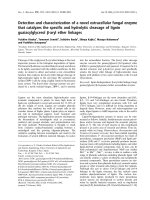
Fig. 1. (left) Photographic view of the grown KLCC crystal and (right) prepared sample immersed in mineral oil to avoid the effect of humidity on the crystal surface.
LiCl (99.998%), and CeCl3 (99.99%) powders purchased from Alfa
Aesar were used as starting materials. The natural 6 Li abundance
in natural LiCl is 7.6%. These powders were put in a 10-mm inner
diameter quartz ampule and dried at 250 ◦ C for several hours to remove
any residual moisture. All handling before and after crystal growth
were performed in ultra-low humidity argon-purged glove box to avoid
the effect of humidity. After drying, the ampule was sealed in a vacuum
of 10−6 torr. The sealed ampule containing the KLCC stoichiometric
charge was transferred to the furnace and sintered at 500 ◦ C. To find
out the melting point of the KLCC sample, a vertical transparent furnace
was used. The melting point of the KLCC was found to be about 600 ◦ C.
After sintering and melting, the ampule was transferred to the two-zone
vertical Bridgman furnace for crystal growth. The crystal was grown
at a rate of 0.5 mm/h at a thermal gradient of 10 ◦ C/cm. The KLCC
crystal was cut using a diamond-coated stainless steel wire saw and
optically polished inside a low-humidity glove box. The grown crystal
and polished sample of 5 × 5 × 5 mm3 dimension are shown in Fig. 1.
Because of the hygroscopic nature of the grown KLCC crystal like other
halide scintillators (Zhuravleva et al., 2013), scintillation properties
were measured inside the low-humidity argon-purged glove box.
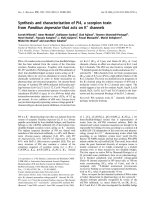
Fig. 2. X-ray-induced emission spectrum of the KLCC crystal.
3. Result and discussion
2.2. Experimental set up
3.1. X-ray induced emission spectrum
An X-ray tube having a W anode with operating voltage of 100 kV
and current of 1.5 mA was used to measure X-ray induced emission
spectrum of the grown crystal. The emission spectrum was recorded
using a calibrated QE65000 ocean optics fiber spectrometer. The pulse
height spectrum, non-proportionality response, decay time, quenching
factor, and pulse shape discrimination were measured with a super
bi-alkali PMT (R6233-100, Hamamatsu) under 𝛾-rays and 𝛼-particles
excitation from various radioactive sources at room temperature. The
KLCC sample was directly coupled to the photocathode of PMT using
optical grease (Ej-550, Eljen technology), covered with several layers of
100-μm thick Teflon tape and irradiated with 𝛾-rays or 𝛼 particles from
various radiation sources. For the light yield measurement, the analog
signal from the PMT was fed to a pre-amplifier (CANBERRA Model,
2005) and then shaped with a spectroscopy amplifier (TC 245, tennelec
Co.). The shaped signals were digitized with a 25-MHz flash analog-todigital converter (FADC25, Notice Co.). Output signals from FADC were
recorded and analyzed with a customized C++-based analysis code,
compiled, and run in ROOT package (So et al., 2008). For analysis of
the fluorescence decay time and pulse shape discrimination, the output
scintillation signal from the PMT was digitized with 400 MHz FADC
(FADC400, NOTICE Co.) (Notice). The FADC400 module samples the
pulse every 2.5 ns for a period of up to 64 μs to fully reconstruct the
decay time signal.
The room temperature X-ray-induced emission spectrum of the
KLCC sample in Fig. 2 shows a broadband emission between 300 nm
and 450 nm, which is attributed to the spin- and parity-allowed 5d â 4f
transition of the Ce3+ ion. In Fig. 2, the transition from the lowest 5d
excited level to the 2 F5∕2 (356 nm) and 2 F7∕2 (370 nm) levels of the 4f1
configuration of Ce3+ ion can be identified (Yang et al., 2009b). The
emission observed for the KLCC crystal best matches with the spectral
sensitivity response of modern photosensors and therefore can be used
as a radiation detector for various applications.
3.2. Energy resolution and relative light yield
The pulse height spectrum of the KLCC sample was measured under
𝛾-rays excitation using 137 Cs radioactive source (Fig. 3). The photopeak in the pulse height spectrum was fitted by Gaussian function (Tasaka, 1972). An energy resolution of 16% full width at half
maximum (FWHM) was obtained at 662 keV 𝛾-rays excitation (see
Fig. 3).
The light yield of the KLCC sample was compared with the reference
LYSO crystal whose absolute light yield is 33,000 ph/MeV (Pepin et al.,
2004; Rooh et al., 2014). Because the channel number is related with
light yield in the pulse height spectrum (Moszynski et al., 1997), the
light yield of the KLCC sample was calculated by considering the
2
Radiation Measurements 141 (2021) 106524
J.Y. Cho et al.
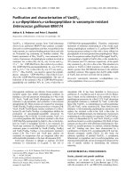
Fig. 5. Non-proportionality response of the KLCC crystal.
Fig. 3. Pulse height spectra of KLCC and LYSO crystals measured under 𝛾-rays
excitation from a 137 Cs source (Kim et al., 2017).
Fig. 6. The fluorescence decay time of the KLCC sample under 𝛾-rays and 𝛼-particles
excitation. The inset (a) and (b) in the figure shows the 𝛾-rays and 𝛼 particles decay
time fitted with three exponential decay time functions, respectively.
Fig. 4. The X-ray-induced emission spectrum of KLCC and LYSO crystals and the
quantum efficiency of R6233-100 PMT.
The deviation of the non-proportional response of the KLCC sample is
less than 7% between 300 keV and 1.1 MeV.
quantum efficiencies of PMT at the peak emissions of LYSO and KLCC
crystals. The X-ray-induced emission spectra of KLCC and LYSO and
the quantum efficiency response of the PMT are shown in Fig. 4 for
comparison. The light yield of KLCC crystal was estimated using the
following equation (Shah, 2010):
𝐿𝑌𝐾𝐿𝐶𝐶 =
𝑃𝐿𝑌 𝑆𝑂 𝑄𝐸𝐾𝐿𝐶𝐶
𝐿𝑌
𝑃𝐾𝐿𝐶𝐶 𝑄𝐸𝐿𝑌 𝑆𝑂 𝐿𝑌 𝑆𝑂
3.4. Fluorescence decay time
The fluorescence decay time was measured using 137 Cs 𝛾-rays and
𝛼-particles excitation source at room temperature. As shown in
Fig. 6. (a), the 𝛾-rays decay time curve of the KLCC crystal was best
fitted by three exponential decay time functions. The obtained values
of the fast, intermediate, and slow decay time components are 70 ns
(67.9%), 360 ns (19.7%), and 2.0 μs (12.4%), respectively. Similarly,
the 𝛼 particles decay time curve of the KLCC crystal was best fitted
by three exponential decay time functions as shown in Fig. 6(b). The
obtained value of the decay components are 80 ns (65.9%), 350 ns
(21.1%), 1.9 μs (13%), for fast, intermediate, and slow decay time
components, respectively. The scintillation decay time in this study
was measured with a spectrally unresolved method, and therefore can
be assigned tentatively to its origin. The fast and intermediate decay
time components (70 ns and 360 ns) can be attributed to the direct
electron hole recombination at Ce+3 and delay transfer of energy to the
Ce+3 , while the slow decay time constant can be attributed to the selftrapped exciton luminescence (Budden et al., 2013). To understand the
luminescence mechanism in this crystal, detailed spectroscopic investigation at room and low temperature are required, which is considered
for future work. The average decay time under 𝛾-rays excitation was
241 Am
(1)
where 𝑃𝐾𝐿𝐶𝐶 and 𝑃𝐿𝑌 𝑆𝑂 represent the photopeak channel number of
each crystal, QE refers to quantum efficiency, and LY is the light yield.
The light yield of the KLCC was estimated to be 21,000 ph/MeV at 662
keV 𝛾-rays excitation.
3.3. Non-proportionality response
The non-proportionality response in the light yield of the KLCC
crystal was studied in a wide energy range from 300 keV to 1.1 MeV
at room temperature. Similar experimental setup was used for the
measurement of pulse height spectrum. The relative light yield as
function of the 𝛾-rays energy for the KLCC crystal was studied using
known 𝛾-rays energies such as 133 Ba (303 keV, 356 keV), 22 Na (511
keV), 137 Cs (662 keV), 54 Mn (835 keV), and 65 Zn (1115 keV). Fig. 5
shows the non-proportionality of light yield (normalized to the value at
662 keV) as a function of different 𝛾-rays energies for the KLCC crystal.
3
Radiation Measurements 141 (2021) 106524
J.Y. Cho et al.
slightly faster than that under 𝛼-particle excitation, which indicates the
possibility that optimized KLCC sample can be used for thermal neutron
detection in various applications via 6 Li(n, 𝛼)3 H reactions.
3.4.1. Pulse shape discrimination
A heavy charged particle causes different excitation than photons
do. These different excitation mechanisms are the basis of pulse shape
discrimination. A heavy charged particle causes greater specific ionization, which makes the scintillator reach higher excited states that take
longer to de-excite. A photon causes lower ionization that takes shorter
time to de-excite. De-excitation causes fluorescence, which via PMT
produces electrical pulses. As the 𝛾-rays and 𝛼 particles have different
interaction processes in the KLCC sample, the decay time components
under 𝛾-rays and 𝛼 particles show different values as shown in Fig. 6.
Using these differences, we can distinguish 𝛾-rays and 𝛼 particles. Fig. 6
shows the fluorescence decay time under (a) 𝛾-rays and (b) 𝛼 particles.
The pulse shape discrimination capability of the KLCC was analyzed by
the mean time method. The mean decay time is given by the following
formula (Gerbier et al., 1999; Vuong et al., 2019):
𝑡=
𝛴𝐴𝑖 × 𝑡𝑖
𝛴𝐴𝑖
(2)
,where 𝐴𝑖 is the amplitude of the FADC signal at the time 𝑡𝑖 .
The figure of merit (FOM), which represents the discrimination
ability value, was used to separate 𝛾-rays and 𝛼 particles. FOM is
defined as (Langeveld et al., 2017) :
𝐹 𝑂𝑀 =
|𝑃𝛼 − 𝑃𝛾 |
𝐹 𝑊 𝐻𝑀𝛼 + 𝐹 𝑊 𝐻𝑀𝛾
(3)
where P𝛼 , P𝛾 are the peak positions of 𝛼 particles and 𝛾-rays, respectively. The discrimination ability value using the FOM for mean time
was calculated to be 0.81, as shown in Fig. 7(a).
The 𝛼/𝛽 ratio shows the quenching factor between the emitted scintillation light under the charged particles and 𝛾-rays excitations (DeVol
et al., 2007). It was calculated by the ratio of the peak position of
𝛼 particles and 𝛾-rays considering their corresponding energies. The
measurement of 𝛼/𝛽 ratio was performed with 5.486 MeV 𝛼 particles
from 241 Am and 662 keV 𝛾-rays from 137 Cs radioactive sources. The
𝛼/𝛽 ratio was found to be 0.15, which means that 85% of the light
from 𝛼 particle was quenched during interaction in material compared
with that of 𝛽 particle. Fig. 7(b) shows 𝛼 particles and 𝛾-rays spectrum,
which was used to calculate 𝛼/𝛽 ratio.
Fig. 7. (a) The mean time and (b) the 𝛼/𝛽 ratio spectrum of the KLCC sample with
137 Cs (𝛾-ray) and 241 Am (𝛼 particle) radioactive source.
Declaration of competing interest
The authors declare that they have no known competing financial interests or personal relationships that could have appeared to
influence the work reported in this paper.
4. Conclusion
Acknowledgment
In this study, we presented the scintillation properties of a new
elpasolite inorganic halide scintillation crystal, K2 LiCeCl6 , which was
grown by vertical Bridgman technique. The X-ray- induced emission
spectrum exhibits a broadband between 300 nm and 450 nm, which
is attributed to the spin- and parity-allowed 5d–4f transition of the
Ce3+ ion. The relative light yield was obtained to be 21000 ph/MeV
compared with the LYSO reference crystal. The non-proportionality of
the K2 LiCeCl6 crystal was found to be within 7% in the energy range
of 300 keV to 1.1 MeV. The energy resolution of the K2 LiCeCl6 was
obtained to be 16% (FWHM) which could be improved significantly
after improving the crystal growth quality and the material’s purity.
The K2 LiCeCl6 crystal scintillator has three decay components: 70 ns
(67.9%), 360 ns (19.7%), and 2.0 μs (12.4%) under 𝛾-ray excitation.
Because 𝛼 particles and 𝛾-rays decay times of the K2 LiCeCl6 crystal were
different, the mean time pulse shape discrimination method was used,
obtaining an FOM of 0.81. The 𝛼/𝛽 ratio of the K2 LiCeCl6 crystal was
measured to be 0.15. These preliminary results show that the K2 LiCeCl6
can be applied for the detection of 𝛾-rays and neutron in the 𝛾-rays and
neutrons mixed field using pulse shape discrimination capability.
These investigations were supported by the National Research Foundation of Korea, funded by the Ministry of Science and Technology,
Korea (MEST), (No. 2020M2A8A4025470)
References
Budden, B., Stonehill, L.C., Terry, J.R., Klimenko, A.V., Perry, J.O., 2013. Characterization and investigation of the thermal dependence of Cs2 LiYCl6 : Ce3+ (CLYC)
waveforms. IEEE Trans. Nucl. Sci. 60 (2), 946–951. />2012.2215884.
DeVol, T.A., Theisen, C.D., DiPrete, D.P., 2007. Effect of quench on alpha/beta pulse
shape discrimination of liquid scintillation cocktails. Health Phys. 92 (Suppl. 5),
S105–S111. />Gerbier, G., Mallet, J., Mosca, L., Tao, C., Chambon, B., Chazal, V., Jésus, M.D.,
Drain, D., Messous, Y., Pastor, C., 1999. Pulse shape discrimination and dark
matter search with NaI(Tl) scintillator. Astropart. Phys. 11 (3), 287–302. http://
dx.doi.org/10.1016/S0927-6505(99)00004-3, URL />science/article/pii/S0927650599000043.
Khan, N., Machrafi, R., 2014. Neutron and gamma-ray detection using a Cs2 LiYCl6
scintillator. EPJ Web Conf. 66, 11018. />20146611018.
4
Radiation Measurements 141 (2021) 106524
J.Y. Cho et al.
Kim, H., Rooh, G., Kim, S., 2017. Tl2 LaCl5 (Ce3+ ): New fast and efficient scintillator for X- and 𝛾 -ray detection. J. Lumin. 186, 219–222. />10.1016/j.jlumin.2017.02.042, URL />pii/S0022231316311310.
Langeveld, W.G.J., King, M.J., Kwong, J., Wakeford, D.T., 2017. Pulse shape discrimination algorithms, figures of merit, and gamma-rejection for liquid and solid
scintillators. IEEE Trans. Nucl. Sci. 64 (7), 1801–1809. />TNS.2017.2681654.
Machrafi, R., Khan, N., Miller, A., 2014. Response functions of Cs2 LiYCl6 : Ce scintillator
to neutron and gamma radiation. Radiat. Meas. 70, 5–10. />1016/j.radmeas.2014.07.010, URL />pii/S1350448714002133.
Moszynski, M., Kapusta, M., Mayhugh, M., Wolski, D., Flyckt, S.O., 1997. Absolute light
output of scintillators. IEEE Trans. Nucl. Sci. 44 (3), 1052–1061. />10.1109/23.603803.
Mughabghab, S., 2003. Thermal neutron capture cross sections resonance integrals and
g-factors.
Notice Korea company. [Online]. Available: .
Pejchal, J., Fujimoto, Y., Chani, V., Moretti, F., Yanagida, T., Nikl, M., Yokota, Y.,
Beitlerova, A., Vedda, A., Yoshikawa, A., 2011. Crystal growth and luminescence
properties of Ti-doped LiAlO2 for neutron scintillator. J. Cryst. Growth 318 (1),
828–832. The 16th International
Conference on Crystal Growth (ICCG16)/The 14th International Conference on
Vapor Growth and Epitaxy (ICVGE14). URL />article/pii/S002202481001033X.
Pepin, C., Bérard, P., Perrot, A.-L., Houde, D., Lecomte, R., Melcher, C., Dautet, H.,
2004. Properties of LYSO and recent LSO scintillators for phoswich PET detectors.
IEEE Trans. Nucl. Sci. 51, 789–795. />Rooh, G., Kim, H., Park, H., Kim, S., 2014. Luminescence and scintillation characterizations of cerium doped Cs2 LiGdBr6 single crystal. J. Lumin. 146, 404–407. http:
//dx.doi.org/10.1016/j.jlumin.2013.09.047, URL />science/article/pii/S002223131300611X.
Shah, K.S., 2010. New Scintillation Detectors for PET (Ph.D. thesis). Faculty Appl. Sci.,
Delft Univ. Technol., Delft, The Netherlands.
So, J., Kim, H.J., Kang, H., Lee, S., Kim, S., Kim, K., Lee, M., 2008. The proton energy
response of a LYSO crystal. J. Korean Phys. Soc. 52, />jkps.52.925.
Tasaka, K., 1972. Method of standard spectrum fitting for the analysis of
gamma-ray spectra from semiconductor detectors. J. Nucl. Sci. Technol. 9
(7), 430–432. arXiv:https:
//www.tandfonline.com/doi/pdf/10.1080/18811248.1972.9734872, URL https://
www.tandfonline.com/doi/abs/10.1080/18811248.1972.9734872.
Vuong, P.Q., Kim, H., Park, H., Rooh, G., Kim, S., 2019. Pulse shape discrimination
study with Tl2 ZrCl6 crystal scintillator. Radiat. Meas. 123, 83–87. .
org/10.1016/j.radmeas.2019.02.007, URL />article/pii/S1350448718306747.
Yamaji, A., Yanagida, T., Kawaguchi, N., Fujimoto, Y., Yokota, Y., Watanabe, K.,
Yamazaki, A., Yoshikawa, A., Pejchal, J., 2011. Crystal growth and scintillation
properties of Ce and Eu doped LiSrAlF6 . Nucl. Instrum. Methods Phys. Res.
A 659 (1), 368–372. URL http://
www.sciencedirect.com/science/article/pii/S0168900211017086.
Yang, P., Doty, F.P., Rodriguez, M.A., Sanchez, M.R., Zhou, X., Shah, K.S., 2009a.
The synthesis and structures of elpasolite halide scintillators. MRS Proc. 1164,
1164–L11–05.
Yang, P., Doty, F.P., Rodriguez, M.A., Sanchez, M.R., Zhou, X., Shah, K.S., 2009b.
The synthesis and structures of elpasolite halide scintillators. MRS Proc. 1164,
1164–L11–05.
Yang, P., Zhou, X., Deng, H., Rodriguez, M.A., Feng, P.L., van Loef, E.V.D., Shah, K.S.,
Doty, F.P., 2013. Crystal growth and scintillation properties of Cs2 NaGdBr6 :Ce3+ .
IEEE Trans. Nucl. Sci. 60 (2), 1033–1038. />2251473.
Yokota, Y., Fujimoto, Y., Yanagida, T., Takahashi, H., Yonetani, M., Hayashi, K., Park, I.,
Kawaguchi, N., Fukuda, K., Yamaji, A., Fukazawa, Y., Nikl, M., Yoshikawa, A.,
2011. Crystal growth of Na-Co-doped Ce:LiCaAlF6 single crystals and their optical,
scintillation, and physical properties. Cryst. Growth Des. 11 (11), 4775–4779.
/>Yoshikawa, A., Yanagida, T., Yokota, Y., Kawaguchi, N., Ishizu, S., Fukuda, K.,
Suyama, T., Kim, K.J., Pejchal, J., Nikl, M., Watanabe, K., Miyake, M., Baba, M.,
Kamada, K., 2009. Single crystal growth, optical properties and neutron response
of Ce3+ doped LiCaAlF6 . IEEE Trans. Nucl. Sci. 56 (6), 3796–3799. .
org/10.1109/TNS.2009.2033115.
Zhuravleva, M., Stand, L., Wei, H., Hobbs, C., Boatner, L.A., Ramey, J.O., Shah, K.,
Burger, A., Rowe, E., Bhattacharya, P., Tupitsyn, E., Melcher, C.L., 2013. Hygroscopicity evaluation of halide scintillators. In: 2013 IEEE Nuclear Science
Symposium and Medical Imaging Conference. 2013 NSS/MIC, pp. 1–5. http://dx.
doi.org/10.1109/NSSMIC.2013.6829669.
5