Testing the potential of using fine quartz for dating loess in South Island, New Zealand
Bạn đang xem bản rút gọn của tài liệu. Xem và tải ngay bản đầy đủ của tài liệu tại đây (2.93 MB, 9 trang )
Radiation Measurements 155 (2022) 106788
Contents lists available at ScienceDirect
Radiation Measurements
journal homepage: www.elsevier.com/locate/radmeas
Testing the potential of using fine quartz for dating loess in South Island,
New Zealand
´ ska b, A. Micallef c, d, A. Timar-Gabor a, b, *
A. Avram a, b, Z. Kabacin
a
Faculty of Environmental Science and Engineering, Babes-Bolyai University, Cluj-Napoca, Romania
Interdisciplinary Research Institute on Bio-Nano-Sciences, Environmental Radioactivity and Nuclear Dating Centre, Babes-Bolyai University, Cluj-Napoca, Romania
c
Helmholtz Centre for Ocean Research, GEOMAR, Kiel, Germany
d
Marine Geology & Seafloor Surveying, Department of Geosciences, University of Malta, Malta
b
A R T I C L E I N F O
A B S T R A C T
Keywords:
Quartz
Polymineral fine grains
Luminescence
Electron spin resonance
New Zealand loess
The applicability of optically stimulated luminescence (OSL) dating on quartz from South Island, New Zealand is
hampered by the poor behaviour of the targeted signals. However, most OSL dating studies have been focused on
using coarse quartz fractions. Since a previous study conducted from a nearby site demonstrated that coarse
quartz (63–90, 90–125, 125–180 and 180–250 μm) is not suitable for OSL dating, we attempt using fine quartz
here. Therefore, the standard SAR protocol was applied on 4–11 μm quartz extracted from a loess/paleosol
section. Unlike the coarser fractions, the OSL signal of fine quartz displayed satisfactory characteristics which
allowed estimating ages ranging from 0.3 ± 0.04 ka to 16 ± 1 ka. In order to understand the differences between
the two quartz fractions, we characterise fine (4–11 μm) as well as the usually used coarser grain sizes (˃ 63 μm)
of quartz by electron spin resonance (ESR). No significant differences are reported in qualitative terms between
the grain sizes investigated and calibration quartz. We report a higher abundance of intrinsic defects in the fine
grain fraction; however, this is typical for quartz from other regions as well, that was amenable for OSL dating.
As such, the differences between the fine quartz fraction and the coarse fraction is not yet understood. In
addition, two elevated temperature post-infrared infrared protocols (pIRIR225 and pIRIR290) were applied and
polymineral grains extracted from the same samples. Despite residual dose corrections being performed using a
modern analogue, pIRIR ages overestimate quartz ages by 19–122% in the case of the application of the pIRIR225
protocol and by 25–217% in the case of the application of the pIRIR290 protocol. The effect could not be cir
cumvented by the application of a test dose with a magnitude of 50% of the equivalent dose in the pIRIR290
protocol. In the case of the application of pIRIR290 protocol, dose recovery tests ratios vary from 1.07 ± 0.06 to
1.23 ± 0.05. While not ideal, these results cannot fully explain the differences reported between the ages ob
tained by fine quartz OSL and the polymineral fine grains pIRIR methods.
1. Introduction
Loess deposits of New Zealand are considered important archives for
paleoclimate reconstruction of the southern hemisphere (Alloway et al.,
2007), thus recent studies have been centred in establishing
high-resolution chronologies.
Optically Stimulated Luminescence Dating (OSL) represents one of
the most used dating techniques for Quaternary climate reconstruction.
Its applicability has been successful for loess deposits located over both
northern and southern hemispheres, respectively (Roberts 2008, 2015).
However, since luminescence dating has been perceived to be chal
lenging for loess sediments from South Island of New Zealand, few OSL
studies have been reported so far (e.g., Holdaway et al., 2002; Rowan
et al., 2012; Sohbati et al., 2016; Micallef et al., 2021; Brezeanu et al.,
2021). Even though quartz is considered the preferred dosimeter when
young sediments have to be dated due to the higher bleachability of the
signal, it is well known that South Island quartz suffers from major
problems that restrain its application, namely the weak sensitivity of the
signal, with the signal originating from many dim grains and the poor
behaviour exhibited in the single aliquot regenerative dose (SAR) pro
tocol (Preusser et al., 2006). These issues have been attributed by the
aforementioned study to the short sedimentation history of the mineral
grains, as it was reported that quartz sensitisation can be achieved by
repeated irradiation/bleaching cycles. A recent study by Brezeanu et al.
* Corresponding author. Faculty of Environmental Science and Engineering, Babes-Bolyai University, Cluj-Napoca, Romania.
E-mail address: (A. Timar-Gabor).
/>Received 22 November 2021; Received in revised form 23 March 2022; Accepted 13 May 2022
Available online 17 May 2022
1350-4487/© 2022 The Authors. Published by Elsevier Ltd. This is an open access article under the CC BY-NC-ND license ( />
A. Avram et al.
Radiation Measurements 155 (2022) 106788
(2021) confirmed that the OSL signals of coarse (˃63 μm) quartz dis
played low-sensitivity and a significant sensitivity-changes during the
repeated SAR cycles. Despite these limitations, there are few OSL studies
that reported ages on quartz in New Zealand (e.g., Holdaway et al.,
2002; Nichol et al., 2003; Rowan et al., 2012; Hornblow et al., 2014;
Sohbati et al., 2016). Holdaway et al. (2002) reported luminescence ages
on 90–125 μm quartz that were in agreement with 14C ages for colluvial
sediments from Otago, South Island of New Zealand. Later, Rowan et al.
(2012) have successfully obtained a luminescence chronology for gla
ciofluvial sediments from Canterbury Plains of South Island using
180–211 μm quartz. Moreover, a more recent study conducted by Soh
bati et al. (2016) reported a good agreement between 40 and 63 μm
quartz SAR-OSL and pIRIR290 luminescence ages in the attempt to refine
palaeorockfall chronologies in New Zealand using luminescence dating.
Since the applicability of luminescence dating on quartz grains
extracted from New Zealand sediments is not always a viable solution,
other luminescence studies conducted on South Island considered that
using infrared stimulated luminescence (IRSL) signal on coarse K-rich
feldspars (Preusser et al., 2005) or on polymineral fine grains (e.g.,
Berger et al., 2001, 2002; Hormes et al., 2003; Rother et al., 2009;
Almond et al., 2001, 2007; Shulmeister et al., 2010) is a more appro
priate solution for obtaining luminescence chronologies. It is well
known that the IRSL signal of feldspars suffers from anomalous fading
and thus recent studies have developed measurement protocols that are
able to circumvent fading. Such protocols consist of a double IR stimu
lation and they are known as post infrared-infrared stimulated lumi
nescence protocols, pIRIR225 and pIRIR290. Even though, these pIRIR
protocols have been successfully applied on coarse K-feldspars as well as
on polymineral fine grains extracted from loess deposits all over the
world (e.g., Roberts 2008; Buylaert et al., 2009, 2011; Thiel et al., 2011;
ăsken et al., 2017; Zhang et al.,
Vasiliniuc et al., 2012; Yi et al., 2016; Bo
2018; Veres et al., 2018; Avram et al., 2020; Avram et al., 2022), their
potential has not been fully explored for New Zealand sediments. Only
three dating studies have reported pIRIR290 luminescence ages (Sohbati
et al., 2016; Micallef et al., 2021; Brezeanu et al., 2021) and two studies
presented pIRIR225 (Micallef et al., 2021; Brezeanu et al., 2021) chro
nologies on loess extracted from South Island of New Zealand.
To our knowledge, all quartz luminescence ages reported so far in
literature were determined using coarse grains quartz (>63 μm), in this
study we attempt for the first time to apply SAR-OSL protocol on fine
(4–11 μm) quartz extracted from loess in the Canterbury Plains of South
Island, New Zealand. pIRIR225 as well as pIRIR290 protocols have been
applied on polymineral fine grains extracted from the same samples.
3. Methodology
3.1. Sample preparation
Stainless steel tubes were used for collecting the luminescence
sample. The minerals of interest were extracted under subdued red light
laboratory conditions. The material from the end of each tube was
removed and used for gamma spectrometry measurements. The material
from the inner part of the tube was used for 4–11 μm quartz and poly
mineral grains extraction. In the first step of sample preparation the
calcium carbonates and the organic matter were removed by employing
a treatment with hydrochloric acid (10% concentration) and hydrogen
peroxide (10% concentration followed by 30%). Minerals with di
ameters smaller than 63 μm were separated by wet sieving. The fine
(4–11 μm) polymineral mixture was obtained after Stoke’s law settling
followed by centrifugation in distilled water (Frechen et al., 1996; Lang
et al., 1996). A 10 days treatment with hexafluorosilicic acid was
employed in order to isolate the fine (4–11 μm) quartz fraction from the
polymineral combination. The extraction procedure for quartz fractions
larger than 63 μm (63–90 μm, 90–125 μm, 125–180 μm, 180–250 μm) is
described in Brezeanu et al. (2021). Both fine quartz and polymineral
grains were mounted on aluminium disks for luminescence
measurements.
3.2. Analytical facilities
Luminescence investigations were carried out using a Risø TL-OSL
reader (model DA-20) equipped with an automated detection and
stimulation head (DASH) (Lapp et al., 2015). The intensity of the blue
(470 nm) and infrared (850 nm) LEDs deliver 80 and 300 mW/cm2,
respectively. Luminescence signals were detected by using PDM
9107Q-AP-TTL-03 (160–630 nm) photomultiplier tubes (Thomsen et al.,
2006). A 7.5-mm-thick Hoya U-340 UV filter was used for quartz signal
determination while the polymineral signals were detected by using a
blue filter combination (Schott BG39 + Corning 7–59, with transmission
between 320 and 460 nm). A radioactive source of 90Sr–90Y was used for
laboratory irradiation. The beta source was calibrated using
gamma-irradiated fine (4–11 μm) calibration quartz (Hansen et al.,
2015).
The polymineral fine grains aliquots used for residual dose and dose
recovery measurements were exposed to window light under natural
conditions in order to remove the natural signal.
ESR measurements were performed on an X band Bruker EMX Plus
Spectrometer. All samples were placed in quartz glass tubes filled by
maintaining the same volume, with a mass between 100 and 200 mg,
and the measurements were normalized to 100 mg for inter-comparison.
Each sample was measured 3 times and rotated in the cavity between the
measurements. Exposure of samples to sunlight during measurements
was restricted to a minimum. Measurements were carried out at 90 K (in
liquid nitrogen) for Al-h and Ti centres, and at room temperature for E′
and “peroxy” centres. Al-h and “peroxy” spectra were acquired using the
following settings: 3350 ± 200 G scanned magnetic field, modulation
amplitude 1 G, modulation frequency 100 kHz, microwave power 2 mW,
conversion time 50 ms, time constant 40 ms. For Ti measurements the
settings were: 3490 ± 110 G scanned magnetic field, modulation
amplitude 1 G, modulation frequency 100 kHz, microwave power 10.0
mW, conversion time 10 ms, time constant 20.48 ms, and 10 scans per
measurement. For E′ spectra the settings were: 3363 ± 10 G scanned
magnetic field, modulation amplitude 0.1 G, modulation frequency 100
kHz, microwave power 0.02 mW, conversion time 30 ms, time constant
20.48 ms, and 3 scans per measurement. Baseline correction was per
formed using Bruker’s Xenon software.
2. Site description
The foothills of the Southern Alps as well as the lowlands of the
Canterbury Plains represent the regions with the widest distribution of
loess deposits in South Island of New Zealand (Yates et al., 2018).
The investigated site (44.014973 ◦ S, 171.891569 ◦ E) is located on
the southern part of the Canterbury Plains and the eastern side of the
South Island of New Zealand. Three modern rivers namely Rakaia,
Rangitata and Ashburton flow perpendicularly to the eastern coastal cliff
in the Canterbury Plains, discharging into the Pacific Ocean. The loess
section is situated less than 1 km away (Fig. S1) from the site investi
gated by Brezeanu et al. (2021) and therefore a more detailed descrip
tion of the area can be found in their study.
Luminescence investigations have been performed on seven samples
collected at a resolution of 20 cm. The uppermost sample, NZ 6 was
collected from a depth of 10 cm while the last sample NZ 12 was
collected at a depth of 130 cm.
ESR analysis presented in this study have been performed on sample
NZ3 as various grain sizes were available from that specific sample,
collected from the loess profile investigated by Brezeanu et al. (2021).
3.3. Equivalent dose determination
Quartz equivalent dose determination has been carried out by using
2
A. Avram et al.
Radiation Measurements 155 (2022) 106788
the standard Single Aliquot Regenerative dose (SAR) protocol (Murray
and Wintle 2000, 2003) whereas polymineral fine grains equivalent
doses were measured by applying two elevated temperature
post-infrared infrared stimulation protocols, namely pIRIR225 (Roberts
2008; Buylaert et al., 2009) and pIRIR290 (Buylaert et al., 2011a; Thiel
et al., 2011). The protocols are outlined in Table S1.
For quartz measurements optical stimulation has been performed
using blue-light emitting diodes for 40 s at 125 ◦ C. The net continuous
wave optically stimulated luminescence (CW-OSL) signal was integrated
over the first 0.308 s of the decay curve minus an early background
subtraction in order to reduce the influence of medium and slow com
ponents (Cunningham and Wallinga, 2010). A test dose of 17 Gy was
used for sensitivity change correction. Thermal treatments consisted of a
preheat temperature of 220 ◦ C for 10 s and a cutheat of 180 ◦ C. A high
temperature bleach (280 ◦ C for 40 s) was performed at the end of each
SAR cycle. In order to assess the robustness of the SAR protocol, the
intrinsic performance tests (recycling and recuperation) (Murray and
Wintle 2003) were integrated in every measurement. The purity of
quartz luminescence signals was checked through the IR depletion test,
where an IR stimulation step was added prior to OSL measurement in the
last cycle of the SAR protocol (Duller 2003). Only the aliquots with
recycling and IR depletion ratios within 10% deviation from unity were
considered suitable and used for equivalent dose determination. Recu
peration ratios less than 2% of the natural signal were considered
acceptable.
Further analysis consisted of equivalent dose determination on pol
ymineral fine grains using two elevated temperature post-infrared
infrared stimulation protocols based on a SAR procedure namely
pIRIR225 (Roberts 2008; Buylaert et al., 2009) (Table S1b) and pIRIR290
(Buylaert et al., 2011; Thiel et al., 2011) (Table S1c). A thermal preheat
of 250 ◦ C (pIRIR225) or 325 ◦ C (pIRIR290) was incorporated prior to IR
stimulation in every SAR cycle. After the heating step, an IR stimulation
at 50 ◦ C for 200 s was performed in order to minimise the charge that is
susceptible to fading. The signal of interest was recorded as a result of IR
stimulation at a temperature of 225 ◦ C (pIRIR225) and 290 ◦ C (pIRIR290),
respectively. At the end of each measurement cycle a high-temperature
bleach for 100 s at 290 ◦ C (pIRIR225) and 325 ◦ C (pIRIR290) was
involved. Sensitivity change corrections have been made by employing a
test dose of 17 Gy unless otherwise stated.
4. Results and discussion
4.1. Luminescence properties – quartz
Equivalent doses on fine quartz were determined by interpolating the
sensitivity corrected natural OSL signal onto the dose response curve.
Fig. 1 shows a representative SAR growth curve and OSL decay curves
for a single aliquot of fine quartz extracted from sample NZ 7. The
natural and regenerative OSL signal exhibits a similar pattern to the
decay measured for calibration quartz during the first seconds of stim
ulation, which is accepted as being dominated by the fast component
(Hansen et al., 2015). The dose response curve was best described by a
sum of two saturating exponential functions. Recycling and IR depletion
ratios were within 10% deviation from unity which demonstrates that
sensitivity corrections are properly made and the quartz signals are
pure. Recuperation ratio was less than 2% indicating that the growth
curves pass very close to the origin and thermal transfer during the
repeated SAR cycle is negligible.
4.1.1. Preheat plateau
The dependency of the equivalent doses on the preheat temperature
was investigated through the preheat plateau test. Sample NZ 7 was
divided in sets of five aliquots. For each set, a preheat temperature
ranging from 180 to 280 ◦ C was applied. A test dose cutheat of 180 ◦ C
was employed throughout the measurements. As can be seen from
Fig. S2, the equivalent doses do not display any significant variation
over the investigated interval of preheat temperatures. The results of the
intrinsic SAR tests were satisfactory for all the aliquots measured.
4.1.2. Dose recovery test
Further, a dose recovery test has been performed on six samples (NZ
6, NZ 7, NZ 8, NZ 9, NZ 10 and NZ 11) in order to investigate whether the
SAR protocol can successfully determine a known laboratory dose given
prior to any thermal treatment (Murray and Wintle, 2003). Sets of five
aliquots from each sample were used. The natural signals were bleached
by a repeated exposure to blue LEDs for 100 s at room temperature with
a pause of 10 ks? The aliquots were irradiated with a beta dose chosen to
approximate the natural dose and measured by using the SAR protocol in
the same manner as measuring the equivalent dose. Fig. S3 represents
the results of the dose recovery test. As can be seen, the dose recovery
results for all samples documented here were satisfactory indicating that
the SAR protocol can successfully recover laboratory doses up to 46 Gy.
3.4. Dosimetry
4.1.3. Equivalent doses
The measured quartz equivalent doses are summarized in Table 1.
The OSL equivalent doses range from 1.3 ± 0.1 Gy obtained for sample
NZ 6 collected from a depth of 10 cm–46 ± 1 Gy for sample NZ 11 which
was collected from a depth of 109 cm.
High-resolution gamma spectrometry was used for the determination
of specific radionuclide activities, using a well-type HPGe detector. In
order to reach the equilibrium of 222Rn with its parent 226Ra, samples
were stored for 1 month before measurements. The annual dose rates
were derived following the conversion factors tabulated by Gu´
erin et al.
(2011). An alpha efficiency factor of 0.04 ± 0.02 was taken into account
for 4–11 μm quartz while for the polymineral fine grains the assumed
alpha efficiency value was 0.08 ± 0.02 (Rees-Jones, 1995). These values
are consistent with the latter determined efficiency factors of 0.035 ±
0.003 determined by Lai et al. (2008) for 4–11 μm quartz as well as the
polymineral fine grains efficiency factors of 0.10 ± 0.014 determined by
Schmidt et al. (2018) for pIRIR290 and 0.11 ± 0.02 for pIRIR225 pro
tocol (Kreutzer et al., 2014), respectively. The time averaged water
content was assumed to be 15% with a relative error of 25%. The water
content was chosen to represent the mean value of the sediment mois
ture over the entire depositional history. Similar values were used for
dating sediments from Canterbury Plains and Banks Peninsula, respec
tively (Rowan et al., 2012; Sohbati et al., 2016; Brezeanu et al., 2021).
The cosmic dose rate was estimated as function of depth, altitude and
geomagnetic latitude using the formula proposed by Prescott and Hutton
(1994). Given the size of fine grains (4–11 μm), any dose rate derived
from internal alpha activity was assumed to be negligible. The specific
radionuclide activities and annual doses are presented in Table 1.
4.2. Luminescence properties – polymineral fine grains
Equivalent doses measured on polymineral fine grains were deter
mined by interpolating the natural sensitivity corrected IRSL signal onto
the dose response curve constructed for each sample using both pIRIR
protocols. Fig. 2 displays a representative growth curve of sample NZ 7
constructed by applying pIRIR225 (Fig. 2a) and pIRIR290 (Fig. 2b) pro
tocols, respectively. A comparison between the decay curve of the nat
ural signal and the pattern of a regenerative signal is shown in the insets
of Fig. 2. The dose response curves constructed using both pIRIR pro
tocols were best fitted using a sum of two saturating exponential func
tions. The measured equivalent doses obtained on pIRIR225 protocol
range from 7.5 ± 0.5 Gy for the youngest sample NZ 6 to 79 ± 2 Gy for
sample NZ 10. On the other hand, pIRIR290 equivalent doses vary be
tween 23 ± 2 Gy for sample NZ 6 and 121 ± 5 Gy for sample NZ10. As
previous studies report a possible influence of the magnitude of the test
dose on the pIRIR290 laboratory growth curves (Yi et al., 2016; Colarossi
3
A. Avram et al.
Table 1
Summary of the SAR-OSL, pIRIR225 and pIRIR290 luminescence ages. The age uncertainties were determined following Aitken and Alldred (1972). The uncertainties associated with the luminescence and dosimetry data
are random; the uncertainties mentioned on the optical ages are the overall uncertainties. The systematic errors taken into account include: 2% beta source calibration, 3% conversion factors, 5% attenuation and etching
factors, 3% gamma spectrometer calibration, 15% cosmic radiation, 25% water content. All uncertainties represent 1σ. Specific activities were measured using gamma spectrometry and the ages were determined
considering 15% water content; adopted alpha efficiency factor was 0.04 ± 0.02 for 4–11 μm quartz and 0.08 ± 0.02 for polymineral 4–11 μm fine grains, respectively (Rees-Jones, 1995). The contribution of cosmic
radiation was taken into account and calculated accordingly to Prescott and Hutton (1994). Equivalent doses presented in this table are not corrected for residuals. Equivalent doses presented in italic for NZ 8 and NZ 12
pIRIR290 were determined using a test dose of 50 Gy, amounting to about 50% of the equivalent dose. n represents the number of accepted aliquots out of the total number of aliquots measured The measured residual was
taken into account for calculation of pIRIR ages, while pIRIR ages calculated using modern analogue correction are denoted by (*). For the sake of completeness IR50 ages (uncorrected and corrected for fading) were
calculated based on the signals collected during the pIRIR225 protocol. g2days values for IR50 were measured for samples NZ7, NZ9 and NZ11 and assumed to be 3%/decade in the case of the rest of the samples.
4
Sample
code
Depth
(cm)
Equivalent dose (Gy)
Radionuclide concentration (Bq/kg)
Annual dose (Gy/ka)
4–11 μm
quartz
pIRIR225
pfg
pIRIR290
pfg
Ra-226
Th-232
K-40
4–11 μm
quartz
pIRIR225
pfg
NZ 6
10
607 ± 17
4.1 ± 0.07
50 ± 2
42 ± 1
604 ± 16
NZ 8
50
38 ± 2
30 ± 1
NZ 9
67
85
NZ 11
109
NZ 12
129
63 ± 1
n = 10/10
79 ± 2
n = 10/10
71 ± 1
n = 10/10
70 ± 1
n = 10/10
29 ± 0.2
NZ 10
39 ± 3
n = 8/10
43 ± 1
n = 10/10
46 ± 1
n = 10/10
41 ± 2
n = 10/10
23 ± 2
n = 9/10
111 ± 5
n = 9/10
114 ± 5
n = 10/10
113 ± 3
n=10/10
107 ± 6
n = 10/10
121 ± 5
n = 10/10
90 ± 2
n = 10/10
99 ± 4
n = 10/10
103 ± 3
n=10/10
48 ± 2
30
7.5 ± 0.5
n = 10/10
72 ± 1
n = 10/10
72 ± 1
n = 10/10
42 ± 2
NZ 7
1.3 ± 0.1
n = 9/10
26 ± 0.3
n = 14/14
32 ± 2
n = 7/10
Age (ka)
4–11 μm
quartz
Age (ka) (MA) *
pIRIR225
pfg
pIRIR290
pfg
pIRIR225
pfg
pIRIR290
pfg
IR50 pfg
no fading
correction
IR50 pfg
fading
corrected
4.6 ± 0.07 4.6 ± 0.07 0.3 ± 0.04
1.2 ± 0.2
4 ± 0.5
4.0 ± 0.06
4.5 ± 0.07 4.5 ± 0.07 6.3 ± 0.6
15 ± 1
23 ± 2
14 ± 1
20 ± 2
6.8 ± 0.6
9.1 ± 0.9
567 ± 17
2.9 ± 0.05
3.7 ± 0.07 3.7 ± 0.07 11 ± 1
19 ± 2
29 ± 3
17 ± 2
24 ± 2
12 ± 1
16 ± 2
22 ± 1
524 ± 14
2.9 ± 0.05
3.1 ± 0.05 3.1 ± 0.05 13 ± 2
19 ± 2
32 ± 3
18 ± 2
27 ± 3
10 ± 1
12 ± 1
47 ± 3
30 ± 1
535 ± 13
3.5 ± 0.07
3.9 ± 0.08 3.9 ± 0.08 12 ± 1
20 ± 2
30 ± 3
19 ± 2
26 ± 3
16 ± 1
20 ± 2
41 ± 1
26 ± 2
443 ± 14
3.0 ± 0.06
3.3 ± 0.07 3.3 ± 0.07 16 ± 1
21 ± 2
26 ± 2
19 ± 2
20 ± 2
15 ± 1
20 ± 2
34 ± 1
27 ± 2
485 ± 13
3.0 ± 0.05
3.3 ± 0.05 3.3 ± 0.05 14 ± 1
21 ± 2
29 ± 3
19 ± 2
23 ± 2
12 ± 1
15 ± 2
pIRIR290
pfg
Radiation Measurements 155 (2022) 106788
A. Avram et al.
Radiation Measurements 155 (2022) 106788
solar simulator for samples collected from Chinese loess. Based on the
aforementioned information, the residual dose corrections should be
cautiously evaluated especially when dealing with young samples.
The assessment of the residual level has been made on five aliquots of
each sample. The natural signal has been erased by exposing the aliquots
to sunlight for 30 days prior to measurements. Residual doses obtained
using pIRIR225 protocol range from 2.1 ± 0.4 Gy for the youngest sample
with a measured equivalent dose of 7.5 ± 0.5 Gy to 3.2 ± 0.3 Gy for a
sample with a measured equivalent dose of 63 ± 1 Gy. On the other
hand, the pIRIR290 residual doses vary from 4 ± 1 Gy for the youngest
sample with a measured equivalent dose of 23 ± 2 Gy to 6.5 ± 1 Gy for a
sample with a measured equivalent dose of 107 ± 6 Gy. The values
obtained on each sample are displayed in Table S2. Similar values of
residual dose were obtained by Brezeanu et al. (2021) for samples with
comparable measured equivalent doses of ~84 Gy and ~120 Gy,
respectively. In their study, Brezeanu et al. (2021) reported that a con
stant residual dose of ~4 ± 1 Gy has been reached after 48 h exposure to
sunlight for pIRIR225 protocol while in the case of pIRIR290 protocol, a
constant level of ~10 ± 1 Gy was achieved after 96 h of bleaching for
New Zealand loess. They mentioned that such values are in line with
those measured after a 30 days exposure to sunlight.
Since it is still questionable whether the natural bleaching condition
can be thoroughly replicated in laboratory, it is advisable to use a
modern analogue sample for residual dose corrections, as well. In this
case, the NZ 6 sample was used as a modern sample. As such, an
equivalent dose of 7.5 ± 0.5 Gy was measured using pIRIR225 protocol
and 22.8 ± 1.5 Gy using pIRIR290 protocol, respectively. Based on the
laboratory residuals, we considered these values as the maximum re
sidual doses. Thus, the residual dose correction for age calculation was
performed by using both laboratory and modern analogue doses
(Table 1).
Fig. 1. Representative sensitivity-corrected dose response curve constructed for
one aliquot of sample NZ 7 on 4–11 μm quartz using SAR-OSL protocol. The
sensitivity corrected natural signals (stars) are interpolated on the dose
response curves. IR depletion point is presented as an up triangle while the
recycling points are presented as inverse triangles. The inset shows the pattern
of a typical quartz decay curve which is compared with the decay of a regen
erative dose as well as with the OSL decay of calibration quartz.
et al., 2018) we have additionally investigated the effect of using a test
dose with a magnitude of 50% of the equivalent doses (50 Gy) for two
samples (NZ 8 and NZ 12). The results obtained are presented in Table 1
and are indistinguishable from the values obtained by using a test dose
of 17 Gy pIRIR measured equivalent doses for each sample investigated
here are labelled in Table 1.
4.2.2. Dose recovery test
The reliability of the measurement protocols was achieved through a
dose recovery test (Murray 1996; Wallinga et al., 2000) on five aliquots
from samples NZ 6, NZ 7 and NZ 8. The natural signals were removed by
exposing the aliquots to window light for 30 days in order to reach a
residual level as described in the previous section. Then, the aliquots
were irradiated with known laboratory doses that were chosen to
approximate the measured equivalent dose. To quantify the accuracy of
the protocols to measure laboratory given doses, a ratio between the
recovered dose, corrected for the measured residual value and the lab
oratory given dose was calculated. The results of the dose recovery tests
4.2.1. Residual doses
It is well known that pIRIR signals are more difficult to reset than
OSL signals (e.g., Buylaert et al., 2009, 2012; Thiel et al., 2011). Many
studies reported residual doses of a few Grays obtained even after long
exposure of the aliquots to sunlight or solar simulator (e.g., Buylaert
et al., 2011a, 2012; Stevens et al., 2011; Murray et al., 2012; Yi et al.,
2016, 2018; Avram et al., 2020, 2022; Brezeanu et al., 2021). Moreover,
from long-term bleaching experiments using pIRIR290 protocol Yi et al.
(2016, 2018) reported that for pIRIR290 protocol, a constant residual
dose of ~6 ± 1 Gy and ~4 ± 1 Gy is achieved after 300 h beaching in
Fig. 2. Representative sensitivity-corrected dose response curves constructed for one aliquot of sample NZ 7 on (a) 4–11 μm polymineral fine grains using the
pIRIR225 protocol and (b) 4–11 μm polymineral using pIRIR290 protocol. The sensitivity corrected natural signals (stars) are interpolated on the dose response curves.
The recycling points are presented as inverse triangles. Insets show typical decay curves. For polymineral fine grains, the natural CW-OSL signals are compared to
regenerated signals induced by a beta dose approximately equal to the equivalent dose.
5
A. Avram et al.
Radiation Measurements 155 (2022) 106788
for both pIRIR protocols are showed in Fig. 3. As it can be seen, dose
recovery ratios obtained for pIRIR225 protocol range from 0.98 ± 0.02
for sample NZ 7 to 1.01 ± 0.06 for sample NZ 6 while the pIRIR290
protocol dose recovery ratios vary from 1.07 ± 0.06 obtained for sample
NZ 6 to 1.23 ± 0.05 calculated for sample NZ 8. These results showed
that pIRIR225 protocol can successfully recover known doses over the
dose interval investigated here, while for pIRIR290 protocol some degree
of overestimation is observed for doses as large as about 100 Gy. A
recent study conducted by Avram et al. (2022) showed that pIRIR290
dose recovery ratios overestimate unity between 12% and 46% for given
doses that range from ~100 Gy to ~850 Gy.
Some previous studies attributed the poor results of the pIRIR290
dose recovery test to the incorrect measurement of the residual dose (e.
g., Thomsen et al., 2008; Buylaert et al., 2012). In order to circumvent
the potential contribution due to inaccurate estimation for residual
doses, a dose recovery test can be carried out by adding laboratory beta
doses on top of the natural dose. As such, dose recovery test results are
further determined as a ratio between the measured dose and the sum of
the natural and additional irradiated dose (equivalent dose + given dose
on top) (Buylaert et al., 2011b; Yi et al., 2018).
In this study, five aliquots from samples NZ 6 (measured De = 23 ± 2
Gy), NZ 7 (measured De = 111 ± 5 Gy), NZ 8 (measured De = 114 ± 5
Gy) and NZ 9 (measured De = 107 ± 6 Gy) were irradiated on top of the
natural signal with a beta dose of 100 Gy. The results for each the
measured dose, corrected using the equivalent dose for all samples are
represented in Fig. 3. These results confirm our previous observation
that in the case of pIRIR290 protocol, a dose overestimation occurs in this
dose range.
et al., 2011; Veres et al., 2018). Based on extensive investigations carried
out on fading rates of pIRIR signals of polymineral fine grains from loess
in Serbia (Avram et al., 2020), as well as based on the very low values
obtained for the fading rates of pIRIR225 signals of polymineral fine
grains form loess at a nearby site in New Zealand (Brezeanu et al., 2021),
we have tested for fading only the pIRIR225 signals here. Five aliquots
from sample NZ 7, NZ 9 and NZ 11 were used in this respect. These
aliquots were previously used for a dose recovery test. A beta dose of
100 Gy was used in the experiment, while the test dose magnitude was
kept as in the equivalent dose measurements. To test the reproducibility
of the measurements four consecutive prompt reads-out were applied
prior to delayed measurements. A preheat treatment was included prior
to storage. Storage time ranging between 2 h and 2 days were used. The
results of the fading test are presented in Table S3. For all samples, the
variation of the measured fading rates obtained on different aliquots is
small. For sample NZ 7 a g-value of 1.06 ± 0.16%/decade was measured
whereas for sample NZ 11 the average measured fading rate was 1.03 ±
0.28. On the other hand, a negative fading rate value of − 0.04 ± 0.03
was measured for sample NZ 9. Such low values for the fading rates are
considered to be laboratory artefact (Vasiliniuc et al., 2012) and the
pIRIR225 ages do not need any correction for fading (Avram et al., 2020,
2022; Brezeanu et al., 2021). Therefore, in the further sections are dis
cussed the uncorrected pIRIR ages.
4.3. ESR investigations
As the poor luminescence properties of coarse quartz in the region
are well known (Preusser et al., 2006) and were characterised in detail at
a nearby site (Brezeanu et al., 2021), it is important to gain a better
understanding of the intrinsic and extrinsic defects that exist in the fine
fraction compared to the coarser fractions given that it was shown above
that the first is amenable to the application of OSL dating, contrary to
the latter. In this respect, ESR analysis have been performed on different
grain sizes of quartz (4–11 μm, 63–90 μm, 90–125 μm, 125–180 μm and
180–250 μm) extracted from sample NZ 3 which was collected from the
nearby loess profile investigated by Brezeanu et al. (2021), as this was
the only sample from which sufficient amount of quartz of different
grain sizes could be extracted for analysis. In view of the proximity of the
sites (less than 1 km apart, see Fig. S1), the sedimentary sequences are
expected to be similar. This is confirmed by geological mapping carried
out at the sites and in between, which confirms identical outcrops
(Micallef et al., 2021). At such it is reasonable to assume that the source
rocks of the sedimentary material at both sites are similar. Luminescence
properties of coarser fractions of quartz were thoroughly described in
Brezeanu et al. (2021) while 4–11 μm quartz fraction of sample NZ 3
presents similar luminescence characteristics as those displayed by the
investigated samples from this study (NZ 6-NZ 12), and an equivalent
dose of 29 ± 3 Gy was determined by measuring 8 aliquots.
Fig. 4 presents the ESR spectra of the different grain sizes of quartz
compared to calibration quartz, a 180–250 μm quartz fraction separated
from aeolian sand from Rømø, Jutland, Denmark, provided by Risø
National Laboratory (Hansen et al., 2015) and investigated by ESR in
Timar-Gabor (2018). No significant differences regarding the presence
of paramagnetic species were observed between the investigated NZ3
sample and the calibration quartz.
The intensities of ESR signals were given in Table 2. The intensity of
Al-h signal was determined from peak-to-peak amplitude measurements
between the top of the first peak (g = 2.018) to the bottom of the last
peak (g = 1.993) (Toyoda and Falgu`eres, 2003). For the Ti centre the
intensities were obtained using “options A, B and D” described in Duval
and Guilarte (2015) and Duval et al. (2017), associated with a mixture of
Ti–H and Ti–Li centres. Option A was measured from the top of g =
1.979 to the bottom of the peak around g = 1.913–1.915, option B as a
peak-to-peak amplitude of the signal at g = 1.931 and option D as a
peak-to-baseline amplitude of the signal g = 1.913–1.915. The intensity
of the E’ signal was evaluated from the peak-to-peak height of the signal,
4.2.3. Fading
Feldspars are known to suffer from anomalous fading phenomenon
(Wintle 1973; Spooner 1992, 1994), which is described as the lumi
nescence signal loss under ambient temperature. The percentage of the
signal that was lost over a decade can be quantified in term of fading
rates (Aitken 1985).
The pIRIR290 natural signal is considered to be a stable signal since
Thiel et al. (2011) reported for the first time that the natural pIRIR290
signal for an old sample is in saturation and thus the ages do not need
any further fading corrections. Later, these findings were confirmed by
other studies (e.g., Stevens et al., 2011; Buylaert et al., 2011a; Thomsen
Fig. 3. The results of dose recovery test using pIRIR225 protocol and pIRIR290
protocol on 4–11 μm polymineral aliquots from sample NZ 6, NZ 7 and NZ 8.
Dose recovery test result when 100 Gy was added on top of the natural accrued
dosed of samples NZ 6, NZ 7, NZ 8 and NZ 9 are also depicted. Five aliquots
were used for each datapoint. The solid line represents y(x) = x function while
the dotted line represent 10% deviation from this dependence.
6
A. Avram et al.
Radiation Measurements 155 (2022) 106788
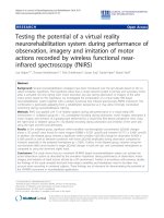
Fig. 4. ESR spectra of Al-h (a), Ti (b), “peroxy”(c), and E′ (d) centres, for quartz fractions 4–11, 63–90, 90–125, 125–180, 180–250 μm from sample NZ 3, and for
calibration quartz.
Table 2
ESR signal intensities of Al-h, Ti (option A, B, D), “peroxy”, and E’ centres, for fractions 4–11, 63–90, 90–125, 125–180, 180–250 μm and calibration quartz.
Sample
Al
st err
Ti A
st err
Ti B
st err
Ti D
st err
peroxy
st err
E′
st err
4–11 μm
63–90 μm
90–125 μm
125–180 μm
180–250 μm
Calibration quartz
1.5939
0.5596
0.5641
0.7602
0.7250
1.7286
0.0056
0.0041
0.0082
0.0073
0.0105
0.0158
0.0769
0.2141
0.1778
0.1557
0.1304
0.0968
0.0112
0.0047
0.0104
0.0028
0.0222
0.0028
0.0429
0.0687
0.0611
0.0842
0.0934
0.0598
0.0080
0.0067
0.0072
0.0065
0.0005
0.0040
0.0610
0.1295
0.1143
0.0966
0.0774
0.0576
0.0070
0.0045
0.0094
0.0033
0.0117
0.0024
2.2026
0.5018
0.4660
0.4569
0.4598
0.5107
0.0099
0.0102
0.0016
0.0053
0.0093
0.0132
0.4766
0.1571
0.1317
0.1430
0.0943
0.4435
0.0073
0.0062
0.0020
0.0008
0.0030
0.0196
and for “peroxy” signal it was determined from the peak-to-peak height
from g ≈ 2.003 to g ≈ 2.009 (Odom and Rink, 1989).
ESR spectra of Al-h centre (Fig. 4a) show a significant contribution
from the “peroxy” signal, which is relatively strong in these samples
(Fig. 4c). Al-h and “peroxy” signals are considerably stronger in 4–11 μm
quartz, compared to other fractions, about 3 and 4 times higher than in
the case of 63–90 μm fraction, for Al-h and “peroxy” respectively.
Interestingly, Ti signals are very weak in all of the investigated samples,
especially in the 4–11 μm fraction (Fig. 4b), for which the intensity
amounts to 40–60% of the intensity observed for 63–90 μm fraction. E′
signal intensity in the smallest fraction is about 3 times bigger than in
63–90 μm fraction, and reduces with increasing grain size (Fig. 4d).
General trends observed in the case of fine grains compared to the coarse
ones – a higher intensity of Al-h, “peroxy” and E’ signals, as well as very
low intensity of Ti centres, are the same as reported in Timar-Gabor
(2018) for samples which displayed a very good OSL behaviour (quartz
from loess from Roxolany, Ukraine (Anechitei-Deacu et al., 2018) and
Stayky, Ukraine (Veres et al., 2018). This suggests that the cause of the
poor OSL properties of the investigated coarse grained quartz samples
might be connected with some non-paramagnetic species, which cannot
be detected by ESR spectroscopy.
5. Luminescence ages
Luminescence ages obtained by using SAR-OSL protocol on fine
quartz as well as pIRIR225 and pIRIR290 protocols, respectively, are
presented in Table 1 along with the dosimetry data. Only the pIRIR ages
calculated with the modern analogue correction are discussed in this
section.
Fine quartz luminescence ages range from 0.3 ± 0.04 ka for sample
NZ 6 which was collected from the uppermost part of the section to 13 ±
2 ka for sample NZ 9.
The pIRIR225 ages calculated using a residual dose correction based
on the modern analogue sample range between 14 ± 1 ka for sample NZ
7 and 18 ± 2 ka for sample NZ 9 for pIRIR225 protocol. On the other
hand, luminescence ages measured using pIRIR290 protocol, using the
same residual correction vary from 20 ± 2 ka for sample NZ 7 to 27 ± 3
ka for sample NZ 9. As can be seen, the pIRIR290 luminescence ages are
slightly higher than those measured using pIRIR2225 protocol. Such age
discrepancy between the two pIRIR protocols over this age interval has
been previously observed in several studies, such as on European loess
(Zhang et al., 2018; Avram et al., 2020) and on New Zealand loess
(Micallef et al., 2021; Brezeanu et al., 2021), respectively.
7
A. Avram et al.
Radiation Measurements 155 (2022) 106788
The two sets of pIRIR ages calculated based on modern analogue
correction along with the fine quartz SAR-OSL ages are presented in
Fig. S4 as function of depth. An age reversal can be observed between
sample NZ 9 and NZ 10, and occur between a depth of ~70 cm and
~130 cm. Such age reversal has been previously observed at the same
depths from a nearby loess site by Brezeanu et al. (2021) as well as by
others in the Canterbury region (e.g., Berger et al., 2001; Almond et al.,
2007; Rowan et al., 2012).
As can be seen from Fig. S4, an age discrepancy between the three
sets of ages is displayed. Based on dose recovery test results as well as on
the previous findings (e.g., Veres et al., 2018; Constantin et al., 2019;
Avram et al., 2020, 2022), we interpret the pIRIR290 ages as being
overestimated. Moreover, the SAR-OSL fine quartz and pIRIR225 ages are
not in agreement even though the behaviour in the SAR procedure was
satisfactory for both minerals. The pIRIR225 age for sample NZ 7 (14 ± 1
ka) collected from a depth of 30 cm is similar with that obtained by
Brezeanu et al. (2021) for sample NZ 2 (14 ± 1 ka) which was collected
from the same depth. Such overlapping was also found for samples
collected from a depth of ~50 and ~130 cm, respectively. As there is
evidence that reliable age up to ~50 ka can be obtained on fine quartz
(e.g., Timar-Gabor and Wintle 2013; Avram et al., 2020), the differences
between the OSL and pIRIR225 ages reported here require further in
vestigations. IR50 ages estimated based on signals collected during the
application of the pIRIR225 protocol support this conclusion.
A. Avram and A. Timar-Gabor acknowledge the financial support of
the research project EEA-RO–NO–2018-0126.
A. Micallef acknowledges the financial support from the European
Research Council (ERC) under the European Union’s Horizon 2020
research and innovation programme (grant MARCAN 677898).
Appendix A. Supplementary data
Supplementary data to this article can be found online at https://doi.
org/10.1016/j.radmeas.2022.106788.
References
Aitken, M.J., Alldred, J.C., 1972. The assessment of error limits in thermoluminescent
dating. Archaeometry 14, 257–267.
Aitken, M.J., 1985. Thermoluminescence Dating. Academic Press, London, p. 360.
Alloway, B.V., Lowe, D.J., Barrell, D.J.A., Newnham, R.M., Almond, P.C., Augustinus, P.
C., Bertler, N.A.N., Carter, L., Litchfield, N.J., McGlone, M.S., Shulmeister, J.,
Vandergoes, M., Williams, P., NZ-INTIMATE Members, 2007. Towards a climate
event stratigraphy for New Zealand over the past 30 000 years (NZ-INTIMATE
project). J. Quat. Sci. 22 (1), 9–35. />10.1002/jqs.1079.
Almond, P.C., Moar, N.T., Lian, O.B., 2001. Reinterpretation of the glacial chronology of
South Westland, New Zealand. N. Z. J. Geol. Geophys. 44, 1–15. />10.1080/00288306.2001.9514917.
Almond, P.C., Shanhun, F.L., Rieser, U., Shulmeister, J., 2007. An OSL, radiocarbon and
tephra isochron-based chronology for birdlings flat loess at Ahuriri Quarry, Banks
Peninsula, Canterbury, New Zealand. Quateranary Geochronology 2, 4–8. https://
doi.org/10.1016/j.quageo.2006.06.002.
Anechitei-Deacu, V., Timar-Gabor, A., Constantin, D., Trandafir-Anothi, O., Del Valle, L.,
Fornos, J.J., Gomez-Pujol, L., Wintle, A.G., 2018. Assessing the maximum limit of
SAR-OSL dating using quartz of different grain sizes. Geochronometria 45, 146–159.
/>Avram, A., Constantin, D., Veres, D., Kelemen, S., Obreht, I., Hambach, U., Markovi´c, S.
B., Timar-Gabor, A., 2020. Testing polymineral post-IR IRSL and quartz SAR-OSL
protocols on Middle to Late Pleistocene loess at Batajnica, Serbia. Boreas 49,
615–663. />Avram, A., Constantin, D., Hao, Q., Timar-Gabor, A., 2022. Optically stimulated
luminescence dating of loess in South-Eastern China using quartz and polymineral
fine grains. Quat. Geochronol. 67, 101226 />quageo.2021.101226.
Berger, G.W., Pillans, B.J., Tonkin, P.J., 2001. Luminescence chronology of loesspaleosol sequences from Canterbury, South Island, New Zealand. N. Z. J. Geol.
Geophys. 44, 501–516. />Berger, G.W., Pillans, B.J., Bruce, J.G., McIntosh, P.D., 2002. Luminescence chronology
of loess-paleosol sequences from southern South Island, New Zealand. Quat. Sci. Rev.
21, 18991913. />Bă
osken, J., Klasen, N., Zeeden, C., Obreht, I., Markovi´c, S.B., Hambach, U., Lehmkuhl, F.,
2017. New luminescence-based geochronology framing the last two glacial cycles at
the southern limit of European Pleistocene loess in Stala´c (Serbia). Geochronometria
44, 150–161. />Buylaert, J.P., Murray, A.S., Thomsen, K.J., 2009. Testing the potential of an elevated
temperature IRSL signal from K-feldspar. Radiat. Meas. 44, 560–565. https://doi.
org/10.1016/j.radmeas.2009.02.007.
Buylaert, J.P., Huot, S., Murray, A.S., Van Den Haute, P., 2011a. Infrared stimulated
luminescence dating of an Eemian (MIS 5e) site in Denmark using K-feldspar. Boreas
40, 46–56. />Buylaert, J.P., Thiel, C., Murray, A., Vandenberghe, S., Yi, S., Lu, H., 2011b. IRSL and
post-IR IRSL residual doses recorded in modern dust samples from the Chinese loess
plateau. Geochronometria 38, 432–440. />Buylaert, J.-P., Jain, M., Murray, A.S., Thomsen, K.J., Thiel, C., Sohbati, R., 2012.
A robust feldspar luminescence dating method for Middle and Late Pleistocene
sediments. Boreas 41, 435–451. />x.
Brezeanu, D., Avram, A., Micallef, A., CintaPinzaru, S., Timar-Gabor, A., 2021.
Investigations on the luminescence properties of quartz and feldspars extracted from
loess in the Canterbury Plains, New Zealand South Island. Geochronometria 48,
46–60. />Colarossi, D., Duller, G.A.T., Roberts, H.M., 2018. Exploring the behaviour of
luminescence signals from feldspars: implications for the single aliquot regenerative
dose protocol. Radiat. Meas. 109, 35–44. />radmeas.2017.07.005.
Constantin, D., Veres, D., Panaiotu, C., Anechitei-Deacu, V., Groza, S.M., Begy, R.C.,
Kelemen, S., Buylaert, J.-P., Hambach, U., Markovic, S.B., Gerasimenko, N., TimarGabor, A., 2019. Luminescence age constraints on the Pleistocene-Holocene
transition recorded in loess sequences across SE Europe. Quat. Geochronol. 49,
71–77. />Cunningham, A.C., Wallinga, J., 2010. Selection of integration time intervals for quartz
OSL decay curves. Quat. Geochronol. 5, 657–666. />quageo.2010.08.004.
6. Summary and conclusions
In this study the SAR-OSL protocol has been applied for the first time
on fine quartz alongside pIRIR225 and pIRIR290 protocols on polymineral
fine grains for dating seven samples of loess from an exposure in
Southern Canterbury Plains South Island of New Zealand. Luminescence
behaviour of fine quartz in the SAR procedure was investigated in the
regard of IR depletion test, preheat plateau test and dose recovery tests,
respectively. The satisfactory results that have been obtained for all the
investigated tests have led to obtaining for the first-time fine quartz
luminescence ages for the investigated loess profile. Moreover, two sets
of pIRIR ages have been also determined on polymineral fine grains
extracted from the same samples. All three sets of ages range up to 13 ±
2 ka (fine quartz), 18 ± 2 ka (pIRIR225) and 27 ± 3 ka (pIRIR290),
respectively suggesting that loess from the investigated profile was
accumulated during the last glacial maximum. As coarse quartz fractions
were not amenable for OSL dating ESR investigations were performed on
different grain sizes of quartz. The main ESR impurity defects (Al and Ti
centres) as well as the most dominant intrinsic defects (E′ and “peroxy”)
showed trends similar to those previously reported for samples charac
terised by a very good OSL behaviour, namely a higher intensity of Al-h,
“peroxy” and E’ signals, and much lower intensity of Ti signals observed
in the case of fine grains compared to the coarse grains. The lack of
significant differences in ESR signals between the samples suitable for
OSL dating such as calibration quartz and other samples previously
investigated and the New Zealand samples which display a poor lumi
nescence behaviour suggest that the factors leading to the different OSL
characteristics might be connected with some non-paramagnetic spe
cies, which cannot be detected by ESR spectroscopy.
Declaration of competing interest
The authors declare that they have no known competing financial
interests or personal relationship that could have appeared to influence
the work reported in this paper.
Acknowledgements
This study was funded by the European Research Council (ERC)
under the European Union’s Horizon 2020 research and innovation
programme ERC-2015-STG (grant agreement No [678106]).
8
A. Avram et al.
Radiation Measurements 155 (2022) 106788
Roberts, H., 2008. The development and application of luminescence dating to loess
deposits: a perspective on the past, present and future. Boreas 37, 483–507. https://
doi.org/10.1111/j.1502-3885.2008.00057.x.
Roberts, H.M., 2015. Luminescence dating, loess. In: Rink, W.J., Thompson, J.W. (Eds.),
Encyclopedia of Scientific Dating Methods. Springer, pp. 425–430.
Rowan, A.V., Roberts, H.M., Jones, M.A., Duller, G.A.T., Covey-Crump, S.J.,
Brocklehurst, S.H., 2012. Optically stimulated luminescence dating of glaciofluvial
sediments on the Canterbury Plains, South Island, New Zealand. Quat. Geochronol.
8, 10–22. />Rother, H., Shulmeister, J., Rieser, U., 2009. Stratigraphy, optical dating chronology
(IRSL) and depositional model of pre-LGM glacial deposits in the Hope Valley, New
Zealand. Quat. Sci. Rev. 117. />Schmidt, C., Bă
osken, J., Kolb, T., 2018. Is there a common alpha-efficiency in
polymineral samples measured by various infrared stimulated luminescence
protocols? Geochronometria 45, 160–172. />Shulmeister, J., Thackray, G.D., Rieser, U., Hyatt, O.M., Rother, H., Smart, C.C., Evans, D.
J., 2010. The stratigraphy, timing and climatic implications of glaciolacustrine
deposits in the middle Rakaia Valley, South Island, New Zealand. Quat. Sci. Rev. 29,
2362–2381. />Sohbati, R., Borella, J., Murray, A., Quigley, M., Buylaert, J.P., 2016. Optical dating of
loessic hillslope sediments constrains timing of prehistoric rockfall, Christchurch,
New Zealand. J. Quat. Sci. 31, 678–690. />Stevens, T., Markovi´c, S.B., Zech, M., Hambach, U., Sümegi, P., 2011. Dust deposition
and climate in the Carpathian Basin over an independently dated last glacialinterglacial cycle. Quat. Sci. Rev. 30, 662–681. />quascirev.2010.12.011.
Spooner, N.A., 1992. Optical dating—preliminary-results on the anomalous fading of
luminescence from feldspars. Quat. Sci. Rev. 11, 139–145. />0277-3791(92)90055-D.
Spooner, N.A., 1994. The anomalous fading of infrared-stimulated luminescence from
feldspars. Radiat. Meas. 23, 625–632. />90111-2.
Thiel, C., Buylaert, J.P., Murray, A., Terhorst, B., Hofer, I., Tsukamoto, S., Frechen, M.,
2011. Luminescence dating of the Stratzing loess profile (Austria) – testing the
potential of an elevated temperature post-IR IRSL protocol. Quat. Int. 234, 23–31.
/>Thomsen, K.J., Bøtter-Jensen, L., Denby, P.M., Moska, P., Murray, A.S., 2006.
Developments in luminescence measurement techniques. Radiat. Meas. 41, 768–773.
/>Thomsen, K.J., Murray, A.S., Jain, M., 2011. Stability of IRSL signals from sedimentary
K-feldspar samples. Geochronometria 38, 1–13. />Timar-Gabor, A., Wintle, A.G., 2013. On natural and laboratory generated dose response
curves for quartz of different grain sizes from Romanian loess. Quat. Geochronol. 18,
34–40. />Timar-Gabor, A., 2018. Electron spin resonance characterisation of sedimentary quartz of
different grain sizes. Radiat. Meas. 120, 59–65. />radmeas.2018.06.023.
Toyoda, S., Falgu`eres, C., 2003. The method to represent the ESR signal intensity of the
aluminum hole center in quartz for the purpose of dating. Adv. ESR Appl. 20, 7–10.
Veres, D., Tecsa, V., Gerasimenko, N., Zeeden, C., Hambach, U., Timar-Gabor, A., 2018.
Short-term soil formation events in last glacial east European loess, evidence from
multi-method luminescence dating. Quat. Sci. Rev. 34–51. />j.quascirev.2018.09.037, 200.
Vasiliniuc, Ș., Vandenberghe, D.A.G., Timar-Gabor, A., Panaiotu, C., Cosma, C., 2012.
Testing the potential of elevated temperature post-IR IRSL signals for dating
Romanian loess. Quat. Geochronol. 10, 75–80. />quageo.2012.02.014.
Wallinga, J., Murray, A., Duller, G., 2000. Underestimation of equivalent dose in singlealiquot optical dating of feldspars caused by preheating. Radiat. Meas. 32, 691–695.
/>Wintle, A.G., 1973. Anomalous fading of thermoluminescence in mineral samples.
Nature 245, 143–144. />Yi, S., Buylaert, J.P., Murray, A.S., Lu, H., Thiel, C., Zeng, L., 2016. A detailed post-IR
IRSL dating study of the Niuyangzigou loess site in northeastern China. Boreas 45,
644–657. />Yi, S., Li, X., Han, Z., Lu, H., Liu, J., Wu, J., 2018. High resolution luminescence
chronology for Xiashu Loess deposits of Southern China. J. Asian Earth Sci. 155,
188–197. />Yates, K., Fenton, C.H., Bell, D.H., 2018. A review of the geotechnical characteristics of
loess and loess-derived soils from Canterbury, South Island, New Zealand. Eng. Geol.
236, 11–21. />Zhang, J., Rolf, C., Wacha, L., Tsukamoto, S., Durn, G., Frechen, M., 2018. Luminescence
dating and palaeomagnetic age constraint of a last glacial loess-paleosol sequence
from Istria, Croatia. Quat. Int. 494, 19–33. />quaint.2018.05.045.
Duller, G.A.T., 2003. Distinguishing quartz and feldspars in single grain luminescence
measurements. Radiat. Meas. 37, 161–165. />(02)00170-1.
Duval, M., Guilarte, V., 2015. ESR dosimetry of optically bleached quartz grains
extracted from Plio-Quaternary sediment: evaluating some key aspects of the ESR
signals associated to the Ti-centers. Radiat. Meas. 78, 28–41. />10.1016/j.radmeas.2014.10.002.
Duval, M., Arnold, L.J., Guilarte, V., Demuro, M., Santonja, M., P´
erez-Gonz´
alez, A., 2017.
Electron spin resonance dating of optically bleached quartz grains from the Middle
Palaeolithic site of Cuesta de la Bajada (Spain) using the multiple centres approach.
Quat. Geochronol. 37, 82–96. />Frechen, M., Schweitzer, U., Zander, A., 1996. Improvements in sample preparation for
the fine grain technique. Ancient TL 14, 15–17.
Gu´erin, G., Mercier, N., Adamiec, G., 2011. Dose-rate conversion factors: update. Ancient
TL 29, 5–8.
Hansen, V., Murray, A., Buylaert, J.P., Yeo, E.Y., Thomsen, K., 2015. A new irradiated
quartz for beta source calibration. Radiat. Meas. 81, 123–127. />10.1016/j.radmeas.2015.02.017.
Hornblow, S., Quigley, M., Nicol, A., Van Dissen, R., Wang, N., 2014. Paleoseismology of
the 2010 Mw 7.1 Darfield (Canterbury) earthquake source, Greendale fault, New
Zealand. Tectonophysics 637, 178–190. />tecto.2014.10.004.
Holdaway, R.N., Roberts, R.G., Beavan-Athfield, N.R., Olley, J.M., Worthy, T.H., 2002.
Optical dating of quartz sediments and accelerator mass spectrometry 14C dating of
bone gelatin and moa eggshell: a comparison of age estimates for non-archaeological
deposits in New Zealand. J. Roy. Soc. N. Z. 32, 463–505. />03014223.2002.9517705.
Hormes, A., Preusser, F., Denton, G., Hajdas, I., Weiss, D., Stocker, T.F., Schlüchter, C.,
2003. Radiocarbon and luminescence dating of overbank deposits in outwash
sediments of the Last Glacial Maximum in North Westland, New Zealand. N. Z. J.
Geol. Geophys. 46, 95–106. />Kreutzer, S., Schmidt, C., DeWitt, R., Fuchs, M., 2014. The a-value of polymineral fine
grain samples measured with the post-IR IRSL protocol. Radiat. Meas. 63, 1829.
/>Lai, Z.P., Ză
oller, L., Fuchs, M., Brückner, H., 2008. Alpha efficiency determination for
OSL of quartz extracted from Chinese loess. Radiat. Meas. 43, 767–770. https://doi.
org/10.1016/j.radmeas.2008.01.022.
Lapp, T., Kook, M., Murray, A.S., Thomsen, K.J., Buylaert, J.P., Jain, M., 2015. A new
luminescence detection and stimulation head for the Risø TL/OSL reader. Radiat.
Meas. 81, 178–184. />Lang, A., Lindauer, S., Kuhn, R., Wagner, G.A., 1996. Procedures used for optically and
infrared stimulated luminescence dating of sediments in Heidelberg. Ancient TL 14,
7–11.
Micallef, A., Marchis, R., Saadatkhah, N., Pondthai, P., Everett, M.E., Avram, A., TimarGabor, A., Cohen, D., Preca Trapani, R., Weymer, B.A., Wernette, P., 2021.
Groundwater erosion of coastal gullies along the Canterbury coast (New Zealand): a
rapid and episodic process controlled by rainfall intensity and substrate variability.
Earth Surf. Dyn. 9, 1–18. />Murray, A.S., 1996. Developments in optically stimulated luminescence and phototransferred thermoluminescence dating of young sediments: application to a 2000years of flood deposits. Geochem. Cosmochim. Acta 60, 565–576. />10.1016/0016-7037(95)00418-1.
Murray, A.S., Wintle, A.G., 2000. Luminescence dating using an improved single-aliquot
regenerative-dose protocol. Radiat. Meas. 32, 57–73. />S1350-4487(99)00253-X.
Murray, A.S., Wintle, A.G., 2003. The single aliquot regenerative dose protocol: potential
for improvements in reliability. Radiat. Meas. 37, 377–381. />10.1016/S1350-4487(03)00053-2.
Murray, A.S., Thomsen, K.J., Masuda, N., Buylaert, J.P., Jain, M., 2012. Identifying wellbleached quartz using the different bleaching rates of quartz and feldspar
luminescence signals. Radiat. Meas. 47, 688–695. />radmeas.2012.05.006.
Nichol, S.L., Lian, O.B., Carter, C.H., 2003. Sheet-gravel evidence for a late Holocene
tsunami run-up on beach dunes, Great Barrier Island, New Zealand. Sediment. Geol.
155, 129–145.
Odom, A.L., Rink, W.J., 1989. Natural accumulation of Schottky-Frenkel defects:
implications for a quartz geochronometer. Geology 17 (1), 55–58. />10.1130/0091-7613(1988)017<0055:NAOSFD>2.3.CO;2.
Prescott, J.R., Hutton, J.T., 1994. Cosmic ray contributions to dose rates for
luminescence and ESR dating: large depths and long term variations. Radiat. Meas.
23, 497–500. />Preusser, F., Andersen, B.G., Denton, G.H., Schlüchter, C., 2005. Luminescence
chronology of Late Pleistocene glacial deposits in north Westland, New Zealand.
Quat. Sci. Rev. 24, 2207–2227. />Preusser, F., Ramseyer, K., Schlüchter, C., 2006. Characterisation of low OSL intensity
quartz from the New Zealand Alps. Radiat. Meas. 41, 871–877. />10.1016/j.radmeas.2006.04.019.
Rees-Jones, J., 1995. Optical dating of young sediments using fine-grain quartz. Ancient
TL 13, 9–13.
9