Báo cáo khoa học: A novel mannitol teichoic acid with side phosphate groups ofBrevibacterium sp.VKM Ac-2118 ppt
Bạn đang xem bản rút gọn của tài liệu. Xem và tải ngay bản đầy đủ của tài liệu tại đây (240.65 KB, 6 trang )
A novel mannitol teichoic acid with side phosphate groups
of
Brevibacterium sp.
VKM Ac-2118
Natalia V. Potekhina
1
, Alexander S. Shashkov
2
, Lyudmila I. Evtushenko
3
, Ekaterina Yu. Gavrish
3
,
Sofya N. Senchenkova
2
, Andrey A. Stomakhin
4
, Anatolii I. Usov
2
, Irina B. Naumova
1,
*
and Erko Stackebrandt
5
1
School of Biology, M. V. Lomonosov Moscow State University, Russia;
2
N. D. Zelinsky Institute of Organic Chemistry, Russian
Academy of Sciences, Moscow, Russia;
3
Institute of Biochemistry and Physiology of Microorganisms, Russian Academy of Sciences,
Pushchino, Moscow Region, Russia;
4
B. A. Engelhardt Institute of Molecular Biology, Russian Academy of Science, Moscow,
Russia;
5
DSMZ-Deutsche Sammlung von Mikroorganismen und Zellkulturen GmbH, Braunschweig, Germany
ThecellwallofBrevibacterium sp. VKM Ac-2118 isolated
from a frozen (mean annual temperature )12 °C) late Plio-
cene layer, 1.8–3 Myr, Kolyma lowland, Russia, contains
mannitol teichoic acid with a previously unknown structure.
This is 1,6-poly(mannitol phosphate) with the majority of
the mannitol residues bearing side phosphate groups at
O-4(3). The structure of the polymer was established by
chemical methods, NMR spectroscopy, and MALDI-TOF
mass spectrometry.
Keywords: teichoic acids; poly(mannitol phosphate); side
phosphate groups; NMR; Brevibacterium.
Structural variants of teichoic acids, the anionic polymers
of the cell walls of Gram-positive bacteria, are extremely
numerous because they differ in the polyol and sugar (or
amino sugar) composition, the types of phosphodiester
bonds, configurations of the glycosidic bonds, the types of
bonds between the monosaccharide components in oligo-
saccharide units, and O-acyl substituents.
Among the cell wall teichoic acids studied so far, four
structural types can be distinguished depending on the
composition of the main chain: poly(polyol phosphates) (I),
poly(glycosylpolyol phosphates) (II), poly(polyol phos-
phate–glycosyl phosphates) (III) and poly(polyol phos-
phate–glycosylpolyol phosphates) (IV) [1]. The polymers of
types I and II are the most abundant cell wall teichoic acids.
Poly(polyol phosphate) chains containing glycerol, erythr-
itol, ribitol, arabinitol, and mannitol have been identified [1].
The interest that has arisen during recent years in these
polymers stems from their taxonomic significance for
Gram-positive bacteria, especially actinomycetes. The spe-
cies-specificity of teichoic acids has been demonstrated for
the genera Nocardiopsis [1], Glycomyces [2,3], Nocardioides
[4,5] and Actinomadura [6–8]. Presumably, the structures of
cell wall teichoic acids can be used as an additional
chemotaxonomic marker for the attribution of new species
of Gram-positive bacteria to the Brevibacterium genus.
Poly(mannitol phosphate) teichoic acids were found in
different species of the genus Brevibacterium. Some strains
of B. linens and B. epidermidis contain unsubstituted and
partially substituted poly(mannitol phosphate) chains (with
monosaccharides as the substituents) together with poly(gly-
cerol phosphate) chains [9,10]. The cell wall of B. iodinum
was shown to contain poly(mannitol phosphate) chain with
most of the mannitol residues acylated at positions 4 and 5
by pyruvic acid. In addition, about half of the mannitol
residues bear a-glycopyranosyl residues at O-2[11].
In the present work, we report the elucidation of the
structure of a cell wall component of Brevibacterium VKM
Ac-2118, which was found to be a new variant of mannitol
teichoic acids.
Materials and methods
The strain VKM Ac-2118 was isolated from a sample of
permafrost sediments, 48.8 m deep, recovered from a frozen
(mean annual temperature )12 °C) late Pliocene layer, 1.8–
3 Myr, Kolyma lowland, Russia. Samples were obtained as
described by Shi et al. [12] and kept frozen at )20 °Cbefore
study. The methods used for studying phenotypical
characteristics were described previously [13]. The 16S
rRNA gene was amplified by PCR using prokaryotic 16S
rDNA universal primers and purified as described [13].
16S rDNA was sequenced using a Big Dye Terminator Kit
(Perkin Elmer) with a model ABI-310 automatic DNA
Sequencer (Perkin Elmer) according to the manufacturer’s
protocol. Nucleotide substitution rates were calculated as
described [14] and the phylogenetic tree was constructed by
the neighbor-joining method [15] with
CLUSTALW
software
[16]. Three topologies were evaluated by bootstrap analysis
of the sequence data with the same software.
To obtain cell wall, the culture of Brevibacterium sp.
VKM Ac)2118 was grown on a pepton/yeast medium [17]
for 12–18 h on a shaker at 28 °C. The biomass was collected
Correspondence to N. V. Potekhina, School of Biology, M. V.,
Lomonosov Moscow State University, 119992 Moscow, Russia.
Fax: + 7 095 9394309, E-mail:
Abbreviation: PME, phosphomonoesterase.
Enzyme: phosphomonoesterase (EC 3.1.3.1).
*Deceased suddenly on 18 August 2003.
(Received 14 April 2003, revised 1 August 2003,
accepted 12 September 2003)
Eur. J. Biochem. 270, 4420–4425 (2003) Ó FEBS 2003 doi:10.1046/j.1432-1033.2003.03832.x
at the logarithmic growth phase. The cells were harvested by
centrifugation, washed with 0.95% NaCl and cell walls were
obtained as described [18]. The cell wall preparation was
heated in 2% SDS for 5 min at 100 °C, washed several
times with water, and freeze-dried.
The teichoic acid was extracted with 10% trichloroace-
tic acid at 4 °C for 24 h. The mixture was centrifuged and
the cell wall was repeatedly treated with 10% trichloro-
acetic acid under the same conditions. The supernatants
were combined, dialysed against distilled water, and
freeze-dried to yield a crude preparation. Fractional
precipitation of teichoic acids was carried out by the
addition of ethanol (two volumes) to the combined
supernatant and the precipitate that formed was removed
by centrifugation for 24 h at 0 °C, 10 000 g,15min.To
the supernatant, more ethanol (two volumes) was added
and the precipitate was collected by centrifugation
(as above). This was redissolved in water, centrifuged,
the supernatant was dialysed and freeze-dried. Thus, two
teichoic acid preparations were isolated as follows:
teichoic acid precipitated with two volumes of ethanol
(preparation I) and teichoic acid precipitated with four
volumes of ethanol (preparation II).
Descending chromatography and electrophoresis were
performed on a Filtrak FN-13 paper. Electrophoresis was
performed in a pyridinium acetate buffer (pH 5.6) to
separate phosphoric esters [7]. The following solvent systems
were used for descending paper chromatography: (a)
propan-1-ol/aqueous NH
3
(specific gravity 0.88)/water
(6 : 3 : 1, v/v/v) for the separation of isomeric phosphoric
esters; (b) pyridine/benzene/butan-1-ol/water (3 : 1 : 5 : 3,
v/v/v/v); (c) butan-1-ol/acetic acid/water (4 : 1 : 5, v/v/v)
for the separation of mannitol, glycerol, and monosaccha-
rides; and (d) pyridine/ethyl acetate/acetic acid/water
(5 : 5 : 1 : 3, v/v/v/v) for the separation of amino sugars.
Detection of compounds was carried out using the following
spray reagents: the molybdate reagent for phosphoric esters,
ninhydrin for amino sugars, 5% AgNO
3
in aqueous NH
3
for polyols and sugars, and aniline hydrogenphthalate for
reducing sugars.
Acid hydrolysis of the teichoic acid was carried out with
2
M
HCl for 3 h at 100 °C and 40% HF for 24 h at 20 °C;
alkaline hydrolysis was performed with 1
M
NaOH for 3 h
at 100 °C, hydrolysis with alkaline phosphatase (EC 3.1.3.1)
from calf intestinal mucosa. (Sigma) was performed in
ammonium acetate buffer (pH 10.4) at 37 °C for 2 h [2].
The polyol/phosphorus molar ratios were determined as
described by Potekhina et al.[2].
Mannitol was isolated on a column (1.3 · 75 cm) with
TSK HW-40(S) in 1% acetic acid using a Knauer differ-
ential refractometer. Optical rotation was measured with a
PU-5 polarimeter (Russia).
NMR spectra were recorded using a Bruker DRX-500
spectrometer for 2–3% solutions in D
2
Oat30°Cwith
acetone (d
H
2.225 and d
C
31.45) as the internal standard
and 80% H
3
PO
4
measured separately. One-dimensional
1
H NMR spectra were obtained with a presaturation of
the HDO signal for 1 s two-dimensional spectra were
obtained using standard pulse sequences from the
BRUKER
software.
Mass spectrometric analysis (MALDI-TOF MS) of
teichoic acid (preparation I) was carried out in the positive
reflection mode using a KOMPACT MALDI 4 (Kratos
Analytical) mass spectrometer. 2,5-Dihydroxybenzoic (gen-
tisic) acid was used as a matrix.
Results and discussion
The strain under study had growth, morphological, and
chemotaxonomic characteristics [meso-isomer of diamino-
pimelic acid in the cell wall, menaquinone MK-9 (H-4),
lack of mycolic acids, and the presence of teichoic acids]
typical of the genus Brevibacterium [10]. Colony pigmen-
tation was similar to that of B. linens, which is the only
orange-pigmented species of the genus, but the strain
differed from the type strain of B. linens in a number of
physiological properties (not presented). Phylogenetic
analysis based on 16S rDNA sequence confirmed that
the strain VKM Ac-2118 belongs to the genus Brevibac-
terium. It showed 92.6–97.5% 16S rDNA sequence simi-
larities to type strains of the known species of the genus
and grouped together with B. linens DSM 20425
T
(X77451), B. iodinum NCDO 613
T
(X76567), B. epidermi-
dis NCDO 2286
T
(X76565) and B. casei NCDO 2048
T
(X76564) in a tight cluster with a 100% bootstrap repli-
cation value (not presented), exhibiting the highest 16S
rDNA sequence similarities of 97.6 and 96.6% to B. casei
NCDO 2048
T
and B. linens DSM 20425
T
, respectively. In
addition, the strain differed from all the above species in
the composition of cell wall components.
The teichoic acid (crude preparation) was isolated from
the cell wall containing 2% of organic phosphate. Upon
acid and alkaline hydrolysis, a polyol and its phosphates
were formed together with glycerol monophosphate and
trace amounts of glycerol bisphosphate. The polyol was
identified as mannitol from its mobility on paper chroma-
tography in solvent systems 2 and 3, which coincided with
that of an authentic sample. Additional proof was obtained
from
13
C NMR spectroscopic studies of the polyol isolated
by preparative paper chromatography following hydrolysis
of the teichoic acid with 40% hydrofluoric acid. The
chemical shifts for the carbon atoms of the polyol coincided
completely with those for the authentic mannitol [19]
(Table 1).
The absolute configuration of mannitol is
D
as deduced
from its optical rotation, [a
20
D
þ 22:5 (approximately
1, 0.03
M
borax) (cf. [a
20
D
þ 24: 0for
D
-mannitol [20]).
Degradation of the teichoic acid
Acid degradation of the trichloroacetic acid extract (crude
preparation) yielded mannitol phosphates and glycerol
phosphates. Therefore, it was necessary to establish whether
these compounds originate from the same or from different
teichoic acids present simultaneously in the cell wall, which
has been demonstrated for other brevibacteria [9].
Electrophoresis of the crude preparation did not reveal
the presence of two different polymers. An attempt has been
undertaken to separate teichoic acids by fractional precipi-
tation with ethanol. Two preparations were obtained; those
precipitated with two and four volumes of ethanol (prepar-
ation I and preparation II, respectively). Acid hydrolysates
of these preparations differed in the compositions. The
essential factor is that they differed in the ratios of glycerol
Ó FEBS 2003 A novel mannitol teichoic acid (Eur. J. Biochem. 270) 4421
phosphates and mannitol phosphates. Mannitol teichoic
acid was virtually the only component of preparation I. This
was free from sugars, which suggested the absence of
glycosyl substituents in the chain. It was this preparation
that was subjected to subsequent structural analysis.
Elucidation of the primary structure of teichoic acids by
chemical methods involves their degradation under differ-
ent conditions, structural analysis of the fragments formed
and reconstruction of the structure of the original polymer
[21].
Alkaline hydrolysis of the polymer furnished three major
phosphates which were isolated by paper electrophoresis.
Phosphate 1 (P1), with the electrophoretic mobility relative
to that of glycerol phosphate (m
GroP
) equal to 0.7, was
treated with phosphomonoesterase (PME) to yield mannitol
and inorganic phosphate in an 0.96 : 1 molar ratio. Thus, P1
is mannitol monophosphate. Phosphate 2 (P2, m
GroP
1.21)
yielded mannitol and inorganic phosphate in a 1 : 2 molar
ratio upon treatment with PME. Thus, P2 is mannitol
bisphosphate. Phosphate 3 (P3, m
GroP
1.5) was hydrolyzed
under the action of PME to yield mannitol and inorganic
phosphate in a 1 : 2.8 molar ratio, which suggests that P3
is mannitol trisphosphate.
Acid hydrolysis of the teichoic acid yielded the same three
major phosphates as those formed upon alkaline hydrolysis.
The presence of mannitol mono- and bisphosphates among
the degradation products of the polymer suggested that the
teichoic acid under study is of the poly(mannitol phosphate)
type and the formation of identical phosphates upon
alkaline and acid degradations corroborated the absence
of glycosyl substituents in the chain [22].
However, the formation of mannitol trisphosphate upon
degradation of the polymer could not be rationalized in
the framework of ordinary pathways of hydrolysis of poly
(polyol phosphate) chains. This could occur in the case
where a secondary hydroxy group of mannitol was substi-
tuted by a phosphate-bearing group.
Alkaline hydrolysis of the teichoic acid with the structure
of poly(mannitol phosphate) with monophosphate side
units is depicted in Fig. 1.
As can be seen, the polymer contains two nonequiva-
lent phosphate groups, A and B, vicinal to the free
hydroxy groups of the polyol. Hydrolysis of the chain
occurs via transient five-membered cyclophosphates. Clea-
vage of the phosphodiester bond A results in scission of
the poly(mannitol phosphate) chain, and the cyclophos-
phate formation involves either one hydroxy group (OH-2
or OH-5) of mannitol or both to yield 1,2(5,6)-cyclophos-
phate or 1,2;5,6-bis(cyclophosphate), respectively. Cyclo-
phosphates are known to be unstable in alkaline media
and their opening results in isomeric phosphates [22].
Cleavage of the phosphate B does not occur under
alkaline conditions and the phosphate group remains
linked to the mannitol residue in the same position. Thus,
mannitol trisphosphate is a hydrolysis product of the
phosphate groups A.
The mechanism of acid hydrolysis is essentially the same,
although the phosphate group migration can occur in
polyol phosphates via transient cyclophosphates.
Esterification of one of the secondary hydroxy groups of
mannitol by phosphoric acid was confirmed by quantitation
of the total phosphate (P
total
) and that liberated under the
action of PME (P
PME
). The P
total
:P
PME
molar ratio was
found to be equal to 2:1.
Teichoic acid was also investigated independently by
NMR spectroscopy. The most abundant signals in the
Fig. 1. Pathways of alkaline hydrolysis of the mannitol teichoic acid of
Brevibacterium sp. VKM Ac-2118.
Table 1.
13
C NMR data of the mannitol teichoic acid of Brevibacterium sp. VKM Ac-2118 (d, p.p.m.; JÆHz
)1
, acetone, d, 31.45 p.p.m. br, broadened).
Residue
Carbon atoms
C-1 C-2 C-3 C-4 C-5 C-6
Mannitol 64.5 72.1 70.5 70.5 72.1 64.5
-1)-Mannitol-(6-P- 68.65 70.65 69.8 69.8 70.65 68.65
-1)-Mannitol-(6-P-
4
j
P
68.3 70.25 70.7 75.3 71.1 68.8
br.
3
J
P-1(6),C-2
2.9
3
J
P-4,C-3
7.5
2
J
P-4,C-4
6.1
3
J
P-6,C-5
2.9 br.
Mannitol-(6-P-
4
j
P
64.3 71.1 69.95 75.1 70.7 68.8
4422 N. V. Potekhina et al. (Eur. J. Biochem. 270) Ó FEBS 2003
13
C NMR spectrum of the teichoic acid (Table 1) corres-
ponded to 1,6-poly(mannitol phosphate) chain (the signals
for the –CH
2
OPO
3
–atd 68.3 and 68.8) bearing a phosphate
group at C-5(2) or C-4(3) (a signal at d 75.3).
The
31
P NMR spectrum of the polymer contained a
broadened signal with a maximum intensity at d 0.8. The
31
P NMR spectrum of the preparation recorded in the
presence of 0.25
M
EDTA contained two major signals of
nearly the same intensities at d 0.4 and 1.6 together with
minor signals belonging probably to the side phosphate
group in the mannitol residue at the growing chain end
and/or to the internal phosphate(s) bound to the mannitol
unit devoid of the side phosphate group.
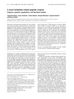
The two dimensional heteronuclear
1
H/
31
P heteronuclear
multiple quantum coherence (HMQC) spectrum (Fig. 2)
revealed a single correlation of the former signal (d 0.4)
with the methine proton resonating at d 4.37. The second
most abundant peak at d 1.6 had cross-peaks with the
protons resonating at d 4.25 (H-6), 4.18 (H-1), 4.09 (H-1¢),
4.08 (H-5), and 4.01 (H-6¢), which suggests the presence of
two phosphate groups, one of which was linked to a
methine group, and the second being involved in the 1,6-
poly(mannitol phosphate) chain of the polymer.
The
1
H NMR spectrum (Table 2) was interpreted using
two dimensional
1
H/
1
H COSY and two dimensional
1
H/
13
C
HSQC (heteronuclear single quantum coherence) spectros-
copy. The HSQC spectrum (Fig. 3) revealed unequivocally
that the signals for the carbon atoms at d 68.8 and 68.3
belong to the –CH
2
OP– groups.
In the COSY spectrum, a correlation was found between
one of the protons of the –CH
2
OP– group at d4.25 and (a)
the second proton of this group at d 4.01 and (b) the proton
Fig. 2.
1
H/
31
CHMQCspectrumofthe
mannitol teichoic acid of Brevibacterium sp.
VKM Ac-2118. The protons H-1, 1¢,4,5,6,
and 6¢ give cross-peaks with phosphorus are
marked at the
1
H NMR spectrum.
Table 2.
1
H NMR date of the mannitol teichoic acid of Brevibacterium sp. VKM Ac-2118 (d, p.p.m.; JÆHz
)1
,acetone,d, 2.225 p.p.m).
Residue
Proton atoms
H-1 H-1¢ H-2 H-3 H-4 H-5 H-6 H-6¢
Mannitol 3.87 3.675 3.76 3.8 3.8 3.76 3.87 3.675
J
1,1
¢ 11.8 J
1
¢
,2
6.1 J
2,3
8.4
J
1,2
2.7
-1)-Mannitol-(6-P- 4.18 4.07 3.97 3.92 3.92 3.97 4.18 4.07
-1)-Mannitol-(6-P-
4
j
P
4.18 4.09 3.91 3.93 4.37 4.08 4.25 4.01
Mannitol-(6-P-
4
j
P
3.85 3.78 3.90 3.94 4.34 4.08 4.17 4.06
Ó FEBS 2003 A novel mannitol teichoic acid (Eur. J. Biochem. 270) 4423
of the –CHO– group at d 4.08 (the corresponding carbon
atom resonates at d 71.1). In turn, there was a correlation
between the latter proton and the proton of a –CHOP–
group at d 4.37 (the corresponding carbon atom resonates at
d 75.3). Thus, the phosphomonoester group is located at
position 4 of mannitol [if numbering is that the protons
resonating at d 4.25 and 4.01 belong to –C(6)H
2
OP– group]
or at position 3 [if numbering is that these protons belong to
–C(1)H
2
OP– group]. In other words, the localization site of
phosphate depends on the mannitol residue numbering.
Taking into account the data on the biosynthesis of ribitol
teichoic acids [23], we assume that mannitol 6-phosphate
that is the monomeric unit of the polymer being studied.
Based on this assumption and on the presence of signals
for the terminal units of the chain, we can localize the
phosphate side group at position 4 of mannitol with greater
certainty from the following observations. Some of the
minor signals for the terminal units belong unambiguously
to the –CH
2
OH group (C-1) and the –CH
2
OP– group (C-6).
Other signals of the terminal unit of the growing chain were
assigned using data from COSY and HSQC spectra
(Table 1), which shows that the phosphomonoester group
is linked with C-4.
The presence of the phosphate substituent at the methine
group was also confirmed by analysis of the MALDI-TOF
mass spectrum (Fig. 4). Thus the most abundant of the
basic peaks are those differing by 324 Da (Man-ol P
2
)or
347 Da (Man-ol P
2
+ Na). The maximum mass recorded
corresponds to 19 mannitol phosphate units. However, the
ratio of the integral intensities of peaks for the terminal and
internal residues in the
13
C NMR spectrum suggests the
presence of 6–7 units on average. With account of possible
broad distribution of oligomeric chains according to masses,
these data on chain length estimations should not be
regarded as contradictory.
Thus, the presence of a phosphate group at the mannitol
methine group is established by several independent meth-
ods: (a) by identification of mannitol trisphosphate as a
degradation product of the teichoic acid; (b) by quantitation
of P
total
and P
PME
(ratio 2 : 1) using treatment of the
polymer with phosphomonoesterase; (c) by detection of a
low-field signal at d 75.3 in the
13
C NMR spectrum. The
spectrum of the polymer treated with PME devoid
completely of the above-mentioned signal corresponded to
unsubstituted 1,6-poly(mannitol phosphate) (Table 1); and
(d) by establishing the molecular mass of the repeating unit
of the polymer equal to 324 Da (MALDI-TOF MS), i.e. the
presence of a phosphate group linked to a methine group of
mannitol.
The results presented here show that the cell wall
of Brevibacterium sp. VKM Ac-2118 contains a
1,6-poly(mannitol phosphate) chain with phosphate groups
attached as side groups to O-4(3) of mannitol residues.
In addition, small amounts of glycerol mono- and bis-
phosphate, and a glycerol phosphodiester containing
Fig. 3.
1
H/
13
C HSQC spectrum of the manni-
tol teichoic acid of Brevibacterium sp. VKM
Ac-2118. The signals at 4.18, 4.09/68.3 and
4.25, 4.01/68.8 belong to the –CH
2
OP–
groups; the signals at 4.37/75.3 belongs to the
–CH(4)OP– group.
Fig.4.TheMALDI-TOFMSoftheteichoicacidofBrevibacterium
sp.VKM Ac-2118.
4424 N. V. Potekhina et al. (Eur. J. Biochem. 270) Ó FEBS 2003
glucosamine were detected in preparation II. The latter
yielded glycerol mono- and bisphosphates and glucosamine
upon acid hydrolysis and proved to be identical with
the alkaline hydrolysis product of the teichoic acid of
Streptomyces rutgersensis var. castelarens [18].
The presence of 1,3-poly(glycerol phosphate) chains
substituted partially with a-N-acetylglycosamine at O-2is
also possible, as can be deduced from NMR spectroscopic
and alkaline hydrolysis data of this preparation.
It is noteworthy that the side phosphate groups impart an
additional negative charge to the polymer, which seems to
be important for functioning of the cell wall on the whole.
These seem to be of special importance considering the
halotolerant properties of Brevibacterium sp.VKM Ac-2118
[24]. Recently, it has been shown that the cell wall of an
alkophilic bacillum comprised three polymers with mark-
edly pronounced acidic properties: polyglucuronic, teich-
uronic and polyglutamic acids [25].
Acknowledgements
This work was supported by grants from INTAS (no. 01–2040) and the
Russian Foundation for Basic Research (no. 01-04-49854).
References
1. Naumova, I.B., Shashkov, A.S., Tul’skaya, E.M., Streshinskaya,
G.M.,Kozlova,Y.I.,Potekhina,N.V.,Evtushenko,L.I.&
Stackebrandt, E. (2001) Cell wall teichoic acids: structural
diversity, species-specificity in the genus Nocardiopsis,andchemo-
taxonomic perspective. FEMS Microbiol. Rev. 25, 269–284.
2. Potekhina, N.V., Tul’skaya, E.M., Naumova, I.B., Shashkov, A.S.
& Evtushenko, L.I. (1993) Erythritolteichoic acid in cell wall of
Glycomyces tenuis VKM Ac-1250. Eur. J. Biochem. 218, 371–375.
3. Potekhina, N.V., Tul’skaya, E.M., Shashkov, A.S., Taran, V.V.,
Evtushenko, L.I. & Naumova, I.B. (1998) Taxonomic specificity
of cell wall teichoic acids of actinomycetes of Glycomyces genus.
Microbiologiya (Moscow) 67, 330–334.
4. Shashkov, A.S., Tul’skaya, E.M., Evtushenko, L.I. & Naumova,
I.B. (1999) Cell wall teichoic acid of Nocardioides albus VKM
Ac-805. Biochemistry (Moscow) 64, 1544–1549.
5. Shashkov, A.S., Tul’skaya, E.M., Evtushenko, L.I., Gratchev,
A.A. & Naumova, I.B. (2000) Structure of teichoic acid of
Nocardioides luteus VKM Ac-1246
T
cell wall. Biochemistry
(Moscow) 65, 509–514.
6. Potekhina, N.V., Shashkov, A.S. & Naumova, I.B. (1996) The cell
wall teichoic acid of Actinomadura madura contains poly
(galactosyl-1,2-glycerol phosphate) and poly-(3-O-methylgalacto-
syl-1,2-glycerol phosphate). Microbiologiya (Moscow) 65,522–
526.
7. Potekhina, N.V., Naumova, I.B., Shashkov, A.S. & Terekhova,
L.P.(1991)Structuralfeaturesofcellwallteichoicacidand
peptidoglycan of Actinomadura cremea INA 292. Eur. J. Biochem.
199, 313–316.
8. Shashkov, A.S., Potekhina, N.V., Naumova, I.B., Evtushenko,
L.I. & Widmalm, G. (1999) Cell wall teichoic acid of Actinoma-
dura viridis VKM Ac-1315
T
. Eur. J. Biochem. 262, 688–695.
9. Fiedler, F. & Bude, A. (1989) Occurrence and chemistry of cell
wall teichoic acids in the genus Brevibacterium. J. Gen. Microbiol.
135, 2837–2846.
10. Fiedler, F., Schaeffler, M.J. & Stackebrandt, E. (1981) Biochem-
ical and nucleic acid hybridisation studies on Brevibacterium linens
and related strains. Arch. Microbiol. 129, 85–93.
11. Anderton, W.J. & Wilkinson, S.G. (1985) Structural studies of
mannitol teichoic acid from the cell wall of Bacterium NCTC 9742.
Biochem. J. 226, 587–599.
12. Shi, T., Reevs, R., Gilichinsky, D. & Friedmann, E.I. (1997)
Characterization of viable bacteria from Siberian permafrost by
16S rDNA sequencing. Microbial Ecol. 33, 169–179.
13. Evtushenko, L.I., Taran, V.V., Akimov, V.N., Kroppenstedt,
R.M., Tiedje, J.M. & Stackebrandt, E. (2000) Nocardiopsis tropica
sp. nov., Nocardiopsis trehalosi sp. nov., nom. rev. & Nocardiopsis
dassonvillei subsp. albirubida subsp. nov., comb. nov. Int. J. Syst.
Evol. Microbiol. 50, 73–81.
14. Kimura, M. & Ohta, T. (1972) On the stochastic model for esti-
mation of mutation distance between homologous proteins.
J. Mol. Evol. 2, 87–90.
15. Saitou, N. & Nei, M. (1987) The neighbour-joining method: a new
method for reconstructing phylogenetic trees. Mol. Biol. Evol. 4,
406–425.
16. Thompson, J.D., Higgins, D.G. & Gibson, T.J. (1994) CLUSTAL
W: Improving the sensitivity of progressive multiple sequence
alignment through sequence weighting, position specific gap
penalties and weight matrix choice. Nucleic Acids Res. 22, 4673–
4680.
17. Naumova, I.B., Kuznetsov, V.D., Kudrina, K.S. & Bezzubenko-
va, A.P. (1980) The occurrence of teichoic acids in streptomycetes.
Arch. Microbiol. 126, 71–75.
18. Tul’skaya, E.M., Vylegzhanina, K.S., Streshinskaya, G.M.,
Shashkov, A.S. & Naumova, I.B. (1991) 1,3-Poly (glycerol phos-
phate) chains in the cell wall of Streptomyces rutgersensis var.
Castelarense. Biochim. Biophys. Acta 1074, 237–242.
19. Bock, K. & Pedersen, C. (1983) Carbon 13 nuclear magnetic
resonance spectroscopy of monosaccharides. Adv. Carbohydr.
Chem. Biochem. 41, 27–66.
20. Merck & Co. Inc. (1989) The Merck Index, 11th edn. p. 901.
Rahway, NJ, USA.
21. Archibald, A.R. (1972) Teichoic acids. In Methods in Carbo-
hydrate Chemistry (Whistler, R.L., ed.) Vol 6, pp. 162–172.
Academic Press, London, New York.
22. Kelemen, M.V. & Baddiley, J. (1961) Structure of the intracellular
glycerol teichoic acid from Lactobacillus casei ATCC 7469. Bio-
chem. J. 80, 246–254.
23. Baddiley, J., Buchanan, J.G. & Carss, B. (1957) The configuration
of the ribitol phosphate residue in citidine diphosphate ribitol.
J. Chem. Soc. 1869–1876.
24. Smirnov, A.V., Kulakovskaya, T.V. & Kulaev, I.S. (2002) Exo-
polyphosphatase of the halotolerant bacterium Brevibacterium
sp.strain VKM Ac-2118 grown at normal and enhanced salinity.
Doklady Acad. Nauk (Moscow) 386, 284–286.
25. Aono, R. (1990) The poly-a-and-b-1,4-glucuronic acid moiety of
teichuronopeptide from the cell wall of the alkalophilic Bacillus
strain C-125. Biochem. J. 270, 363–367.
Ó FEBS 2003 A novel mannitol teichoic acid (Eur. J. Biochem. 270) 4425